Blood Pressure Level Is Associated With Changes in Plasma Aβ1-40 and Aβ1-42 Levels: A Cross-sectional Study Conducted in the Suburbs of Xi'an ...
←
→
Page content transcription
If your browser does not render page correctly, please read the page content below
ORIGINAL RESEARCH
                                                                                                                                                 published: 04 June 2021
                                                                                                                                         doi: 10.3389/fnagi.2021.650679

                                          Blood Pressure Level Is Associated
                                          With Changes in Plasma Aβ1–40 and
                                          Aβ1–42 Levels: A Cross-sectional
                                          Study Conducted in the Suburbs of
                                          Xi’an, China
                                          Meilin She 1,2† , Suhang Shang 1† , Ningwei Hu 1 , Chen Chen 1 , Liangjun Dang 1 , Ling Gao 1 ,
                                          Shan Wei 1 , Kang Huo 1 , Jingyi Wang 3 , Jin Wang 1 * and Qiumin Qu 1 *
                                          1
                                            Department of Neurology, The First Affiliated Hospital of Xi’an Jiaotong University, Xi’an, China, 2 Department of Neurology,
                                          Yulin Hospital of Traditional Chinese Medicine, Shaanxi, China, 3 Huyi Hospital of Traditional Chinese Medicine, Xi’an, China

                                          Objectives: Amyloid-β (Aβ) deposition in the brain is the hallmark of Alzheimer’s disease
                                          (AD) pathology. Hypertension is a risk factor for AD, but the effects of hypertension on
                            Edited by:    Aβ deposition are not fully determined. Considering peripheral Aβ closely relates to Aβ
                 Nibaldo C. Inestrosa,    deposition in the brain, we investigated the relationships between blood pressure (BP)
    Pontificia Universidad Católica de
                                          level and plasma Aβ concentrations.
                           Chile, Chile

                      Reviewed by:        Methods: One-thousand and sixty-nine participants (age above 45) from a village
                      Colin Masters,      in the suburbs of Xi’an, China were enrolled. Questionnaires and validated Chinese
   University of Melbourne, Australia
                                          versions of the Mini-Mental State Examination (MMSE) were used to collect information
                     Fernando Goni,
  New York University, United States      about vascular risk factors and assess cognition function. The apolipoprotein E (ApoE)
                  *Correspondence:        genotype was detected using PCR and sequencing. Plasma Aβ levels were measured
                            Jin Wang      using ELISA. The associations between BP and plasma Aβ levels were analyzed by using
                  drwangjin@163.com
                           Qiumin Qu
                                          multivariate linear regression.
               quqiumin@xjtufh.edu.cn     Results: Plasma Aβ1–40 level was higher in high BP group than that in normal BP group
    †
        These authors have contributed    (53.34 ± 8.50 pg/ml vs. 51.98 ± 8.96 pg/ml, P = 0.013), in high SBP group than
                   equally to this work   that in normal SBP group (53.68 ± 8.69 pg/ml vs. 51.88 ± 8.80 pg/ml, P = 0.001)
           Received: 07 January 2021
                                          and in high MABP group than that in normal MABP group (54.05 ± 8.78 pg/ml vs.
             Accepted: 06 May 2021        52.04 ± 8.75 pg/ml, P = 0.001). After controlling for the confounding factors, SBP
            Published: 04 June 2021       (b = 0.078, P < 0.001), DBP (b = 0.090, P = 0.008) and MABP (b = 0.104, P < 0.001)
                            Citation:     correlated with plasma Aβ1–40 level positively in ApoE ε4 non-carriers, but not ApoE
    She M, Shang S, Hu N, Chen C,
       Dang L, Gao L, Wei S, Huo K,
                                          ε4 carriers.
          Wang J, Wang J and Qu Q         Conclusions: Elevated BP levels were associated with increased plasma Aβ1–40 levels in
      (2021) Blood Pressure Level Is
 Associated With Changes in Plasma        middle-aged and elderly ApoE ε4 non-carriers.
         Aβ1–40 and Aβ1–42 Levels: A
                                          Keywords: Alzheimer’s disease, plasma β-amyloid level, blood pressure, apolipoprotein E, hypertension
 Cross-sectional Study Conducted in
         the Suburbs of Xi’an, China.
  Front. Aging Neurosci. 13:650679.       Abbreviations: BP, blood pressure; SBP, systolic blood pressure; DBP, diastolic blood pressure; MABP, mean arterial
    doi: 10.3389/fnagi.2021.650679        blood pressure.

Frontiers in Aging Neuroscience | www.frontiersin.org                              1                                            June 2021 | Volume 13 | Article 650679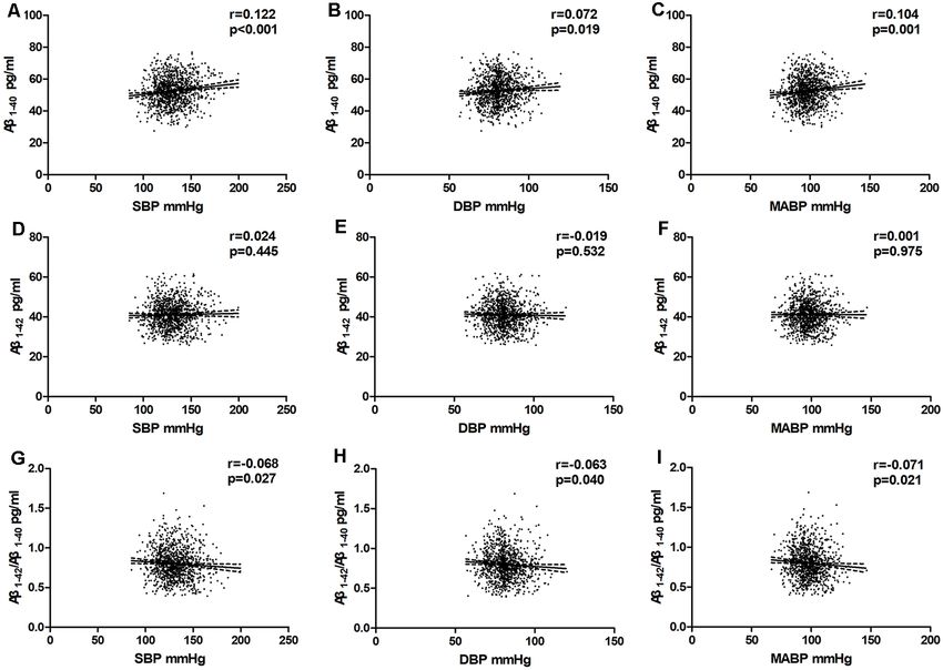
She et al.                                                                                                     Blood Pressure and Plasma Amyloid-β Levels

INTRODUCTION
Alzheimer’s disease (AD) is the most common cause of dementia,
affecting more than 33.9 million people worldwide (Barnes and
Yaffe, 2011). The deposition of amyloid-β (Aβ) in the brain is the
main pathological characteristic of AD (Karran et al., 2011), and
the amyloid cascade hypothesis is widely considered to underlie
the pathogenesis of AD (Karran et al., 2011). Studies have found
that elevated blood pressure (BP) levels in midlife may be related
to the development and progression of AD in later life (Kivipelto
et al., 2001; Qiu et al., 2005; Gottesman et al., 2017; Walker et al.,
2019). According to recent studies, hypertension is associated
with an increased Aβ burden in the brain (Ingmar and Deborah,
2013). Based on accumulating evidence, elevated BP may impair                 FIGURE 1 | Flow chart of participant selection. Aβ, amyloid-β.
the clearance of Aβ and increase Aβ production in both the
peripheral circulation and the central nervous system (Faraco                (3) had missing covariates, or at least one aberrant plasma
and Iadecola, 2013).                                                         Aβ1–40 or Aβ1–42 level. The flow chart of study is shown
    Plasma Aβ, the source of which is mainly brain efflux                    in Figure 1.
via low-density lipoprotein receptor-related protein-1 (LRP1)                    Among the 1, 699 residents living in the village and older than
through the blood-brain barrier (BBB) or glymphatic system                   45 years, 227 were taking anti-hypertension medicine, 329 had
(Roberts et al., 2014), is closely related to brain Aβ deposition            missing covariates, and 74 had at least one aberrant plasma
(Vergallo et al., 2019). A complex equilibrium is believed to                Aβ1–40 or Aβ1–42 level (exceeding ± 3 SDs from the mean). After
exist between the amyloid burden in the brain and plasma Aβ                  all the exclusions, a final count of 1, 069 subjects was included
levels in both animal models and healthy individuals (DeMattos               in the study. The protocols used were reviewed and approved
et al., 2002b; Giedraitis et al., 2007). The continuous translocation        by the Ethics Committee of the First Affiliated Hospital of Xi’an
of Aβ from the brain parenchyma to the peripheral blood is                   Jiaotong University (No: XJTU1AF2014LSK-111).
essential for preventing Aβ accumulation and reducing the Aβ
burden in the brain (DeMattos et al., 2001; Matsuoka et al.,                 Data Collection
2003). Plasma Aβ levels were recently reported to be associated              Subjects completed standardized questionnaires of general
with the incidence of AD (Ertekin-Taner et al., 2008; Lambert                information to collect demographic data (age, sex, and education
et al., 2009; Pérez-Grijalba et al., 2019). However, the relationship        levels) and lifestyle habits (alcohol abuse, self-reported smoking
between the blood pressure (BP) level and plasma Aβ level                    history as current/former/never, and physical activity level)
is currently unclear. In the present study, we investigated the              and underwent tests to determine the levels of multiple
relationships between the parameters of BP and plasma Aβ                     laboratory markers. We also recorded the medical history of
levels in middle-aged and older individuals in the general                   cardiovascular disease, and transient ischaemic attack (TIA)
population.                                                                  or stroke. Additionally, we measured height, weight, BMI
                                                                             {which was calculated as [weight (kg)]/[height (m)2 ]}, and the
MATERIALS AND METHODS                                                        pulse rate. The following vascular risk factors were measured:
                                                                             hypertension (defined as a mean systolic blood pressure
Study Population                                                             measurement ≥140 mm Hg or diastolic blood pressure ≥
This is an ongoing population-based study designed to determine              90 mm Hg, a self-reported medical diagnosis, or use of
the potential vascular factors for AD in the general population.             antihypertensive drug therapy), diabetes (fasting serum glucose
We used the cluster sampling method from October 2014 to                     level ≥7.0 mmol/L, or use of diabetic medication or insulin.),
March 2015 to make a face-to-face questionnaire survey on all                and hyperlipidemia (fasting serum cholesterol concentration
the permanent residents in Qubao village, Huyi District, Xi’an               >5.18 mmol/L, serum triglyceride concentration >1.70 mmol/L,
City, and conducted a household survey on the disabled. The                  serum LDL cholesterol concentration >3.37 mmol/L, serum
lifestyle and population composition of the village are similar              HDL cholesterol concentration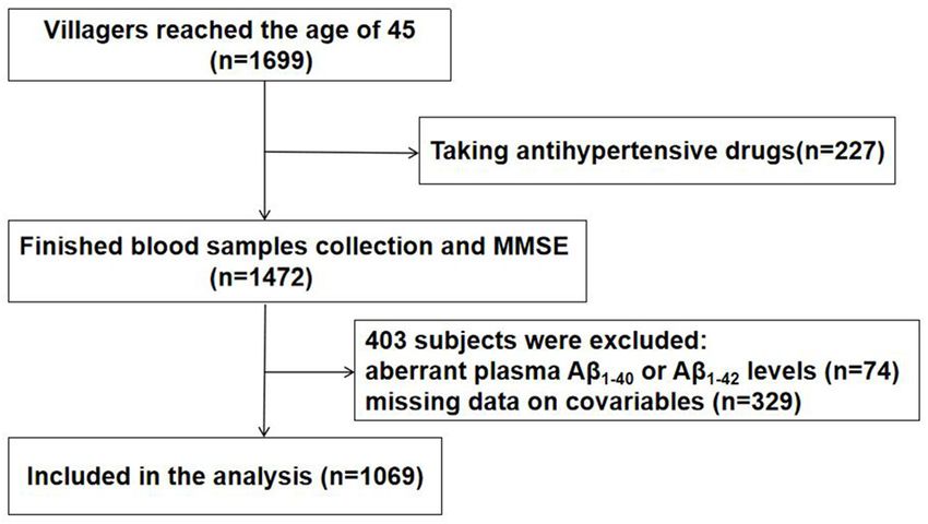
She et al.                                                                                            Blood Pressure and Plasma Amyloid-β Levels

the cut-off value set by Katzman et al. (1988) as the criterion          5.0. Quantitative variables are reported as the means ± SD
for cognitive impairment; specifically, the cut-off value was            or medians (interquartile ranges), and qualitative variables are
≤17 for the uneducated, ≤20 for individuals with primary school          reported as numbers (percentages). Unpaired Student’s t-tests
education, and ≤24 for individuals educated at the junior high           were used to analyses data with an approximately normal
school level or above.                                                   distribution, the Mann-Whitney U-test was used to compare
                                                                         data with skewed distributions, and the c2 or Fisher’s exact
BP Measurements                                                          test was used for categorical data. However, plasma triglyceride
Blood pressure measurements were obtained during the                     and fasting blood glucose (FBG) levels were log transformed
inclusion interview for this study. Two brachial blood pressure          prior to analysis, as they displayed skewed distributions.
measurements were recorded twice in a seated position after              Then, unpaired Student’s t-tests were used to compare the
subjects had rested for at least 10 min. The instruments were            differences in plasma Aβ levels in the subgroups stratified by
a manual mercury sphygmomanometer with an appropriate-                   BP parameters. Partial correlation coefficients and multivariate
sized cuff (Shanghai Medical Instruments Co. Shanghai, China).           linear regression models were used to evaluate the associations
Korotkoff phases 1 and 5 established the levels of systolic blood        between BP levels and Aβ levels after adjusting for the
pressure (SBP) and diastolic blood pressure (DBP), respectively.         confounding factors. Model 1 was adjusted for age and sex,
The average of two measurements was used for analysis. The               and model 2 was additionally adjusted for the BMI, pulse rate,
mean arterial pressure (MABP) was defined as [(SBP + DBP)/3].            ApoEε4 carrier status, log-transformed fasting blood glucose
   Four variables, BP, SBP, DBP, and MABP, were used as                  level, log-transformed triglyceride level, total cholesterol level,
indicators of the blood pressure level. A high BP was defined as a       high-density lipoprotein level, smoking status, drinking status,
mean SBP ≥140 mm Hg or DBP ≥ 90 mm Hg. A high SBP was                    physical activity level, stroke, transient ischaemic attack, and
defined as a mean SBP ≥140 mm Hg. A high DBP was defined as              heart disease. The linear correlation and regression analyses were
a mean DBP ≥ 90 mm Hg. A high MABP was defined as a mean                 performed to explore the potential effect of ApoE genotype on
MABP ≥ 105 mm Hg.                                                        the relationships. Potential confounders identified in previous
                                                                         studies that might affect BP and plasma Aβ levels were
Detection of Plasma β-Amyloid Levels                                     considered. A P value of
She et al. Blood Pressure and Plasma Amyloid-β Levels TABLE 1 | Characteristics of the total study population and the population stratified by blood pressure. Characteristic Total sample (n = 1,069) Normal BP group (n = 625) High BP group (n = 444) P-Value Age, y (mean ± SD) 57.4 ± 9.2 55.8 ± 8.7 59.6 ± 9.3
She et al. Blood Pressure and Plasma Amyloid-β Levels
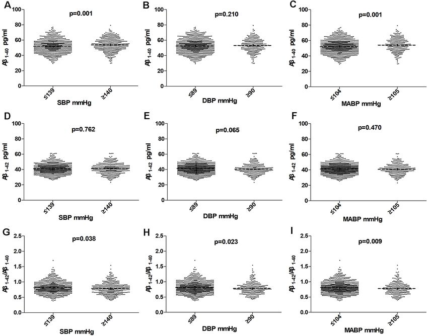
FIGURE 2 | Comparision of Aβ1–40 , Aβ1–42 , and Aβ1–42 /Aβ1–40 in subjects according to SBP (A,D,G), DBP (B,E,H), MABP (C,F,I) in the total population. Horizontal
lines represent the mean and 95% CI. Abbreviations: BP, blood pressure; SBP, systolic blood pressure; DBP, diastolic blood pressure; MABP, mean arterial blood
pressure.
middle-aged and older villagers, even after controlling for other detected using ELISA, which has been demonstrated as an
confounding factors. However, the association was only observed accurate and dependable method (Katzman et al., 1988). We
in ApoE ε4 non-carriers, but not ApoE ε4 carriers. did a multiple analysis to adjusted for almost all identified
Several publications have suggested an association between potential confounder factors, including the ApoE genotype
plasma Aβ and BP levels (Fujiwaraa et al., 2003; Abdullah et al., (Rodrigue et al., 2013; Giau et al., 2015), BMI (Qiu et al.,
2009; Lambert et al., 2012; Ruiz et al., 2013; Wang et al., 2018). 2005), MMSE score, and serum TC, TG and HDL-c levels
However, a consistent conclusion has not been drawn. A positive (Matsuzaki et al., 2011). The relationships between plasma Aβ
correlation between SBP and plasma Aβ1–40 levels (Abdullah level and BP levels did not change. These results were similar
et al., 2009; Lambert et al., 2012) or a negative correlation to the three-city study by Lambert et al. (2012) which showed
between SBP and plasma Aβ1–40 levels (Abdullah et al., 2009), that elevated BP levels were associated with decreased plasma
as well as a positive correlation between DBP and plasma Aβ1–42 /Aβ1–40 ratio.
Aβ1–42 levels (Fujiwaraa et al., 2003) have been reported. These The mechanism underlying the association between plasma
inconsistencies are likely due to the use of different inclusion Aβ and BP levels is not well understood. One possible
criteria, exclusion criteria, and test methods. In those cross- mechanism is that BP may affect the deposition of Aβ in
sectional studies, investigators either used a case-control study the brain. It has been reported that hypertension is associated
design with a small sample size (Lambert et al., 2012; Ruiz et al., with Aβ deposition in the brain in individuals with normal
2013) or only explored plasma Aβ1–42 and BP levels (Fujiwaraa cognition. Animal experiments also observed a direct effect of
et al., 2003). hypertension on the deposition of Aβ in the brain (Cifuentes
Unlike the previous studies, our present study used a random et al., 2015; Faraco et al., 2016). In PET imaging studies of
cluster sampling method with a large sample size consisting of middle-aged to old adults with normal cognition, Langbaum
middle-aged and elderly individuals in the general population. et al. revealed an association between elevated SBP and fibrillary
All enrolled residents lived in the selected village for over Aβ levels in the brain (Langbaum et al., 2012). In addition,
3 years with permanent residency. Plasma Aβ levels were Rodrigue et al. (2013) observed a significant correlation between
Frontiers in Aging Neuroscience | www.frontiersin.org 5 June 2021 | Volume 13 | Article 650679She et al. Blood Pressure and Plasma Amyloid-β Levels
FIGURE 3 | Partial linear correlations between plasma Aβ and SBP, DBP, MABP in total participants. Partial linear correlations of blood pressure (BP) with plasma
Aβ1–40 levels (A–C) and plasma Aβ1–42 levels (D–F) and Aβ1–42 /Aβ1–40 (G–I) are shown in the figure. Partial correlation coefficients and P values were obtained after
adjustment for age, sex, BMI, pulse rate, log-transformed fasting blood glucose, log-transformed TG, TC, HDL-C, smoking, drinking, physical activity level, stroke,
transient ischaemic attack (TIA), and heart disease.
TABLE 2 | Multiple linear regression models analysis of blood pressure components and plasma Aβ1–40 , Aβ1–42 , and Aβ1–42 /Aβ1–40 ratio in total study subjects.
Aβ1– 40 Aβ1–42 Aβ1– 42 /Aβ1– 40
β p β p β p
Model 1
SBP 0.056 0.001 0.008 0.507 −0.001 0.063
DBP 0.052 0.104 −0.017 0.418 −0.001 0.103
MABP 0.065 0.008 −0.002 0.894 −0.001 0.062
Model 2
SBP 0.068She et al. Blood Pressure and Plasma Amyloid-β Levels
TABLE 3 | Characteristics of the subpopulation stratified by ApoE ε4 carrier status.
Characteristic Subpopulation (n = 961a ) ApoEε4 non-carriers (n = 808) ApoEε4 carriers (n = 153) P-Value
Age, y (mean ± SD) 57.6 ± 9.2 57.5 ± 9.2 58.1 ± 9.2 0.509
Female, % 556 (57.9%) 453 (56.1%) 103 (67.3%) 0.010
Educational level, y 6.1 ± 3.5 6.1 ± 3.4 5.9 ± 3.6 0.435
Medical history, n (%)
Diabetes mellitus 114 (11.9%) 95 (11.8%) 19 (12.4%) 0.817
Heart disease 50 (5.2%) 40 (5.0%) 10 (6.5%) 0.418
Stroke or TIA 74 (7.7%) 62 (1.7%) 12 (7.8%) 0.461
Dyslipidemia 489 (50.9%) 403 (49.9%) 86 (56.2%) 0.151
Blood pressure, mmHg
SBP 131.2 ± 17.2 131.4 ± 17.2 130.3 ± 17.3 0.477
DBP 81.3 ± 9.6 81.5 ± 9.6 80.2 ± 9.4 0.120
MABP 97.9 ± 11.2 98.1 ± 11.2 96.9 ± 11.3 0.213
Personal history, n(%)
Smoking 297 (30.9%) 259 (32.1%) 38 (24.8%) 0.132
Drinking 146 (15.2%) 130 (16.1%) 16 (10.5%) 0.075
Lack of physical activity 176 (18.3%) 148 (18.3%) 28 (13.3%) 0.996
BMI, kg/m2 (mean ± SD) 25.0 ± 3.2 25.0 ± 3.1 25.3 ± 3.5 0.338
Pulse rate, bpm(mean ± SD) 74.9 ± 7.9 74.8 ± 8.0 75.4 ± 7.7 0.417
Fasting blood glucose, mmol/l 5.40 (5.06, 5.76) 5.40 (5.06, 5.77) 5.71 (5.07, 5.78) 0.582
TG, mmol/l 1.44 (1.04, 2.01) 1.43 (1.04, 2.01) 1.50 (1.06, 2.09) 0.577
TC, mmol/l 5.04 ± 0.96 5.03 ± 0.97 5.13 ± 0.94 0.217
HDL-c, mmol/l 1.42 ± 0.31 1.42 ± 0.30 1.40 ± 0.32 0.501
Aβ1– 40 , pg/ml 52.45 ± 8.94 52.36 ± 8.90 52.93 ± 9.20 0.468
Aβ1– 42 , pg/ml 41.16 ± 6.70 41.01 ± 6.68 41.94 ± 6.78 0.116
Aβ1– 42 /Aβ1– 40 0.81 ± 0.19 0.81 ± 0.19 0.81 ± 0.19 0.602
Numbers are mean (SE) or count (%). P values obtained from t test or χ 2 test or ANOVA in comparison of ApoE ε4 non-carriers and ApoE ε4 carriers. a Data available for 961 participants,
961 participants agreed to perform gene testing. Abbreviations: TIA, transient ischemic attack; BMI, body mass index; TG, triglyceride; TC, total cholesterol; HDL-c, high-density
lipoprotein cholesterol.
transport. Research shows that hypertension damages the In the subgroup analysis stratified according to the ApoE
integrity of vascular endothelial cells and affects the vascular ε4 status, the association between plasma Aβ and BP levels
wall (Faraco and Iadecola, 2013). It has been hypothesized were only observed in ApoE ε4 non-carriers, but not in ApoE
(Shah et al., 2012) that the vasoactivity of Aβ in combination ε4 carriers. The underlying mechanism is not clear. As the
with hypertension could hardly destroy the integrity of the strongest genetic risk factor for AD, the ApoE ε4 allele was
vascular wall and reduce the clearance of Aβ in the brain closely associated with the decrease of cerebral spinal flow Aβ
which would lead to an inflammatory response and cell and the increase of aggregation and deposition of cerebral Aβ
death. Despite all of this, the authors suggested that vascular in the brain (Liu et al., 2013; Giau et al., 2015). ApoE ε4 might
integrity was an important part of the early trajectory. compromise the effects of BP on plasma Aβ levels. Recent articles
Importantly, blood pressure and plasma Aβ levels were reported that the association between plasma Aβ levels and
measured 10–20 years before the diagnosis of AD, which Aβ deposition in the brain was exactly observed in ApoE ε4
indicates that early intervention on elevated BP might be non-carriers (Swaminathan et al., 2014; Tateno et al., 2017).
very important for reducing AD caused by hypertension. This Additionally, Katsuya et al. reported that the prevalence of
conclusion is also suggested by our results. Moreover, the hypertension is lower in ApoE ε4 carriers (Katsuya et al., 2002).
elevated BP might impair the vascular clearance of Aβ and These indicated that the relationship between BP and plasma Aβ
increase its cleavage from APP in both peripheral and the levels is dependent on ApoE ε4 states.
central nervous system to further facilitate the onset of AD In this study, we measured plasma Aβ levels using an ELISA
(Ingmar and Deborah, 2013). kit marketed by Yuanye Co (China). Compared to Aβ levels
We did not find an association between BP and plasma mentioned in other publications, the Aβ42 levels we determined
Aβ1–42 levels, which might be due to the different physiological are higher. We speculate this discrepancy might be due to
roles of Aβ1–40 and Aβ1–42 . Aβ1–42 is insoluble and prone to the heterogeneity of the different populations studied and the
fibrillate and deposit in senile plaques with greater neurotoxicity measurement methods used and standards of Aβ that have not
(Verbeek et al., 1997). While, Aβ1–40 is soluble, and has been validated to each other. Compared to the INNO-BIA assay,
direct physiological or toxic effects on the blood vessel wall the ELISA Aβ40 levels measures are slightly lower, while the
(Niwa et al., 2000). The levels of Aβ1–40 , but not Aβ1–42 , are Aβ42 levels are slightly higher (Barnes and Yaffe, 2011). Our
markedly increased in patients with ischemic stroke (Lee et al., previous studies using the same ELISA kit produced credible
2005) and diffuse SVD (Gomis et al., 2009). These indicated results (Jiang et al., 2017; Gao et al., 2018; Wang et al., 2018);
that Aβ1–40 is more closely relate to vascular disease than however, we acknowledge more rigorous data are needed to
Aβ1–42 . validate the comparisons.
Frontiers in Aging Neuroscience | www.frontiersin.org 7 June 2021 | Volume 13 | Article 650679She et al. Blood Pressure and Plasma Amyloid-β Levels
0.831
0.390
0.554
0.968
0.542
0.741
r, the partial correlation coefficient. β, the unstandardized regression coefficient. Model 1: adjusted for age and sex. Model 2: adjusted for age, sex, BMI, pulse rate, log-transformed fasting blood glucose, log-transformed triglyceride,
CONCLUSION
p
In summary, in this population-based cross-sectional study, we
Aβ1– 42 /Aβ1–40
found that elevated BP levels were associated with increased
0.001
0.001
0.001
0.001
−4.153
TABLE 4 | Partial linear correlation analysis and multiple linear regression models of blood pressure components and plasma Aβ1–40 , Aβ1–42 , and Aβ1–42 /Aβ1–40 ratio in subjects stratified by ApoE ε4 status.She et al. Blood Pressure and Plasma Amyloid-β Levels
REFERENCES Katsuya, T., Baba, S., Ishikawa, K., Mannami, T., Fu, Y., Inamoto, N., et al. (2002).
Epsilon 4 allele of apolipoprotein E gene associates with lower blood pressure
Abdullah, L., Luis, C., Paris, D., Mouzon, B., Ait-Ghezala, G., Allen, E., et al. in young Japanese subjects: the Suita Study. J. Hyperten. 20, 2017–2021.
(2009). High serum Abeta and vascular risk factors in first-degree relatives of doi: 10.1097/00004872-200210000-00021
Alzheimer’s disease patients. Mol. Med. 15, 95–100. doi: 10.2119/molmed.2008. Katzman, R., Zhang, M., Ouang, Y., Liu, W., and Yu, E. (1988). A Chinese
00118 version of the MINI-mental state examination; impact of illiteracy in a
Barnes, D. E., and Yaffe, K. (2011). The projected effect of risk factor Shanghai dementia survey. J. Clin. Epidemiol. 41, 971–978. doi: 10.1016/0895-
reduction on Alzheimer’s disease prevalence. The Lancet Neurolol. 10, 819–828. 4356(88)90034-0
doi: 10.1016/S1474-4422(11)70072-2 Kivipelto, M., Helkala, E. L., Laakso, M. P., Hanninen, T., Hallikainen, M.,
Cifuentes, D., Poittevin, M., Dere, E., Broquères-You, D., Bonnin, P., Alhainen, K., et al. (2001). Midlife vascular risk factors and Alzheimer’s
Benessiano, J., et al. (2015). Hypertension accelerates the progression of disease in later life: longitudinal, population based study. BMJ 322, 1447–1451.
Alzheimer-like pathology in a mouse model of the disease. Hypertension 65, doi: 10.1136/bmj.322.7300.1447
218–224. doi: 10.1161/HYPERTENSIONAHA.114.04139 Lambert, J. C., Dallongeville, J., Ellis, K. A., Schraen-Maschke, S., Lui, J., Laws, S.,
DeMattos, R. B., Bales, K. R., Cummins, D. J., Dodart, J. C., Paul, S. M., et al. (2012). Association of plasma Aßpeptides with blood pressure in the
Holtzman, D. M., et al. (2001). Peripheral anti-A beta antibody alters CNS elderly. PLoS One 6:e18536. doi: 10.1371/journal.pone.0018536
and plasma A beta clearance and decreases brain A beta burden in a mouse Lambert, J. C., Schraen-Maschke, S., Richard, F., Fievet, N., Rouaud, O., Berr, C.,
model of Alzheimer’s disease. Proc. Natl. Acad. Sci. U S A 98, 8850–8855. et al. (2009). Association of plasma amyloid β with risk of dementia: the
doi: 10.1073/pnas.151261398 prospective three-city study. Neurology 73, 847–853. doi: 10.1212/WNL.
DeMattos, R. B., Bales, K. R., Cummins, D. J., Paul, S. M., and Holtzman, D. M. 0b013e3181b78448
(2002a). Brain to plasma amyloid-beta efflux: a measure of brain amyloid Langbaum, J. B., Chen, K., Launer, L. J., Fleisher, A. S., Lee, W., Liu, X., et al.
burden in a mouse model of Alzheimer’s disease. Science 295, 2264–2267. (2012). Blood pressure is associated with higher brain amyloid burden and
doi: 10.1126/science.1067568 lower glucose metabolism in healthy late middle-age persons. Neurobiol. Aging
DeMattos, R. B., Bales, K. R., Parsadanian, M., O’Dell, M. A., Foss, E. M., 33:827. doi: 10.1016/j.neurobiolaging.2011.06.020
Paul, S. M., et al. (2002b). Plaque-associated disruption of CSF and plasma Lee, P. H., Bang, O. Y., Hwang, E. M., Lee, J. S., Joo, U. S., Mook-Jung, I., et al.
amyloid-beta (Abeta) equilibrium in a mouse model of Alzheimer’s disease. (2005). Circulating beta amyloid protein is elevated in patients with acute
J. Neurochem. 81, 229–236. doi: 10.1046/j.1471-4159.2002.00889.x ischemic stroke. J. Neural Transm. 112, 1371–1379. doi: 10.1007/s00702-004-
Ertekin-Taner, N., Younkin, L. H., Yager, D. M., Parfitt, F., Baker, M. C., 0274-0
Asthana, S., et al. (2008). Plasma amyloid beta protein is elevated in late-onset Liu, C. C., Liu, C. C., Kanekiyo, T., Xu, H., and Bu, G. (2013). Apolipoprotein E and
Alzheimer disease families. Neurology 70, 596–606. doi: 10.1212/01.WNL. Alzheimer disease: risk, mechanisms and therapy. Nat. Rev. Neurol. 9, 106–118.
0000278386.00035.21 doi: 10.1038/nrneurol.2012.263
Faraco, G., and Iadecola, C. (2013). Hypertension: a harbinger of stroke and Matsuoka, Y., Saito, M., Lafrancois, J., Saito, M., Gaynor, K., Olm, V., et al.
dementia. Hypertension 62, 810–817. doi: 10.1161/HYPERTENSIONAHA.113. (2003). Novel therapeutic approach for the treatment of Alzheimer’s disease by
01063 peripheral administration of agents with an affinity to beta amyloid. J. Neurosci.
Faraco, G., Park, L., Zhou, P., Luo, W., Paul, S. M., Anrather, J., et al. (2016). 23, 29–33doi: 10.1523/JNEUROSCI.23-01-00029.2003
Hypertension enhances Aβ-induced neurovascular dysfunction, promotes β- Matsuzaki, T., Sasaki, K., and Hata, J. (2011). Association of Alzheimer disease
secretase activity and leads to amyloidogenic processing of APP. J. Cereb. Blood pathology with abnormal lipid metabolism: the Hisayama study. Neurology 77,
Flow Metab. 36, 241–252. doi: 10.1038/jcbfm.2015.79 1068–1075. doi: 10.1212/WNL.0b013e31822e145d
Fujiwaraa, Y., Takahashib, M., Tanakac, M., Hoshid, T., Someyab, T., Shinkai, S., Niwa, K., Younkin, L., Ebeling, C., Turner, S. K., Westaway, D., Younkin, S.,
et al. (2003). Relationships between plasma β-amyloid peptide 1–42 and et al. (2000). Abeta 1–40-related reduction in functional hyperaemia in mouse
atherosclerotic risk factors in community-based older populations. Gerontology neocortex during somatosensory activation. Proc. Natl. Acad. Sci. U S A 97,
49, 374–379. doi: 10.1159/000073765 9735–9740. doi: 10.1073/pnas.97.17.9735
Gao, L., Jiang, Y., Wei, S., Shang, S., Li, P., Chen, C., et al. (2018). The Pérez-Grijalba, V., Arbizu, J., Romero, J., Prieto, E., Pesini, P., Sarasa, L.,
level of plasma amyloid-40 is correlated with peripheral transport proteins et al. (2019). Plasma Aβ42/40 ratio alone or combined with FDG-PET can
in cognitively normal adults: a population-based cross-sectional study. accurately predict amyloid-PET positivity: a cross-sectional analysis from
J. Alzheimers Dis. 65, 951–961. doi: 10.3233/JAD-180399 the AB255 Study. Alzheimers Res. Ther. 11:96. doi: 10.1186/s13195-019
Giau, V. V., BagyinszkY, E., An, S. S., and Kim, S. Y. (2015). Role of apolipoprotein -0549-1
E in neurodegenerative diseases. Neuropsychiatr. Dis. Treat. 11, 1723–1737. Qiu, C., Winblad, B., and Fratiglioni, L. (2005). The age-dependent relation of
doi: 10.2147/NDT.S84266 blood pressure to cognitive function and dementia. The Lancet Neurolol. 4,
Giedraitis, V., Sundelof, J., Irizarry, M. C., Garevik, N., Hyman, B. T., 487–499. doi: 10.1016/S1474-4422(05)70141-1
Wahlund, L. O., et al. (2007). The normal equilibrium between CSF and plasma Roberts, K. F., Elbert, D. L., Kasten, T. P., Patterson, B. W., Sigurdson, W. C.,
amyloid beta levels is disrupted in Alzheimer’s disease. Neurosci. Lett. 427, Connors, R. E., et al. (2014). Amyloid-beta efflux from the central nervous
127–131. doi: 10.1016/j.neulet.2007.09.023 system into the plasma. Ann. Neurol. 76, 837–844. doi: 10.1002/ana.24270
Gomis, M., Sobrino, T., Ois, A., Milla’n, M., Rodríguez-Campello, A., Rodríguez- Rodrigue, K. M., Rieck, J. R., Kennedy, K. M., Devous, M. D., Diaz-
Gonza’lez, R., et al. (2009). Plasma beta-amyloid 1–40 is associated Arrastia, R., Park, D. C., et al. (2013). Risk factors for β-amyloid deposition
with the diffuse small vessel disease subtype. Stroke 40, 3197–3201. in healthy aging: vascular and genetic effects. JAMA Neurol. 70, 600–7606.
doi: 10.1161/STROKEAHA.109.559641 doi: 10.1001/jamaneurol.2013.1342
Gottesman, R. F., Schneider, A. L. C., Zhou, Y., Coresh, J., Green, E., Gupta, N., Ruiz, A., Pesini, P., Espinosa, A., Pérez-Grijalba, V., Valero, S., Sotolongo-
et al. (2017). Association between midlife vascular risk factors and estimated Grau, O., et al. (2013). Blood amyloid beta levels in healthy, mild cognitive
brain amyloid deposition. JAMA 317, 1443–1450. doi: 10.1001/jama.2017.3090 impairment and Alzheimer’s disease individuals: replication of diastolic blood
Ingmar, S., and Deborah, G. (2013). Update on hypertension and Alzheimer’s pressure correlations and analysis of critical covariates. PLoS One 8:e81334.
disease. Neurol. Res. 28, 605–611. doi: 10.1179/016164106X130506 doi: 10.1371/journal.pone.0081334
Jiang, Y., Shang, S., Li, P., Chen, C., Dang, L., Wang, J., et al. (2017). Pulse pressure Shah, N. S., Vidal, J. S., Masaki, K., Petrovitch, H., Ross, G. W., Tilley, C.,
is associated with plasma amyloid-β transport dysfunction. J. Hypertension 36, et al. (2012). Midlife blood pressure, plasma-amyloid and the risk for
569–579. doi: 10.1097/HJH.0000000000001565 Alzheimer disease the honolulu asia aging study. Hypertension 59, 780–786.
Karran, E., Mercken, M., and De Strooper, B. (2011). The amyloid cascade doi: 10.1161/HYPERTENSIONAHA.111.178962
hypothesis for Alzheimer’s disease: an appraisal for the development Swaminathan, S., Risacher, S. L., Yoder, K. K., West, J. D., Shen, L., Kim, S., et al.
of therapeutics. Nat. Rev. Drug Discov. 10, 698–712. doi: 10.1038/ (2014). Association of plasma and cortical amyloid beta is modulated by APOE
nrd3505 epsilon4 status. Alzheimers Dement. 10, e9–e18. doi: 10.1016/j.jalz.2013.01.007
Frontiers in Aging Neuroscience | www.frontiersin.org 9 June 2021 | Volume 13 | Article 650679She et al. Blood Pressure and Plasma Amyloid-β Levels Tateno, A., Sakayori, T., Kim, W. C., Koeda, M., Kumita, S., Suzuki, H., et al. Wang, J., Qiao, F., Shang, S., Li, P., Chen, C., Dang, L., et al. (2018). Elevation (2017). Effect of apolipoprotein E phenotype on the association of plasma of plasma amyloid-β level is more significant in early stage of cognitive amyloid beta and amyloid positron emission tomography imaging in Japan. impairment: a population- based cross-sectional study. J. Alzheimers Dis. 64, Alzheimers Dement. 9, 51–56. doi: 10.1016/j.dadm.2017.08.002 61–69. doi: 10.3233/JAD-180140 Verbeek, M. M., Eikelenboom, P., and de Waal, R. M. (1997). Differences between the pathogenesis of senile plaques and congophilic angiopathy in Alzheimer Conflict of Interest: The authors declare that the research was conducted in the disease. J. Neuropathol. Exp. Neurol. 56, 751–761. doi: 10.1097/00005072- absence of any commercial or financial relationships that could be construed as a 199756070-00001 potential conflict of interest. Vergallo, A., Mégret, L., Lista, S., Cavedo, E., Zetterberg, H., Blennow, K., et al. (2019). Plasma amyloid β 40/42 ratio predicts cerebral amyloidosis Copyright © 2021 She, Shang, Hu, Chen, Dang, Gao, Wei, Huo, Wang, Wang in cognitively normal individuals at risk for Alzheimer’s disease. Alzheimers and Qu. This is an open-access article distributed under the terms of the Creative Dement. 15, 764–775. doi: 10.1016/j.jalz.2019.03.009 Commons Attribution License (CC BY). The use, distribution or reproduction in Walker, K. A., Sharrett, A. R., Wu, A., Schneider, A. L. C., Albert, M., other forums is permitted, provided the original author(s) and the copyright owner(s) Lutsey, P. L., et al. (2019). Association of midlife to late-life blood pressure are credited and that the original publication in this journal is cited, in accordance patterns with incident dementia. JAMA 322, 535–545. doi: 10.1001/jama.2019. with accepted academic practice. No use, distribution or reproduction is permitted 10575 which does not comply with these terms. Frontiers in Aging Neuroscience | www.frontiersin.org 10 June 2021 | Volume 13 | Article 650679
You can also read