Usefulness of serum hyaluronic acid levels as a predictor of incidence of hand osteoarthritis analyzed by longitudinal analysis from the Iwaki ...
←
→
Page content transcription
If your browser does not render page correctly, please read the page content below
www.nature.com/scientificreports
OPEN Usefulness of serum hyaluronic
acid levels as a predictor
of incidence of hand osteoarthritis
analyzed by longitudinal analysis
from the Iwaki cohort
Tatsuro Saruga1*, Eiji Sasaki1, Ryo Inoue2, Daisuke Chiba1, Seiya Ota1, Hiroki Iwasaki1,
Ryoko Uesato1, Shigeyuki Nakaji2 & Yasuyuki Ishibashi1
The factors predicting hand osteoarthritis (HOA) in patients remain unknown. We aimed to investigate
the usefulness of serum hyaluronic acid (sHA) levels in predicting HOA progression from a 6-year
longitudinal epidemiological study. A total of 417 participants in the Iwaki cohort were followed-up
over 6 years. Hand and knee radiographs taken at baseline and follow-up were scored according to
Kellgren–Lawrence grades and Kallman score. Participants were classified into the HOA group and the
non-HOA group. sHA levels at baseline were determined by ELISA. Correlations between sHA levels,
the number of involved joints, and Kallman score were estimated. Factors related to the incidence or
progression of HOA over 6 years were analyzed. The prevalence of HOA was 19.9% at baseline, and
3.6 ± 2.1 joints were involved. sHA levels in the HOA group at baseline were significantly higher than in
the non-HOA group (p < 0.001) and correlated with the number of involved joints (r = 0.399, p < 0.001)
and Kallman score (r = 0.540, p < 0.001). The incidence rate was 14.5%, and the progression rate was
46.1% over 6 years. Higher sHA levels at baseline were the risk factor of HOA incidence. Thus, sHA
levels predicted the incidence of HOA over 6 years.
Abbreviations
HOA Hand osteoarthritis
sHA Serum hyaluronic acid
BMI Body mass index
DIP Distal interphalangeal
PIP Proximal interphalangeal
IP Interphalangeal
CMC Carpometacarpal
KL Kellgren–Lawrence classification
ROC Receiver operating characteristic
AUC Area under the curve
sCOMP Serum cartilage oligomeric matrix protein
Hand osteoarthritis (HOA) is a common disease in the elderly, and its prevalence as reported radiographically in
population-based studies is 29–89% in the middle-aged1–5. HOA causes chronic pain and disabilities that lead to
serious problems in activities of daily living. It also has a significant impact on socio-economic status. Although
early detection of higher-risk patients is necessary to begin a preventive approach, patients could not recognize
the severity of their HOA until it progressed and caused serious pain and disabilities. Moreover, the natural
history of this disease and therapeutic strategies for preventing incidence or progress have not been established.
While there are several potential problems regarding the high prevalence and progressive activity of this disease,
1
Department of Orthopedic Surgery, Hirosaki University Graduate School of Medicine, 5 Zaifu‑cho, Hirosaki,
Aomori 036‑8562, Japan. 2Department of Social Medicine, Hirosaki University Graduate School of Medicine,
Hirosaki, Japan. *email: suntatsurous@hirosaki‑u.ac.jp
Scientific Reports | (2021) 11:4074 | https://doi.org/10.1038/s41598-021-83693-0 1
Vol.:(0123456789)www.nature.com/scientificreports/
radiographs cannot detect minute changes at an early stage. Hence, an easier quantitative evaluation of disease
activity needs to be established.
As the evaluation tool of synovitis, serum biomarkers have attracted attention. Biomarkers are measured from
blood and urine, and many substances that specifically reflect the condition of bone, cartilage, and synovitis
have been reported6,7. Biomarkers are suggested as a diagnostic tool and severity predictors of knee OA (KOA),
as well as possibly a prognostic predictor8. Among them, serum hyaluronic acid (sHA) is strongly related to
symptoms and progression of OA since it reflects the state of synovitis. It is gaining attention as a biomarker
for OA severity and a predictor of OA progression. Regarding finger OA, it was revealed that higher sHA levels
were correlated with the number of osteoarthritic joints in a population-based cohort s tudy9 and progression
of joint space narrowing from longitudinal observations focusing on the p atients10. However, there has been no
longitudinal evaluation of the relationship between long-term radiographic changes in HOA and sHA levels in
epidemiological studies. It is unclear whether sHA levels could be a predictor of incidence or progress of HOA.
This study aimed to investigate whether sHA levels could reflect the severity and number of involved joints in
HOA. Furthermore, we examined the predictive power of sHA levels in determining the incidence or progress
of HOA in a longitudinal cohort study. We hypothesized that higher sHA levels at baseline could predict these
over 6 years.
Methods
Subjects were voluntary participants from the Iwaki Health Promotion Project of 2008 and 2014, a community-
based program to prevent lifestyle diseases and improve average life expectancy by performing general health
checkups and prophylactic interventions11,12. It is an annual program that has been performed in the general
population living in the Iwaki area of Hirosaki City, located in western Aomori prefecture, Japan, since 2005.
This cohort study allows the evaluation of many kinds of diseases and disorders from various perspectives and
research into the risk factors of locomotive disability. The study protocol was approved by the institutional review
board of the Hirosaki University School of Medicine and performed in accordance with the relevant guidelines
and regulations. Informed consent was obtained from all patients before enrollment in the study.
Subjects. Altogether, 887 volunteers from approximately 12,000 residents participated in this study in 2008.
They were recruited via phone calls from public health nurses and an advertisement in the mass media. Those
who had renal failure, liver failure, rheumatoid arthritis, malignant tumors, and incomplete questionnaires were
excluded from the study. Those who did not undergo radiographic examination were also excluded. A total of
724 participants (273 male and 451 female) were enrolled at baseline. Among them, 408 participants (142 male,
266 female) were followed-up in the Iwaki 2014 cohort. The follow-up rate was 56.3%. Height and body weight
were measured, and body mass index (BMI) was calculated.
Measurement of sHA levels. Blood samples were taken from all participants early in the morning for
biochemical examination at baseline and follow-up. Blood sampling was performed before breakfast because
eal13. The levels of sHA were determined using the Hyaluronan Assay Kit
circulating sHA increases following a m
(Seikagaku Corporation, Tokyo, Japan)9. The change in sHA levels over 6 years was defined as ΔsHA.
Radiographic diagnosis. Radiographs were taken for joint evaluation: postero-anterior view of bilateral
hands and antero-posterior view of weight-bearing bilateral knees. The following regions were evaluated from
each joint group by trained orthopedic surgeons (RU, HI). The second to fifth distal interphalangeal (DIP),
proximal interphalangeal (PIP), thumb interphalangeal (IP), and carpometacarpal (CMC), and scapho-trapezial
joints for each hand were graded according to the Kellgren–Lawrence classification (KL)14. Radiographic OA
was defined as KL grade ≥ 2. Participants with at least one involved joint at baseline were assigned to the HOA
group, while those without radiographic HOA were in the non-HOA (nHOA) group. Similarly, the presence of
KOA was also evaluated based on the KL scale in both knee radiographs and defined as OA with KL grade 2
or more. Furthermore, the severity of HOA was also scored according to the Kallman scoring s ystems15. Kall-
man scoring system can semi-quantitatively evaluate the progress of HOA from X-ray images, and is used as an
evaluation tool for HOA in various fields such as clinical practice and epidemiology. Individual hand joints were
assessed for the presence of osteophytes (graded 0–3), joint space narrowing (0–3), subchondral sclerosis (0–1),
subchondral cysts (0–1), lateral deformity (0–1), and the collapse of central joint cortical bone (0–1) with a total
of 208 points. To investigate the intra-observer reliability of the scale, 20 randomly selected hand radiographs
were scored by the same reader, and two orthopedists (RU and HI) also scored the 20 radiographs to assess the
inter-observer reliability. The intra- and inter-observer reliability was assessed by the k-statistic, and they were
0.78 and 0.77, respectively.
In this longitudinal study, participants in the nHOA group with an increasing number of involved joints for
6 years were classified into the incident-HOA (iHOA) group, and participants in the HOA group with an increas-
ing number of involved joints for 6 years were classified into the progression-HOA (pHOA) group. The progres-
sion by at least one KL grade of KOA for 6 years was also evaluated and defined as the progression of KOA. Both
HOA and nHOA groups were further divided into two groups based on the progression of KOA in each group.
Statistical analysis. Data input and calculations were performed with SPSS ver. 12.0J (SPSS Inc., Chicago,
IL, USA). In the baseline data, Chi-square testing was performed between HOA and non-HOA groups to com-
pare sex, knee OA, and smoking status. The Mann–Whitney U test was performed to compare age, BMI, and
sHA levels at baseline. Spearman’s correlation coefficients were estimated for age, number of involved joints,
Scientific Reports | (2021) 11:4074 | https://doi.org/10.1038/s41598-021-83693-0 2
Vol:.(1234567890)www.nature.com/scientificreports/
Non-HOA (n = 331) HOA (n = 77) p value (non-HOA vursus HOA)
Age (y) 55.2 ± 9.9 66.1 ± 7.9 < 0.001
Female (%) 61.5 80.2 < 0.001
BMI (kg/m2) 22.9 ± 3.0 23.5 ± 3.0 0.071
Knee OA (%) 19.1 57 < 0.001
Smoking (%) 33.4 7.3 < 0.001
sHA (ng/mL) 55.7 ± 27.6 92.3 ± 52.1 < 0.001
Table 1. Brief summary of participants. Comparison using Mann–Whitney U test and Chi-square test. Values
are given as means ± standard deviations. p values < 0.05 significant.
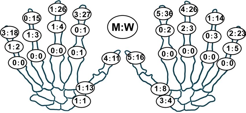
Figure 1. Prevalence (%) of hand osteoarthritis on radiography in each joint in men (M, left) and women (W,
right).
Kallman score, and sHA levels. Analysis of variance (ANOVA) and the Tukey method was performed to com-
pare ages and sHA levels in each KL grade of KOA and the presence of HOA and KOA.
In the longitudinal analysis over 6 years, the baseline levels of sHA and ΔsHA were compared using ANOVA
and Tukey method among the four groups of the non-HOA group, classified by HOA incidence and KOA pro-
gression and the four groups of the HOA group, classified by HOA progression and KOA progression. Further-
more, logistic regression analysis was performed with a model in which the presence of incidence or progression
of HOA was a dependent variable, while baseline levels of sHA or ΔsHA and relevant factors like age and presence
of knee OA were independent variables. A receiver operating characteristic (ROC) analysis was performed to
determine whether the baseline levels of sHA at baseline could predict the incidence or progress of HOA for
6 years. We calculated the areas under the curve (AUC). The optimal cut-off point was the highest Youden index
value (sensitivity + specificity − 1). A p value below 0.05 was considered to be statistically significant.
Ethics approval and consent to participate. The Ethics Committee of the Hirosaki University Gradu-
ate School of Medicine approved the study, and all participants provided written informed consent before par-
ticipation.
Results
Seventy-seven of 408 participants (18.9%) were classified into the HOA group (Table 1). The HOA group was
older (p < 0.001) and had a higher proportion of females. The prevalence of knee OA was higher (p < 0.001), but
no significant difference was observed in BMI. The prevalence of HOA at baseline was 10.6% in males and 22.9%
in females. Comparing the prevalence of HOA among interphalangeal joints in all cases, the prevalence in the
thumb CMC joint, the thumb IP joint, and the DIP joints were high (Fig. 1). The mean levels of baseline sHA
were 65.3 ± 35.9 (ng/ml). There was a significant correlation between the levels of baseline sHA and the number
of involved joints, and the correlation coefficient was 0.399 (p < 0.001) (Fig. 2A). Similarly, there was a significant
correlation between the levels of baseline sHA and higher baseline Kallman score, with a correlation coefficient
of 0.540 (p < 0.001) (Fig. 2B). Furthermore, the correlation between sHA levels and age was significant, with a
correlation coefficient of 0.631 (p < 0.001) in the non-HOA group and 0.578 (p < 0.001) in the HOA group. In
the HOA group, ages were significantly higher with and without KOA. Ages were also significantly higher in
participants with KL2 or higher grades of KOA.
The mean levels of baseline sHA were 55.7 ± 27.6 (ng/ml) in the non-HOA group and 92.3 ± 52.1 (ng/ml) in
the HOA group, which was significantly higher than in the non-HOA group (p < 0.001) (Fig. 3A). Levels of sHA
Scientific Reports | (2021) 11:4074 | https://doi.org/10.1038/s41598-021-83693-0 3
Vol.:(0123456789)www.nature.com/scientificreports/
A r=0.399, pwww.nature.com/scientificreports/
Figure 4. Serum hyaluronan levels among the incidence and progression of HOA and progression of KOA
(value ± standard error). P values < 0.05 significant (*) in analysis of variance (ANOVA) and Tukey method.
Figure 5. Amount of change in serum hyaluronan levels (ΔsHA) among the incidence and progression of HOA
and progression of KOA (value ± standard error). There were no significant changes in the analysis of variance
(ANOVA) and the Tukey method.
the levels of sHA in participants with HOA progression were significantly higher than participants with none of
HOA progression (Fig. 4B). In the levels of ΔsHA, there were no significant differences in each group (Fig. 5).
Logistic regression analysis showed that the levels of baseline sHA were significantly correlated with the
incidence of sHA, even considering the effect of KOA (Table2). In the HOA group, the levels of sHA or ΔsHA
had no correlation with the progression of HOA (Table3). From the ROC curve, the levels of baseline sHA had
a high predictive ability (AUC = 0.657, p < 0.001) for the incidence of HOA in which the cut-off level was 46.6
(ng/ml) (sensitivity = 0.75.0, specificity = 0.45.2) with an odds ratio of 3.63 (Fig. 6).
Discussion
This is the first population-based longitudinal study to examine the relationship between HOA and sHA levels.
From this epidemiological study, it was revealed that, regardless of KOA, sHA levels were higher in participants
with HOA and correlated with the number of involved joints and Kallman score. Furthermore, the longitudinal
analysis showed that the incidence of HOA over 6 years was associated with the levels of baseline sHA, which
meant that higher sHA levels could predict the incidence of HOA in the future. Regarding the relationship
Scientific Reports | (2021) 11:4074 | https://doi.org/10.1038/s41598-021-83693-0 5
Vol.:(0123456789)www.nature.com/scientificreports/
sHA ΔsHA
B p value Odds 95% CI B p value Odds 95% CI
Age 0.03 0.066 1.03 0.98–1.06 0.04 0.004 1.04 1.01–1.08
KOA 0.67 0.031 0.51 0.28–0.94 0.721 0.017 0.485 0.29–0.88
sHA 0.074 0.047 1.08 1.00–1.16
ΔsHA 0.03 0.443 1.03 0.96–1.11
Table 2. Examination of factors related to the incidence of HOA joints. Logistic regression analysis was
performed with a model, in which the presence of incidence of HOA as a dependent variable, the baseline
sHA/ΔsHA, the relevant factor were age, presence of knee OA as the independent variables. p values < 0.05
significant.
sHA ΔsHA
B p value Odds 95% CI B p value Odds 95% CI
Age 0.07 0.079 0.93 0.86–1.01 0.05 0.174 0.951 0.89–1.02
KOA 1.06 0.075 0.73 0.45–0.92 1.06 0.074 0.88 0.52–0.88
sHA 0.07 0.216 1.07 0.96–1.12
ΔsHA 0.05 0.286 1.06 0.96–1.16
Table 3. Examination of factors related to the progression of HOA joints. Logistic regression analysis
was performed with a model, in which the presence of the progression of HOA as a dependent variable,
the baseline sHA/ΔsHA, the relevant factor were age, presence of knee OA as the independent variables. p
values < 0.05 significant.
Figure 6. The predictability of incidence (A) and progression (B) of HOA by the baseline sHA levels in the
receiver operating characteristic curve. AUCarea under the curve, p statistical significance.
between sHA levels and HOA, similar results were obtained in past cross-sectional studies10,16, but their validity
as a predictor in the longitudinal analysis was not sufficiently investigated.
HA is a glycosaminoglycan found in many joint tissues and an important component of articular cartilage
and synovium10. It is a marker for synovitis and joint inflammation and is influenced by a variety of factors such
as food intake, activity levels, and the presence of disease17,18. Therefore, measurement of sHA is performed
using blood collected after an overnight fast with less influence of exercise and food. The levels of sHA have been
considered a promising biomarker for diagnosing OA and the disease burden19–21. Higher sHA levels have been
associated with higher KL grades of the knee and hip joints6,9,22–24. In HOA, the burden of osteophyte, joint space
narrowing, and the number of involved joints were all related to sHA l evels19. Although the statistical significance
of sHA in the HOA group was not demonstrated in the CARRIAGE family study where the association between
sHA and HOA was reported for the first time25, Filcova reported a significant association with sHA in erosive
Scientific Reports | (2021) 11:4074 | https://doi.org/10.1038/s41598-021-83693-0 6
Vol:.(1234567890)www.nature.com/scientificreports/
HOA compared to non-erosive HOA in HOA patients10. In normal joints, functional and metabolic activities
of hyaluronic acid depend on its high levels and high molecular w eight26. During inflammation, reactive free
radicals from neutrophils in synovial fluid damage and depolymerize HA; this leads to a reduction in its high
molecular weight27–29. This contributes to a reduction in synovial fluid viscosity and to the dispersion of HA
fragments and disaccharide monomers into the c irculation30,31. Soluble pro-inflammatory cytokines, including
interleukin-1 and tumor necrosis factor-α, can also be responsible for the production of HA in synovial fl uid32.
Small HA oligosaccharides in the joint combine with high molecular mass HA and interfere with the normal
chondrocyte-matrix interactions33,34. They also activate the production and activity of matrix metalloproteinases
and nitric oxide synthesis by articular chondrocytes and inflammatory cells35,36. Furthermore, monomers of HA
can bind Toll-like receptor 2 on macrophages and activate inflammatory r esponses37. This process is involved
in the pathogenesis of OA, and it can be inferred that the increased levels of sHA in HOA patients can reflect
synovial inflammation and destruction of OA cartilage. Moreover, Chen demonstrated that increased levels
of sHA in HOA patients is associated with hand s ymptoms25. However, there is still a lack of sufficient studies
analyzing biomarkers in HOA.
We evaluated only knee joints as a confounding factor for this analysis at baseline. Previously, from the per-
spective of generalized OA, we revealed the sHA levels, which were strongly related to knee and hand OA, from
the cross-sectional a nalysis9. Moreover, this study showed that the elderly had osteoarthritis joints in multiple
sites even when they were asymptomatic. Ideally, evaluating the presence and severity of OA in more joints would
further increase the accuracy of the analysis. However, in epidemiological studies, it would not be feasible and
reasonable to perform imaging of multiple sites in view of ethical consideration for exposure to radiation in
asymptomatic volunteers and time consumption.
In this study, there was a significant correlation between sHA levels and the number of involved joints in
HOA. Furthermore, a correlation between sHA levels and Kallman scores was found. It has been reported that
there is a significant correlation between radiological HOA severity and finger pain3, and also that serum carti-
lage oligomeric matrix protein (sCOMP), a type of synovial biomarker, showed association with decreased hand
function16. In knee OA and hip OA, the association between radiographic severity and sHA has been s hown17,23.
From this study, the relationship between radiographic severity of HOA and levels of sHA was also suggested.
Moreover, it is suggested that the incidence of HOA tended to increase in patients with high levels of sHA, and
sHA levels had a predictive ability from the ROC curve. From the Youden index, the cut-off level was calculated
as 46.6 (ng/ml). However, this level was lower than the mean levels of sHA in the non-HOA group. Therefore, this
cut-off level was considered useful as only a screening tool rather than a diagnostic tool. Filcova reported that a
2-year follow-up study of 88 HOA patients who visited the hospital revealed that Kallman scores increased 2 years
later in patients with high levels of sHA10. It is considered that the degree of synovitis and cartilage damage may be
associated with these correlations. The knee is the largest among weight-bearing joints and has a large volume of
cartilage and synovium. Although the individual sizes of finger joints are very small, their number is significant,
resulting in large cartilage and synovial volume. Therefore, it seems that association with sHA was also shown in
HOA. In our study, the high levels of sHA were correlated with the incidence of HOA. In participants with the
progression of HOA for 6 years, Although the baseline levels of sHA tended to be high, they had no statistical
correlation with the progression of HOA. The HOA group in this study included 77 participants, and additional
follow-up focused on the more samples of the HOA group may show a more detailed association of the levels of
sHA with the progression of HOA. It is important to note that symptoms of HOA do not necessarily coincide
with radiographic findings. In daily practice, there are older adults who live without pain and activities of daily
living (ADL) restrictions, even though their KL grade is high with a significant number of HOA joints, while
patients with low KL grades may develop pain and joint swelling and have a great limitation in ADL. Therefore,
it is considered necessary to evaluate both symptoms and prognosis when considering the pathology of HOA.
This study has several limitations. First, we did not evaluate the patient-reported outcome scales (PROMs)
(for example, Disability of the Arm, Shoulder, and Hand scores or Hand20) and hand function such as grip
strength, handedness, pain, and range of motion at the finger joint. We intend to perform longitudinal observa-
tions, including PROMs and functional examinations. Secondly, we did not investigate detailed evaluations of
erosion in radiographic images. We assessed joints using anterior–posterior radiographs of the hand. Strictly
speaking, it may have been better to use lateral views to assess OA in the hand joints38. However, in previous
cohort studies, the anterior–posterior view was used to assess OA in all the hand joints1–3,5,39,40; thus, comparing
the prevalence of OA among them might be beneficial. Third, the intake of hyaluronic acid supplements has
not been evaluated. Fourth, we did not evaluate the pharmacological therapies that the patients received during
the 6 years. Indeed, the uptake of non-steroidal anti-inflammatory drugs or the local injection of steroids in
the knee of patients also with knee OA could have reduced the systemic inflammation, with a secondary effect
on sHA levels41. Fifth, we did not evaluate the hip or other joint OA. The levels of sHA were affected by several
OA joints. On the other hand, previous studies have reported that only the knees and fingers were associated
with sHA levels9. In the future, we intend to analyze the relationship between sHA levels and HOA incidence or
progression, including hip and other affected joints prospectively.
Despite these limitations, our results show that the high baseline levels of sHA tend to increase the incidence
of HOA. In addition, a significant correlation between the number of involved joints and Kallman score to sHA
levels was also seen, supporting the previous report that sHA plays an important role in the pathogenesis of
HOA. This study is the first report from a long-term longitudinal epidemiological study of the general population
concerning the relationship between sHA levels and HOA.
Scientific Reports | (2021) 11:4074 | https://doi.org/10.1038/s41598-021-83693-0 7
Vol.:(0123456789)www.nature.com/scientificreports/
Conclusion
Serum hyaluronic acid levels correlated significantly with the presence of HOA, the number of joints involved,
and the Kallman score. In the longitudinal study, sHA levels were associated with the incidence of HOA after
6 years, suggesting its usefulness as a predictor of HOA incidence.
Data availability
The datasets used and analyzed in the current study are available from the corresponding author on reasonable
request.
Received: 1 July 2020; Accepted: 1 February 2021
References
1. Dahaghin, S. et al. Prevalence and pattern of radiographic hand osteoarthritis and association with pain and disability (the Rot-
terdam study). Ann. Rheum Dis. 64, 682–687 (2005).
2. Zhang, Y. et al. Lower prevalence of hand osteoarthritis among Chinese subjects in Beijing compared with white subjects in the
United States: The Beijing Osteoarthritis Study. Arthritis Rheum. 48, 1034–1040 (2003).
3. Kodama, R. et al. Prevalence of hand osteoarthritis and its relationship to hand pain and grip strength in Japan: The third survey
of the ROAD study. Mod. Rheumatol. 26, 767–773 (2016).
4. Dillon, C. F., Hirsch, R., Rasch, E. K. & Gu, Q. Symptomatic hand osteoarthritis in the United States: Prevalence and functional
impairment estimates from the third US National Health and Nutrition Examination Survey, 1991–1994. Am. J. Phys. Med. Rehabil.
86, 12–21 (2007).
5. Haara, M. M. et al. Osteoarthritis of finger joints in Finns aged 30 or over: Prevalence, determinants, and association with mortality.
Ann. Rheum. Dis. 62, 151–158 (2003).
6. Garnero, P. et al. Cross sectional evaluation of biochemical markers of bone, cartilage, and synovial tissue metabolism in patients
with knee osteoarthritis: Relations with disease activity and joint damage. Ann. Rheum. Dis. 60, 619–626 (2001).
7. Ishijima, M. et al. Relationships between biomarkers of cartilage, bone, synovial metabolism and knee pain provide insights into
the origins of pain in early knee osteoarthritis. Arthritis Res. Ther. 13, R22 (2011).
8. Golightly, Y. M. et al. Biomarkers of incident radiographic knee osteoarthritis: Do they vary by chronic knee symptoms?. Arthritis
Rheum. 63, 2276–2283 (2011).
9. Sasaki, E. et al. Serum hyaluronan levels increase with the total number of osteoarthritic joints and are strongly associated with
the presence of knee and finger osteoarthritis. Int. Orthotop. 37, 925–930 (2013).
10. Filkova, M. et al. Serum hyaluronic acid as a potential marker with a predictive value for further radiographic progression of hand
osteoarthritis. Osteoarthr. Cartil. 17, 1615–1619 (2009).
11. Chiba, D. et al. Meniscal extrusion seen on ultrasonography affects the development of radiographic knee osteoarthritis: A 3-year
prospective cohort study. Clin. Rheumatol. 36, 2557–2564 (2017).
12. Ota, S. et al. Symptomatic bone marrow lesions induced by reduced bone mineral density in middle-aged women: A cross-sectional
Japanese population study. Arthritis Res. Ther. 21, 113 (2019).
13. Engström-Laurent, A. Changes in hyaluronan concentration in tissues and body fluids in disease states. CIBA Found. Symp. 143,
233–240 (1989).
14. Kellgren, J. H. & Lawrence, J. S. Radiological assessment of osteoarthrosis. Ann. Rheum. 16, 494–502 (1957).
15. Kallman, D. A., Wigley, F. M., Scott, W. W. Jr., Hochberg, M. C. & Tobin, J. D. New radiographic grading scales for osteoarthritis
of the hand. Reliability for determining prevalence and progression. Arthritis Rheum. 32, 1584–1591 (1989).
16. Aslam, I. et al. Associations between biomarkers of joint metabolism, hand osteoarthritis, and hand pain and function: The Johnston
County Osteoarthritis Project. J. Rheumatol. 41, 938–944 (2014).
17. Inoue, R. et al. Knee osteoarthritis, knee joint pain and aging in relation to increasing serum hyaluronan level in the Japanese
population. Osteoarthr. Cartil. 19, 51–57 (2011).
18. Fraser, J. R. & Gibson, P. R. Mechanisms by which food intake elevates circulating levels of hyaluronan in humans. J. Intern. Med.
258, 460–466 (2005).
19. Kraus, V. B., Kepler, T. B., Stabler, T., Renner, J. & Jordan, J. First qualification study of serum biomarkers as indicators of total
body burden of osteoarthritis. PLoS One 5, e9739 (2010).
20. Van, W. E., DeGroot, J., Lems, W. F., Oostveen, J. C. & Lafeber, F. P. Serum and urinary biochemical markers for knee and hip-
osteoarthritis: A systematic review applying the consensus BIPED criteria. Osteoarthr. Cartil. 18, 605–612 (2010).
21. Bauer, D. C. et al. Classification of osteoarthritis biomarkers: A proposed approach. Osteoarthr. Cartil. 14, 723–727 (2006).
22. Davis, C. R. et al. Can biochemical markers serve as surrogates for imaging in knee osteoarthritis?. Arthritis Rheum. 56, 4038–4047
(2007).
23. Elliott, A. L. et al. Serum hyaluronan levels and radiographic knee and hip osteoarthritis in African Americans and Caucasians in
the Johnston County Osteoarthritis Project. Arthritis Rheum. 52, 105–111 (2005).
24. Sharif, M. et al. Serum hyaluronic acid level as a predictor of disease progression in osteoarthritis of the knee. Arthritis Rheum.
38, 760–767 (1995).
25. Chen, H. C., Shah, S., Stabler, T. V., Li, Y. J. & Kraus, V. B. Biomarkers associated with clinical phenotypes of hand osteoarthritis
in a large multigenerational family: The CARRIAGE family study. Osteoarthr. Cartil. 16, 1054–1059 (2008).
26. Balazs, E. A., Watson, D., Duff, I. F. & Rosemans, S. Hylaluronic acid in synovial fluid. Molecular parameters of hyaluronic acid
in normal and arthritis human fluids. Arthritis Rheum. 10, 357–376 (1967).
27. Baker, M. S., Green, S. P. & Lowther, D. A. Changes in the viscosity of hyaluronic acid after exposure to a myeloperoxidase-derived
oxidant. Arthritis Rheum. 32, 461–467 (1989).
28. Al-Assaf, S., Navaratnam, S., Parsons, B. J. & Phillips, G. O. Chain scission of hyaluronan by peroxynitrite. Arch. Biochem. Biophys.
411, 73–82 (2003).
29. Saari, H., Sorsa, T. & Konttinen, Y. T. Reactive oxygen species and hyaluronate in serum and synovial fluid in arthritis. Int. J. Tissue
React. 12, 81–89 (1990).
30. Belcher, C., Yaqub, R., Fawthrop, F., Bayliss, M. & Doherty, M. Synovial fluid chondroitin and keratan sulphate epitopes, glycosa-
minoglycans, and hyaluronan in arthritic and normal knees. Ann. Rheum. Dis. 56, 299–307 (1997).
31. Dahl, L. B., Dahl, I. M., Engström-Laurent, A. & Granath, K. Concentration and molecular weight of sodium hyaluronate in
synovial fluid from patients with rheumatoid arthritis and other arthropathies. Ann. Rheum. Dis. 44, 817–822 (1985).
32. Nishida, Y., D’Souza, A. L., Thonar, E. J. & Knudson, W. Stimulation of hyaluronan metabolism by interleukin-1alpha in human
articular cartilage. Arthritis Rheum. 43, 1315–1326 (2000).
33. Maleski, M. P. & Knudson, C. B. Hyaluronan-mediated aggregation of limb bud mesenchyme and mesenchymal condensation
during chondrogenesis. Exp. Cell Res. 225, 55–66 (1996).
Scientific Reports | (2021) 11:4074 | https://doi.org/10.1038/s41598-021-83693-0 8
Vol:.(1234567890)www.nature.com/scientificreports/
34. Ohno, S., Im, H. J., Knudson, C. B. & Knudson, W. Hyaluronan oligosaccharide-induced activation of transcription factors in
bovine articular chondrocytes. Arthritis Rheum. 52, 800–809 (2005).
35. Iacob, S. & Knudson, C. B. Hyaluronan fragments activate nitric oxide synthase and the production of nitric oxide by articular
chondrocytes. Int. J. Biochem. Cell Biol. 38, 123–133 (2006).
36. Knudson, W. et al. Hyaluronan oligosaccharides perturb cartilage matrix homeostasis and induce chondrocytic chondrolysis.
Arthritis Rheum. 43, 1165–1174 (2000).
37. Scheibner, K. et al. Hyaluronan fragments act as an endogenous danger signal by engaging TLR2. J. Immunol. 15, 1272–1281
(2006).
38. Eaton, R. G., Lane, L. B., Littler, J. W. & Keyser, J. J. Ligament reconstruction for the painful thumb carpometacarpal joint: A long-
term assessment. J. Hand Surg. Am. 9, 692–699 (1984).
39. Yoshida, S. et al. Comparison of the prevalence of radiographic osteoarthritis of the knee and hand between Japan and the United
States. J. Rheumatol. 29, 1454–1458 (2002).
40. Bernard, T. E., Wilder, F. V., Aluoch, M. & Leaverton, P. E. Job-related osteoarthritis of the knee, foot, hand, and cervical spine. J.
Occup. Environ. Med. 52, 33–38 (2010).
41. Risburd, M. V. & Shanpio, I. M. Role of cytokines in intervertebral disc degeneration: Pain and disc content. Nat. Rev. Rheumatol.
10, 44–56 (2014).
Acknowledgements
We are extremely grateful to all the participants in the Iwaki Health Promotion Project and to the staff of our
department, who conducted the interviews and collected the data. We would like to thank Editage (http://www.
editage.jp) for the English language editing.
Author contributions
All authors were involved with the design of the study, interpretation of data, critical revising of the manuscript,
and approving the final version for submission. T.S., E.S., and H.I. were primarily responsible for the data acquisi-
tion, and T.S. primarily did the initial analysis of the data and drafted the manuscript. Figure 1 was drawn by T.S.
T.S. and E.S. take full responsibility of the integrity of the work from inception to the finished article.
Funding
This study was supported in part by a Grant-in-Aid from the Japanese Society for the Promotion of Science
(nos. 21500676, 18K16606, 18K09091), Health Labor Sciences Research Grant, JOA-Subsidized Science Project
Research from the Japanese Orthopedic Association, and the Centre of Innovation Program from the Japan
Science and Technology Agency (JPMJCE1302). We would like to thank Editage (www.editage.jp) for English
language editing.
Competing interests
The authors declare no competing interests.
Additional information
Correspondence and requests for materials should be addressed to T.S.
Reprints and permissions information is available at www.nature.com/reprints.
Publisher’s note Springer Nature remains neutral with regard to jurisdictional claims in published maps and
institutional affiliations.
Open Access This article is licensed under a Creative Commons Attribution 4.0 International
License, which permits use, sharing, adaptation, distribution and reproduction in any medium or
format, as long as you give appropriate credit to the original author(s) and the source, provide a link to the
Creative Commons licence, and indicate if changes were made. The images or other third party material in this
article are included in the article’s Creative Commons licence, unless indicated otherwise in a credit line to the
material. If material is not included in the article’s Creative Commons licence and your intended use is not
permitted by statutory regulation or exceeds the permitted use, you will need to obtain permission directly from
the copyright holder. To view a copy of this licence, visit http://creativecommons.org/licenses/by/4.0/.
© The Author(s) 2021
Scientific Reports | (2021) 11:4074 | https://doi.org/10.1038/s41598-021-83693-0 9
Vol.:(0123456789)You can also read