Neuroanatomy of melanocortin-4 receptor pathway in the mouse brain
←
→
Page content transcription
If your browser does not render page correctly, please read the page content below
Open Life Sciences 2020; 15: 580–587
Rapid Communication
Kun Wang#, Wei Mao#, Xiaoyu Zhang#, Yufei Zhao#, Kuikui Fan, Deng Pan, Haodong Liu,
Penghui Li, Rihan Hai, Chenguang Du*
Neuroanatomy of melanocortin-4 receptor
pathway in the mouse brain
https://doi.org/10.1515/biol-2020-0063 MC4R protein levels were found in the ARC and ventrome-
received February 14, 2020; accepted May 1, 2020 dial hypothalamic nucleus, while they were significantly
Abstract lower in the parabrachial nucleus and NTS. The lowest
Objective ‒ Melanocortin-4 receptors (MC4Rs) are key MC4R protein levels were found in the PVN; there was no
regulators of energy homeostasis and adipose deposition difference in the protein levels between the area postrema
in the central nervous system. Considering that MC4R and RPa.
expression regions and function-related research mainly Conclusions ‒ These data provide a basic character-
focus on the paraventricular nucleus (PVN), little is ization of MC4R-expressing neurons and protein dis-
known about their distribution throughout the mouse tribution in the mouse brain and may aid further
brain, although its messenger RNA distribution has been research on its role in energy homeostasis.
analyzed in the rat. Therefore, MC4R protein localization Keywords: melanocortin-4 receptor, hypothalamus, im-
in mouse neurons was the focus of this study. munofluorescence, Western blot, central nervous system
Methods ‒ MC4R protein distribution was assessed in
mice through immunofluorescence and Western blotting.
Results ‒ MC4R was differentially expressed throughout
the arcuate nucleus (ARC), nucleus of the solitary tract 1 Introduction
(NTS), raphe pallidus (RPa), medial cerebellar nucleus,
intermediolateral nucleus, and brainstem. The highest The melanocortin system in the central nervous system (CNS)
plays an important role in regulating appetite and energy
homeostasis [1]. As a potential mediator, the central
melanocortin system regulates food intake [2], energy
expenditure, and body weight via distinct projection patterns
to melanocortin receptors (MCRs) in hypothalamic and extra-
# These authors contributed equally to this work. hypothalamic nuclei [3]. These effects are mediated mainly via
G protein-coupled melanocortin-4 receptor (MC4R) activation
and stimulated by the α melanocyte-stimulating hormone
* Corresponding author: Chenguang Du, College of Veterinary
[4,5]. An MC4R deficiency results in obesity and many features
Medicine, Inner Mongolia Agricultural University, Hohhot 010018,
China; Vocational and Technical College, Inner Mongolia
of the metabolic syndrome, including insulin resistance,
Agricultural University, Baotou 014109, China; Inner Mongolia Key hyperinsulinemia, and increased visceral adiposity [6].
Laboratory of Basic Veterinary Science, Hohhot 010018, China, MC4Rs are expressed throughout various mamma-
tel: +86-135-1481-0049, fax: +86-135-1481-0049, lian tissues; in particular, they are expressed in human
e-mail: duc@imau.edu.cn skin, hair, eyes, and in the brain [7]. Over the last
Kun Wang: Institute of Cereal and Oil Crops, Hebei Academy of
decade, functional MC4R research focused on the
Agriculture and Forestry Sciences, Shijiazhuang 050000, China
Wei Mao: College of Veterinary Medicine, Inner Mongolia paraventricular nucleus (PVN) [8]. However, there are
Agricultural University, Hohhot 010018, China additional neuronal populations, in which MC4R activa-
Xiaoyu Zhang, Yufei Zhao, Rihan Hai: Vocational and Technical tion controls the metabolic and cardiovascular functions.
College, Inner Mongolia Agricultural University, Baotou 014109, That also has been identified in other regions of the
China
brain. In addition to the nucleus tractus solitarius (NTS),
Kuikui Fan, Deng Pan, Haodong Liu, Penghui Li: College of
Veterinary Medicine, Inner Mongolia Agricultural University, Hohhot
MC4R expression has also been located in the arcuate
010018, China; Inner Mongolia Key Laboratory of Basic Veterinary nucleus (ARC) and intermediolateral nucleus (IML) [9].
Science, Hohhot 010018, China These findings suggest that MC4R expression in other
Open Access. © 2020 Kun Wang et al., published by De Gruyter. This work is licensed under the Creative Commons Attribution 4.0 Public
License.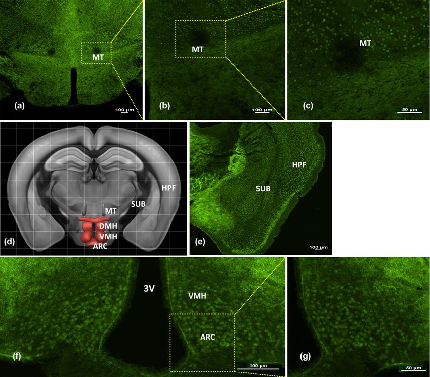
Neuroanatomy of melanocortin-4 receptor 581
neuronal populations, besides the PVN, is important in mouse brains were removed, then postfixed in the same
body weight regulation and may be involved in altering fixative overnight at 4°C, followed by cryoprotection in
energy expenditure as well as food intake. 30% sucrose solution at 4°C before being processed for
MC4R-green fluorescent protein (GFP) transgenic immunocytochemistry. Brains (n = 3) were subsequently
mice have recently been used to identify the MC4R sliced as 30 µm sections, encompassing the hypothalamus,
distribution. However, through this approach, the recog- with a cryostat microtome (Slee MNT, Mainz, Germany).
nition of MC4R expression is based on the MC4R-GFP The sections were collected into four serially ordered
specific fluorescent signal; this, along with a high cost, sets of sections in 0.01 M phosphate-buffered saline (PBS).
made this approach unsuited for public laboratories. After an initial blocking step (3% normal donkey serum in
Moreover, although MC4R-expressing PVN neurons pro- 0.01 M PBS containing 1% Triton X-100), the sections were
mote satiety by projecting to and activating neurons in incubated with primary antibodies F rabbit anti-MC4R
the NTS [10], the MC4R distribution pattern in the CNS (1:1,000, ab24233, Abcam) overnight at 4°C. Next, the
has not been explored in detail. Furthermore, neuroana- sections were washed and incubated with Alexa Fluor 647
tomical MC4R studies have been limited by the avail- Donkey anti-Rabbit IgG (1:1,000, ab150075, Abcam) for 2 h at
ability of the MC4R-specific antibodies for the brain, about 25°C. After washes, the sections were mounted on
whereas few studies have shown messenger RNA levels. frosted gelatin-coated slide and air-dried. All immunofluor-
Exploring MC4R expression in regions other than the escence experiments used the free-floating method under
PVN will assist the research on energy homeostasis 300 rpm wobbling with an Eppendorf ThermoMixer C.
regulation. Accordingly, this study investigates the As a negative control, we used the same samples and the
expression pattern of MC4R neurons and protein levels same protocol without primary antibody. Fluorescent
in the CNS using specific antibody labeling. neuronal cells were visualized and imaged using a Nikon
C2 confocal microscope (Japan) with 4× and 10× objectives.
2 Materials and methods
2.3 Western blot
2.1 Animal husbandry
With the remaining animals, those not used for immuno-
fluorescence, brains were removed into ice-cold 0.9%
C57/BL6j wild-type male mice (WT, 8–9 weeks old) were saline and sliced into 200 µm sections (bregma: −4.84 to
obtained from Charles River Laboratories (Beijing, China). −7.48 mm) with a vibrating microtome. PVN, VMH, ARC,
The mice (n = 5) were maintained in a normal environment PBN, NTS, AP, and raphe pallidus (RPa) tissues (n = 2)
on a 12 h light/dark cycle (lights on 06:00–18:00) with were collected by capillary glass tube with an aperture of
ad libitum access to food and water and were fed a 2 mm under stereomicroscope.
standard diet. Total protein was extracted for the expression of
MC4R. Specifically, brain tissue sections were cut into
Ethical approval: The research related to animal use has small pieces and powdered with liquid nitrogen, placed
been complied with all the relevant national regulations into a homogenization solution containing ice-cold RIPA
and institutional policies for the care and use of animals, (2333s, Beijing, China) and protease inhibitors (complete
and has been approved by the Institutional Animal Care EDTA-free protease inhibitor cocktail; Roche Diagnostics,
and Use Committee (IACUC) of the Inner Mongolia Alameda, CA), and then briefly sonicated (15 s/time,
University, in compliance with NIH guidelines (Protocol 90 V, 3 times). Protein concentrations were determined
number – SYCK-002, approved in July 2014). by BCA protein assay (Thermo Scientific, MA, USA), and
approximately 30 µg of total protein was electrophoresed
through a 12% running and a 5% stacking SDS-PAGE and
wet transferred to a nitrocellulose transfer membrane
2.2 Immunofluorescence (66485, Pall). Membranes were blocked with 5% nonfat
dry milk and then incubated with primary antibodies and
Animals were deeply anesthetized and transcardially the corresponding secondary antibodies: rabbit anti-
perfused with ice-cold 0.9% saline, followed by 4% MC4R (1:1,000, ab24233, Abcam) and rabbit anti-GAPDH
paraformaldehyde-borate fixative (pH 7.4) for 20 min. The (1:10,000, ab181602, Abcam) overnight at 4°C. The
582 Kun Wang et al.
membranes were subsequently washed followed by neurons exited the CNS, suggesting that MC4R may be
incubation with the corresponding IRDye® 800LT essential in regulating processes of CNS.
Goat anti-Rabbit (1:10,000; 926-32211, Odyssey) for 1 h
at about 25°C. Detection and quantification were per-
formed using an Infrared Imaging System (Odyssey; LI-
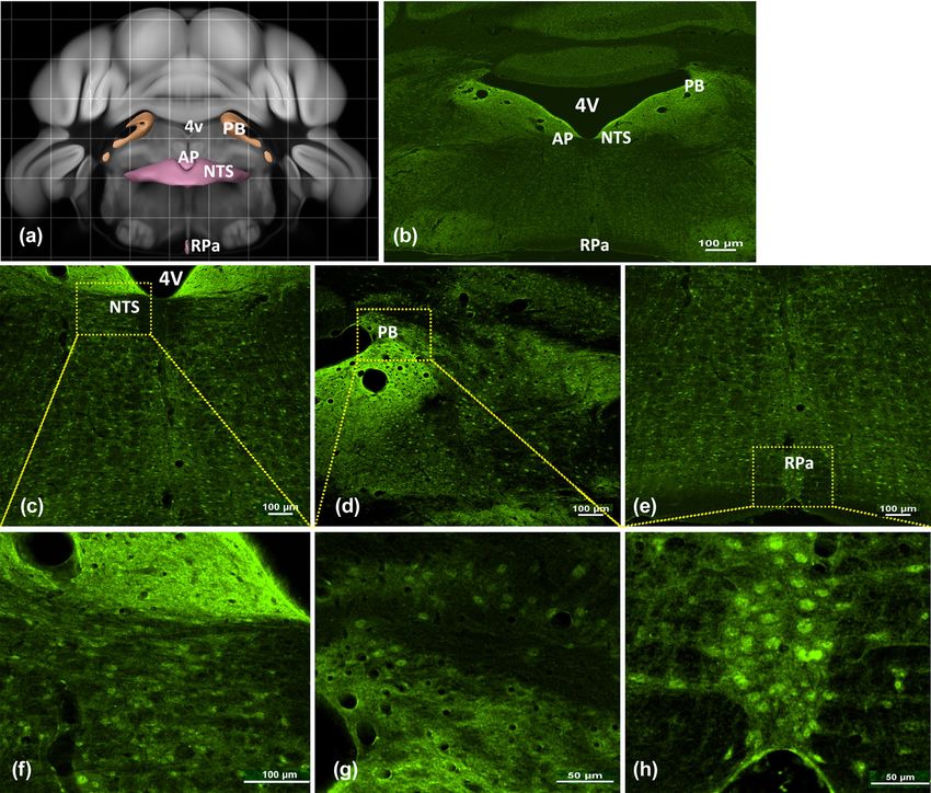
COR Biosciences). Band intensities were determined 3.2 MC4R expression in the MT, VMH,
using the median background method. The MC4R levels and ARC
were normalized to GAPDH.
Positive MC4R staining was observed in coronal sections,
as expected. Strong MC4R immunoreactivity was detected
in the medial terminal nucleus layer (Figure 2a–c), and
2.4 Statistics MC4R was homogenously expressed around the third
ventricle (3V) throughout all areas of the forebrain. In the
Data analysis was done using Prism 7.0 software subiculum, stellate-like distributions of MC4R immuno-
(GraphPad, San Diego, CA, USA). Data are presented as reactivity were observed throughout, suggesting that
means ± standard errors of the means. Comparisons these nerve cells expressed MC4R. Similarly, MC4R
between the expression levels of MC4R in different staining was intense around the hippocampal formation
tissues were made by repeated-measures ANOVA. (Figure 2e); immunopositive neurons were largely con-
Results with P values less than 0.05 were considered fined to ARC areas (Figure 2f and g). Throughout the
statistically significant. hypothalamus, a structure central to neuroendocrine
function, MC4R immunoreactivity was evident in the
ventromedial hypothalamic nucleus (VMH) and ARC.
3 Results
3.3 MC4R expression in the NTS, PB,
3.1 MC4R expression in the DG-mo, CB,
and RPa
hypothalamus, and MY
Within rhombencephalon regions, MC4R expression was
In sagittal brain sections, a very high density of MC4R- prominent in cells surrounding the fourth ventricle (4V)
labeled neurons was detected in the dentate gyrus (Figure 3b). MC4R labeling was detected in the NTS (Figure
molecular layer (DG-mo; Figure 1a). Other areas of the 3c and f) with robust expression surrounding the 4 V.
brain showed moderate signal intensity. The cerebellum Notably, intense nerve cell MC4R immunoreactivity was
(CB; Figure 1b) also showed a very strong MC4R seen throughout the parabrachial nucleus (PBN) (Figure 3d
expression. Intense MC4R immunoreactivity was found and g). Furthermore, compared with other hindbrain
along the medulla (Figure 1c), an area influencing blood regions, a large distribution was observed in the RPa
pressure and respiration. Within the region next to the (Figure 3e and h). MC4R staining was not intense in the
medulla, homogenous MC4R staining was found on the area postrema, but was evident in a region likely involved
cervical enlargement region (Figure 1d) and hypotha- in energy intake control. The present data show intense
lamus (Figure 1h and i). Labeling was also detected MC4R immunoreactivity throughout the hindbrain.
within the rhombencephalon, specifically the medulla
oblongata (MY; Figure 1j). As expected, we did not
observe any staining with the negative control
(Figure 1g). 3.4 MC4R protein expression levels in key
Compared with other hindbrain regions, an abun- nuclei of energy metabolism
dant MC4R-positive signal was observed in the unique
architecture of the IML (Figure 1k) that transmits via MC4R immunoreactivity was detected in the ARC (Figure 4a)
parasympathetic preganglionic fibers. MC4R signaling and dorsal root ganglion in the same manner as previously
neurons were observed in both the medulla and spinal reported with MC4R-GFP [11,12]. Protein was isolated from
cord regions. Overall, projections of MC4R-expressing the PVN, VMH, ARC, PBN, NTS, AP, and RPa and separated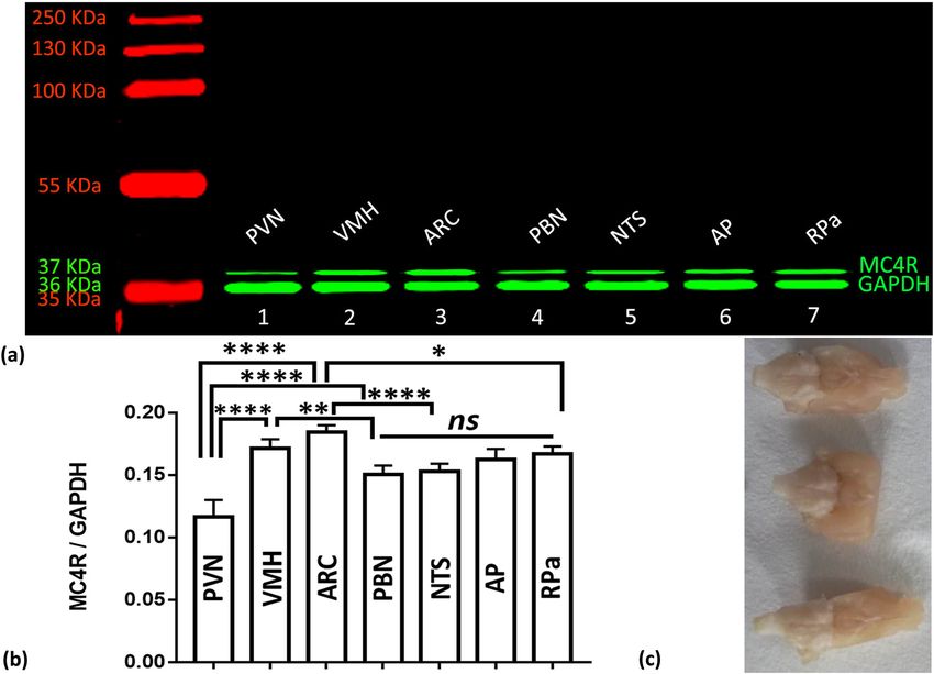
Neuroanatomy of melanocortin-4 receptor 583
Figure 1: MC4R expression in the mouse CNS. Representative images of sagittal mouse brain sections showing MC4R (green) expression by
immunofluorescence. (a) MC4R signal-labeled neurons were detected in the DG-mo. (b) The CB showed MC4R-positive cells. (c) MC4R labeling
in the medulla oblongata is shown. (d) MC4R labeling in the cervical enlargement is shown. (e) Sagittal left-to-right views of the whole brain are
shown. (f) The Brain Map–Brain Explorer 2 from the Allen Institute for Brain Science. (g) Negative control. (h and i) MC4R expression in the
hypothalamus is shown. (j) MC4R expression in the MY is shown. (k) MC4R expression in the IML is shown. Abbreviations: CB, cerebellum;
DG-mo, dentate gyrus molecular layer; IML, intermediolateral nucleus; MC4R, melanocortin-4 receptor; MY, medulla. Scale bars: 100 µm.
by SDS-PAGE (Figure 4c). The highest MC4R protein addition to many other CNS regions. Several extra-
expression was observed in the ARC and VMH, with a hypothalamic areas showed considerable MC4R expres-
significantly lower expression in the PBN and NTS; the sion, including the hippocampus, cerebral cortex, CB,
lowest amount was observed in the PVN. There was no DG-mo, MY, as well as several brainstem and spinal cord
difference in protein expression between the AP and RPa. nuclei. Overall, this study provides a foundation for
A significant difference was observed between the exploring the neurochemical phenotype of central
MC4R protein levels in the ARC and those in the AP, RPa melanocortinergic neurons.
(P = 0.0037 and 0.0030, respectively), and PVN (P < MC4R expression is located in the PVN [14] and ARC
0.0001). The VMH, compared with the PBN (P = 0.0070) [3] regions in mice, regulating energy metabolism in
and NTS (P = 0.0239), demonstrated significantly higher different ways [15]. Specifically, MC4R is a critical factor
MC4R levels than the PVN (P < 0.0001) and the AP and for maintaining hypothalamic appetite regulation [16].
RPa (P = 0.9778). No significant differences were Furthermore, the MC4R expression pattern also suggests
observed between the NTS and PBN (P = 0.9987), or a specialized role in energy homeostasis regulation. For
AP and RPa (P = 0.9778, Figure 4b). instance, MC4R knockout mice lack the ability to
properly maintain energy homeostasis and, for this
reason, may be more prone to obesity [11].
4 Discussion The hypothalamus is a critical structure for control-
ling food intake and energy expenditure [17], implicating
Here, we describe that the MC4Rs are expressed in the the melanocortin system as a signaling pathway that
VMH and ARC region, as previously reported [13], in drives energy homeostasis. The melanocortin system is584 Kun Wang et al. Figure 2: MC4R expression in the mouse forebrain. A picture collage of the representative coronal mouse brain sections showing the MC4R distribution within forebrain regions. (a) MC4R expression in the MT is shown. (d) The Brain Map–Brain Explorer 2 from the Allen Institute for Brain Science. (e) MC4R expression in the SUB and HPF is shown. (f) MC4R expression in ARC of the hypothalamus is shown. (b, c and g) Higher magnification of the left images is shown. Abbreviations: ARC, arcuate nucleus; DMH, dorsomedial hypothalamic; HPF, hippocampal formation; MC4R, melanocortin-4 receptor; MT, medial terminal nucleus of the accessory optic tract; SUB, subiculum; VMH, ventromedial hypothalamic nucleus; 3 V, third ventricle. Scale bars: 50 or 100 µm. involved in amylin-induced food intake suppression and energy metabolism. These data, together with recent thermogenesis activation in both the hypothalamus and evidence indicating that MC4R modulates BAT thermo- brown adipose tissue (BAT) via modulation of acetyl-CoA genic activity during energy expenditure in the hypotha- carboxylase phosphorylation and uncoupling protein-1 lamus, suggest that MC4R is essential for energy expression [3]. The current findings agree with previous metabolism [3,10]. The evolution of endocrine and results suggesting regional hypothalamic MC4R expres- neuronal MCR-mediated circuits has been shaped by sion, with the highest MC4R levels near the 3V and the coevolution of the MCR gene family, the proopiome- posterior brain regions [18]. Furthermore, marked MC4R lanocortin (POMC) gene, and genes that code for downregulation was observed in an animal model of polypeptides that interact with the receptors as reverse obesity [11], in agreement with the phenotype of the agonists (i.e., agouti gene-related protein [AGRP]). AGRP MC4R knockout mice [12], suggesting a role for MC4R in [19], neuropeptide Y [20], and POMC act on MC4R [21]
Neuroanatomy of melanocortin-4 receptor 585
Figure 3: MC4R expression in the mouse hindbrain. Representative micrographs showing MC4R expression in the mouse brain in hindbrain
regions. (a) The Brain Map–Brain Explorer 2 from the Allen Institute for Brain Science. (b) The coronal–caudal to rostral view of the
whole brain. (c) MC4R expression in the NTS is shown. (d) MC4R expression in the PB is shown. (e) MC4R expression in the RPa is shown.
(f–h) Higher magnification of the upper images. Abbreviations: AP, area postrema; MC4R, melanocortin-4 receptor; NTS, nucleus of the
solitary tract; PB, parabrachial nucleus; RPa, raphe pallidus nucleus; 4V, fourth ventricle. Scale bar: 50 or 100 µm.
and are involved in mediating energy homeostasis. important regulatory center of neuroendocrine activity and
Hence, the central melanocortin system is implicated in the sympathetic nerve drive, while the hypothalamic ARC is
energy homeostasis with many gene interactions. involved in anterior pituitary endocrine function regulation,
Here, Western blotting and immunohistochemistry many metabolic processes, and complex behaviors. Robust
results elucidated the MC4R expression pattern throughout staining indicated a marked MC4R expression in the
the mouse brain, providing a comparatively comprehensive hippocampus, ARC, PBN, NTS, RPa, and AP; expression
neuroanatomical study of MC4R localization and protein was also found in the VMH, DMH, and IML, but very little
levels. Notably, a region-specific MC4R expression pattern immunoreactivity was found medial to the PVN [22] and
was demonstrated. Comparable to other studies, the MC4R brainstem. These patterns agree with a previous study [3,23].
expression was highly centralized with intense ARC im- Importantly, MC4R expression was particularly high in the
munoreactivity. Similarly, immunoreactivity was found in ARC, a structure hypothesized to be important in metabolism
the PVN [15], terminating around the 3V. The PVN is an and other functions related to energy consumption [24]. The586 Kun Wang et al.
Figure 4: MC4R protein expression in the mouse brain. Western blot analysis results of various brain regions. (a) A representative blot of
MC4R monomers (37 kDa, upper band) and GAPDH (36 kDa, lower band) proteins in the PVN, VMH, ARC, PBN, NTS, AP, and RPa of the
mouse brain is shown. (b) MC4R band intensities were scanned, quantified, and normalized to the corresponding GAPDH level.
(c) Materials of brain for Western blotting. *indicates P < 0.05; **indicates P < 0.01; ***indicates P < 0.001; ****indicates P < 0.0001; and
ns indicates no significance when compared with the brain regions. Abbreviations: ARC, arcuate nucleus; AP, area postrema; GAPDH,
glyceraldehyde-3-phosphate dehydrogenase; MC4R, melanocortin-4 receptor; NTS, nucleus of the solitary tract; PBN, parabrachial
nucleus; PVN, paraventricular nucleus; RPa, raphe pallidus nucleus; VMH, ventromedial hypothalamus.
heterogeneous MC4R expression pattern throughout the expressed in the dense nerve network throughout the
brain suggests various functional roles for MC4R, in addition brain. Furthermore, MC4Rs demonstrated region-specific
to its role in homeostasis similar to findings in the MC4R-GFP results within individual structures. Thus, by investi-
mouse reports [23,25]. Our study agrees with and expands on gating the local relationship between MC4R expression
previously published results, providing detailed data on the and specific anatomical regions, a detailed distribution
distribution of MC4R in the CNS. and morphological characterization of MC4R protein in
the brain were provided that could aid further research
into its function.
5 Conclusion Acknowledgments: This work was supported by the
National Natural Science Foundation of China (31660701);
This study comprehensively characterized MC4R distri- Beef Cattle Industry Innovation Team, Hebei Modern
bution and, using direct labeling with specific anti- Agricultural Industry Technology System (HBCT2018130201);
bodies, found brain areas with high expression. MC4Rs and the Innovation project of Hebei Academy of Agricultural
were densely expressed in metabolism-processing re- and Forestry Sciences (2019-4-4-3).
gions surrounding the 3V and in brain regions associated
with the hypothalamus, including the ventricles, mid- Author contributions: K. W., W. M., X. Z., and Y. Z.
brain and hindbrain regions. Regionally, MC4R protein designed and oversaw the study, data collection, and analysis
expression was highest in the ARC, where it was strongly and drafted the manuscript. K. F., D. P., H. L., P. L., and R. H.Neuroanatomy of melanocortin-4 receptor 587
contributed to the study design and data analysis. C. D. is the [12] Huszar D, Lynch CA, Fairchild-Huntress V, Dunmore JH,
guarantor of this work and takes responsibility for the Fang Q, Berkemeier LR, et al. Targeted disruption of the
integrity of the data and the accuracy of the data analysis. All melanocortin-4 receptor results in obesity in mice. Cell.
1997;88(1):131–41.
authors critically reviewed and approved the final version of
[13] Mountjoy KG, Mortrud MT, Low MJ, Simerly RB, Cone RD.
the manuscript. Localization of the melanocortin-4 receptor (MC4-R) in
neuroendocrine and autonomic control circuits in the brain.
Conflict of interest: The authors state no conflict of Mol Endocrinol. 1994;8(10):1298–308.
interest. [14] Podyma B, Sun H, Wilson EA, Carlson B, Pritikin E, Gavrilova O,
et al. The stimulatory G protein Gsalpha is required in
melanocortin 4 receptor-expressing cells for normal energy
Data availability statement: The datasets generated dur- balance, thermogenesis, and glucose metabolism. J Biol
ing and/or analyzed during the current study are available Chem. 2018;293(28):10993–1005.
from the corresponding author on reasonable request. [15] Rodriguez EM, Blazquez JL, Guerra M. The design of barriers in
the hypothalamus allows the median eminence and the
arcuate nucleus to enjoy private milieus: the former opens to
the portal blood and the latter to the cerebrospinal fluid.
References Peptides. 2010;31(4):757–76.
[16] Michael NJ, Simonds SE, van den Top M, Cowley MA,
[1] Girardet C, Butler AA. Neural melanocortin receptors in obesity Spanswick D. Mitochondrial uncoupling in the melanocortin
and related metabolic disorders. Biochim Biophys Acta. system differentially regulates NPY and POMC neurons to
2014;1842(3):482–94. promote weight-loss. Mol Metab. 2017;6(10):1103–12.
[2] Baldini G, Phelan KD. The melanocortin pathway and control of [17] Fan K, Li Q, Pan D, Liu H, Li P, Hai R, et al. Effects of amylin on
appetite-progress and therapeutic implications. J Endocrinol. food intake and body weight via sympathetic innervation of
2019;241(1):R1–33. the interscapular brown adipose tissue. Nutr Neurosci.
[3] Li X, Fan K, Li Q, Pan D, Hai R, Du C. Melanocortin 4 receptor- 2020;25:1–13.
mediated effects of amylin on thermogenesis and regulation of [18] Morgan DA, Mcdaniel LN, Yin T, Khan M, Jiang J, Acevedo MR,
food intake. Diabetes/Metabolism Res Rev. 2019;35(5):e3149. et al. Regulation of glucose tolerance and sympathetic activity
[4] Ghamari-Langroudi M, Digby GJ, Sebag JA, Millhauser GL, by MC4R signaling in the lateral hypothalamus. Diabetes.
Palomino R, Matthews R, et al. G-protein-independent coupling of 2015;64(6):1976–87.
MC4R to Kir7.1 in hypothalamic neurons. Nature. [19] Liu Y, Huang Y, Liu T, Wu H, Cui H, Gautron L.
2015;520(7545):94–8. Lipopolysacharide rapidly and completely suppresses AgRP
[5] Yeo GS, Farooqi IS, Challis BG, Jackson RS, O’Rahilly S. The neuron-mediated food intake in male mice. Endocrinology.
role of melanocortin signalling in the control of body weight: 2016;157(6):2380–92.
evidence from human and murine genetic models. QJM. [20] Vella KR, Ramadoss P, Lam FS, Harris JC, Ye FD, Same PD, et al.
2000;93(1):7–14. NPY and MC4R signaling regulate thyroid hormone levels
[6] Do CJ, Da SA, Rushing JS, Pace B, Hall JE. Differential control of during fasting through both central and peripheral pathways.
metabolic and cardiovascular functions by melanocortin-4 Cell Metab. 2011;14(6):780–90.
receptors in proopiomelanocortin neurons. Am J Physiol Regul [21] Balthasar N, Dalgaard LT, Lee CE, Yu J, Funahashi H,
Integr Comp Physiol. 2013;305(4):R359–68. Williams T, et al. Divergence of melanocortin pathways in the
[7] Dores RM, Londraville RL, Prokop J, Davis P, Dewey N, control of food intake and energy expenditure. Cell.
Lesinski N. Molecular evolution of GPCRs: melanocortin/ 2005;123(3):493–505.
melanocortin receptors. J Mol Endocrinol. 2014;52(3):T29–42. [22] Pan D, Fan K, Li Q, Liu H, Li P, Hai R, et al. Response of the
[8] Hill JW, Faulkner LD. The role of the melanocortin system in expression of oxytocin neurons to ghrelin in female mice. Exp
metabolic disease: new developments and advances. Brain Res. 2020;238:1085–95.
Neuroendocrinology. 2017;104(4):330–46. [23] Cawley NX, Yanik T, Woronowicz A, Chang W, Marini JC,
[9] Cui H, Sohn JW, Gautron L, Funahashi H, Williams KW, Loh YP. Obese carboxypeptidase E knockout mice exhibit
Elmquist JK, et al. Neuroanatomy of melanocortin-4 receptor multiple defects in peptide hormone processing contributing
pathway in the lateral hypothalamic area. J Comp Neurol. to low bone mineral density. Am J Physiol Endocrinol Metab.
2012;520(18):4168–83. 2010;299(2):E189–97.
[10] Shah BP, Vong L, Olson DP, Koda S, Krashes MJ, Ye C, et al. [24] Rossi J, Balthasar N, Olson D, Scott M, Berglund E, Lee CE,
MC4R-expressing glutamatergic neurons in the paraventri- et al. Melanocortin-4 receptors expressed by cholinergic
cular hypothalamus regulate feeding and are synaptically neurons regulate energy balance and glucose homeostasis.
connected to the parabrachial nucleus. Proc Natl Acad Sci Cell Metab. 2011;13(2):195–204.
USA. 2014;111(36):13193–8. [25] Gautron L, Lee CE, Lee S, Elmquist JK. Melanocortin-4 receptor
[11] Wang T, Takikawa Y, Satoh T, Yoshioka Y, Kosaka K, expression in different classes of spinal and vagal primary
Tatemichi Y, et al. Carnosic acid prevents obesity and hepatic afferent neurons in the mouse. J Comp Neurol.
steatosis in ob/ob mice. Hepatol Res. 2011;41(1):87–92. 2012;520(17):3933–48.You can also read