Hypolipogenic Effect of Shikimic Acid Via Inhibition of MID1IP1 and Phosphorylation of AMPK/ACC - MDPI
←
→
Page content transcription
If your browser does not render page correctly, please read the page content below
International Journal of
           Molecular Sciences

Article
Hypolipogenic Effect of Shikimic Acid Via Inhibition
of MID1IP1 and Phosphorylation of AMPK/ACC
Moon Joon Kim † , Deok Yong Sim † , Hye Min Lee , Hyo-Jung Lee and Sung-Hoon Kim *
 College of Korean Medicine, Kyung Hee University, Seoul 02447, Korea; pigcross@naver.com (M.J.K.);
 simdy0821@naver.com (D.Y.S.); glansy555@gmail.com (H.M.L.); hyonice77@naver.com (H.-J.L.)
 * Correspondence: sungkim7@khu.ac.kr; Tel.: +82-2-961-9233
 † These authors contributed equally to this work.

 Received: 4 January 2019; Accepted: 28 January 2019; Published: 29 January 2019

 Abstract: Although shikimic acid from Illicium verum has antioxidant, antibacterial, anti-inflammatory,
 and analgesic effects, the effect of shikimic acid on lipogenesis has not yet been explored. Thus,
 in the present study, hypolipogenic mechanism of shikimic acid was examined in HepG2, Huh7 and
 3T3-L1 adipocyte cells. Shikimic acid showed weak cytotoxicity in HepG2, Huh7 and 3T3-L1 cells,
 but suppressed lipid accumulation in HepG2, Huh7 and 3T3-L1 cells by Oil Red O staining. Also,
 shikimic acid attenuated the mRNA expression of de novo lipogenesis related genes such as FAS,
 SREBP-1c, and LXR-α in HepG2 cells by RT-PCR analysis and suppressed the protein expression
 of SREBP-1c and LXR-α in HepG2 and 3T3-L1 cells. It should be noted that shikimic acid activated
 phosphorylation of AMP-activated protein kinase (AMPK)/Aacetyl-coenzyme A carboxylase (ACC)
 and reduced the expression of MID1 Interacting Protein 1 (MID1IP1) in HepG2, Huh7 and 3T3-L1
 cells. Conversely, depletion of MID1IP1 activated phosphorylation of AMPK, while overexpression
 of MID1IP1 suppressed phosphorylation of AMPK in HepG2 cells. However, AMPK inhibitor
 compound c did not affect the expression of MID1IP1, indicating MID1IP1 as an upstream of AMPK.
 Taken together, our findings suggest that shikimic acid has hypolipogenic effect in HepG2 and
 3T3-L1 cells via phosphorylation of AMPK/ACC and inhibition of MID1IP1 as a potent candidate for
 prevention or treatment of fatty liver and hyperlipidemia.

 Keywords: shikimic acid; MID1IP1; AMPK; hepatocellular carcinoma (HCC); 3T3-L1; lipogenesis

1. Introduction
     Fatty liver disease is caused by excessive fat accumulation, leading to progressive liver fibrosis
and cirrhosis with features of metabolic syndrome including insulin resistance [1,2]. Excessive intake of
alcohol or fatty food can induce alcoholic or nonalcoholic fatty liver diseases (NAFLD) [3] by promoting
de novo fatty acid synthesis through downregulation of AMP-activated protein kinase (AMPK),
an important hepatic transcriptional regulator, and then its downstream acetyl CoA carboxylase
(ACC) [4,5].
     It is well documented that De Novo lipogenesis (DNL) can induce hepatic steatosis and/or
hypertriglyceridemia, and also cause steatohepatitis by saturated fatty acids including palmitate [6].
Also, DNL is regulated mainly by two key transcription factors such as sterol regulatory
element-binding protein 1c (SREBP1c), which is activated by insulin and liver X receptor α (LXR-α),
and carbohydrate regulatory element-binding protein (ChREBP) [7,8].
     The MID1 Interacting Protein 1 (MID1IP1) also known as MIG12 or S14 has been implicated in
lipogenesis in mammals [9]. Hence, Kim et al. [10] reported that MID1IP1 regulates and binds to
acetyl-CoA carboxylase (ACC), the first committed enzyme in fatty acid (FA) synthesis, and induces

Int. J. Mol. Sci. 2019, 20, 582; doi:10.3390/ijms20030582                          www.mdpi.com/journal/ijms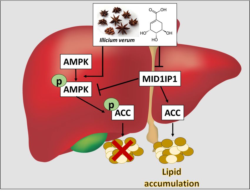
Int. J. Mol. Sci. 2018, 19, x FOR PEER REVIEW 2 of 11
Int. J. Mol. Sci. 2019, 20, 582 2 of 11
acetyl‐CoA carboxylase (ACC), the first committed enzyme in fatty acid (FA) synthesis, and induces
polymerizationduring
ACC polymerization duringincreased
increasedhaptic
haptic FAFA synthesis.
synthesis. Furthermore,
Furthermore, Inoue
Inoue et al.et[11]
al. [11] claimed
claimed that
that MID1IP1
MID1IP1 regulates
regulates LXRLXR ligand
ligand and and glucose,
glucose, resulting
resulting in triglyceride
in triglyceride accumulation
accumulation andandfattyfatty liver.
liver.
Since shikimic acid is a natural compound isolated from the Japanese plant, plant, Illicium
Illicium verum
verum [12],
[12],
and seeds
seeds of of Liquidambar
Liquidambarstyraciflua
styraciflua(sweetgum)
(sweetgum) abundant
abundant in inNorth America
North [13][13]
America and and
Chinese star
Chinese
aniseanise
star (Illicium verum),verum),
(Illicium shikimicshikimic
acid hasacid
been has
usedbeen
as a base
usedmaterial for production
as a base material for of production
oseltamivir
(Tamiflu)
of [14]. Also,
oseltamivir though
(Tamiflu) shikimic
[14]. Also, acid is known
though to have
shikimic acidanti‐diabetic
is known [15], antibacterial
to have [16], anti‐
anti-diabetic [15],
inflammatory[16],
antibacterial [17], analgesic [18],[17],
anti-inflammatory antioxidant [18],antioxidant
analgesic [18], and anti‐thrombogenic [19] effects, [19]
[18], and anti-thrombogenic its
hypolipogenic mechanism has never been reported. Thus, in the present study,
effects, its hypolipogenic mechanism has never been reported. Thus, in the present study, hypolipogenic hypolipogenic
mechanism of shikimic acid was elucidated in HepG2 and Huh7 hepatocellular carcinoma HCC cells
3T3‐L1 adipocytes in association with AMPK/ACC
and 3T3-L1 AMPK/ACC and and MID1IP1
MID1IP1 signaling
signaling axis.
axis.
2. Results
2.1. Shikimic Acid
2.1. Shikimic Acid Exerted
Exerted Weak
Weak Cytotoxicity
Cytotoxicity in
in HepG2
HepG2 and
and Huh7
Huh7 Cells
Cells and
and 3T3-L1
3T3‐L1 Cells
Cells
The
The cytotoxicity
cytotoxicity of
ofshikimic
shikimicacid
acid(Figure
(Figure1b,c)
1b,c)was
wasevaluated
evaluatedininHepG2
HepG2 and Huh7
and Huh7 cells and
cells and3T3-L1
3T3‐
cells by MTT assay after the cells were treated with various concentrations of shikimic acid
L1 cells by MTT assay after the cells were treated with various concentrations of shikimic acid (0, (0, 10,
10,
20,
20, 40,
40, 80,
80, 160 μM). As
160 µM). As shown
shown inin Figure
Figure 1b,c,
1b andthec,viability of HepG2
the viability and Huh7
of HepG2 or 3T3-L1
and Huh7 cells cells
or 3T3‐L1 was
maintained up to 70% of untreated control even at the concentration of 160
was maintained up to 70% of untreated control even at the concentration of 160 μM.µM.
Figure
Figure 1. Chemicalstructure
1. Chemical structureofofshikimic
shikimic acid
acid and
and its its effect
effect on on cytotoxicity
cytotoxicity in HepG2,
in HepG2, Huh7Huh7
and and
3T3
3T3 cells. (a) Chemical structure of shikimic acid. (b) Cytotoxicity of shikimic acid in
cells. (a) Chemical structure of shikimic acid. (b) Cytotoxicity of shikimic acid in HepG2 and Huh7HepG2 and
Huh7 cells.
cells. The The
cells cellstreated
were were treated with shikimic
with shikimic acid foracid
24 hfor 24cytotoxicity
and h and cytotoxicity was evaluated
was evaluated by
by 3‐(4,5‐
3-(4,5-dimethylthiazol-2-yl)-2,5-diphenyltetrazolium bromide MTT assay (c) Cytotoxicity
dimethylthiazol‐2‐yl)‐2,5‐diphenyltetrazolium bromide MTT assay (c) Cytotoxicity of shikimic of shikimic
acid in 3T3-L1 cells. Results represent means ± S.D. from three independent experiments. * p < 0.05,
acid in 3T3‐L1 cells. Results represent means ± S.D. from three independent experiments. * p < 0.05, **
** p < 0.01 versus control.
p < 0.01 versus control.
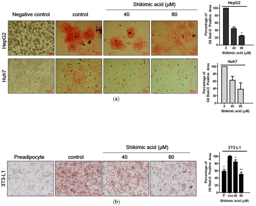
2.2. Shikimic Acid Reduced the Number of Lipid Droplets in HCCs
2.2. Shikimic Acid Reduced the Number of Lipid Droplets in HCCs
To confirm the hypolipidemic effect of shikimic acid, Oil Red O staining was conducted in
To confirm
shikimic the hypolipidemic
acid-treated effect in
HCC cells. As shown of Figure
shikimic
2a, acid, Oil Red were
lipid droplets O staining was conducted
significantly decreased in
in
shikimic acid‐treated HCC cells. As shown in Figure 2a, lipid droplets were significantly decreased
a concentration-dependent manner in HepG2 and Huh7 cells by shikimic acid. Similarly, shikimic acid
in a concentration‐dependent
reduced lipid accumulation inmanner in HepG2 and
3T3-L1 adipocytes Huh7
as well cells by
(Figure shikimic acid. Similarly, shikimic
2b).
acid reduced lipid accumulation in 3T3‐L1 adipocytes as well (Figure 2b).Int. J. Mol. Sci. 2019, 20, 582 3 of 11
Int. J. Mol. Sci. 2018, 19, x FOR PEER REVIEW 3 of 11
Figure 2. Effect of shikimic acid on lipid accumulation by Oil Red O staining in HepG2 and 3T3-L1 cells.
Figure 2. Effect of shikimic acid on lipid accumulation by Oil Red O staining in HepG2 and 3T3‐L1
(a) Effect of shikimic acid on lipid accumulation in HepG2 cells by Oil red staining. Scale bar = 200 µm.
cells. (a) Effect of shikimic acid on lipid accumulation in HepG2 cells by Oil red staining. Scale bar =
(b) Effect of shikimic acid on lipid accumulation in 3T3-L1 cells. Shikimic acid was treated for 24 h in
200 μm. (b) Effect of shikimic acid on lipid accumulation in 3T3‐L1 cells. Shikimic acid was treated
HCCs and 3T3-L1 cells. Scale bar = 100 µm. P: Preadipocyte. All experiments were independently
for 24 h in HCCs and 3T3‐L1 cells. Scale bar = 100 μm. P: Preadipocyte. All experiments were
performed at least three times. * p < 0.05, ** p < 0.01.
independently performed at least three times. * pInt. J.
Int. J. Mol.
Mol. Sci.
Sci. 2018, 20, x582
2019, 19, FOR PEER REVIEW 44 of
of 11
11
86
Figure 3. Effect of MID1PI1 depletion on proliferation and lipogenesis-related genes. (a) Expression
87 Figure 3. Effect of MID1PI1 depletion on proliferation and lipogenesis-related genes. (a) Expression
level of MID1IP1 in different cell lines. β-actin was used as loading control. (b) Depletion level of
88 level of MID1IP1 in different cell lines. β-actin was used as loading control. (b) Depletion level of
MID1IP1 for 48 h in HepG2 cells by qRT-PCR. (c) Effect of MID1PI1 depletion on proliferation in HepG2
89 MID1IP1 for 48
cells by MTT h in(d,e)
assay. HepG2 cells
Effect of by qRT-PCR.
MID1PI1 (c) Effect
depletion of MID1PI1
on the depletion
mRNA level on proliferation
of SREBP-1c and FAS inin
90 HepG2 cells by
HepG2 cells byRT-qPCR
MTT assay. (d,e) Effect
analysis. of MID1PI1
All experiments depletion
were on the mRNA
independently (e) atof
level
performed SREBP-1c
least and
three times.
91 FAS in HepG2 cells by RT-qPCR analysis. All experiments were independently performed at least
92 2.4. Shikimic Acid Downregulated MID1IP1 Expression Level by Phosphorylation of AMPK in HCCs
three times.
and Adipocytes
93 2.4. Shikimic Acidexamine
To further Downregulated MID1IP1 Expression
the hypolipogenic effect ofLevel by Phosphorylation
shikimic acid, westernofblot
AMPKwasinconducted
HCCs and to
94 Adipocytes
estimate the expression level of lipogenesis-related proteins such as p-AMPKα, AMPKα, p-ACC, ACC,
95 MID1IP1, LXR-αexamine
To further and SREBP-1c in HepG2 cells,
the hypolipogenic Huh7
effect cells and acid,
of shikimic 3T3-L1 adipocytes
western blot after shikimic acid
was conducted to
96 treatmentthe
estimate forexpression
24 h. Shikimic
level acid reduced the expression
of lipogenesis-related proteinslevel of as
such MID1P1, LXR-α
p-AMPKα, and SREBP-1c.
AMPKα, p-ACC,
97 However,
ACC, shikimic
MID1IP1, acid significantly
LXR-α and SREBP-1c upregulated
in HepG2phosphorylation
cells, Huh7 cells of and
AMPKα andadipocytes
3T3-L1 ACC in HepG2
after
98 cells and acid
shikimic adipocytes (Figure
treatment 4a,b).
for 24 h. Shikimic acid reduced the expression level of MID1P1, LXR-α and
99 SREBP-1c. However, shikimic acid significantly upregulated phosphorylation of AMPKα and ACC
100 in HepG2 cells and adipocytes (Figure 4a,b).Int. J. Mol. Sci. 2019, 20, 582 5 of 11
Int. J. Mol. Sci. 2018, 19, x FOR PEER REVIEW 5 of 11
Figure 4. Effect of shikimic acid on lipid metabolism related molecules in HCC and 3T3-L1 cells.
Figure 4. Effect of shikimic acid on lipid metabolism related molecules in HCC and 3T3‐L1 cells.
Lipogenesis-related proteins were evaluated by Western blotting after treatment of shikimic acid for
Lipogenesis‐related proteins were evaluated by Western blotting after treatment of shikimic acid for
24 h in HCCs (a) and 3T3-L1 preadipocytes and adipocytes (b). P: Preadipocyte. All experiments were
24 h in HCCs (a) and 3T3‐L1 preadipocytes and adipocytes (b). P: Preadipocyte. All experiments were
independently performed at least three times. * p < 0.05, ** p < 0.01, *** p < 0.001.
independently performed at least three times. * p < 0.05, ** p < 0.01, *** p < 0.001.
2.5. Pivotal Role of MID1IP1 in Shikimic Acid Regulated Lipogenesis in HepG2 and AML-12 Cells
2.5. Pivotal Role of MID1IP1 in Shikimic Acid Regulated Lipogenesis in HepG2 and AML‐12 Cells
To examine the role of MID1IP1 in shikimic acid-regulated lipogenesis-related genes,
To examine
overexpression the role of
or depletion MID1IP1
plasmid in shikimic
of MID1IP1 acid‐regulated
and AMPK lipogenesis‐related
inhibitor compound genes,
c were used in
overexpression
AML-12 and HepG2or depletion plasmid
cells. As shown of MID1IP1
in Figure and AMPK of
5a, overexpression inhibitor compoundphosphorylation
MID1IP1-reduced c were used in
AML‐12
of AMPK by and HepG2
shikimic acidcells. Asin shown
(80 µM) AML-12 incellsFigure
(Figure 5a, overexpression
5b), whereas depletionofof MID1IP1
MID1IP1‐reduced
activated
phosphorylation of of AMPK/ACC
AMPK by shikimic acid (80 μM) in AML‐12 cells (Figure 5b), whereas
in HepG2 cells (Figure 5c). However, AMPK inhibitor compound c depletion
of MID1IP1
did not affectactivated phosphorylation
expression of MID1IP1 in of AMPK/ACC
HepG2 in HepG2
cells (Figure 5d). cells (Figure 5c). However, AMPK
inhibitor compound c did not affect expression of MID1IP1 in HepG2 cells (Figure 5d)Int. J. Mol. Sci. 2019, 20, 582 6 of 11
Int. J. Mol. Sci. 2018, 19, x FOR PEER REVIEW 6 of 11
Figure 5. Close relationship between MID1IP1 and AMPK in HepG2 cells. (a) Overexpression level of
Figure 5. Close relationship between MID1IP1 and AMPK in HepG2 cells. (a) Overexpression level of
MID1IP1 in AML-12 cells. (b) MID1IP1 overexpression for 48 h disturbed phosphorylation of AMPK
MID1IP1 in AML‐12 cells. (b) MID1IP1 overexpression for 48 h disturbed phosphorylation of AMPK
in AML-12 cells. (c) Depletion of MID1IP1 (0, 40, 80 nM) activated phosphorylation of AMPK in
in AML‐12 cells. (c) Depletion of MID1IP1 (0, 40, 80 nM) activated phosphorylation of AMPK in
HepG2 cells. (d) AMPK inhibitor compound c did not affect phosphorylation of AMPK in HepG2 cells.
HepG2 cells. (d) AMPK inhibitor compound c did not affect phosphorylation of AMPK in HepG2
All experiments were independently performed at least three times. * p < 0.05, ** p < 0.01.
cells. All experiments were independently performed at least three times. * p < 0.05, ** p < 0.01.
3. Discussion
3. Discussion
Herein, hypolipogenic mechanism of shikimic acid from I. verum was examined in HCC cells and
3T3-L1Herein, hypolipogenic
adipocyte mechanism
cells. It is well known ofthatshikimic
products acid
by from I. verum
hepatic de novowas examined esterification
lipogenesis, in HCC cells
and
of 3T3‐L1
plasma adipocyte
free fatty acidscells. It is welldietary
or increased known fatthat
intakeproducts by hepatic
are critically involved de in
novo lipogenesis,
development of
esterification
NAFLD [3,20].ofShikimic
plasma free
acid fatty
showedacids or cytotoxicity
weak increased dietary
in HCC fatcells
intake
andare critically
3T3-L1 cells. involved in
To confirm
development of NAFLD [3,20]. Shikimic acid showed weak cytotoxicity in HCC cells and
hypolipogenic effect of shikimic acid, Oil red O staining assay that has been applied for evaluation of 3T3‐L1 cells.
To confirm
liver hypolipogenic
steatosis effect of shikimic
and lipid metabolism [21] wasacid, Oil redin
conducted O lipogenic
staining assay
HepG2,thatweak
has been applied
lipogenic for
Huh7
evaluation
and of liver
adipogenic steatosis
3T3-L1 and lipidacid
cells. Shikimic metabolism
suppressed [21] was
lipid conducted in
accumulation in lipogenic
HepG2 and HepG2,
3T3-L1weak
cells,
lipogenic hypolipogenic
implying Huh7 and adipogenic
potential 3T3‐L1 cells. Shikimic
of shikimic acid. acid suppressed lipid accumulation in HepG2
and 3T3‐L1 cells, implying hypolipogenic potential of shikimic acid.Int. J. Mol. Sci. 2019, 20, 582 7 of 11
Accumulating evidences reveal that SREBP-1c primarily regulates FAS, whereas liver X receptors
(LXR)Int.regulate transcription
J. Mol. Sci. 2018, 19, x FOR PEERofREVIEW
SREBP-1c through LXR response element (LXRE) for cholesterol 7 of 11
homeostasis and lipogenesis [22–24]. Consistently, RT-qPCR analysis showed that shikimic acid
attenuated Accumulating
the mRNA evidences expression reveal
of de that SREBP‐1c
novo primarily regulates
lipogenesis-related genesFAS, suchwhereas
as FAS,liver X
SREBP-1c,
receptors
and LXR-α in (LXR)
HepG2regulate transcription
cells. Likewise, of SREBP‐1c
shikimic through LXR
acid attenuated the response element (LXRE)
protein expression for
of SREBP-1c
cholesterol homeostasis and lipogenesis [22–24]. Consistently, RT‐qPCR analysis showed that
and LXR-α in HepG2 and 3T3-L1 cells, indicating shikimic acid inhibits lipogenesis-related genes both
shikimic acid attenuated the mRNA expression of de novo lipogenesis‐related genes such as FAS,
at mRNA and protein levels.
SREBP‐1c, and LXR‐α in HepG2 cells. Likewise, shikimic acid attenuated the protein expression of
Emerging evidence shows that AMPK, a sensor of cellular energy charge and a “metabolic master
SREBP‐1c and LXR‐α in HepG2 and 3T3‐L1 cells, indicating shikimic acid inhibits lipogenesis‐related
switch”, enhances
genes both at mRNA fatty acid
andoxidation by lowering the concentration of malonyl coenzyme A (malonyl
protein levels.
CoA) andEmerging
also modulatesevidence shows that AMPK,ofa malonyl
the concentration sensor ofCoA by concurrently
cellular energy charge phosphorylating
and a “metabolic and
inhibiting
masteracetyl
switch”, CoA carboxylase
enhances fatty acid(ACC)
oxidationalpha or beta the
by lowering [25,26]. Notably,
concentration shikimiccoenzyme
of malonyl acid activated
A
(malonyl CoA)
phosphorylation and also
of AMPK and itsmodulates
downstream the ACCconcentration
in HepG2,ofHuh7 malonyl CoA by
and 3T3-L1 concurrently
cells, demonstrating
phosphorylating
the critical and inhibiting acetyl CoA carboxylase (ACC) alpha or beta [25,26]. Notably,
role of pAMPK/pACC.
Recent studies revealphosphorylation
shikimic acid activated that MID1IP1,ofknown AMPK as andMIG12
its downstream
or S14, ACC in HepG2,
activates ACC Huh7 and acid
for fatty
3T3‐L1 cells, demonstrating the critical role of pAMPK/pACC.
synthesis and also controls triglyceride accumulation in fatty liver [9,10,27]. Interestingly, shikimic acid
Recent studies reveal that MID1IP1, known as MIG12 or S14, activates ACC for fatty acid
attenuated expression of MID1IP1 in HepG2 cells at mRNA and protein levels, implying antiadipogenic
synthesis and also controls triglyceride accumulation in fatty liver [9,10,27]. Interestingly, shikimic
potential
acid of shikimicexpression
attenuated acid. Conversely,
of MID1IP1knockdown
in HepG2ofcells MID1IP1 activated
at mRNA phosphorylation
and protein of AMPK,
levels, implying
whereas overexpression of MID1IP1 reduced phosphorylation
antiadipogenic potential of shikimic acid. Conversely, knockdown of MID1IP1 activatedof AMPK in HepG2 cells. In contrast,
AMPK inhibitor compound
phosphorylation of AMPK, c did not affect
whereas the expression
overexpression of MID1IP1,
of MID1IP1 reducedindicating that MID1IP1
phosphorylation of AMPK can be
an upstream
in HepG2ofcells. AMPK. Nonetheless,
In contrast, it is still compound
AMPK inhibitor necessary ctodid perform further
not affect experiments
the expression for detailed
of MID1IP1,
indicating
mechanism usingthatIP,MID1IP1
genomecan be an by
editing upstream
way ofof AMPK. Nonetheless,
CRISRP/Caspase9 it is RNA
assay, still necessary to perform
editing methods and in
further experiments for detailed
animal study for future clinical trials. mechanism using IP, genome editing by way of CRISRP/Caspase9
assay, RNA
Overall, ourediting
findings methods
provide andevidence
in animalthat
study for future
shikimic acidclinical
has a trials.
hypolipogenic effect in HepG2 and
Overall, our findings provide evidence that shikimic acid has a hypolipogenic effect in HepG2
3T3-L1 cells via phosphorylation of AMPK/ACC and inhibition of MID1IP1 as a potential candidate
and 3T3‐L1 cells via phosphorylation of AMPK/ACC and inhibition of MID1IP1 as a potential
for prevention or treatment of fatty liver and hyperlipidemia (Figure 6).
candidate for prevention or treatment of fatty liver and hyperlipidemia (Figure 6).
Figure 6. Schematic
Figure diagram
6. Schematic ofof
diagram hypolipogenic
hypolipogenic mechanism
mechanism ofofshikimic
shikimic acid
acid viavia inhibition
inhibition of MID1IP1
of MID1IP1
and phosphorylation
and phosphorylationof AMPK/ACC
of AMPK/ACCin inhepatocellular carcinomas
hepatocellular carcinomas (HCCs).
(HCCs). Black
Black arrow
arrow for activation
for activation
and Tand
barTfor
barinhibition.
for inhibition.
4. Materials andand
4. Materials Methods
Methods
4.1. Reagents, Antibodies
4.1. Reagents, andand
Antibodies Plasmids
Plasmids
Shikimic acid,
Shikimic Oil-red-O
acid, Oil‐red‐Opowder,
powder,SREBP-1c(SREBF1), LXR-αand
SREBP‐1c(SREBF1), LXR‐α and β-actin
β‐actin were
were purchased
purchased from from
Sigma‐Aldrich
Sigma-Aldrich (St.(St. Louis,
Louis, MO,USA).
MO, USA).Shikimic
Shikimic acid
acid was
wasdissolved in in
dissolved distilled water
distilled according
water to to
according
manufacturer’s instruction. Lipofectamine 2000 reagent was purchased from Invitrogen (Carlsbad,
manufacturer’s instruction. Lipofectamine 2000 reagent was purchased from Invitrogen (Carlsbad, CA,Int. J. Mol. Sci. 2019, 20, 582 8 of 11
USA). Roswell Park Memorial Institute (RPMI) 1640, Dulbecco’s modified Eagle’s medium (DMEM),
and fetal bovine serum (FBS) were purchased from Welgene (Daegu, Gyeongsangbuk-do, Korea).
Antibodies of p-AMPKα, AMPKα, p-ACC and ACC were purchased from Cell Signaling Technology
(Beverly, MA, USA). Antibody of MID1IP1 was purchased from Abcam (Abcam, Cambridge,
Cambridgeshire, United Kingdom). Primers for MID1IP1, SREBP-1c, LXR-α and FAS were obtained
from Bioneer (Bioneer, Daejun, Korea). MID1IP1 siRNA and overexpression plasmids were purchased
from Addgene (Addgene, Cambridge, MA, USA).
4.2. Cell Lines and Culture
HepG2 liver hepatocellular cancer (ATCC® HB-8065™) was purchased from the American Type
Culture Collection (ATCC, Manassas, VA, USA). Huh7 liver hepatocellular cancer, AML-12 liver normal
cells and preadipocyte 3T3-L1 cells were obtained from the Korean Cell Line Bank (KCLB, Seoul, Korea).
HepG2 cells, AML-12 and preadipocyte 3T3-L1 cells were cultured in DMEM supplemented with 10%
FBS and 1% antibiotics, and Huh7 cells were maintained in RPMI 1640. All cells were incubated at
37 ◦ C under condition of relative humidity and 5% CO2 .
4.3. Cell Viability Assay
The cytotoxicity of shikimic acid was evaluated in HepG2, Huh7 and 3T3-L1 cells by using
colorimetric 3-(4,5-dimethylthiazol-2-yl)-2,5-diphenyltetrazolium bromide (MTT) assay (Sigma,
St. Louis, MO, USA). Briefly, cells were treated by various concentrations (0, 10, 20, 40, 80, 160 µM) of
shikimic acid for 24 h and then were exposed to MTT (1 mg/mL) for 2 h. Then optical density (OD)
was measured using a microplate reader (Tecan, Switzerland) at a wavelength of 570 nm. Cell viability
was calculated as a percentage of viable cells in a shikimic acid-treated group versus untreated control.
4.4. Adipogenic Differentiation Induction
The preadipocyte 3T3-L1 cells were incubated onto 6-well plates at 0.8 × 10⁵ cells/well in
DMEM supplemented with 10% FBS and 1% antibiotics for two days. To induce differentiation,
3T3-L1 preadipocytes were incubated in DMEM with 1 µM dexamethasone, and 1 µg/mL of insulin,
and 0.5 mM isobutylmethylxanthine (IBMX) (Sigma-Aldrich, St. Louis, MO, USA) for two days and
were replaced by fresh normal medium containing 1 µg/mL of insulin.
4.5. Oil-Red-O Staining
The 3T3-L1 cells were treated with or without shikimic acid (40, 80 µM), fixed with 4%
paraformaldehyde at room temperature for 30 min and washed with distilled water twice and 60%
isopropanol. The cells were stained for 20 min at room temperature by immersion with Oil-Red-O
solution (Sigma-Aldrich, St. Louis, MO, USA) and then were washed with distilled water four times.
The plate was photographed using a camera connected to an Axio observer a1 inverted microscope
(Zeiss, Germany).
4.6. Western Blotting
For protein extraction, HCCs or 3T3-L1 cells treated with or without shikimic acid (40, 80 µM)
were lysed with RIPA lysis buffer (Thermo) with protease inhibitor. Twenty to thirty micrograms
of total protein were separated on SDS-PAGE and electrotransferred to nitrocellulose blotting
membranes (Amersham Biosciences, Buckinghamshire, UK). The membranes were blocked with
3% non-fat dry milk in TBST and probed with antibodies of SREBP-1c, LXR-α, p-AMPKα, AMPKα,
p-ACC, ACC and β-actin at 4 ◦ C. After washing, the membrane was incubated with horseradish
peroxidase (HRP)-conjugated secondary antibodies, and protein expression was examined by enhanced
chemiluminescence (ECL) (GE Health Care Biosciences, Piscataway, NJ, USA).Int. J. Mol. Sci. 2019, 20, 582 9 of 11
4.7. Quantitative Real-Time Polymerase Chain Reaction (qRT-PCR)
The total RNA of cells was isolated from HepG2 cells using QIAZOL lysis reagent (QIAZEN,
Venlo, Netherlands). After synthesis process of cDNA by using M-MLV reverse transcriptase (Promega,
WI, USA), the mRNA levels were measured by qRT-PCR with the light cycler TM instrument (Roche
Applied Sciences, IN, USA) according to manufacturer’s protocol. The mRNA level of GAPDH was
used to normalize the expression of genes of interest. Primers of MID1IP1, SREBP-1c and FAS were
purchased from Bioneer. The sequences of these primers used are as follows (Table 1):
Table 1. Primers used for quantitative real-time PCR (qPCR) in this study.
Sense Antisense
MID1IP1 50 GGC GAC ACC TTT CCT GGA CT30 50 GAT GGC TGA GGG TGC TCT GT30
SREBP-1c 50 CCA TGG ATG CAC TTT CGA A30 50 CCA GCA TAG GGT GGG TCA A30
FAS 50 GCT GCT CCA CGA ACT CAA ACA CCG30 50 CGG TAC GCG ACG GCT GCC TG30
Each sample was tested in triplicates, and relative gene expression data were analyzed by means
of the 2−∆CT method.
4.8. RNA Interference
The AML-12 cells were transfected with MID1IP1 overexpression or siRNA plasmid using
X-treme-transfection reagent (Sigma-Aldrich) according to manufacturer’s protocol for next experiment.
The mixtures of the MID1IP1 overexpression or siRNA plasmid and X-treme-transfection reagent were
incubated for 25 m, and the cells were incubated at 37 ◦ C for 48 h before exposure to 80 µM of shikimic
acid for 24 h.
4.9. Statistical Analysis
The data were expressed as means ± standard deviation (SD) of three replications per experiment.
Analysis of variance (ANOVA) was conducted to determine the significant differences between two
groups. p < 0.05 was considered significant.
5. Conclusions
Shikimic acid has hypolipogenic effect in HepG2 and 3T3-L1 cells via phosphorylation of
AMPK/ACC and inhibition of MID1IP1 as a potent candidate for prevention or treatment of fatty liver
and hyperlipidemia.
Author Contributions: M.J.K. and D.Y.S. designed and performed several experiments. H.M.L. and H.-J.L.
supported experiments. S.-H.K. supervised the experiments and wrote the MS.
Funding: This work was supported by the National Research Foundation of Korea (NRF) Grant funded by the
Korea Government (MEST) (no. 2017R1A2A1A17069297).
Conflicts of Interest: The authors declare no conflict of interest.
Abbreviations
MID1IP1 MIG12, Midline-1-interacting G12-like protein
AMPK Adenosine 30 ,50 -cyclic monophosphate (cAMP)-activated protein kinaseivated protein kinase
SREBP-1c Sterol regulatory element-binding protein 1
FAS Fatty acid synthase
ACC Acetyl-coA carboxylase
LXR-α Liver X receptor alpha
HCC Human Hepatocellular CarcinomaInt. J. Mol. Sci. 2019, 20, 582 10 of 11
References
1. Day, C.P.; James, O.F. Steatohepatitis: A tale of two “hits”? Gastroenterology 1998, 114, 842–845. [CrossRef]
2. Geisler, C.E.; Renquist, B.J. Hepatic lipid accumulation: Cause and consequence of dysregulated
glucoregulatory hormones. J. Endocrinol. 2017, 234, R1–R21. [CrossRef]
3. de Castro, G.S.; Calder, P.C. Non-alcoholic fatty liver disease and its treatment with n-3 polyunsaturated
fatty acids. Clin. Nutr. 2018, 37, 37–55. [CrossRef] [PubMed]
4. Viollet, B.; Guigas, B.; Leclerc, J.; Hebrard, S.; Lantier, L.; Mounier, R.; Andreelli, F.; Foretz, M. AMP-activated
protein kinase in the regulation of hepatic energy metabolism: From physiology to therapeutic perspectives.
Acta Physiol. (Oxf) 2009, 196, 81–98. [CrossRef] [PubMed]
5. Postic, C.; Girard, J. Contribution of de novo fatty acid synthesis to hepatic steatosis and insulin resistance:
Lessons from genetically engineered mice. J. Clin. Investig. 2008, 118, 829–838. [CrossRef] [PubMed]
6. Yue, Y.; Zhang, L.; Zhang, X.; Li, X.; Yu, H. De novo lipogenesis and desaturation of fatty acids during
adipogenesis in bovine adipose-derived mesenchymal stem cells. In Vitro Cell. Dev. Biol. Anim. 2018, 54,
23–31. [CrossRef] [PubMed]
7. You, M.; Fischer, M.; Deeg, M.A.; Crabb, D.W. Ethanol induces fatty acid synthesis pathways by activation of
sterol regulatory element-binding protein (SREBP). J. Biol. Chem. 2002, 277, 29342–29347. [CrossRef]
8. Wang, L.; Zhang, T.; Wang, L.; Cai, Y.; Zhong, X.; He, X.; Hu, L.; Tian, S.; Wu, M.; Hui, L.; et al. Fatty
acid synthesis is critical for stem cell pluripotency via promoting mitochondrial fission. EMBO J. 2017, 36,
1330–1347. [CrossRef]
9. Aipoalani, D.L.; O’Callaghan, B.L.; Mashek, D.G.; Mariash, C.N.; Towle, H.C. Overlapping roles of the
glucose-responsive genes, S14 and S14R, in hepatic lipogenesis. Endocrinology 2010, 151, 2071–2077.
[CrossRef]
10. Kim, C.W.; Moon, Y.A.; Park, S.W.; Cheng, D.; Kwon, H.J.; Horton, J.D. Induced polymerization of
mammalian acetyl-CoA carboxylase by MIG12 provides a tertiary level of regulation of fatty acid synthesis.
Proc. Natl. Acad. Sci. USA 2010, 107, 9626–9631. [CrossRef]
11. Inoue, J.; Yamasaki, K.; Ikeuchi, E.; Satoh, S.; Fujiwara, Y.; Nishimaki-Mogami, T.; Shimizu, M.; Sato, R.
Identification of MIG12 as a mediator for stimulation of lipogenesis by LXR activation. Mol. Endocrinol. 2011,
25, 995–1005. [CrossRef] [PubMed]
12. Wang, G.W.; Hu, W.T.; Huang, B.K.; Qin, L.P. Illicium verum: A review on its botany, traditional use,
chemistry and pharmacology. J. Ethnopharmacol. 2011, 136, 10–20. [CrossRef] [PubMed]
13. Kramer, M.; Bongaerts, J.; Bovenberg, R.; Kremer, S.; Muller, U.; Orf, S.; Wubbolts, M.; Raeven, L. Metabolic
engineering for microbial production of shikimic acid. Metab. Eng. 2003, 5, 277–283. [CrossRef] [PubMed]
14. Estevez, A.M.; Estevez, R.J. A short overview on the medicinal chemistry of (-)-shikimic acid. Mini Rev. Med.
Chem. 2012, 12, 1443–1454. [CrossRef] [PubMed]
15. Al-Malki, A.L. Shikimic acid from Artemisia absinthium inhibits protein glycation in diabetic rats. Int. J. Biol.
Macromol. 2018, 122, 1212–1216. [CrossRef] [PubMed]
16. Tripathi, P.; Rawat, G.; Yadav, S.; Saxena, R.K. Shikimic acid, a base compound for the formulation of
swine/avian flu drug: Statistical optimization, fed-batch and scale up studies along with its application as
an antibacterial agent. Antonie Van Leeuwenhoek 2015, 107, 419–431. [CrossRef]
17. Rabelo, T.K.; Guimaraes, A.G.; Oliveira, M.A.; Gasparotto, J.; Serafini, M.R.; de Souza Araujo, A.A.;
Quintans-Junior, L.J.; Moreira, J.C.F.; Gelain, D.P. Shikimic acid inhibits LPS-induced cellular
pro-inflammatory cytokines and attenuates mechanical hyperalgesia in mice. Int. Immunopharmacol. 2016, 39,
97–105. [CrossRef]
18. Sun, J.Y.; You, C.Y.; Dong, K.; You, H.S.; Xing, J.F. Anti-inflammatory, analgesic and antioxidant activities of
3,4-oxo-isopropylidene-shikimic acid. Pharm. Biol. 2016, 54, 2282–2287. [CrossRef]
19. Veach, D.; Hosking, H.; Thompson, K.; Santhakumar, A.B. Anti-platelet and anti-thrombogenic effects of
shikimic acid in sedentary population. Food Funct. 2016, 7, 3609–3616. [CrossRef]
20. Doege, H.; Grimm, D.; Falcon, A.; Tsang, B.; Storm, T.A.; Xu, H.; Ortegon, A.M.; Kazantzis, M.; Kay, M.A.;
Stahl, A. Silencing of hepatic fatty acid transporter protein 5 in vivo reverses diet-induced non-alcoholic
fatty liver disease and improves hyperglycemia. J. Biol. Chem. 2008, 283, 22186–22192. [CrossRef]
21. Hunt, G.B.; Luff, J.A.; Daniel, L.; Van den Bergh, R. Evaluation of hepatic steatosis in dogs with congenital
portosystemic shunts using Oil Red O staining. Vet. Pathol. 2013, 50, 1109–1115. [CrossRef] [PubMed]Int. J. Mol. Sci. 2019, 20, 582 11 of 11
22. Beltowski, J.; Semczuk, A. Liver X receptor (LXR) and the reproductive system—A potential novel target for
therapeutic intervention. Pharmacol. Rep. 2010, 62, 15–27. [CrossRef]
23. Zhao, C.; Dahlman-Wright, K. Liver X receptor in cholesterol metabolism. J. Endocrinol. 2010, 204, 233–240.
[CrossRef] [PubMed]
24. Beaven, S.W.; Wroblewski, K.; Wang, J.; Hong, C.; Bensinger, S.; Tsukamoto, H.; Tontonoz, P. Liver X receptor
signaling is a determinant of stellate cell activation and susceptibility to fibrotic liver disease. Gastroenterology
2011, 140, 1052–1062. [CrossRef] [PubMed]
25. Ruderman, N.B.; Park, H.; Kaushik, V.K.; Dean, D.; Constant, S.; Prentki, M.; Saha, A.K. AMPK as a metabolic
switch in rat muscle, liver and adipose tissue after exercise. Acta Physiol. Scand. 2003, 178, 435–442. [CrossRef]
[PubMed]
26. Hardie, D.G.; Pan, D.A. Regulation of fatty acid synthesis and oxidation by the AMP-activated protein
kinase. Biochem. Soc. Trans. 2002, 30, 1064–1070. [CrossRef] [PubMed]
27. Colbert, C.L.; Kim, C.W.; Moon, Y.A.; Henry, L.; Palnitkar, M.; McKean, W.B.; Fitzgerald, K.; Deisenhofer, J.;
Horton, J.D.; Kwon, H.J. Crystal structure of Spot 14, a modulator of fatty acid synthesis. Proc. Natl. Acad.
Sci. USA 2010, 107, 18820–18825. [CrossRef] [PubMed]
© 2019 by the authors. Licensee MDPI, Basel, Switzerland. This article is an open access
article distributed under the terms and conditions of the Creative Commons Attribution
(CC BY) license (http://creativecommons.org/licenses/by/4.0/).You can also read