Common Ancestry of Herpesviruses and Tailed DNA Bacteriophages
←
→
Page content transcription
If your browser does not render page correctly, please read the page content below
JOURNAL OF VIROLOGY, Dec. 2005, p. 14967–14970                                                                                Vol. 79, No. 23
0022-538X/05/$08.00⫹0 doi:10.1128/JVI.79.23.14967–14970.2005
Copyright © 2005, American Society for Microbiology. All Rights Reserved.

 Common Ancestry of Herpesviruses and Tailed DNA Bacteriophages
                        Matthew L. Baker,1 Wen Jiang,1 Frazer J. Rixon,2* and Wah Chiu1
   National Center for Macromolecular Imaging, Verna and Marrs McLean Department of Biochemistry and Molecular Biology,
          Baylor College of Medicine, Houston, Texas 77030,1 and MRC Virology Unit, University of Glasgow, Glasgow,
                                             Scotland G11 5JR, United Kingdom2
                                           Received 27 August 2005/Accepted 12 September 2005

            Comparative analysis of capsid protein structures in the eukaryote-infecting herpesviruses (Herpesviridae)
          and the prokaryote-infecting tailed DNA bacteriophages (Caudovirales) revealed a characteristic fold that is
          restricted to these two virus lineages and is indicative of common ancestry. This fold not only serves as a major

                                                                                                                                                 Downloaded from http://jvi.asm.org/ on January 9, 2021 by guest
          architectural element in capsid stability but also enables the conformational flexibility observed during viral
          assembly and maturation. On the basis of this and other emerging relationships, it seems increasingly likely
          that the very diverse collection of extant viruses may have arisen from a relatively small number of primordial
          progenitors.

   There are many unresolved questions concerning the origins            links. A likely place to find evidence for shared ancestry be-
of viruses and their subsequent evolutionary histories (10). In          tween highly diverged viruses is in fundamental structures,
the nature of their genomes, replication mechanisms, and par-            such as the capsid, that are likely to have been established at a
ticle structures, viruses represent a very diverse group of enti-        very early stage in the history of the viruses, predating any
ties, which seems to imply multiple independent origins. To              evolutionary split. In support of this supposition, recent anal-
make order of this diversity, viruses have traditionally been            yses of capsid protein structures have revealed previously un-
grouped using a wide range of physical and biological proper-            suspected relationships among apparently distinct virus fami-
ties (18). With the explosive growth in sequence availability,           lies (1, 3). Here we propose, based on analysis of their capsid
genomic comparison has increasingly been used to supplement              structures, that two lineages of large double-stranded DNA
and extend other classification criteria. However, in such rap-          viruses, the Herpesviridae and Caudovirales, are structurally
idly evolving organisms as viruses, sequence-based methods               and evolutionarily related.
are less effective at uncovering deeply rooted evolutionary                 Potential links between the eukaryote-infecting Herpesviri-

   FIG. 1. A gallery of bacteriophage capsid protein structures determined by either X-ray crystallography or cryoEM. HK97 gp5 (A), mature P22
gp5 (B), procapsid P22 gp5 (C), and T4 gp24 (D) are shown in comparison to HSV-1 VP5 (E). VP5, the 145-kDa capsid protein, was segmented
from an approximately 8-Å cryoEM map of the HSV-1 capsid. The red line demarcates the boundary between the floor domain and the other two
domains of VP5 (upper and middle domains). The N-terminal helix in P22 that has been proposed to undergo refolding is indicated by the arrow
in panel C.

   * Corresponding author. Mailing address: MRC Virology Unit, Uni-
versity of Glasgow, Church St., Glasgow, Scotland G11 5JR, United
Kingdom. Phone: 44 141 330 4025. Fax: 44 141 337 2236. E-mail:
f.rixon@bio.gla.ac.uk.

                                                                    14967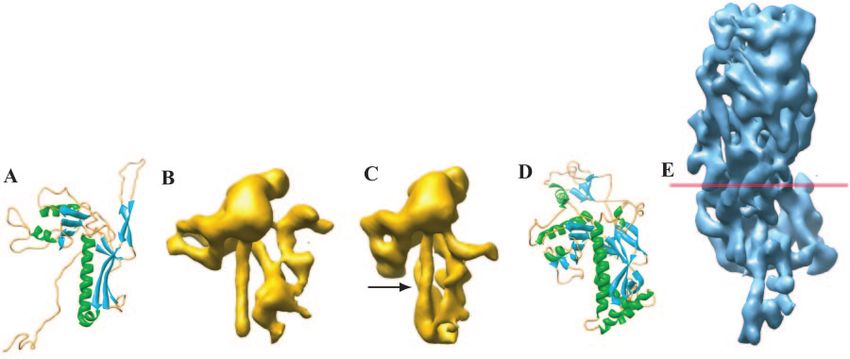
14968 NOTES J. VIROL.
Downloaded from http://jvi.asm.org/ on January 9, 2021 by guest
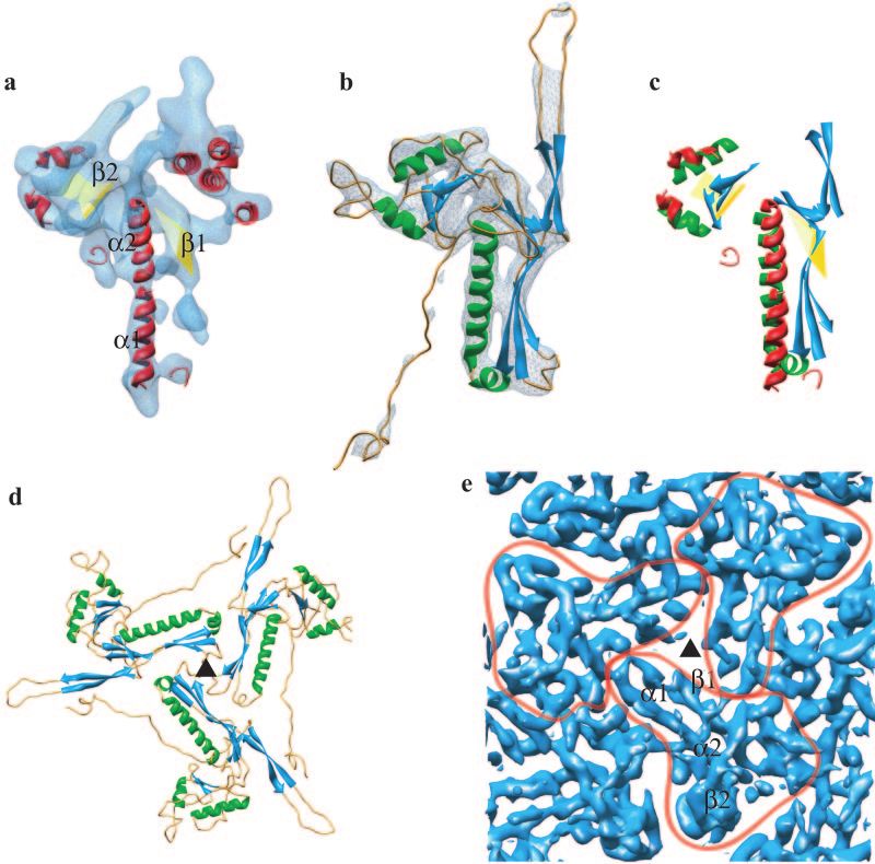
FIG. 2. Match of the secondary structure elements of HSV-1 capsid VP5 and HK97 phage gp5 and their molecular interactions in the capsids.
(a) The isolated VP5 floor domain, in blue, viewed from outside the capsid. SSEhunter identified two long ␣-helices (red; ␣1 and ␣2) adjacent to
a large -sheet (yellow; 1) in the floor domain, as well as a second -sheet (yellow; 2) and several smaller helices flanking ␣1 and ␣2. (b) The
HK97 capsid protein (gp5) shown in the same view reveals a similar structural motif. A simulated density map for gp5 at approximately 8-Å
resolution is shown in pale blue. (c) Alignment of the secondary structure elements by use of Foldhunter (12) demonstrated a clear match between
the floor domain of VP5 and the core structure of gp5. (d) Arrangement of gp5 subunits around a local three-fold axis (Œ) in the HK97 capsid
as viewed from inside the capsid. (e) Organization of the HSV-1 capsid floor as shown in the same view in panel d. Individual VP5 subunit floor
domains are demarcated with the long ␣-helices (␣1, ␣2) and associated -sheets (1, 2) annotated in one subunit.
dae and the prokaryote-infecting Caudovirales have been sug- The crystal structure of the HK97 capsid (19) shows that the
gested previously because of parallels in their capsid assembly capsid protein is roughly triangular (Fig. 1A) and contains a
pathways and similarities between their portal complexes, fold not found in any other protein in the SCOP database
through which DNA enters the capsid (2, 16). While only one (http://scop.mrc-lmb.cam.ac.uk/scop/). This signature fold con-
bacteriophage portal structure has been determined by X-ray sists of three ␣-helices and two -sheets. The sub-nanometer-
crystallography (15), the distinctive 12-fold arrangement of resolution cryoelectron microscopy (cryoEM) structures of
subunits has been reported for several other bacteriophages P22 (13) (Fig. 1B and C) and ⌽29 (14) particles and the X-ray
and also one herpesvirus (17). However, the overall appear- crystal structure of gp24, the T4 pentavalent capsid protein (8)
ance of herpesvirus particles is very different from that of the (Fig. 1D), established that the capsid proteins of these bacte-
Caudovirales and sequence comparison has not provided any riophages follow the same fold design despite having disparate
evidence for common origins in either capsid shell or portal sequences (⬍15% identity).
proteins. To investigate whether evidence for potential rela- Our analysis of an improved (approximately 8-Å) cryoEM
tionships could be detected at the protein structural level, we map of the HSV-1 capsid obtained with a larger data set than
compared the capsid structure of herpes simplex virus type 1 that published previously (20) now reveals that the major cap-
(HSV-1) with those of four bacteriophages, P22, ⌽29, T4, and sid protein, VP5, has the same structural organization in its
HK97, all members of the Caudovirales (7, 13, 14, 19) (Fig. 1). floor (Fig. 1E). Although VP5 is much larger than the bacte-VOL. 79, 2005 NOTES 14969
riophage capsid proteins (Fig. 1), the size disparity is almost relationships across these major biological divides. In light of
entirely accounted for by the middle and upper domains of this and other recent studies (1, 3), it is becoming increasingly
VP5, which form the large penton and hexon towers. The VP5 plausible that extant viruses may have arisen from a relatively
floor domain has very similar dimensions and capsomere spac- small number of primordial progenitors.
ing to the HK97 capsid protein, gp5. Both the overall shape of It is not clear whether the existence of related viruses in-
the HK97 gp5 protein and the disposition of its secondary fecting cells from different domains of life reflects the presence
structural elements are preserved in the VP5 floor domain of a common ancestor that predates the separation of the
(Fig. 2a and b). This is shown by the positional match of the domains or is a result of later adaptation to a new host cell.
␣-helix centroids with ⬍2.5-Å root mean square deviation (Fig. Superficially, the strategy adopted by incoming herpesvirus
2c) and by the close match in the locations of the -sheets. capsids to target and release DNA into eukaryotic nuclei ap-
Additionally, the relative orientations and molecular interfaces pears analogous to that employed by tailed bacteriophages to
of the subunits are retained, with the ␣-helices and -sheets infect bacterial cells. Although this resemblance may be coin-
from the three subunits giving rise to a common architecture at cidental, it is conceivable that if eukaryotic cells are the prod-
the three-fold axes in the floors of HK97 and HSV-1 (Fig. 2d ucts of symbiosis, as has been postulated, it actually reflects the
Downloaded from http://jvi.asm.org/ on January 9, 2021 by guest
and e). Thus, the exhibited fold can be considered a structural retention of an ancient pathway of infection. In this case, the
signature for these viruses, which is analogous to the well- common ancestor of the Caudovirales and Herpesviridae would
known -sandwich fold of many RNA virus particles (9) or the predate the incorporation of the prokaryotic derived nucleus
double -barrel of some DNA virus capsids (1). into the eukaryotic cell.
CryoEM “snapshots” of HSV-1 capsids undergoing angular-
ization during maturation show extensive structural rearrange- This investigation was stimulated by Roger Hendrix of Pittsburgh
ments in the floor domain (11). Similarly, P22 capsid proteins University. We thank Duncan McGeoch for critically reading the
undergo large movements during maturation, including rota- manuscript.
Research has been supported by the National Institutes of Health
tion of the -sheet about the long ␣-helix and refolding of and National Science Foundation and by the UK Medical Research
another ␣-helix, resulting in capsid expansion by over 100 Å in Council and the UK-Texas Bioscience Initiative of the Department of
external diameter (13). The similar dispositions of secondary Trade and Industry.
structural elements in P22 and HSV-1 raise the possibility that REFERENCES
an equivalent rotation produces the changes in the floor do- 1. Benson, S. D., J. K. H. Bamford, D. H. Bamford, and R. M. Burnett. 2004.
main seen during HSV-1 maturation. Since the signature cap- Does common architecture reveal a viral lineage spanning all three domains
sid protein fold has been found in all of the sufficiently well- of life? Mol. Cell 16:673–685.
2. Casjens, S., and J. King. 1975. Virus assembly. Annu. Rev. Biochem. 44:
studied capsids that undergo reconfiguration, it is likely that its 555–611.
development was an important factor in meeting the poten- 3. Coulibaly, F., C. Chevalier, I. Gutsche, J. Pous, J. Navaza, S. Bressanelli, B.
Delmas, and F. A. Rey. 2005. The birnavirus crystal structure reveals struc-
tially conflicting demands imposed by the need to maintain tural relationships among icosahedral viruses. Cell 120:761–772.
capsid stability while allowing for conformational changes as- 4. Davison, A. J. 1992. Channel catfish virus: a new type of herpesvirus. Virol-
sociated with virus maturation. ogy 186:9–14.
5. Davison, A. J. 2002. Evolution of the herpesviruses. Vet. Microbiol. 86:69–
The observation that herpesviruses and tailed bacterio- 88.
phages are related through their capsid protein structures trig- 6. Davison, A. J., B. L. Trus, N. Q. Cheng, A. C. Steven, M. S. Watson, C.
gers the need for a re-evaluation of evolutionary evidence from Cunningham, R. M. Le Deuff, and T. Renault. 2005. A novel class of her-
pesvirus with bivalve hosts. J. Gen. Virol. 86:41–53.
sequence comparisons. The divergence between the evolution- 7. Fokine, A., P. R. Chipman, P. G. Leiman, V. V. Mesyanzhinov, V. B. Rao, and
arily distant fish, mollusk, and mammalian herpesviruses is so M. G. Rossmann. 2004. Molecular architecture of the prolate head of bac-
teriophage T4. Proc. Natl. Acad. Sci. USA 101:6003–6008.
great that their common ancestry cannot be deduced from 8. Fokine, A., P. G. Leiman, M. M. Shneider, B. Ahvazi, K. M. Boeshans, A. C.
sequence similarity (4, 6). The best candidate for a protein that Steven, L. W. Black, V. V. Mesyanzhinov, and M. G. Rossmann. 2005.
is specific to herpesviruses is a packaging protein, the putative Structural and functional similarities between the capsid proteins of bacte-
riophages T4 and HK97 point to a common ancestry. Proc. Natl. Acad. Sci.
terminase encoded by HSV-1 gene UL15. In the past it has not USA 102:7163–7168.
been considered diagnostic for Herpesviridae, as it is distantly 9. Harrison, S. C. 2001. Principles of virus structure, p. 53–85. In D. M. Knipe,
related to an equivalent function identified in certain Caudovi- P. M. Howley, D. E. Griffin, R. A. Lamb, M. A. Martin, B. Roizman, and
S. E. Strauss (ed.), Fields virology, 4th ed., vol. 1. Lippincott-Raven, Phila-
rales (5). However, in light of the evolutionary link demon- delphia, Pa.
strated through their capsid protein folds, we can now interpret 10. Hendrix, R. W. 1999. Evolution: the long evolutionary reach of viruses. Curr.
Biol. 9:R914–R917.
the occurrence of related packaging proteins as independent 11. Heymann, J. B., N. Q. Cheng, W. W. Newcomb, B. L. Trus, J. C. Brown, and
evidence for common ancestry of these two viral lineages. A. C. Steven. 2003. Dynamics of herpes simplex virus capsid maturation visual-
The arrangement of subunit proteins in the portal and the ized by time-lapse cryo-electron microscopy. Nat. Struct. Biol. 10:334–341.
12. Jiang, W., M. L. Baker, S. J. Ludtke, and W. Chiu. 2001. Bridging the
characteristic folds of the capsid proteins are sufficiently dis- information gap: computational tools for intermediate resolution structure
tinctive to suggest that each evolved only once. Both are fun- interpretation. J. Mol. Biol. 308:1033–1044.
damental components of the capsid structure, which represents 13. Jiang, W., Z. L. Li, Z. X. Zhang, M. L. Baker, P. E. Prevelige, and W. Chiu.
2003. Coat protein fold and maturation transition of bacteriophage P22 seen
one of the defining features of any virus. When considered at subnanometer resolutions. Nat. Struct. Biol. 10:131–135.
together with the retained homology in the terminase protein, 14. Morais, M. C., K. H. Choi, J. S. Koti, P. R. Chipman, D. L. Anderson, and
M. G. Rossmann. 2005. Conservation of the capsid structure in tailed
they provide a compelling molecular evidence-based argument dsDNA bacteriophages: the pseudoatomic structure of ⌽29. Mol. Cell 18:
in support of a common origin for the particle-packaging com- 149–159.
plex in the Caudovirales and Herpesviridae. This linkage be- 15. Simpson, A. A., Y. Z. Tao, P. G. Leiman, M. O. Badasso, Y. N. He, P. J.
Jardine, N. H. Olson, M. C. Morais, S. Grimes, D. L. Anderson, T. S. Baker,
tween the most abundant set of bacteriophages and a major and M. G. Rossmann. 2000. Structure of the bacteriophage 29 DNA pack-
family of eukaryotic viruses highlights the growing evidence of aging motor. Nature 408:745–750.14970 NOTES J. VIROL.
16. Steven, A. C., and P. G. Spear. 1997. Herpesvirus capsid assembly and Pringle, and R. B. Wickner (ed.). 2000. Virus taxonomy: seventh report of
envelopment, p. 312–351. In W. Chiu, R. M. Burnett, and R. Garcea (ed.), the International Committee on Taxonomy of Viruses. Academic Press, San
Structural biology of viruses. Oxford University Press, New York, N.Y. Diego, Calif.
17. Trus, B. L., N. Q. Chen, W. W. Newcomb, F. L. Homa, J. C. Brown, and A. C. 19. Wikoff, W. R., L. Liljas, R. L. Duda, H. Tsuruta, R. W. Hendrix, and J. E.
Steven. 2004. Structure and polymorphism of the UL6 portal protein of Johnson. 2000. Topologically linked protein rings in the bacteriophage HK97
herpes simplex virus type 1. J. Virol. 78:12668–12671. capsid. Science 289:2129–2133.
18. van Regenmortel, M. H. V., C. M. Fauquet, D. H. L. Bishop, E. B. Carstens, 20. Zhou, Z. H., M. Dougherty, J. Jakana, J. He, F. J. Rixon, and W. Chiu. 2000.
M. K. Estes, S. M. Lemon, J. Maniloff, M. A. Mayo, D. J. McGeoch, C. R. Seeing the herpesvirus capsid at 8.5 Å. Science 288:877–880.
Downloaded from http://jvi.asm.org/ on January 9, 2021 by guestJOURNAL OF VIROLOGY, Dec. 2005, p. 14471–14472 Vol. 79, No. 23
0022-538X/05/$08.00⫹0 doi:10.1128/JVI.79.23.14471–14472.2005
Copyright © 2005, American Society for Microbiology. All Rights Reserved.
SPOTLIGHT
Articles of Significant Interest Selected from This Issue by the Editors
Novel MMTV RNA Export Protein Identified
Mouse mammary tumor virus (MMTV) previously has been classified as a simple retrovirus with an unknown mechanism
for export of unspliced and partially spliced RNAs. Mertz et al. (p. 14737–14747) report a novel protein called regulator
of export of MMTV mRNA (Rem). Rem facilitates nuclear export of unspliced RNAs via the Crm1 pathway using a
unique, self-regulatory C terminus. The identification of an accessory protein encoded by a doubly spliced RNA suggests
that MMTV is the first murine complex retrovirus to be documented. Manipulation of the MMTV genome may provide
mouse models for human retroviral diseases, such as AIDS.
SARS-CoV Group-Specific ORFs Encode Nonessential Functions for Replication in Cell Culture and Mice
The highly pathogenic SARS coronavirus (SARS-CoV) encodes several unique group-specific open reading frames
(ORFs). The functions of these ORFs in replication and pathogenesis are unknown. Yount et al. (p. 14909–14922) now
show that several of the SARS-CoV group-specific ORFs can be deleted without altering replication in culture or in mice.
Either the group-specific ORFs play little role in replication in vivo or the mouse model is insufficient for discerning the
role of the group-specific ORFs in disease pathogenesis.
High-Level Herpes Simplex Virus Gene Expression during Reactivation Requires Secondary Infections
Herpes simplex virus (HSV) gene expression during reactivation from latency is sensitive to inhibition of viral DNA
synthesis. This and other observations have led to the suggestion that regulation of gene expression during reactivation
differs from that during productive infection. Pesola et al. (p. 14516–14525) found that inhibiting viral encapsidation or
viral DNA synthesis results in similar reductions in expression of immediate-early and early genes after reactivation of
latently infected ganglia. This finding is consistent with the rapid detection of infectious virus following a reactivating
stimulus. Thus, high-level HSV gene expression during reactivation appears to require secondary infections, which has
implications for viral regulatory mechanisms.
Cytomegalovirus Engages a DNA Damage Response Player To Inhibit Apoptosis
Cytomegalovirus (CMV) evades the host cell response to infection by expressing vMIA, a mitochondrion-localized
inhibitor of apoptosis that lacks sequence relatedness to Bcl-2 proteins but may act at a similar level. Smith and Mocarski
(p. 14923–14932) show that vMIA activity depends on an amphipathic ␣-helical motif at the carboxyl terminus that
specifically binds DNA damage response protein GADD45␣. Addition of GADD45 family proteins to cells increases the
potency of vMIA- or Bcl-xL-mediated cell death suppression. Concordantly, inhibition of GADD45 function compro-
mises vMIA activity. This work suggests a link between the DNA damage response and suppression of apoptosis by viral
as well as cellular inhibitors.
Viral Env Determines HTLV Distinct T-Cell Transformation Tropism
HTLV-1 and HTLV-2 are highly related complex retroviruses that infect various cell types but only immortalize or
transform distinct T-lymphocyte populations in culture. HTLV-1 has a preferential tropism for CD4⫹ T lymphocytes,
whereas HTLV-2 preferentially transforms CD8⫹ T lymphocytes. Xie and Green (p. 14536–14545) used infectious
HTLV-1 and HTLV-2 recombinants to identify the viral env gene as a major genetic determinant of the distinct HTLV
T-cell transformation tropism in vitro. These findings provide strong evidence implicating a postentry contribution of Env
to transformation tropism and ultimately the distinct pathobiologies associated with HTLV-1 and HTLV-2 infections.
1447114472 SPOTLIGHT J. VIROL.
A Model for Human Papillomaviruses and Skin Cancer
Although it has been clearly demonstrated that high-risk human papillomaviruses (HPVs) of the genus alpha are
associated with cervical cancer, the role of HPV types of the genus beta in human skin cancer is debated. Dong et al. (p.
14899–14908) show that transgenic mice expressing oncogenes from genus beta HPV type 38 in the skin display
hyperplasia, dysplasia, and an increased susceptibility to chemical-induced carcinogenesis. This work provides further
support for the role of HPV type 38 and possibly other HPV types of this genus in human skin carcinogenesis.
Differentiation State of Human Airway Epithelia Influences ACE2 Expression and Susceptibility to SARS
Coronavirus Infection
Human airway epithelial cells are a site of SARS coronavirus (SARS-CoV) replication during the course of SARS-CoV
infection in vivo. Jia et al (p. 14614–14621) discovered that the state of airway epithelial cell differentiation positively
correlates with ACE2 receptor expression and SARS-CoV infection and replication in these cells. ACE2 is most
abundantly expressed on the apical surface of ciliated cells, and SARS-CoV enters and exits predominantly from the
apical surface of polarized airway epithelia. This work provides new insights into the pathogenesis of pulmonary disease
caused by SARS-CoV and the more common NL63-CoV and suggests new targets for coronavirus therapeutics.
Interaction of Rotavirus with Myeloid Human Dendritic Cells
The human immune response to rotavirus is poorly understood. Children infected with rotavirus have very few circulating
rotavirus-specific T cells that secrete gamma interferon. Narváez et al. (p. 14526–14535) now show that rotavirus infects
small numbers of immature dendritic cells. Rotavirus is not a strong stimulus for dendritic cell maturation but can prime
these cells to stimulate allogeneic naive CD4⫹ T cells to secrete Th1 cytokines. Further work is needed to establish why
rotavirus does not induce a strong Th1 response in acutely infected children.
Pathogenesis of Influenza Viruses with the Haemagglutinin and Neuraminidase of the 1918 Pandemic Virus
Histopathological analyses of lung tissues from individuals who died from primary influenza pneumonia in 1918 reveal
heavy infiltrates of leukocytes. The basis of the severe pulmonary damage caused by the 1918 pandemic influenza virus
is largely unknown. Tumpey et al (p. 14933–14944) investigated the pathogenesis in mice of a recombinant influenza virus
possessing the 1918 hemagglutinin and neuraminidase. Lung tissue from mice infected with the 1918 recombinant virus
displayed a predominant neutrophil infiltrate and a moderate increase in macrophages shortly before death of the
infected mice. Using cell depletion techniques, both cell types were found to be crucial in controlling the growth and
promoting clearance of this highly virulent virus.
Common Ancestry of Herpesviruses and Tailed DNA Bacteriophages
Evolutionary links between herpesviruses and tailed bacteriophages have been suggested based on similarities in capsid
assembly and DNA packaging mechanisms. However, to date, there has been no direct structural evidence for such a
relationship. Baker et al. (p. 14967–14970) now show that the arrangement of secondary structural elements in the floor
domain of the herpes simplex virus major capsid protein affords a close match with those in the capsid proteins of several
tailed bacteriophages. These findings suggest that contemporary viruses may be descended from a relatively small number
of ancient progenitors.You can also read