NEW USE OF DRUG, GALANTHAMINE AS BOTH AMYLOID-BETA INHIBITOR AND ACHE INHIBITOR - GROUP MEMBER INSTRUCTOR SCHOOL LOCATION - 丘成桐中学科学奖
←
→
Page content transcription
If your browser does not render page correctly, please read the page content below
New use of drug, Galanthamine as both
Amyloid-Beta inhibitor and AChE inhibitor

                    Group Member

                       Wenkai Fan

                       Instructor
           Professor Wang Shu Dr. Mingjun Teng

                         School
               Beijing National Day School

                        Location
                      Beijing, China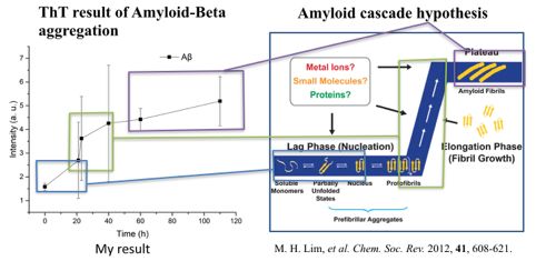
Abstract ............................................................................................................................... 3
Table 1 List of Abbreviations .............................................................................................4
1. Introduction ....................................................................................................................1
  1.1 Introduction of Alzheimer’s Disease ....................................................................................1
    1.1.1 The Cholinergic Hypothesis ...............................................................................................1
    1.1.2 The Aβ hypothesis of Alzheimer’s Disease........................................................................ 2
    1.1.3 The Tau theory ...................................................................................................................3
  1.2 Function of the drug ..............................................................................................................3
    1.2.1 Common types of AChE inhibitor ....................................................................................... 4
  1.3 The propose of this research ...............................................................................................4
2. Experimental section ...................................................................................................6
  2.1 Synthesis of Galanthamine-Me .............................................................................................6
    2.1.1 Meterials and agents ........................................................................................................ 6
  2.2 Measurements ......................................................................................................................6
  2.3 The process of synthesize ..................................................................................................6
    2.3.1 Neutralization reaction ...................................................................................................... 7
  2.4 The bio-assay and experiment for microscopic properties. .............................................8
    2.4.1 Detoxification of Aβ42 ....................................................................................................... 8
  2.5 The Circular Dichroism(CD) Spectroscopy .........................................................................9
    Preparation of CD Spectroscopy ................................................................................................9
  2.6 The ThT Fluorescence Measurement ...................................................................................9
    2.6.1Molecular mechanism of Thioflavin-T binding to amyloid fibrils ..........................................9
    2.6.2The preparation and condition of ThT Fluorescence test ................................................. 10
    2.6.3Condition for the Aβ protein Used in the Aggregation Assay ............................................10
3. Results and discussion .............................................................................................12
  3.1 The result of synthesis ........................................................................................................12
  3.2 Cell Assay Result .................................................................................................................13
  3.3 CD result discussion ...........................................................................................................15
  3.4ThT result with TEM addition ...............................................................................................16
Conclusions .....................................................................................................................19
Reference .......................................................................................................................... 21
Acknowledgements ..........................................................................................................22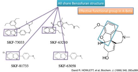
Abstract
The rapid research on the culprits of Alzheimer’s Disease theorized in the theory of Amyloid- Beta
protein. But, there is a lack of a multi-functional drug to help stop or reverse the disease.
Galanthamine, as the second generation of AChE inhibitor, shares the similar structure of
Benzofuran rings with the existing Alzheimer’s treating drug. Has the potential to act both function
in the human brain. In this research, the author focuses on the decoration of active site alongside the
Benzofuran structure, which could complement on the further decoration on this structure. Besides,
the advance in understanding across Alzheimer’s Disease research, this application allows the
design better cost-effective method to simulate alternative strategies to modulate neural function
inside the synapse of the human brain.       Adoption of such approaches should provide a wider
understanding on the synapse terminals across neural networks.
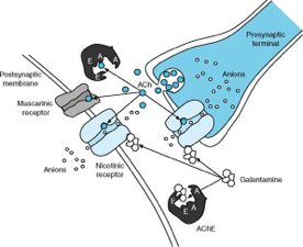
Table 1 List of Abbreviations

                    Aβ                      Amyloid Beta Protein aggregation

                  AChE                       Acetylcholinesterase EC 3.1.1.7

                   Glu                                   glutamate

                  CH3I                                Iodomethane
              RAZADYNE          (4aS,6R,8aS)- 5,6,9,10,11,12- hexahydro- 3-methoxy- 11-
                                methyl- 4aH- [1]benzofuro[3a,3,2-ef] [2] benzazepin- 6-ol

                  GABA                            gamma-aminobutyric acid

                 CH₂Cl₂                             dichloromethane
                 Na₂CO₃                              Sodium Carbonate

                 CH3CN                                 Acetonitrile
                  PBS                         Phosphate buffered saline
                  NaOH                               Sodium hydroxide

                   ThT                                 Thioflavin T

             Galanthamine-Me     (4aS,6R,8aS)- 5,6,9,10,11,12- hexahydro- 3-methoxy- 11-
                                dimethyl- 4aH- [1]benzofuro[3a,3,2-ef] [2] benzazepin- 6-ol

               Galantamine                            Galanthamine

               Acetylcholine

                                              !
1. Introduction
1.1 Introduction of Alzheimer’s Disease
Over a century ago, the German scientist, Alois Alzheimer, reported this brain anomaly in the 37th
Meeting of South-West German Psychiatrists 37th Meeting of South-West German Psychiatrists. At
that time, people dis not know the cause of this disease. He describe this special disease as “A
peculiar severe disease process of the cerebral cortex”(Hanns Hippius, ncbi). Five years after his
death, this disease was first reported as plaques and neurofibrillary in the cerebral cortex soon
named as Alzheimer’s Disease. One major cost of dementia is the problem of diagnosis. Dementia
patients are often diagnosed in the medial or late stages of AD, in which the disease already become
irreversible. With the aging population around the globe, the number of AD patients is likely to be
increased to 131 million by 2050. An estimate cost from the WHO will be US$818 billion on the
worldwide cost of dementia[1].
Currently, there is a need for developing medicines that would slow progression, halt, or prevent
AD and other related dementias from occurring. Although scientists do not yet know the underlying
cause of this typical disease, several possible culprits has brought to help the understanding of the
disease[2].
1.1.1 The Cholinergic Hypothesis
Smith firstly brought up the cholinergic hypothesis in 1918. In this theory, he postulated that the
synthesis of acetylcholine, a neurotransmitter, was low in the neocortex of the brain in AD
patients[3,4]. In supporting this theory, the level of choline acetyltransferase was found to reduce
the neural function in the hippocampus and frontal cortex, and cholinergic neuron counts in the
nucleus basalis was generally lowered in AD condition.
As extensively reviewed in this theory, acetylcholinesterase (AChE) is a key enzyme in the
cholinergic nervous system. The function of AChE enzyme, is to return back the Acetylcholine
inside synapses. In a AD patient brain, the concentration of Acetylcholine reduced as the other types
of neuron deteriorate. Beside the neuron, the source for synthesizing and the production of the
Acetylcholine. Both the acetylcholine-synthesizing enzyme and the functional acetylcholine-
hydrolyzing enzyme enzyme are also effected. However, when the production of acetylcholine
reduce, the AChE enzyme will keep functioning though returning back the neurons. Hence, the
acetylcholine concentration in a AD patient brain will be significantly reduced .
According to the US national library of medicine, the AChE hypothesis, as the first brought theory
that stand a solitary standpoint. In the researcher of Alzheimer’s Disease, Acetylcholinesterase
enzyme is the most studied proteins and the most designed therapy in the Alzheimer’s field. Data
                                                  !1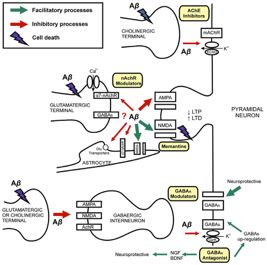
shown in the library indicate about 1500 manuscripts indexed into the PubMed; the vast majority of
reports in the field relate with treatment strategies associated with the use of AChE-I.
1.1.2 The Aβ hypothesis of Alzheimer’s Disease
Beside the AChE hypothesis, the amyloid cascade hypothesis was first brought in 1992 by professor
Hardy and Higgins. They postulate that what Dc. Alzheimer saw in a dementia patient brain,
include neurofibrillary tangles, cell loss, vascular damage was caused by amyloid-beta protein.
Inside human brain, the A-beta protein trend to misfiled from the separate of Amyloid Precursor
protein, form soluble oligomers, aggregate in to insoluble plaques. In this hypothesis. Though
which specific protein in the plaques are unknown to be toxic, but the oligomers reprovide to be the
most danger to the neurons. In this theory, the production, accumulation or disposal of beta-amyloid
protein from the plaque is believe to the main cause of Alzheimer’s Disease. As the fundamental
base of the research, the A-beta hypothesis will be further elaborate in the following part[3].
The plaque of the A-beta protein contain many types, that is unknown of which is toxic to the brain
cells. In this hypothesis, it is believed that the release of Cytokines when microglia triggering up the
inflammatory that damage neurons, this release also damage the synapse from functioning.
In diagram, when the toxic oligomer of Amyloid plaques formed inside the brain, superficial,
cerebral cortex, it will back the transformation of neurons by destroying the vesicle contain the
neurotransmitters, AMPA and NMDA as two types of neurotransmitter carrier, are inhibited by the
Amyloid protein, where the original terminals disfunction and deteriorate.

                          !
          Figure1 The mechanism of synaptic disfunction cause by Amyloid-Beta protein
                                                   !2
1.1.3 The Tau theory
From the timeline of the research, Tau protein aggregation hypothesis was raised as the third
hypothesis on the cause of AD. This protein was originally a component of tangles. Where the
health Tau protein are the component of the made on a series of track, and it stabilized the tube
when Molecule are carried along axon, this series of track is named as microtubes. But in
Alzheimer’s Disease, the Tau protein is no longer the health proteins but modified causing it to
dissociate from the microtubules. The aggregation of the protein adopt abnormal shape and move
form the axon to the cell body.
Like the A-beta theory, Tau proteins come from different forms, it’s still unclear of which on
contribute to the disease. And like beta, these forms either remain, or stick together and deposit. as
the tangle that Dr. Alzheimer saw. Eventually, these process kill the neuron. In the race brain
experiment, the researcher also find that the modified Tau protein can spread pathology across the
brain, there make the healthy Tau protein start to misfiled as well. The patten of spread though the
different brain regions share similar characteristics as the changing symptoms from early to late
stage of this research. Galanthamine is a natural product belonging to the Amaryllidaceae family of
alkaloids which was discovered in the early 1950s. The molecule can be natural extracted from
plant, with the function of maintaining the Acetylcholine concentration, RAZATYNE
(Galanthamine Hydro-bromide) was first used to treat nerve pain and poliomyelitis[5].
!
Figure 2 The mechanism of Galanthamine binding to AChE enzyme [4]: 1) Nootropic Agents,
Parasympathomimetics, 2) Cholinesterase Inhibitors.
1.2 Function of the drug
The main function of Galanthamine is though enhancing the cholinergic function. It is achieved
!3though two ways: firstly, it maintains acetylcholine concentration though competitive binding on
the acetyl cholinesterase enzyme, and act as an allosteric potentiator to help the neurotransmitter to
enter both nicotinic and muscarinic acetylcholine receptors. Secondly, it direct inhibits the
hydrolysis of the neurotransmitter hence increase Acetylcholine concentration inside synaptic cleft.
1.2.1 Common types of AChE inhibitor
Galantamine is not the only cholinesterase inhibitor. As figure three shown below, Donepezil,
Rivastigmine, and Tacrine are included in the common types of AChE inhibitor and the use in the
treatment of several brain disease. But, one feature that distinct Galanthamine from all other drugs,
is the way Galanthamine bind with the enzyme inside synapse though competitive blocking. As
previous example mentioned, the direct binding to the AChE enzyme distinguished Galanthamine
form the other AChE inhibitors[6].
!
Table 2 Pharmacology of common types of AChE inhibitors and it’s relation to Alzheimer’s.
1.3 The propose of this research
The cause of Alzheimer’s Disease have been long brought to the world these last centuries, yet is
still no drug design that can reverse or stop the deterioration of mental disorder. Though out the
research on the the molecule Galanthamine, the researcher find that this molecule share the similar
Benzofuran with some existing Alzheimer’s Drugs, like Benzofuran-chalcone hybrids, has been
proven to be effective on inhibiting the Amyloid-beta aggregation. On top of this, Galanthamine is
!4registered as AChE enzyme inhibitor. So, the researcher would like to see if this molecule can be
applied to the both theories. Which, it’s assumed that Galanthamine will success in inhibiting the
self-assembly of Amyloid-Beta oligomer. Therefore, when the drug entering the synapse terminal in
brain areas like cerebral cortex, this drug may share multi-functions to help further modify drug on
this.
To testify suitable active site for the modification, the N-CH3 site is identified as a independent site
else than the Benzofuran. Hence, the synthesis on decorating this site is taken.
!52. Experimental section
2.1 Synthesis of Galanthamine-Me
2.1.1 Meterials and agents
All organic solvents were purchased from Beijing Chemical Works and used without further
purification. Other chemicals were purchased from Acros, Sigma-Aldrich Chemical Company or
Alfa-Aesar and used as received. Aβ42, the purity of the peptides was greater than 98%. The cell
line PC12 was purchased from cell culture center of Institute of Basic Medical Sciences, Chinese
Academy of Medical Sciences (Beijing, China). Deionized water (18.2 MΩ·cm) was obtained from
a Milli-Q system (Millipore, Bedford, MA). Fetal bovine serum (FBS) was purchased from Sijiqing
Biological Engineering Materials (Hangzhou, China). Modified RPMI 1640 (RP 1640) was
purchased from HyClone/Thermofisher (Beijing, China). 3-(4,5-dimethylthiazol-2-yl)-2,5-
diphenyltetrazolium bromide (MTT).
2.2 Measurements
The 1H NMR and 13C NMR spectra were measured on Bruker Avance 400 or 600 MHz
spectrometers. High resolution mass spectra (HR-MS) were taken on a Bruker 9.4T Solarix FT-
ICR-MS spectrometer. Fluorescence spectra were recorded on Varioskan Flash (Thermo Scientific
Company, USA). The absorbance for MTT analysis was measured on a microplate reader (BIO-
TEK Synergy HT, USA) at a wavelength of 570 nm. Cell counting was performed on an automated
cell counter (Countess, Invitrogen). CD spectra were measured on a Hitachi Jasco-J815 circular
dichroism spectrophotometer. TEM images were recorded on a Hitachi S-7700 transmission
electron microscope.
2.3 The process of synthesize
Since the document shows that the Benzofuran structure in Galathamine (HBr) is effective
in treating the Alzheimer’s Disease. The effect of another functional group in this compound
is unclear. So, a set of synthesis is designed, with the goal of outlining the effect of another
functional group—benzazepin. To inhibit the function of benzazepin and only exhibit the
effect of Benzofuran, one common way is though methylation. After methylation, the lose of
charge in benzazepin site reduce the water solubility of RAZATYNE compound. Hence, the
related reaction from this site is been limited. Ideally, if the effect of the drug is not reduced
significantly, the functional group of Benzofuran is effective in reversing the aggregation of
Amyloid-Beta protein. The synthesis of the compound was follow by three steps.
Neutralization, extraction, and methylation.
!62.3.1 Neutralization reaction
The drug purchased were the hydro-bromide Galanthamine(RAZATYNE). To obtain pure
Galanthamine, Sodium Carbonate is used as the base to neutralize hydrogen bromide. Compare to
the strong based that are usually used in the reaction. The salt is suitable without affiliate the
structure of the compound. Hence, 1.2 time of the standard requirement for the neutralization of
Sodium Carbonate is been added into the round-bottom flask. To ensure the complete of reaction,
the reaction ti me is been set to be 30 minute. Consider the polarity of the compound, deionized
water are consider as the common solvent for both RAZATYNE and Sodium Carbonate.
!
Figure 3 the mechanism of the neutralization reaction
The product after neutralization, Galanthamine-Me, is non polar. Due to insolubility in deionized
water, the extraction process can help filter out the excess Sodium Carbonate, and isolate deionized
water.
Consider the solvent used to dissolve Galanthamine, dichloromethane is used in the extraction. Due
to higher density of the prance face, the extraction can help extract the Galanthamine to the
substratum level.
Before the process of methylation, to obtain the substance and calculate the percentage yield from
the neutralization, Decompress distillation is required to gather the results.
With a 250ml round-bottom flask with knowing mass, add the solution obtain from the last step not
the separate funnel. The mechanism of distillation is though the reduce of boiling point by
Decompression of air pressure inside the flask. Hence, require less heat and speed up the reaction.
Considering the common types of organic solvent like dichloromethane, have the boiling point
lower than the water, with the addition decompression of air pressure. It’s less time consuming to
get the galanthamine powder. Hence, calculate for the yield of the product.
After taking out the product by using Acetonitrile into a new flask, the last reaction will undergo the
overnight reaction though magnet stirring. Then, the methylation of galanthamine is complete(22h).
!7!
Figure 4 The methylation of the compound galanthamine-Me
After collect the powder form the drying owen, the Galanthamine-Me was splited into smaller
dosage for further testing.
To see the result of synthesis, both H1-NMR and Mass Spectrum are used to testify the structural
formula of the compound.
2.4 The bio-assay and experiment for microscopic properties.
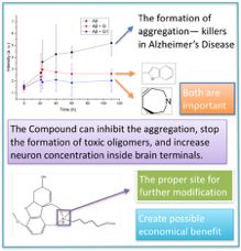
From the Introduction, the Amyoid-Beta protein is toxic during the process of oligmers formation.
If the compound Galanthamine, and Galanthamine-Me are effective, both drug should be able to
reduce the formation of oligmers. Besides, it’s important to see the toxicity of drug to give a
suggesting range of dosage to the future pharmacy design. Hence, the first experiment used in
decting the effectiveness of both drug is the cell assay, as a way to simulate the brain cell after
gettingg an Alzheimer’s Disease. Though it is not the perfect condition lessoning the huamn body,
conducting the cell culture assay is the most direct experienment in a labtoray opreation.
2.4.1 Detoxification of Aβ42
Rat pheochromocytoma PC12 cells were cultured in RP 1640 medium containing 10% fetal bovine
serum under 5% CO2 at 37°C. The cells were seeded at a density of 7,000 cells per well. Aβ42
alone, with Galanthmine-HBr (ten-fold excess) or with Galanthamine-Me(ten-fold excess) was
incubated for 15 h at 37°C prior to the addition to cells. The samples were diluted with RP 1640
medium to Aβ42 concentration of 20 µM. After incubation for 48 h, the mixture in each well was
replaced with MTT (1 mg/mL in medium, 100 µL/well) and incubation for 4 h, Then, the
supernatant was replaced with 100 µL DMSO per well, and the plates were shaken for 10 min.
Absorbance values of formazan were read with a microplate reader at 570 nm. Wells with neither
Galanthamine nor Aβ42 were served as control group.
Though the observation on the average damage of the cells(7000 cell per wall), the Cell Culture
result will suggest the effect in inhibiting the formation of toxic oligomers.
!82.5 The Circular Dichroism(CD) Spectroscopy
Though the Cell Culture suggest the effect of inhibiting the formation of toxic oligomers inside the
human brain. The Cell Assay can not observe the microscopic change of the Amyloid-Beta protein.
Hence, the Circular Dichroism, as the measurement on the secondary structure of protein, is used.
Preparation of CD Spectroscopy
200 µL of the sample was added to a 0.1 cm quartz cell for far-UV (190-260 nm) measurements.
The bandwidth was 2 nm. The scanning speed is 100 nm/min with a response time of 4 s. Each
spectrum was an average of 3 scans.
Consider the Chiral Carbons in the structural formula for compound Galanthamine, the use of CD
spectroscopy may not be the best way to observe the microscopic properties. Hence, it is not expect
in the data discussion that the result diagram are perfect in explaining the inhibition of protein
formation.
2.6 The ThT Fluorescence Measurement
To ensure the proper observations to cassette the microscopic change of compound Galanthamine in
inhibiting the Amyloid-Beta formation. An other measurement, later used as the main measurement
to detect the aggregation of A-Beta protein— the Thioflavin-T fluorescence measurement is taken.
2.6.1Molecular mechanism of Thioflavin-T binding to amyloid fibrils
Since its first description in 1959, the fluorescent dye Thioflavin-T (ThT) has become among the
most widely used “gold standards” for selectively staining and identifying amyloid fibrils both in
vivo and in vitro.
Figure 5: Common experimental techniques employing ThT. (a) Structure
of ThT (top). The two planer segments of ThT whose mutual rotation
defines its chirality are also shown (bottom).
In addition to the instrument for ThT measurement, the picture below draw the general detection
!9pattern. When the twist in the two planner segment of ThT occur, the mutual rotation stabilized the
spinning electron on the planner segment. The fluorescence light is collect as following:
!
Figure 6 the principle of the optical system
!
Figure 7 the varioskan flash instrument
2.6.2The preparation and condition of ThT Fluorescence test
In a 96 slot plate, 3 parallel experiment for pure A-beta, galanthamine hydro-bromide,
galanthamine-Me were inject to the slot. Eachneaction(200µL) were mixed with 20µL of 20 µM
ThT in 10mM phosphate buffer (PBS), pH 7.4, each trial were recorded every 24 hours in total
time 110h. The incubation timer the individual fluorescence are 5 minutes. The wavelength in the
measurement were at λex = 452 nm and λem = 485 nm, the equipment used were shown above. [15]
2.6.3Condition for the Aβ protein Used in the Aggregation Assay
In preparing the solution for the Aggregation Assay[16], NaOH is first added as the solvent of the
protein. After create a weak base environment for the protein, the PBS is used as the buffer to
simulate human brain condition for the A-Beta protein aggregation. Two set of aggregation assay
used the 1,5,10 time excessive Galanthamine, and Galanthamine-Me to assess the inhibition.
!10Incubation
(5min)
Temperature agitation assay
Aβ42(control group) Preparation A:were dissolved in 50 37 + ThT
mM sodium hydroxide (10% of the
final volume), followed by addition of
90 of PBS.
Aβ42 protein with 1 Preparation B:were dissolved in 60 37 + ThT
time, 5 times, 10 mM sodium hydroxide (10% of the
times excessive final volume), followed by addition of
RAZATYNE 90% of PBS. 1,510µL of Galanthamine
is added.
Aβ42 protein with 1 Preparation B:were dissolved in 60 37 + ThT
time, 5 times, 10 mM sodium hydroxide (10% of the
times excessive final volume), followed by addition of
RAZATYNE 90% of PBS. 1,510µL of
Galanthamine-Me is added.
Table 3: The condition for aggregation assay
!113. Results and discussion
3.1 The result of synthesis
Figure 8: the H1-NMR representation
From the result receive and interpret above, the H1-NMR spectrum suggest that the peak I and K
are the two methyl groups on the product galanthamine-Me. Form the data of chemical shit on
Chemdraw, the research also construct the corresponding peak for all the hydrogens alongside the
compound. Which, based on the result received, the structural formula of the compound is the target
structure.
Then, the mass spectrometry was used to confirm the product after synthesis is the idea product.
The diagram below shows the base peak, the largest segment in the spectrum are the compound
Galanthamine after methylation.
!12Figure 9: Mass Spectrum Result
3.2 Cell Assay Result
Since the synthesis is right. The drug used in the Cell Culture Assay are appropriate to compare the
bio-toxicity.
Figure 9: Cell Assay for Galanthamine-HBr Figure 10: Cell Assay for Galanthamine-Me
!13The Result of Cell Assay collect above indicate that, for all 10 concentration of the both
Galanthamine, and Galanthemine-Me have low bio-tonicity towards human body. The highest
concentration used(200µM) for the cell assay keep a high cell viability. Hence, this drug can go
direct to human brain.
Considering human health as one major concern in the drug design, the cell culture assay is
prepared to hep formulate proper concentration of the drug.
The goal of this experiment is to give a understanding on the concentration of drug appropriate for
human health. This experiment is commonly used in a laboratory condition to give a considerable
range on the dosage. But, this can not replace the clinical trial in determining LD50 or TD50.
Figure 11: Cell Assay for the inhibition of A-Beta
After confirming the relative low damage to human body of the drug. The next cell assay is use to
compare the cell damage with and without the addition of the drug. For the A-Beta control group,
without any addition of the drug, it preformed a great damage to the cell. Which match the Amyloid-
beta theory—the oligomers are the most toxic to the human brain.
The middle and right side cell assay, are the group with the addition of both compound. Both shown
a increase in Cell Viability, represent the less damage the A-Beta generated as the first groups
shows. With a little higher in Cell Viability, the group after methylation preformed better in the
Biocompatibility. From the data of the cell assay, both compound are effective in inhibiting the
Amyloid-Beta protein aggregation.
Since both drug are effect in inhibiting the formation of toxic oligomers during protein aggregation.
The inhibition on the benzazepin did not effect the function of the drug on a cellular level. Yet,
!14without the microscopic detection. It is still unclear to see that the formation of aggregation.
Form this point. As the AChE inhibitor in human brain, this drug can be applied to another
hypothesis—the Amyloid-Beta hypothesis. Further information is required to give a conclusion.
3.3 CD result discussion
To help confirm the microscopic property of the drug, the first instrument the researcher thanked
about, is the Circular Dichroism(CD) Spectroscopy. Which the aggregation can be observed though
the change in the secondary structure of the proteins.
To monitor the formation of Amyloid protein aggregation, the first CD spectrum shows the similar
formation of protein as the introduction mentioned.
Random Beta- sheet
As a control group, the CD spectrum above indicate the formation of A-beta protein, generate a
wavelength around 210-220mm, which is the shift recorded the protein formation.
However, when different concentration of Galanthamine-HBr, and Galanthamine-Me is been
record. The result shows a different pattern—the original A-beta protein absorption is no longer
being observed. However, the the differential absorption of left- and right-handed light for the
compound Galanthamine is been observed— Due to the Chiral Carbon center in Galanthamine, the
spin angular momentum in the drug is overwhelmed. Hence, in this way. It can not distinguish the
!15difference in spin angular momentum between the drug and protein. But, the result for A-Beta
protein is used as a reference to the report the aggregation.
3.4ThT result with TEM addition
In view of the results, the researcher asked the question: to what extent is the inhibition of
aggregation by both galanthamine compound was a general phenomenon. To answer this question,
the result of ThT diagram is assisted by the TEM and CD graph. In the original design, and the
experimental section. The self-assembling of the amyloid-beta protein was considered as a
independent control group. In the research, the researcher collected the data on the growth of A-beta
on the basis of ThT intensity. Which the ThT flash device is designed for the detecting the intensity
of fluorescence by eliminating the background intensity of the fluorescence.
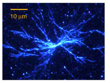
Figure 12: TIRF microscopy image of branched
glucagon fibrils stained with ThT
Since the CD experiment is not well suited due to the chiral carbons in the compound Galathamine.
The advantage of using the ThT intensity as indicator to the protein aggregation—there is no
reaction between Galanthamine and ThT. The Fluorescence used can only bind to the protein.
Hence, it can shows the exact intensity of protein aggregation in any given time. Consider the
mature of aggregation, the experiment time is set to be 100 hours. For each recording, there will be
two parallel group to reduce deviation.
!16Figure 13: The similar pattern o aggregation between the result collect (left), and document
The Diagram above indicate the similar aggregation pattern from monomers to mature fibril
structure of the A-Beta protein.
Fibril structures
toxic oligomers
No Mature
Fibrils
!17On the basis of number, location, and relative abundance of the Amyloid-beta protein(TEM), the
result of both ThT and TEM supported the phenomenon of success inhibition of protein
aggregation.
Since both molecules succeed in the inhibition process, though the observation on the relative
abundance. And the difference in figure 13. The Galanthamine hydro-bromide have a better
inhibition on the aggregation.
In review of this result, the researcher asked for another question: is the Benzofuran the only
functional group inside? By comparing the structure of Galanthamine-HBr and Galanthamine-Me.
The dimethyl nitro group on the seven carbon ring avianize the original structure. Which the weak-
base environment is more suitable for the molecule for the inhibition. But, the dimethyl group in the
Cell Assay shows a better Biocompatibility. Hence, the Benzofuran is not the only active site that
inhibit the Amyloid-beta protein. Since the benzazepin can be decorated without influencing the
function of the drug. The future research can be conduct based on this single site.
Figure 14,15 Comparing the structural formula of Galanthamine compounds
!18Conclusions
By observing microscopically. The molecule galanthamine effective in inhibiting the assembly fibril
structure of Amyloid beta protein. As a original Acetylcholine inhibitor, the galanthamine molecule
share function beyond it’s original medical use as an AChE inhibitor. The research shows that this
molecule can direct interact with the fibril structure aggregation side human brain.
By synthesizing compound with difference functional group on the basis of Benzofuran rings , the
researcher also give thoughtful explanation to the function of active sites inside the brain. Which
have brought a way for the further application of this drug to allow the decoration on these active
site. Beside from modifying into a dimethyl group, the same active site and be attached to another
molecule. Or forming a bilayer with hydrophobic layer to increase it’s solubility in liquid for better
entering the blood-brain barrier, and a hydrophilic inner layer for better dissolve of the
galanthamine molecule.
!
Figure 15:Galathamine as both Amyloid-Beta inhibitor and AChE inhibitor
Despite from the bright future on the drug design base on this benzofuran structure. In face of
treating of Alzheimer Disease, problem and limitations are needed to identify. With the decoration
of the drug, the researcher question the original function of competitive binding to the original
enzyme—AChE. If this process will keep successful when the lab environment do no allows the
author to simulate synaptic terminals inside the brain. Besides, the less access to modern medical
condition also leave millions of lives in LEDC countries to have no access on diagnosing and
reporting this disease, when the synapse were already broken, the chemical stimulation may not be
!19able to reconstruct certain component in the brain like an nicotinic receptor.
!20Reference
[1] What Is Alzheimer's? Alzheimer's Association. https://www.alz.org/alzheimers-dementia/what-
is-alzheimers. Accessed September 13, 2018.
[2] Decarli C. Concepts, Conjectures, and Hypotheses in Dementia. Alzheimer Disease &
Associated Disorders. 2009;23(3):185. doi:10.1097/wad.0b013e3181b85eae.
O'Neil, M.J. (ed.). The Merck Index - An Encyclopedia of Chemicals, Drugs, and Biologicals. 13th
Edition, Whitehouse Station, NJ: Merck and Co., Inc., 2001., p. 771
[3,4] Figure 2f from: Irimia R, Gottschling M (2016) Taxonomic revision of Rochefortia Sw.
(Ehretiaceae,Boraginales).Biodiversity Data Journal 4: e7720. https://doi.org/10.3897/BDJ.4.e7720.
doi:10.3897/bdj.4.e7720.figure2f.
[5] Decarli C. Concepts, Conjectures, and Hypothesis in dementia. Alzheimer's Disease&
Associated Disorders. 2009;23(3:185)
[6] Nature Video Channel. Inside Alzheimer's disease. YouTube. https://www.youtube.com/ watch?
v=zTd0-A5yDZI. Published January 3, 2017. Accessed September 15, 2018.
[7] Figure 2f from: Irimia R, Gottschling M (2016) Taxonomic revision of Rochefortia Sw.
(Ehretiaceae,Boraginales).Biodiversity Data Journal 4: e7720. https://doi.org/10.3897/BDJ.4.e7720.
doi:10.3897/bdj.4.e7720.figure2f.
[8] Macpherson S. Galanthamine: the new treatment of choice in Alzheimer’s Disease Inpharma
Weekly. 1995;&NA;(1002):3-5. doi:10.2165/00128413-199510020-00002.
[9] GBan, et al, Journal of American Society, 2011, 133, 16958-16969
[10] M. H. Lim, et al. Chem. Soc. Rev. 2012, 41, 608-621.
[11] G. Bitan, et al. J. Am. Chem. Soc. 2011, 133, 16958-16969.
[12] Gordon K Wilcock,et al, Neurol, Psych, 1999: 66; 137-147
[13]M. Biancalana, S. Koide / Biochimica et Biophysica Acta 1804 (2010) 1405–1412
[14]Grudzielanek, S.; Velkova, A.; Shukla, A.; Smirnovas, V.;vTatarek-Nossol, M.; Rehage, H.;
Kapurniotu, A.; Winter, R. J. Mol. Biol. 2007, 370, 372–784.
[15]Bitan, G.; Kirkitadze, M. D.; Lomakin, A.; Vollers, S. S.; Benedek, G. B.; Teplow, D. B. Proc.
Natl. Acad. Sci. U.S.A. 2003, 100, 330–335.
[16]Sharmistha Sinha,† Dahabada H. J. Lopes,et al, J.Am. Chem. Soc. 2011, 133, 16958–16969
!21Acknowledgements
I would like to appreciate the guidance and instructions of my instructor professor Wangshu, who
provided the research lab and resources of Insitute of Chemistry, Chinese Academy of Sciences.
during the activity of Yingcai program.
Dr. Mingjun Teng who offered valuable feedbacks for my academic writing, the China Youth
Association for Science and Technology Center (CYSCC) which is the sponsor of Yingcai program;
Dr. Sun Han who gave me instruction in the experiment.
!22本参赛团队声明所提交的论⽂是在指导⽼师指导下进⾏的研究⼯作和取得的研究成果。
尽本团队所知,除了⽂中特别加以标注和致谢中所罗列的内容以外,论⽂中不包含其他⼈已
经发表或撰写过的研究成果。若有不实之处,本⼈愿意承担⼀切相关责任。
参赛队员: 范⽂楷 指导⽼师: 王树、滕明俊
!1You can also read