Ratios between circulating myeloid cells and lymphocytes are associated with mortality in severe COVID-19 patients
←
→
Page content transcription
If your browser does not render page correctly, please read the page content below
Open Medicine 2021; 16: 351–360
Research Article
Hui Ma#, Xiong Chang Lim#, Qihong Yu#, Yi Li, Yuechuan Li, Wei Jia*
Ratios between circulating myeloid cells and
lymphocytes are associated with mortality in
severe COVID-19 patients
https://doi.org/10.1515/med-2021-0237 lymphocytes were highly correlated with coagulation status
received October 29, 2020; accepted January 13, 2021 and patient mortality in severe COVID-19.
Abstract: Recent studies indicate that host immune Keywords: COVID-19, myeloid cells, lymphocytes, coagu-
responses are dysregulated with either myeloid cell com- lation, D-dimer
partment or lymphocyte composition being disturbed in
COVID-19. This study aimed to assess the impact of SARS-
CoV-2 viral infection on the composition of circulating
immune cells in severe COVID-19 patients. In this retro- 1 Introduction
spective single-center cohort, 71 out of 87 COVID-19 patients
admitted to the intense care unit for oxygen treatment were The novel coronavirus disease-2019 (COVID-19) outbreak,
included in this study. Demographics, clinical features, mediated by the severe acute respiratory syndrome coron-
comorbidities, and laboratory findings were collected on avirus 2 (SARS-CoV-2), was first identified in Wuhan,
admission. Out of the 71 patients, 5 died from COVID-19. China, in December 2019 [1,2]. As with other virulent corona-
Compared with survived patients, deceased patients showed virus infections such as severe acute respiratory syn-
higher blood cell counts of neutrophils and monocytes but drome (SARS) and Middle East respiratory syndrome
lower cell counts of lymphocytes. Intriguingly, the neutro- (MERS), SARS-CoV-2 poses a major health threat to human
phil-to-lymphocyte ratio (NLR), monocyte-to-lymphocyte worldwide [3,4]. With the rapid spread worldwide causing
ratio (MLR), and basophil-to-lymphocyte ratio (BLR) were high morbidity and mortality, it has been declared a pan-
markedly higher in deceased patients compared to survived demic by the World Health Organization. There have been
patients. Furthermore, the lymphocyte counts were nega- 24,257,989 confirmed cases and 827,246 deaths reported as
tively correlated with D-dimer levels, while the ratios of 28 August 2020 [5].
between myeloid cells and lymphocyte (NLR, MLR, and Respiratory compromise characterized by lower respira-
BLR) were positively correlated with D-dimer levels. Our tory tract symptoms, such as fever, dry cough, and dyspnea,
findings revealed that the ratios between myeloid cells and has been reported in patients with COVID-19 [6]. In patients
with severe COVID-19, the disease progresses to acute
respiratory distress syndrome and life-threatening events
including coagulopathy, septic shock, and death [3]. To
reduce the morbidity and mortality rates, it is crucial to
identify key risk factors associated with poorer prognosis.
Basic laboratory tests have been proven crucial in identifica-
tion of high-risk COVID-19 patients [7]. This allows early
# Co-first authors.
intensive care and support to be provided to patients at
high risk of mortality.
* Corresponding author: Wei Jia, Department of Respiratory and The immune systems, including the innate and adap-
Critical Care Medicine, Tianjin Chest Hospital, No. 261, tive immune systems, are commonly disturbed in virus
Taierzhuangnan Road, Jinnan District, Tianjin, 300222, China, infection [8–10]. White blood cells, which are categorized
e-mail: jiaweimr@126.com, tel: +68-22-88185354
into myeloid cells (neutrophils, monocytes, eosinophils,
Hui Ma, Qihong Yu, Yi Li, Yuechuan Li: Department of Respiratory
and Critical Care Medicine, Tianjin Chest Hospital, Tianjin, China
and basophils) and lymphocytes (T cells, B cells, and nat-
Xiong Chang Lim: Yong Loo Lin School of Medicine, National ural killer [NK] cells) based on their cell lineage, constitute
University of Singapore, Singapore, Singapore the first line of defense against invading pathogens
Open Access. © 2021 Hui Ma et al., published by De Gruyter. This work is licensed under the Creative Commons Attribution 4.0 International
License.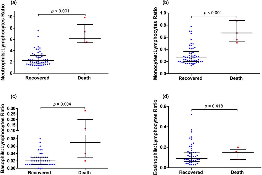
352 Hui Ma et al.
including viruses [11]. In COVID-19, several recent studies
indicate that host immune responses are dysregulated with
either myeloid cell compartment [12–14] or lymphocyte com-
position [15–17] being disturbed. Furthermore, the effective-
ness and concerns over immunosuppressive therapies in
COVID-19 patients have been discussed [18,19]. However,
studies on differential involvement of the myeloid cells
and lymphocytes and their correlation with the coagulation
status and disease severity in COVID-19 patients are scarce
to date.
In this study, we analyzed blood cells, coagulation
parameters, and inflammatory markers from routine clinical
laboratory tests and discovered the differential impact of
SARS-CoV-2 infection on circulating myeloid cells and lym-
phocytes. Furthermore, we found that the ratios between
myeloid cells and lymphocytes were highly correlated with
Figure 1: Study design.
blood coagulation status and disease severity in COVID-19.
transcriptase-polymerase chain reaction (RT-PCR) assay.
2 Materials and methods Peripheral blood was collected and analyzed in the clinical
laboratory of Wuhan Iron & Steel (Group) Company Second
2.1 Patients and study design Staff Hospital. The laboratory test results including full
blood cell counts, coagulation parameters (prothrombin time
This was a retrospective single-center study with a total [PT], international normalized ratio [INR], activated partial
of 87 patients tested positive with COVID-19 between 19 thromboplastin time [APTT], thrombin time [TT], D-dimer,
January and 23 February 2020 in Wuhan Iron & Steel and fibrinogen), and other blood markers (C-reactive protein
(Group) Company Second Staff Hospital. Subsequently, [CRP] and creatinine kinase [CK]) were assessed for both
only 71 patients with comprehensive medical records survived and deceased COVID-19 patients. A full blood
were included in this study (Figure 1). The study was count was performed using the XN-10 automated hema-
conducted in accordance with the principles of Declara- tology analyzer (Sysmex® Corporation), which gener-
tion of Helsinki and approved by the Institutional Review ated the white blood cell differential fluorescence (WDF)
Board of Tianjin Chest Hospital (IRB-SOP-016(F)-001-02). scattergram [21].
The need for informed consent was waived given the
observational and retrospective nature of the study.
All patients were confirmed of COVID-19 either by
2.3 Statistical analysis
SARS-CoV-2 nucleic acid test (63.4%) or by clinical symp-
toms and computerized tomography (CT) scan imaging
All statistical analyses were performed using SPSS 25.0
(36.6%), according to the Diagnosis and Treatment
(IBM Corp., Armonk, NY, USA). A p-value of less than
Protocol for Novel Coronavirus Pneumonia (Trial Version
0.05 is considered statistically significant. Graphs for fig-
4.0) released by the National Health Commission of the
ures were prepared with GraphPad Prism 8.00 (GraphPad
People’s Republic of China [20]. Patient data including
Software, San Diego, CA, USA). Missing data were not
demographics, comorbidities, symptoms, and laboratory
imputed. Continuous data were presented as mean ±
findings were collected during the hospital admission.
standard deviation (SD) or median ± interquartile range
(IQR), and categorical variables were presented as per-
centage. The differences were compared by Student’s
2.2 Laboratory testing t-test, Mann–Whitney U test, χ2 test, or Fisher’s exact
test, depending on the nature of data. The relationship
Patient throat swab specimens were collected for the among biomarkers was assessed using Spearman’s cor-
detection of SARS-CoV-2 via the real-time reverse relation analysis.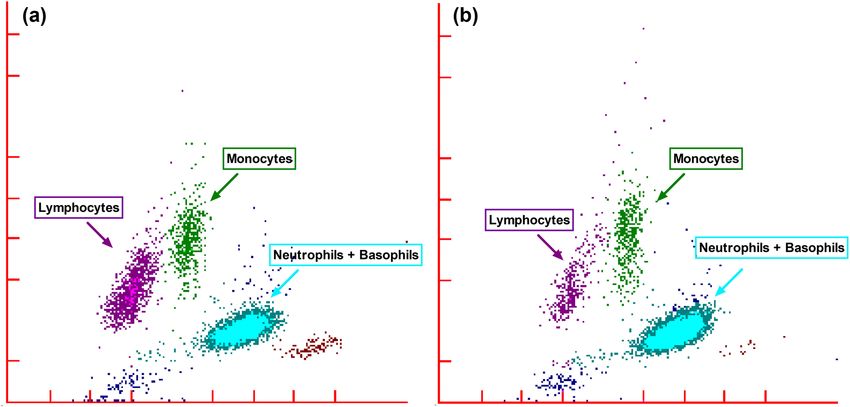
Circulating immune cells in COVID-19 patients 353
3 Results malignancy, cerebrovascular disease, and respiratory dis-
ease. Hypertension was more frequently seen among
the deceased patients than those who recovered (60.0
3.1 Patient characteristics
vs 13.6%; p = 0.008).
From 19 January to 23 February 2020, a total of 71 patients
admitted to Wuhan Iron & Steel (Group) Company
Second Staff Hospital with confirmed COVID-19 and
comprehensive medical records were included in this 3.2 Laboratory parameters of COVID-19
study (Figure 1). Five of these 71 patients died from patients
COVID-19 and 66 patients fully recovered and were
discharged. As shown in Table 1, the median age of Differences in the initial laboratory parameters, including
deceased patients was significantly older than those blood cell counts and biochemical markers, between the
who recovered (74.0 years, IQR: 65.0–83.0 vs 61.0 deceased patients and those who recovered from COVID-
years, IQR: 50.0–71.0; p = 0.025), and there were more 19 are presented in Table 2.
males in the deceased patients (100 vs 40.9%; p = A full blood count and the white blood cell WDF
0.010). Of all COVID-19 patients, nearly half of them scattergram were generated using the XN-10 automated
(n = 34, 47.9%) had at least one chronic medical con- hematology analyzer (Table 2 and Figure S1). Compared
dition, with diabetes being the most common comor- with the survivors, the deceased patients had lower red
bidity followed by hypertension, coronary heart disease, blood cell counts (3.46 × 10¹²/L vs 4.06 × 10¹²/L; p =
0.010) and platelet counts (152.00 × 10⁹/L vs 228.00 ×
109/L; p = 0.004). Interestingly, deceased patients pre-
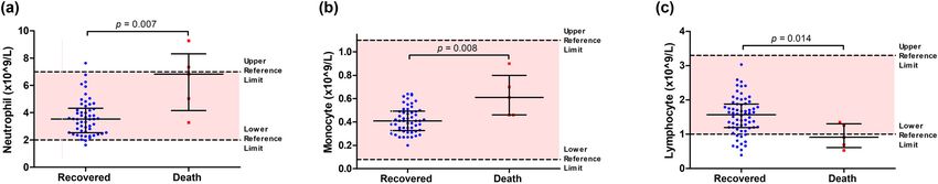
sented with lower lymphocyte counts (0.91 × 10⁹/L vs
Table 1: Baseline characteristics of the study population
1.57 × 10⁹/L; p = 0.014) but higher neutrophil (6.83 ×
Alive Dead p-value
109/L vs 3.53 × 10⁹/L; p = 0.007) and monocyte counts
n = 66 n=5 (0.61 × 10⁹/L vs 0.41 × 10⁹/L; p = 0.008) (Table 2, Figure 2
and Figure S1). No difference in basophil and eosinophil
Demographic
Age (years) 61.0 74.0 0.025
counts was observed between the two patient groups.
(50.0–71.0) (65.0–83.0) All the patients presented with pro-thrombotic
Male (%) 27 (40.9) 5 (100.0) 0.010 states, including relatively lower PT and INR, and higher
Diagnosis D-dimer levels (Table 2). Of all COVID-19 patients with
Nuclear test (%) 42 (63.6) 3 (60.0) 0.871 available data of coagulation tests, 22 out of 31 (70.97%)
Clinical test (%) 24 (36.4) 2 (40.0) 0.871
patients presented with D-dimer levels higher than the
Hospital stay 19.5 6 (3.5–15.0) 0.008
duration (days) (16.0–22.0) normal range. However, no significant difference was
Symptoms observed between survived and deceased patients in
Fever (%) 40 (60.6) 5 (100.0) 0.078 any of the coagulation parameters measured, including
Cough (%) 35 (53.0) 4 (80.0) 0.243 D-dimer levels (2.01 µg/mL, IQR: 1.17–3.00 for deceased
Comorbidities
patients vs 0.57 µg/mL, IQR: 0.30–2.10 for survived patients;
Diabetes 12 (18.2) 1 (20.0) 0.919
mellitus (%)
p = 0.159), likely due to a small number of deceased patients
Hypertension (%) 9 (13.6) 3 (60.0) 0.008 in the current study.
Coronary heart 8 (12.1) 1 (20.0) 0.610 The levels of an inflammatory biomarker, CRP, were
disease (%) elevated in most COVID-19 patients whose CRP data were
Cerebrovascular 2 (3.0) 1 (20.0) 0.069 available, with 18 out of 25 (72.00%) patients presented
disease (%)
with CRP levels higher than the normal range. Although
Respiratory 2 (3.0) 0 (0.0) 0.693
disease (%) higher median CRP levels were found in deceased patients
Malignancy (%) 4 (6.1) 0 (0.0) 0.571 (75.25 mg/L, IQR: 48.55–78.63 vs 29.58 mg/L, IQR: 5.00–74.50;
p = 0.203), the levels did not differ between survived
Continuous data are presented as mean ± SD or median ± IQR, and
and deceased patients. CK levels, however, were signifi-
statistical analysis of continuous data was performed using unpaired
Student’s t-test or Mann–Whitney U test. Categorical variables are pre- cantly higher in deceased patients (105.00 U/L, IQR:
sented as %, with differences between the groups tested with χ2. Bold 69.00–165.00) compared with those who survived (37.00
values indicate significant difference with a p-value of less than 0.05. U/L, IQR: 29.00–51.00; p = 0.019).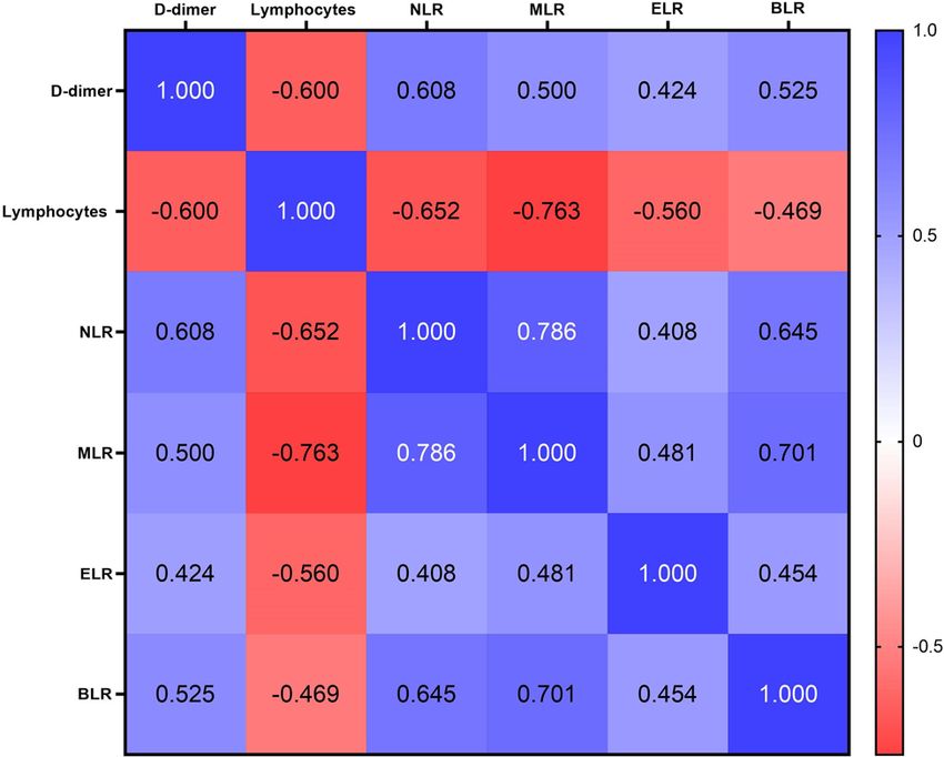
354 Hui Ma et al.
Table 2: Laboratory parameters of COVID-19 patients
Alive Dead p-value
n = 66 n=5
Full blood count indices
RBC count (×1012/L) 4.06 (3.80–4.29) 3.46 (3.15–3.90) 0.010
Hemoglobin (g/L) 125.00 (118.00–133.00) 116.00 (106.00–135.00) 0.237
Hematocrit (%) 36.90 (34.80–39.45) 35.50 (31.35–38.90) 0.261
MCV (fL) 91.70 (89.10–93.90) 92.10 (89.95–101.55) 0.370
MCH (pg) 30.90 (30.00–32.10) 32.10 (29.95–34.75) 0.251
MCHC (g/L) 337.50 (330.50–344.00) 330.00 (319.50–341.00) 0.146
RDW-CV (%) 12.00 (11.60–12.60) 12.50 (11.75–12.95) 0.389
RDW (fL) 46.00 (44.20–48.30) 49.30 (44.05–52.45) 0.153
Platelet (×109/L) 228.00 (184.00–264.00) 152.00 (88.00–178.00) 0.004
PDW (fL) 16.10 (15.80–16.40) 15.90 (15.35–17.00) 0.282
MPV (fL) 8.90 (8.30–9.40) 9.00 (8.75–9.55) 0.567
PCT (%) 0.19 (0.17–0.22) 0.21 (0.09–0.23) 0.836
WBC (×109/L) 5.85 (4.55–6.61) 8.43 (5.38–10.49) 0.056
Neutrophil count (×109/L) 3.53 (2.54–4.27) 6.83 (4.16–8.32) 0.007
Lymphocyte count (×109/L) 1.57 (1.20–1.88) 0.91 (0.61–1.31) 0.014
Monocyte count (×109/L) 0.41 (0.33–0.49) 0.61 (0.46–0.80) 0.008
Basophil count (×109/L) 0.03 (0.02–0.03) 0.06 (0.02–0.18) 0.110
Eosinophil count (×109/L) 0.15 (0.11–0.22) 0.11 (0.08–0.17) 0.256
Coagulation panel
PT (s) 11.00 (10.60–11.30) 11.40 (10.75–14.70) 0.173
INR 0.85 (0.80–0.85) 0.89 (0.81–1.17) 0.268
APTT (s) 32.60 (29.30–38.45) 29.40 (27.30–32.80) 0.190
TT (s) 14.20 (13.70–14.65) 14.80 (13.70–15.40) 0.290
Fibrinogen (g/L) 2.49 (2.07–2.97) 2.43 (1.97–2.93) 0.679
D-dimer (µg/mL) 0.57 (0.30–2.10) 2.01 (1.17–3.00) 0.159
Others
C-reactive protein (mg/L) 29.58 (5.00–74.50) 75.25 (48.55–78.63) 0.203
Creatine kinase (U/L) 37.00 (29.00–51.00) 105.00 (69.00–165.00) 0.019
Data are presented as median ± IQR and statistical analysis was performed using the Mann–Whitney U test. Abbreviations: RBC, red blood
cell; MCV, mean cell volume; MCH, mean corpuscular hemoglobin; MCHC, mean corpuscular hemoglobin concentration; RDW-CV, red cell
distribution width – coefficient of variation; RDW, red cell distribution width; PDW, platelet distribution width; MPV, mean platelet volume;
PCT, plateletcrit; WBC, white blood cell; PT, prothrombin time; INR, international normalized ratio; APTT, activated partial thromboplastin
time; TT, thrombin time. Bold values indicate significant difference with a p-value of less than 0.05.
3.3 Differential influence of SARS-CoV-2 on ELR appeared higher in deceased patients (0.15 vs 0.09;
circulating myeloid cells and p = 0.418), the difference was not statistically significant
lymphocytes (Figure 3d), likely due to the small sample size available
for this study.
Given the different profiles of circulating myeloid cells
and lymphocytes from peripheral blood of survived and
deceased patients (Table 2 and Figure 2), we further ana-
lyzed ratios of these two cell populations, including the 3.4 Correlation of D-dimer levels with
neutrophil-to-lymphocyte ratio (NLR), monocyte-to-lym- circulating immune cell counts
phocyte ratio (MLR), basophil-to-lymphocyte ratio (BLR),
and eosinophil-to-lymphocyte ratio (ELR). Strikingly, as Since both coagulation parameters and the immune sys-
shown in Figure 3, deceased patients had significantly tems are disturbed in COVID-19, we analyzed the correla-
higher ratios of myeloid cells over lymphocytes: NLR tion between D-dimer levels and different immune cells
(6.19 vs 2.27; p < 0.001), MLR (0.67 vs 0.26; p < 0.001), (and their ratios). As shown in the correlation matrix
and BLR (0.07 vs 0.02; p = 0.004). Of note, although the (Figure 4), D-dimer levels were negatively correlatedCirculating immune cells in COVID-19 patients 355 Figure 2: Myeloid cells (a, neutrophils; b, monocytes) and lymphocytes (c) in deceased patients and those recovered from COVID-19. Data are presented as median ± IQR, and statistical analysis was performed using the Mann–Whitney U test. The region highlighted in pink is the normal ranges of the respective cell counts. with lymphocyte counts (r = −0.600; p < 0.001), whereas cells and lymphocyte counts is highly correlated with the there was no correlation between D-dimer levels and pro-thrombotic state in COVID-19. other immune cells including neutrophils, monocytes, basophils, and eosinophils. The correlations between D-dimer levels and the inflammatory marker CRP or the tissue damage marker CK were also analyzed but did not 4 Discussion reach statistical significance. Intriguingly, D-dimer levels in COVID-19 patients were positively correlated with NLR In this single-center retrospective study, we analyzed the (r = +0.608; p = 0.001), MLR (r = +0.500; p = 0.007), ELR clinical features, blood cells, coagulation parameters, (r = +0.424; p = 0.025), and BLR (r = +0.525; p = 0.004). and inflammatory markers of severe COVID-19 patients These findings indicate that the imbalance between myeloid and uncovered that circulating myeloid cells and Figure 3: Ratios of myeloid cells and lymphocytes, including (a) neutrophil:lymphocyte, (b) monocyte:lymphocyte, (c) basophil:lymphocyte, and (d) eosinophil:lymphocyte, in deceased patients and those recovered from COVID-19. Data are presented as median ± IQR, and statistical analysis was performed using the Mann–Whitney U test.
356 Hui Ma et al.
lymphocytes were differentially affected. In particular, of severe COVID-19 patients [12]. Whether this change
the ratios of myeloid cells and lymphocytes were overly is associated with activation of monocytic Toll-like
associated with disease severity and highly correlated receptor 7 by SARS-CoV-2 is unknown. Nevertheless,
with the pro-thrombotic state in COVID-19. uncontrolled activation of innate immune cells including
The deceased patients in this study were older, pre- neutrophils and monocytes may contribute to the detri-
dominantly male, and the majority had hypertension. mental cytokine storm in COVID-19 patients [6,8,37].
These clinical features of severe COVID-19 patients are The majority of lymphocytes are T cells and B cells
in agreement with previous reports [3,4,6]. Ageing com- which constitute the major cellular components of
monly contributes to more severe COVID-19 and higher adaptive immune system [38]. Given the emerging roles
risk of death, which is likely associated with uncontrolled of T cells in COVID-19 and the critical role of B cells in
innate immune responses that cause cytokine storm and producing antibodies [38], the lower levels of lympho-
altered T cell responses [22]. The severity of COVID-19 cytes in deceased COVID-19 patients in the current
was also reported to be inversely correlated with age in study reflect a weaker adaptive immune response in
hospitalized children [23]. Recent studies also suggest fighting against SARS-CoV-2 infection. The significantly
that the phenotype and function of circulating mono- lower lymphocyte levels in deceased patients may be
cytes, a type of innate immune cells, are changed in both attributable to stress-induced apoptosis and exhaus-
aging and COVID-19 [24,25]. On top of the compromised tion of antiviral lymphocytes [39]. In addition, the
immune system in aging population, comorbidities such as levels of circulating NK cells, a type of anti-viral innate
hypertension may explain the higher risk of death in immune cells which constitute a minor population of
elderly COVID-19 patients. lymphocytes, also decrease in severe COVID-19 patients
Activation of the immune systems has been well [15]. As a result, higher ratios of myeloid cells and lym-
documented in viral infections including the SARS, phocytes (NLR, MLR, and BLR) indicate a more severe
MERS, and COVID-19 [26–28]. Neutrophilia and lym- imbalance of innate and adaptive immune responses to
phopenia are associated with more severe disease SARS-CoV-2 infection and consequently an unfavorable
symptoms and death in COVID-19 [29,30]. In agree- outcome in such patients contracted with COVID-19. In
ment, we found higher counts of neutrophil and mono- agreement with our findings, the NLR was reported to
cyte in deceased patients than in survived patients. be positively associated with disease severity in COVID-
In addition, we found lower lymphocyte counts in 19 [40–43]. However, to our knowledge, no prior study
deceased patients. The differential changes in myeloid has systemically assessed the ratios of different myeloid
cells and lymphocytes result in markedly higher NLR, cell populations and lymphocytes in severe COVID-19.
MLR, and BLR in deceased COVID-19 patients (Figure 3).
The higher ratios of myeloid cells and lymphocytes in the
more critically ill patients (deceased) may reflect the
pathogenesis of COVID-19. Activation of neutrophils
releases reactive oxygen species and cytokines as well
as neutrophil extracellular traps, therefore, constitutes
the first line of defense in response to viral infection
[31,32]. Given the expression of virus-recognizing
immune receptor, Toll-like receptor 7 [33], in mono-
cytes, higher levels of monocytes in the blood may
lead to over-reaction to SARS-CoV-2 infection. As Toll-
like receptor 7 has been recently reported to deteriorate
cardiovascular disease [34,35], higher levels of acti-
vated monocytes are expected to increase the risk of
mortality in COVID-19 patients with pre-existing cardi-
ovascular disease [4,6,36]. Indeed, using high-dimen-
sional flow cytometry analysis and single-cell RNA
sequencing, Silvin et al. recently uncovered a disturbed
Figure 4: Correlation matrix showing the strength of correlation
balance between non-classical CD14LowCD16High mono- between D-dimer levels and inflammatory cells and ratios. Values in
cytes and HLA-DRLow classical monocytes (Human Leu- cells are Spearman correlation coefficient. All correlations were
kocyte Antigen-DR isotype) in the peripheral blood statistically significant (p < 0.05).Circulating immune cells in COVID-19 patients 357
Inflammatory responses have been associated with 5 Conclusion
thrombotic and bleeding manifestations in sepsis [44,45].
In the context of COVID-19, early studies have shown the In conclusion, innate and adaptive immune systems were
association between elevated D-dimer levels and mortality affected differentially among severe COVID-19 patients.
[4]. D-dimer is a fibrin degradation product, and it serves as Our findings suggest that the ratios of myeloid cells
a marker of fibrinolytic activity [46]. There is evidence that (except eosinophils) and lymphocytes are highly asso-
D-dimer correlates with proinflammatory cytokine levels in ciated with pro-thrombotic state and disease severity
critically ill patients [47]. In consistence, majority of the in COVID-19 and may serve as potential biomarkers for
patients in this study presented with D-dimer levels higher risk stratification of COVID-19 patients, allowing early
than the normal range. We did not find significant differ- intensive care and support for those of higher risk of
ences in D-dimer and CRP levels between deceased death.
patients and recovered patients, as it is likely because all
the patients included in this study were severe cases. How- Acknowledgments: This study was supported by the
ever, by correlation matrix analysis, we found a significant Healthcare Technology Programme from the Health
correlation between D-dimer levels and the ratios of myeloid Commission of Tianjin (2020XKC07).
cells and lymphocytes (NLR, MLR, BLR, and ELR) as well as
lymphocyte counts (Figure 4). This raises an open question: Disclosure: The author reports no conflicts of interest in
are the elevated D-dimers because of coagulopathy arising this work.
from viral infection, or are the imbalanced innate and adap-
tive immune responses attributable to activation of coagula- Data availability statements: The datasets generated
tion in viral infection? Further studies are warranted. during and/or analyzed during the current study are
Besides coagulation parameters and inflammatory available from the corresponding author on reasonable
markers, we also observed a higher CK level in deceased request.
patients compared with survived patients. Elevated
CK levels were previously reported in the SARS and
MERS outbreak [48]. Although rhabdomyolysis has
been reported as potential late complication associated References
with COVID-19 [49], we did not have data regarding the
symptoms of rhabdomyolysis in patients with elevated [1] Zhu N, Zhang D, Wang W, Li X, Yang B, Song J, et al. A novel
CK levels. The increase in CK levels may be attributed coronavirus from patients with pneumonia in China, 2019. N
Engl J Med. 2020;382(8):727–33.
to hypovolemia that causes renal impairment in COVID-
[2] Gralinski LE, Menachery VD. Return of the coronavirus: 2019-
19 patients [50].
nCoV. Viruses. 2020;12(2):135.
This study has several limitations. First, it is a single- [3] Chen N, Zhou M, Dong X, Qu J, Gong F, Han Y, et al.
center retrospective study with a small number of patients. Epidemiological and clinical characteristics of 99 cases of
Second, laboratory parameters were not complete for each 2019 novel coronavirus pneumonia in Wuhan, China:
individual patient and the number of deceased patients in a descriptive study. Lancet. 2020;395(10223):507–13.
[4] Guan WJ, Ni ZY, Hu Y, Liang WH, Ou CQ, He JX, et al. Clinical
this study is rather small, therefore, may result in under-
characteristics of coronavirus disease 2019 in China. N Engl J
powered statistical analysis. Furthermore, as only clinical Med. 2020;382(18):1708–20.
laboratory data are available for this study, we are not [5] World Health Organization (WHO). Coronavirus disease
able to analyze subpopulations of lymphocytes such as (COVID-2019) situation reports; 2020 [cited 28 August 2020].
T-cells. Multi-center studies with a larger cohort of patients Available from: https://www.who.int/emergencies/diseases/
novel-coronavirus-2019/situation-reports
are therefore required to confirm our findings. Moreover,
[6] Huang C, Wang Y, Li X, Ren L, Zhao J, Hu Y, et al. Clinical
multi-dimensional analysis of subpopulations of immune
features of patients infected with 2019 novel coronavirus in
cells will help to delineate the impact of SARS-CoV-2 infec- Wuhan, China. Lancet. 2020;395(10223):497–506.
tion on innate and adaptive immunity in COVID-19. [7] Sun Y, Koh V, Marimuthu K, Ng OT, Young B, Vasoo S, et al.
Nevertheless, given the fact that the overwhelming Epidemiological and clinical predictors of COVID-19. Clin Infect
cases of COVID-19 lead to shortage of medical resources Dis. 2020;71(15):786–92.
[8] Channappanavar R, Perlman S. Pathogenic human coronavirus
worldwide, our findings may facilitate the development
infections: causes and consequences of cytokine storm and
of diagnostic protocols for patient stratification based immunopathology. Semin Immunopathol. 2017;39(5):529–39.
on the routine laboratory tests available in most clinical [9] Gong F, Dai Y, Zheng T, Cheng L, Zhao D, Wang H, et al.
laboratories. Peripheral CD4+ T cell subsets and antibody response in358 Hui Ma et al.
COVID-19 convalescent individuals. J Clin Invest. [27] Xu Z, Shi L, Wang Y, Zhang J, Huang L, Zhang C, et al.
2020;130(12):6588–99. Pathological findings of COVID-19 associated with acute
[10] Wilk AJ, Rustagi A, Zhao NQ, Roque J, Martínez-Colón GJ, respiratory distress syndrome. Lancet Respir Med.
McKechnie JL, et al. A single-cell atlas of the peripheral 2020;8(4):420–2.
immune response in patients with severe COVID-19. Nat Med. [28] Kothari A, Singh V, Nath UK, Kumar S, Rai V, Kaushal K, et al.
2020;26(7):1070–6. Immune dysfunction and multiple treatment modalities for the
[11] Chaplin DD. Overview of the immune response. J Allergy Clin SARS-CoV-2 pandemic: races of uncontrolled running sweat?
Immunol. 2010;125(Suppl 2):S3–23. Biology. 2020;9(9):243.
[12] Silvin A, Chapuis N, Dunsmore G, Goubet A-G, Dubuisson A, [29] Feng X, Li S, Sun Q, Zhu J, Chen B, Xiong M, et al. Immune-
Derosa L, et al. Elevated calprotectin and abnormal myeloid inflammatory parameters in COVID-19 cases: a systematic
cell subsets discriminate severe from mild COVID-19. Cell. review and meta-analysis. Front Med. 2020;7:301.
2020;182(6):1401–18. [30] Wan S, Xiang Y, Fang W, Zheng Y, Li B, Hu Y, et al. Clinical
[13] Agrati C, Sacchi A, Bordoni V, Cimini E, Notari S, Grassi G, et al. features and treatment of COVID-19 patients in northeast
Expansion of myeloid-derived suppressor cells in patients with Chongqing. J Med Virol. 2020;92(7):797–806.
severe coronavirus disease (COVID-19). Cell Death Differ. [31] Agraz-Cibrian JM, Giraldo DM, Mary FM, Urcuqui-Inchima S.
2020;27:3196–207. Understanding the molecular mechanisms of NETs and their
[14] Schulte-Schrepping J, Reusch N, Paclik D, Baßler K, Schlickeiser S, role in antiviral innate immunity. Virus Res. 2017;228:124–33.
Zhang B, et al. Severe COVID-19 is marked by a dysregulated [32] Muraro SP, De Souza GF, Gallo SW, Da Silva BK, De Oliveira SD,
myeloid cell compartment. Cell. 2020;182(6):1419–40. Vinolo MAR, et al. Respiratory syncytial virus induces the
[15] Market M, Angka L, Martel AB, Bastin D, Olanubi O, classical ROS-dependent NETosis through PAD-4 and necrop-
Tennakoon G, et al. Flattening the COVID-19 curve with natural tosis pathways activation. Sci Rep. 2018;8(1):14166.
killer cell based immunotherapies. Front Immunol. [33] Diebold SS, Kaisho T, Hemmi H, Akira S, Reis e Sousa C. Innate
2020;11:1512. antiviral responses by means of TLR7-mediated recognition of
[16] Chen Z, John, Wherry E. T cell responses in patients with single-stranded RNA. Science. 2004;303(5663):1529–31.
COVID-19. Nat Rev Immunol. 2020;20(9):529–36. [34] de Kleijn DPV, Chong SY, Wang X, Yatim S, Fairhurst AM,
[17] Wang Z, He Y, Shu H, Wang P, Xing H, Zeng X, et al. High- Vernooij F, et al. Toll-like receptor 7 deficiency promotes sur-
fluorescent lymphocytes are increased in patients with COVID- vival and reduces adverse left ventricular remodelling after
19. Br J Haematol. 2020;190(2):e76–8. myocardial infarction. Cardiovasc Res. 2019;115(12):1791–803.
[18] Campbell CM, Guha A, Haque T, Neilan TG, Addison D. [35] Karadimou G, Plunde O, Pawelzik S-C, Carracedo M,
Repurposing immunomodulatory therapies against corona- Eriksson P, Franco-Cereceda A, et al. TLR7 expression is
virus disease 2019 (COVID-19) in the era of cardiac vigilance: associated with M2 macrophage subset in calcific aortic valve
a systematic review. J Clin Med. 2020;9(9):2935. stenosis. Cells. 2020;9(7):1710.
[19] Di Micco P, Di Micco G, Russo V, Poggiano MR, Salzano C, [36] Nishiga M, Wang DW, Han Y, Lewis DB, Wu JC. COVID-19 and
Bosevski M, et al. Blood targets of adjuvant drugs against cardiovascular disease: from basic mechanisms to clinical
COVID19. J Blood Med. 2020;11:237–41. perspectives. Nat Rev Cardiol. 2020;17(9):543–58.
[20] National Health Commission of the People’s Republic of China. [37] Zhou Y, Fu B, Zheng X, Wang D, Zhao C, Qi Y, et al. Pathogenic
Notice on the issuance of a programme for the diagnosis and T-cells and inflammatory monocytes incite inflammatory
treatment of novel coronavirus (2019-nCoV) infected pneu- storms in severe COVID-19 patients. Natl Sci Rev.
monia (Trial Version 4); 2020 [cited 28 August 2020]. 2020;7(6):998–1002.
Available from: http://bgs.satcm.gov.cn/zhengcewenjian/ [38] Tay MZ, Poh CM, Rénia L, MacAry PA, Ng LFP. The trinity of
2020-01-28/12576.html COVID-19: immunity, inflammation and intervention. Nat Rev
[21] Osman J, Lambert J, Templé M, Devaux F, Favre R, Flaujac C, Immunol. 2020;20(6):363–74.
et al. Rapid screening of COVID-19 patients using white blood [39] Zheng M, Gao Y, Wang G, Song G, Liu S, Sun D, et al. Functional
cell scattergrams, a study on 381 patients. Br J Haematol. exhaustion of antiviral lymphocytes in COVID-19 patients. Cell
2020;190(5):718–22. Mol Immunol. 2020;17(5):533–5.
[22] Meftahi GH, Jangravi Z, Sahraei H, Bahari Z. The possible [40] Qun S, Wang Y, Chen J, Huang X, Guo H, Lu Z, et al. Neutrophil-
pathophysiology mechanism of cytokine storm in elderly to-lymphocyte ratios are closely associated with the severity
adults with COVID-19 infection: the contribution of “inflame- and course of non-mild COVID-19. Front Immunol.
aging”. Inflamm Res. 2020;69(9):825–39. 2020;11:2160.
[23] Nathan N, Prevost B, Sileo C, Richard N, Berdah L, [41] Yang AP, Liu JP, Tao WQ, Li HM. The diagnostic and predictive
Thouvenin G, et al. The wide spectrum of COVID-19 clinical role of NLR, d-NLR and PLR in COVID-19 patients. Int
presentation in children. J Clin Med. 2020;9(9):2950. Immunopharmacol. 2020;84:106504.
[24] Merad M, Martin JC. Pathological inflammation in patients with [42] Chan AS, Rout A. Use of neutrophil-to-lymphocyte and
COVID-19: a key role for monocytes and macrophages. Nat Rev platelet-to-lymphocyte ratios in COVID-19. J Clin Med Res.
Immunol. 2020;20(6):355–62. 2020;12(7):448–53.
[25] Pence BD. Severe COVID-19 and aging: are monocytes the key? [43] Vafadar Moradi E, Teimouri A, Rezaee R, Morovatdar N,
Geroscience. 2020;42(4):1051–61. Foroughian M, Layegh P, et al. Increased age, neutrophil-to-
[26] Huang KJ, Su IJ, Theron M, Wu YC, Lai SK, Liu CC, et al. An lymphocyte ratio (NLR) and white blood cells count are asso-
interferon-gamma-related cytokine storm in SARS patients. ciated with higher COVID-19 mortality. Am J Emerg Med.
J Med Virol. 2005;75(2):185–94. 2021;40:11–4.Circulating immune cells in COVID-19 patients 359
[44] Delabranche X, Helms J, Meziani F. Immunohaemostasis: [47] Shorr AF, Thomas SJ, Alkins SA, Fitzpatrick TM, Ling GS.
a new view on haemostasis during sepsis. Ann Intensive D-dimer correlates with proinflammatory cytokine levels and
Care. 2017;7(1):117. outcomes in critically ill patients. Chest. 2002;121(4):1262–8.
[45] Iba T, Levy JH. Inflammation and thrombosis: roles of neutro- [48] Lee N, Hui D, Wu A, Chan P, Cameron P, Joynt GM, et al. A major
phils, platelets and endothelial cells and their interactions in outbreak of severe acute respiratory syndrome in Hong Kong.
thrombus formation during sepsis. J Thrombosis Haemost. N Engl J Med. 2003;348(20):1986–94.
2018;16(2):231–41. [49] Jin M, Tong Q. Rhabdomyolysis as potential late complication
[46] Soomro AY, Guerchicoff A, Nichols DJ, Suleman J, Dangas GD. associated with COVID-19. Emerg Infect Dis J. 2020;26(7):1618.
The current role and future prospects of D-dimer [50] Rivas-García S, Bernal J, Bachiller-Corral J. Rhabdomyolysis as
biomarker. Eur Heart J Cardiovasc Pharmacother. the main manifestation of coronavirus disease 2019.
2015;2(3):175–84. Rheumatology. 2020;59(8):2174–6.360 Hui Ma et al. Appendix Figure S1: Representative white blood cell differential fluorescence (WDF) scattergrams of (a) survived and (b) deceased patients from severe COVID-19.
You can also read