Valerian and valeric acid inhibit growth of breast cancer cells possibly by mediating epigenetic modifications
←
→
Page content transcription
If your browser does not render page correctly, please read the page content below
www.nature.com/scientificreports
OPEN Valerian and valeric acid inhibit
growth of breast cancer cells
possibly by mediating epigenetic
modifications
Fengqin Shi1,2, Ya Li1,2, Rui Han1,2, Alan Fu1, Ronghua Wang1,2, Olivia Nusbaum1, Qin Qin1,
Xinyi Chen2, Li Hou1,2 & Yong Zhu1*
Valerian root (Valeriana officinalis) is a popular and widely available herbal supplement used to
treat sleeping disorders and insomnia. The herb’s ability to ameliorate sleep dysfunction may
signify an unexplored anti-tumorigenic effect due to the connection between circadian factors and
tumorigenesis. Of particular interest are the structural similarities shared between valeric acid,
valerian’s active chemical ingredient, and certain histone deacteylase (HDAC) inhibitors, which
imply that valerian may play a role in epigenetic gene regulation. In this study, we tested the
hypothesis that the circadian-related herb valerian can inhibit breast cancer cell growth and explored
epigenetic changes associated with valeric acid treatment. Our results showed that aqueous valerian
extract reduced growth of breast cancer cells. In addition, treatment of valeric acid was associated
with decreased breast cancer cell proliferation, migration, colony formation and 3D formation
in vitro in a dose- and time-dependent manner, as well as reduced HDAC activity and a global DNA
hypomethylation. Overall, these findings demonstrate that valeric acid can decrease the breast cancer
cell proliferation possibly by mediating epigenetic modifications such as the inhibition of histone
deacetylases and alterations of DNA methylation. This study highlights a potential utility of valeric
acid as a novel HDAC inhibitor and a therapeutic agent in the treatment of breast cancer.
Natural products provide a rich source for the development of novel drugs with anti-tumor activities. Currently,
over a half of the available anticancer drugs were derived from botanical sources1. For example, Taxol (paclitaxel)
is an FDA approved anti-microtubule agent extracted from bark of the Pacific yew tree Taxus brevifolia and has
shown a strong anticancer effect in human cancer2. Considering herbal extracts and their bioactive compounds
are a rich source of therapeutics to human beings, there is a compelling reason to seek novel bioactive compounds
in the herbal extracts.
Valerian root is an herb that has long been found to be effective for treating circadian related sleep disorders.
The name “Valerian” in Latin means “to be strong or healthy”, which can be attributed to its use as an herbal
remedy that has been in practice since ancient Greek and Roman times. Currently, the herb is used mainly as a
treatment for insomnia and is considered among the most popular sleeping aids in E urope3. Valerian has been
approved as a Generally Recognized as Safe (GRAS) food ingredient by the U.S. Food and Drug Administration
(FDA). A recent systematic review of research concluded that “The available evidence suggests that valerian
might improve sleep quality without producing side effects”4.
Disruptions in sleep and circadian rhythm have been shown to be associated with elevated breast cancer
risk5,6. The International Agency for Research on Cancer (IARC) has designated circadian disruption caused
by night shift work as a “2A” carcinogen (probably carcinogenetic) for breast cancer7,8. This designation is reaf-
firmed by the finding of a 40% elevated risk of breast cancer risk among female night shift workers in a recent
meta-analysis6. In breast tumors, many human circadian genes have been found to be dysregulated9. Animal
studies have also demonstrated that mice with disruptions in their molecular clocks showed increased breast
cancer risk10 and accelerated growth rates of malignant t umors11.
Given the connection between circadian factors and tumorigenesis, the herb’s ability to ameliorate sleep
dysfunction may signify a completely unexplored underlying anti-tumorigenic capacity. Valeric acid, a short
1
Department of Environmental Health Sciences, Yale University School of Public Health, New Haven, CT 06520,
USA. 2Department of Oncology and Hematology, Dongzhimen Hospital, Beijing University of Chinese Medicine,
Beijing 100700, China. *email: yong.zhu@yale.edu
Scientific Reports | (2021) 11:2519 | https://doi.org/10.1038/s41598-021-81620-x 1
Vol.:(0123456789)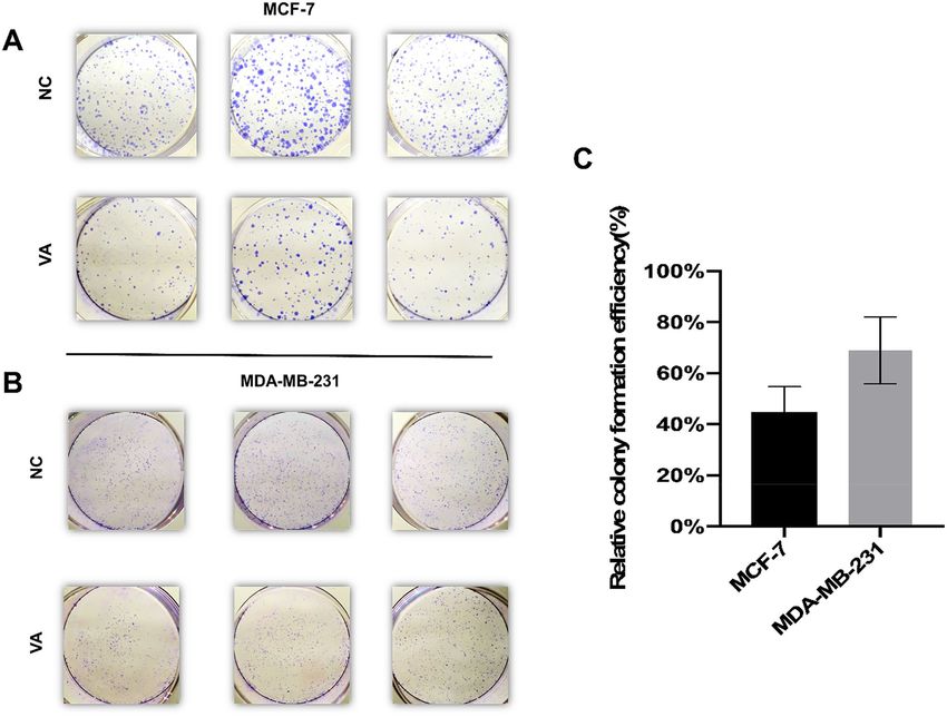
www.nature.com/scientificreports/
Figure 1. Cell proliferation inhibitory effect of valerian extract and valeric acid on breast cancer cells. (A)
A picture of Valerian root. (B) Valerian inhibited the growth of MCF-7 cells in a dose-dependent manner.
MCF-7 cells were incubated with valerian extract in different concentrations (0.75 mg/mL, 1.5 mg/mL, 3 mg/
mL, and 6 mg/mL) and significant inhibition of cell proliferation was observed after 48 h. (C) Cell proliferation
inhibitory effect of valeric acid on MCF-7, MDA-MB-231 and MCF-10A cells at different concentrations
(1 mM, 2.5 mM, 5 mM, and 10 mM) and the inhibition of cell proliferation was measured after 48 h. (D) Cell
proliferation inhibitory effect of valeric acid on MCF-10A, MDA-MB-231 and MCF-7 cells at 24 h, 48 h, and
72 h. Cells were incubated with 5 mM of valeric acid and all experiments were performed in triplicate.
chain fatty acid (SCFA), is a major active chemical ingredient of valerian and shares a high structural similarity
with the known histone deacteylase inhibitor (HDACi) valproic a cid12–14. HDACi is a novel group of anti-cancer
drugs, which may reverse gene-associated DNA methylation and influence gene expression l evels15. Considering
the role of circadian factors in breast cancer tumorogenesis and the potential HDAC inhibiting function, we
tested the hypothesis that valeric acid could provide therapeutic benefits against human breast cancer through
epigenetic modifications, including DNA methylation and histone deacetylation in the current study.
Results
Valerian and valeric acid reduce proliferation of breast cancer cells. Figure 1A shows a picture of
Valerian root. Compared to the control group, cell proliferation rates of valerian treated MCF-7 cells (0.75 mg/mL,
1.5 mg/mL, 3 mg/mL, 6 mg/mL) were significantly inhibited at 48 h by 10.39 ± 0.12% (p = 0.0038), 22.16 ± 5.02%
(p < 0.001), 38.22 ± 1.62% (p < 0.001) and 55.01 ± 3.46% (p < 0.001), respectively (Fig. 1B). We then tested anti-
proliferative activity of valeric acid at different concentrations against breast cancer cell lines MCF-7, MDA-
MB-231 and normal breast epithelial cells MCF-10A at 48 h. Results showed that valeric acid (1 mM, 2.5 mM,
5 mM, and 10 mM) inhibited MCF-7 cells by 27.92 ± 1.05% (p = 0.009), 13.36 ± 0.60% (p = 0.037), 25.15 ± 1.83%
(p = 0.006), 50.67 ± 2.57% (p = 0.001), and inhibited MDA-MB-231 cells by 17.07 ± 1.11% (p < 0.001), 26.99 ± 1.21%
(p < 0.001), 37.65 ± 1.35% (p < 0.001), 54.44 ± 0.85% (p < 0.001) at 48 h, respectively (Fig. 1C). Valeric acid at 5 mM
was further tested for anticancer effect at different time points. Specifically, the proliferation of MCF-7 cells was
inhibited by 34.48 ± 3.64% (p = 0.007), 51.95 ± 2.04% (p < 0.001), and 60.02 ± 0.53% (p < 0.001) at 24 h, 48 h, 72 h,
respectively, versus 24.13 ± 0.76% (p = 0.003), 37.65 ± 0.35% (p < 0.001), and 42.16 ± 1.91% (p = 0.001) for MDA-
MB-231; and 17.01 ± 0.58% (p < 0.001), 33.21 ± 3.55% (p = 0.016), and 33.18 ± 3.55% (p = 0.002) for MCF-10A at
the concentration of 5 mM (Fig. 1D). Overall, valeric acid showed strong inhibitory effect on MCF-7 and MDA-
MB-231 breast cancer cell lines and relatively less inhibitory effect on the MCF-10A cell line.
Effect of valeric acid on migration of breast cancer cells. A wound healing assay was performed to
examine the migration and invasiveness of breast cancer cells treated by valeric acid. For MDA-MB-231 cells,
the percentage of wound closure for control, 5 mM and 10 mM groups were 60.48 ± 2.97%, 40.48 ± 2.18% and
Scientific Reports | (2021) 11:2519 | https://doi.org/10.1038/s41598-021-81620-x 2
Vol:.(1234567890)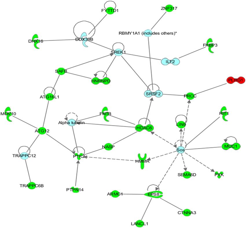
www.nature.com/scientificreports/
Figure 2. Effect of valeric acid on migration of MCF-7 and MDA-MB-231 cells. Monolayer cells were scraped
by a sterile micropipette tip and then treated with various concentrations of valeric acid for 24 h. (A,B)
MDA-MB-231 cells migrated to the wounded region were photographed (100 × magnification). (C,D) MCF-7
cells migrated to the wounded region were photographed (100 × magnification). The wound closure of the cell
cultures was presented in the middle field of each treatment, and data were calculated from three independent
experiments. All experiments were performed in triplicate.
28.10 ± 2.18% (Fig. 2A,B), and 72.86 ± 1.43%, 44.76 ± 2.18% and 25.71 ± 1.43% for MCF-7 (Fig. 2C,D), respec-
tively, with a significant difference (p < 0.001) in all comparisons.
Effect of valeric acid on colony formation of breast cancer cells. The colony formation assay
showed reduced cell colony formation ability of the cells (MCF-7 and MDA-MB-231) in valeric acid treated
groups (Fig. 3A,B). Figure 3C revealed that the calculated relative colony formation efficiency was 44.84 ± 8.05%
for MCF-7 cells treated by valeric acid (p < 0.001) and 68.9 ± 10.72% for MDA-MB-231 cells treated by valeric
acid (p < 0.001).
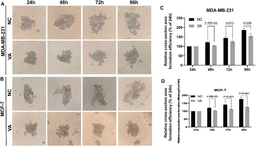
Inhibition of 3D formation by valeric acid. To investigate the impact of valeric acid on cell 3D con-
dition that mimics breast cancer cell growth in vivo, the relative cross-section area formation efficiency was
calculated by the comparison to their original area size (24 h) in each group, respectively. As shown in Fig. 4,
for MDA-MB-231 cells, relative cell cross-section area of valeric acid treated group reached to 103.52 ± 2.77%
at 48 h, and showed a significant difference compared to its counterparts of the control group (121.34 ± 2.61%,
p < 0.001). At 96 h, the trend still remained, relative area of valeric acid treated group reached to 168.54 ± 9.67%
compared to 180.73 ± 8.86% of the control group (p = 0.029). Similarly, a significantly smaller spheroid area was
observed in the valeric acid treated group compared to the control at both 48 h and 96 h (p < 0.001) in experi-
ment using MCF-7 cells.
Effect of valeric acid on HDAC activity. MCF-7 cells were treated with 2.5 mM, 5 mM, and 10 mM of
valeric acid and HDAC activity was measured after 48 h (Fig. 5A). The HDAC activity inhibition ratio of cells
treated with 2.5 mM valeric acid was 40.44 ± 1.88% that showed a significant reduction compared to the control
group (p = 0.028). Similarly, the HDAC activity of MCF-7 cells was inhibited by 53.15 ± 0.68% (p = 0.002) at the
Scientific Reports | (2021) 11:2519 | https://doi.org/10.1038/s41598-021-81620-x 3
Vol.:(0123456789)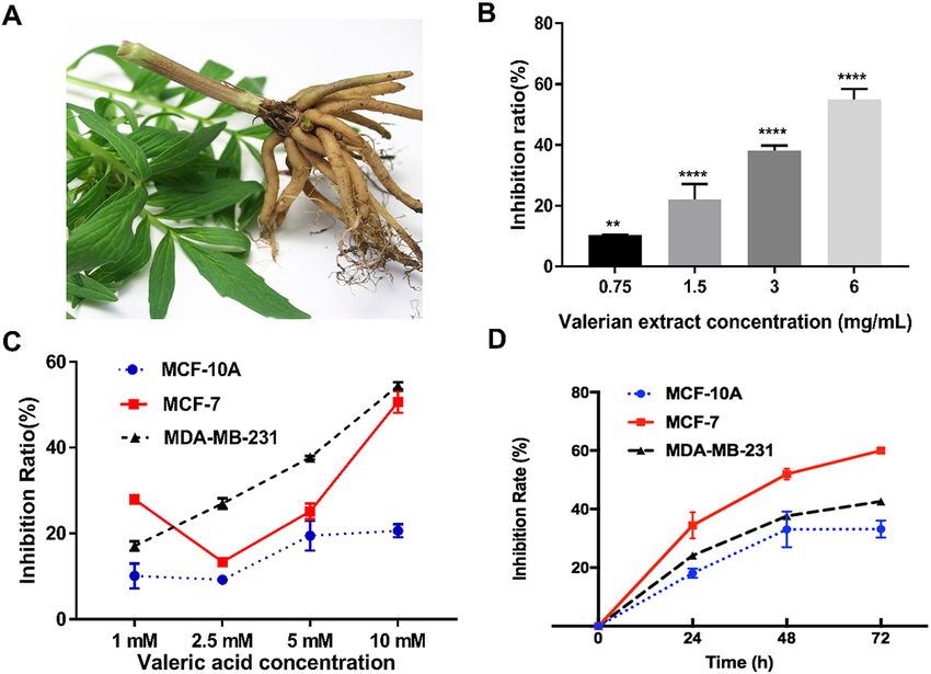
www.nature.com/scientificreports/

                                            Figure 3.  Valeric acid suppresses the colony formation ability of MCF-7 and MDA-MB-231. The representative
                                            colony formation of MCF-7 (A) and MDA-MB-231 (B) cells intervened by valeric acid (VA) and PBS,
                                            respectively. The relative colony formation efficiency showed reduction in number of colonies of both cells (C).
                                            Data are presented as the mean ± standard deviation (SD).

                                            concentration of 5 mM valeric acid and 61.64 ± 2.10% (p < 0.001) at the concentration of 10 mM valeric acid
                                            compared to the control group.

                                            Effect of valeric acid on global DNA methylation. A dose-dependent effect of valeric acid on the global
                                            DNA methylation levels in MCF-7 cells was observed. MCF-7 breast cancer cells were incubated with 2.5 mM,
                                            5 mM, and 10 mM of valeric acid and global DNA methylation was measured at 72 h. Global DNA methylation lev-
                                            els associated with different concentrations of valeric acid were 0.76 ± 0.02% (p < 0.001), 0.53 ± 0.04% (p < 0.001) and
                                            0.34% ± 0.02 (p < 0.001), respectively, versus 1.89 ± 0.03% for the control group in MCF-7 cells (Fig. 5B), indicating
                                            decreased global DNA methylation levels associated with increased valeric acid concentrations.

                                            Epigenetic landscape changes affected by valeric acid. To better understand epigenetic landscape
                                            changes affected by valeric acid, we performed the genome-wide DNA methylation assay followed by network-
                                            based analysis. Our data showed that 1398 CpG sites were differentially methylated across 1075 unique genes
                                            (Supplementary table). Significant CpG sites with methylation differences were determined by adjusted p values
                                            (FDR p < 0.05). Delta-beta ranges from − 0.14 to − 0.52 for hypomethylated sites and from 0.14 to 0.83 for hyper-
                                            methylated sites. Of these sites, 1267 CpG sites were hypermethylated while 131 CpG sites were hypomethylated
                                            in treated cells. According to the reference location of the probes used for each CpG site, we found that 229 CpG
                                            sites with differential methylation are located in either 5′UTR or 3′UTR regulatory regions. Sixty-three genes
                                            have multiple CpG sites with differential methylation and these CpG sites in 57 of these genes are either all
                                            hyper- or hypo-methylated.
                                                Among differentially methylated genes, there were 15 histone-related genes including histone deacetylase 6
                                            (HDAC6) that was hypermethlated. The network identified by the IPA tool as being most significantly associated
                                            with the set of input genes was designated as having functional relevance in “RNA post-transcriptional modifi-
                                            cation, cancer, and developmental disorder” (p = 10–59) as illustrated in Fig. 6. Briefly, 27 genes were present in

          Scientific Reports |   (2021) 11:2519 |                 https://doi.org/10.1038/s41598-021-81620-x                                                      4

Vol:.(1234567890)
www.nature.com/scientificreports/
Figure 4. Valeric acid inhibits the 3D formation of MCF-7 and MDA-MB-231 cells. (A) illustrated the dynamic
changes of 3D formation for MDA-MB-231 cells. (B) showed the dynamic changes of 3D formation for
MCF-7 cells. Their relative cross-section area formation efficiencies were shown in (C,D). Significant reduction
of cross-section area of MDA-MB-231 cells (C) and MCF-7 cells (D) in VA treated groups were observed.
Figure 5. Impact of valeric acid on HDAC activity and global DNA methylation. (A) MCF-7 cells were
incubated at three different concentrations of valeric acid and HDAC activity was measured after 48 h. Valeric
acid significantly inhibited HDAC activity at concentration of 2.5 mM, 5 mM, and 10 mM compared to
the control group (*p < 0.05). (B) The Global DNA methylation assay revealed that valeric acid (2.5 mM, 5 mM,
and 10 mM) significantly decreased the levels of global DNA methylation in MCF-7 cells compared to the
control group (*p < 0.05). All assays were performed in triplicate.
the network, 26 of which were significantly hypermethylated, while 1 of which was hypomethylated in treated
cells. Several putative tumor related genes with oncogeneic role in breast cancer were hypermethylated, such as
HDAC6, NASP, HNRNPC and LIN9.
Scientific Reports | (2021) 11:2519 | https://doi.org/10.1038/s41598-021-81620-x 5
Vol.:(0123456789)www.nature.com/scientificreports/
Figure 6. Network of interactions significantly enriched for genes displaying altered methylation patterns
following treatment with valeric acid. This network was identified by the IPA as being the most significantly
associated with the set of input genes and molecules within this network have functional relevance in “RNA
post-transcriptional modification, Cancer, and Developmental disorder” (p = 10E−59). Of the 27 genes present
in the network, 26 were significantly hypermethylated are shaded in green, while only 1 was hypomethylated
are shaded in red. Each interaction is supported by at least one literature reference identified in the Ingenuity
Pathway Knowledge Base, with solid lines representing direct interactions, and dashed lines representing
indirect interactions.
Discussion
Despite being the most commonly used herb (or herbal remedy) for sleep disorders, the efficacy of valerian root
to treat cancer remained unexplored. We have shown that valerian root and valeric acid (a major compound
of valerian root) can inhibit proliferation, migration, colony formation and 3D formation of breast cancer cells
in a dose- and time-dependent manner. Moreover, this inhibitory effect was significantly decreased on normal
breast epithelial cells compared to breast cancer cells, indicating a cancer-specific action with low side-effects.
These findings support our hypothesis that the sleep-related herb, valerian root, may provide therapeutic benefits
against human breast cancer.
To explore the underlying basis of the functional effect of valeric acid, we also conducted the HDAC activ-
ity assay because valeric acid shares a high structural similarity with the known histone deacteylase inhibitor
(HDACi) valproic a cid12–14. Histone acetylation is an important epigenetic modification and HDAC inhibitors
are among the most promising targets in the development of drugs for cancer t herapy16. More than 15 HDAC
Scientific Reports | (2021) 11:2519 | https://doi.org/10.1038/s41598-021-81620-x 6
Vol:.(1234567890)www.nature.com/scientificreports/
inhibitors have been tested in preclinical and clinical studies. Inhibition of HDAC leads to the acetylation of
non-histone proteins such as tumor suppressor p 5317, which may exhibit antiproliferative p
roperties18,19. Our
data demonstrate that valeric acid significantly decreases HDAC activity in treated breast cancer cells, which
provides a possible mechanism to account for its anticancer efficacy. This result suggests that valeric acid may
serve as a novel HDACi for breast cancer chemotherapy.
Histone acetylation may modify chromatin and most DNA methylation related proteins (DNA methyltrans-
ferase Dnmt1, several methyl-CpG binding proteins, MeCP2, MBD2, and MBD3) are associated with histone
deacetylase20. These enzymes regulate the process of DNA m ethylation15 and the HDAC activity reduced by
valeric acid treatment may lead to alterations of DNA methylation. Our results support this assertion by show-
ing that both global DNA methylation level and gene specific DNA methylation status were affected by valeric
acid. Of particlar interest, genome-wide DNA methylation screening detected 15 histone-related genes, includ-
ing histone deacetylase 6 (HDAC6), to be hypermethylated, which is in congruent with the observed decreased
HDAC activity in valeric acid treated cancer cells.
Hypermethylated HDAC6 gene may have anticancer properties because HDAC6 was significantly overex-
pressed in, not only breast cancer21, but also various other tumor types22. Histone deacetylase 6 (HDAC6) is a
unique member of the HDAC family and plays an important role in regulating the function of HSP90, α-tubulin,
and the a ggresome23 and effects on functional reduction of these downstream oncogenic targets may account
for the growth inhibitory effects of HDAC6 knockdown.
We also searched for transcription factor binding sites in the scouse sequence (50 bases) of HDAC6 5′UTR
that harbors the hypermethylated CpG site using the PROMO tool24 and identified binding sites of 4 transcrip-
tion factors, Pax-5 (GGGCCTG/CCTGCCC), p53 (CCTGCCC), AP-2 alphaA (GCCTGC), and C/EBP beta
(GCAA). All of these 4 transcription factors have been shown to play roles in breast tumorigenesis25–28, which
warrants further investigation on molecular mechanisms of valeric acid efficacy.
In addition to HDAC6, several other putative tumor related genes with oncogeneic role in breast cancer
were hypermethylated after valeric acid treatment in our study. NASP, nuclear autoantigenic sperm protein,
has become an important constituent of “poor prognosis signature” in breast cancer patients29. Depletion of
the histone chaperone tNASP inhibits proliferation and induces apoptosis in prostate cancer PC-3 c ells30 and
human gastric cancer31. HNRNPC, heterogeneous nuclear ribonucleoproteins C, is a RNA binding protein and
its elevated expression has been reported in cancer c ells32, but the function of HNRNPC in tumors is still not
clear. Repression of HNRNPC in breast cancer cells MCF7 and T47D was reported to inhibit cell proliferation
and tumor g rowth33. LIN9 is a part of the MuvB core complex (composed of LIN9, LIN52, LIN37, LIN54, and
RBBP4), which functions as a transcriptional regulator of mitosis. Overexpression of LIN9 has been observed
in the majority of triple-negative breast cancers (TNBCs) and is associated with poor prognosis across breast
cancer subtypes34. Hypermethylation of these putative oncogenes may provide a new mechanism accounting for
the observed anticancer efficacy of valeric acid in addition to the HDAC mediated processes.
Changes or alterations in both histone acetylation and DNA methylation are common targets studied in can-
cer epigenomics because these epigenetic processes are generally involved in cell proliferation, differentiation, and
survival, and are linked to carcinoma progression35,36. Discovering chemicals that can modify these epigenetic
processes is a promising new strategy for drug development in cancer chemotherapy. The functional effects of
valeric acid in the inhibition of histone deacetylase and alteration of DNA methylation make it a promising novel
epigenetic agent for future clinical implications in breast cancer treatment.
Although a profound anticancer effect is observed, findings from this study are still preliminary, which need
to be further confirmed in more breast cancer cell lines and animal models. More mechanistic studies are also
necessary to further evaluate other epigenetic effects of valeric acid. Inhibition rates of valeric acid on MCF-7
cells (not MDA-MB-231) decrease at low concentrations and then increase again at high doses in cell prolif-
eration assay. This might be due to hormone receptor status of these 2 cells. MCF-7 is a breast cancer cell line
with estrogen, progesterone and glucocorticoid receptors and MDA-MB-231 is a triple negative breast cancer
cell line. Agents such as valeric acid may have different types of dose-responses (i.e. bell-shaped and U-shaped
dose–response curves) when used to treat different tumor types. More breast cancer cell lines and doses are
needed to confirm this observation in future study. Nonetheless, as the promising profile of valeric acid were well
established, valeric acid is a potential new therapeutic agent for breast cancer and probably cancer in general,
worthy of further investigation.
Materials and methods
Cell lines and cell culture. Human breast cancer cells MCF-7 and MDA-MB-231, and normal breast epi-
thelial cells MCF-10A were purchased from the American Type Culture Collection (ATCC) (Manassas, VA,
USA). MCF-10A cells were maintained in MEGM (Life Technologies, CA, USA) supplemented with 100 ng/mL
cholera toxin. MCF-7 and MDA-MB-231 cells were cultured in DMEM (Life Technologies, CA, USA) supple-
mented with 10% fetal bovine serum, 1% penicillin–streptomycin. The cells were cultured at 37 °C in a humidi-
fied atmosphere consisting of 5% CO2. The culture medium was changed once every 2 days.
Preparation of valerian extract and valeric acid. Valerian and valeric acid were purchased from Chro-
madex (Irvine, CA, USA). Valerian was received as a powder from the herb pulverized rhizome. To prepare the
extract of valerian for experiments, 250 mg of this powder was diluted in 2 mL of distilled water, heated to 100 °C
for 20 min in a water bath and centrifuged at 1000g for 10 min. The supernatant was filtered through a hydro-
philic membrane containing 0.22 μM pores (Millipore, Billerica, MA, USA). Valeric acid was diluted in culture
medium to obtain working concentrations.
Scientific Reports | (2021) 11:2519 | https://doi.org/10.1038/s41598-021-81620-x 7
Vol.:(0123456789)www.nature.com/scientificreports/
Cell proliferation assay. We first tested the anticancer effect of valerian on the growth of breast cancer
cells using cell proliferation assay. MCF-7 cells were harvested and seeded into each well of 96-well plates at the
concentration of 4000 cells/100 μL, and different concentrations of valerian extract (0.75 mg/mL, 1.5 mg/mL,
3 mg/mL, and 6 mg/mL, final concentrations after dilution with medium) or the same volume of d dH2O (control
group) were added to each well. After incubation for 48 h, we added 20 μL of MTS assay (Promega, Wisconsin,
USA) per well in a dark hood and then incubated plates for 2 h. Optical density (OD) was determined at the
wavelength of 490 nm by a microplate Spectrophotometer (Biotek, Winooski, USA). MCF-7, MDA-MB-231
and MCF-10A cell lines were used to detect the efficacy of valeric acid on the proliferation of breast cancer cells.
Cells were seeded into 96-well plates as described above. 1 mM, 2.5 mM, 5 mM, and 10 mM of valeric acid or
the same volume of ddH2O were added and OD values were measured at different time points. Triplicate were
conducted for each condition at each time point. The proliferation inhibition rate was calculated using the for-
mula as: inhibition ratio = (1 − ODtreated group/ODcontrol group) × 100%. The inhibition ratio was fitted to a non-linear
regression plot to determine the IC50 (half maximal inhibitory concentration) for breast cancer cells treated with
valerian extract or valeric acid.
Wound‑healing assay. In order to understand how valeric acid affects cancer cell migration and con-
firm the cell proliferation results, we performed a wound-healing assay. Approximately 1 × 10⁶ MCF-7 or MDA-
MB-231 cells were added into each well of 6-well tissue culture plates with culture medium containing 10% FBS
and grew to nearly 80–90% confluence. A scratch was gently made using a 100 μL pipette tip in each well and the
cells were washed with PBS twice to remove the detached cells. Cells were then treated with different concentra-
tions of valeric acid or the same amount of ddH2O with 0.1% FBS at 37 °C for 24 h. Wound-healing assay was
conducted in triplicate. Images were taken at 0 h and 24 h and the area of a wound was measured with ImageJ
software (version of 1.52a) and the average extent of wound closure was quantified.
Colony formation assay. To examine relatively long-time (~ 10 days) inhibitory impact of valaric acid on
the growth of breast cancer calle, we conducted a colony formation assay. Approximately 3000 MCF-7 or MDA-
MB-231 cells were seeded into 6-well tissue culture plates with 2 mL complete medium per well. Cells were
treated with 0.85 mM valeric acid or the same amount of ddH2O (control group). Cells were cultured for 10 days
and drug-containing culture medium was renewed once every 3 days. Cells were then fixed by 4% paraformal-
dehyde (FD NeuroTechnologies, USA) and dyed with 0.05% crystal violet (Sigma-Aldrich, USA) at room tem-
perature. Images were taken and the number of colonies with more than 50 cells was counted manually. Relative
colony formation efficiency = (amount of colony in treated group/amount of colony in control group) × 100%.
3D formation assay. We simulated in vivo cancer cell survival when treated with valeric acid by perform-
ing a 3D formation assay. A hanging drop method has been successfully developed for generation of 3D sphe-
roids that enables the growth of cells in a multidimensional and multicellular manner and facilitates cancer
study and more efficient drug s creening37,38. Briefly, MDA-MB-231 or MCF-7 cells were placed into each drop
which contained 20 μl complete medium and 200 cells, respectively. 0.85 mM valeric acid or the same amount
of ddH2O (control group) was then added into each drop. All drops were cultured in a humidified incubator at
37 °C with 5% CO2. Images were taken at different time points (24 h, 48 h, 72 h, 96 h). The cross-section area of
3D spherical formation was calculated by software ImageJ (version of 1.52a).
HDAC colorimetric activity assay. The effect of valeric acid on histone deacetylase (HDAC) activity was
assessed using the Colorimetric HDAC Activity Assay Kit (BioVison, Milpitas, CA, USA) according to the man-
ufacturer’s instructions. Briefly, MCF-7 cells were treated with various concentrations of valeric acid for 48 h,
at which point nuclear protein fractions were prepared using the Epiquik Nuclear Extraction Kit (Epigentek,
USA) following the manufacturer’s protocols. 50 μg of nuclear extracts from treated cells were then diluted in
85 μL ddH2O. 10 μL of 10 × HDAC assay buffer was added followed by the addition of 5 μL of the colorimetric
substrate. Samples were then incubated at 37 °C for 1 h. Subsequently, the reaction was stopped by adding 10
μL of lysine developer and left for an additional 30 min at 37 °C. OD values of samples were read by microplate
spectrophotometer (Biotek, Winooski, USA) at the wavelength of 405 nm. HDAC activity was expressed as rela-
tive OD values per μg of protein sample. The assay was conducted in triplicate.
Global DNA methylation assay. MCF-7 cells were treated with 5 mM valeric acid for 72 h and then
collected for genomic DNA isolation using the QIAamp DNA Blood Mini Kit (Qiagen Sciences, Maryland,
MD, USA). The Global DNA methylation levels were determined using the MethylFlash Global DNA Meth-
ylation Quantification Kit (Epigentek, NY, USA) according to the manufacturer’s instructions. The absorbance
of the samples was measured on amicroplate reader at 450 nm. The experiment was performed in triplicate.
For the OD intensity to be proportional to the amount of methylated DNA, the % 5-mC was calculated using
the following formula: 5-mC% = [(sample OD-negative control OD) × P/(positive control OD-negative control
OD) × S × 2] × 100%, where S represents the amount of input sample DNA (100 ng) and P represents the amount
of input positive control DNA (5 ng).
Genome‑wide DNA methylation array. To further explore epigenetic impact of valeric acid on specific
CpG sites in cancer related genes, we performed a genome-wide DNA methylation analysis. Genomic DNA of
MCF-7 cells treated with 5 mM valeric acid for 72 h was isolated and purified using the QIAamp DNA Blood
Mini Kit (Qiagen Sciences, Maryland, MD, USA) according to the manufacturer’s protocols. 50 ng of genomic
Scientific Reports | (2021) 11:2519 | https://doi.org/10.1038/s41598-021-81620-x 8
Vol:.(1234567890)www.nature.com/scientificreports/
DNA per sample was used for the analysis using the Infinium Human Methylation 450 BeadChips (Illumina,
San Diego, CA USA). A methylation index (β) was used to estimate the methylation level of each CpG site using
the ratio of intensities between methylated and unmethylated cytosines, which is a continuous variable between
0 and 1. 0 corresponds to a completely unmethylated site, while 1 corresponds to a completely methylated site.
Significant methylation differences in methylation levels between treated and control group at each CpG site
were obtained using The Illumina Custom Model.
Network analysis. The Ingenuity Pathway Analysis software (IPA, Ingenuity Systems) (Redwood City,
CA) was used to investigate whether differentially methylated genes were enriched for functional relationships
between treated and control groups. Differentially methylated genes were input into the program and used to
identify experimentally verified interactions using the Ingenuity Knowledge Base, a manually curated database
of functional interactions extracted from peer reviewed p ublications39. p values for individual networks were
obtained by comparing the likelihood of obtaining the same number of genes or greater in a random gene set as
are actually present in the input set using a Fisher’s exact test, based on the hypergeometric distribution.
Statistical analysis. All statistics and figures were generated using the GraphPad Prism 7.00 software
(www.graphpad.com) if not specified. All results were shown as mean ± standard error. All p values presented
were two-sided and p < 0.05 was regarded as statistically significant.
Ethical approval. This article does not contain animal studies and any studies with human participants
performed by any of the authors.
Received: 28 September 2020; Accepted: 6 January 2021
References
1. Cragg, G. M. & Newman, D. J. Plants as a source of anti-cancer agents. J. Ethnopharmacol. 100, 72–79 (2005).
2. Foa, R., Norton, L. & Seidman, A. D. Taxol (paclitaxel): a novel anti-microtubule agent with remarkable anti-neoplastic activity.
Int. J. Clin. Lab. Res. 24, 6–14 (1994).
3. Block, K. I., Gyllenhaal, C. & Mead, M. N. Safety and efficacy of herbal sedatives in cancer care. Integr. Cancer Ther. 3, 128–148.
https://doi.org/10.1177/1534735404265003 (2004).
4. Bent, S., Padula, A., Moore, D., Patterson, M. & Mehling, W. Valerian for sleep: a systematic review and meta-analysis. Am. J. Med.
119, 1005–1012. https://doi.org/10.1016/j.amjmed.2006.02.026 (2006).
5. Viswanathan, A. N. & Schernhammer, E. S. Circulating melatonin and the risk of breast and endometrial cancer in women. Cancer
Lett. 281, 1–7 (2009).
6. Samuelsson, L. B., Bovbjerg, D. H., Roecklein, K. A. & Hall, M. H. Sleep and circadian disruption and incident breast cancer risk:
an evidence-based and theoretical review. Neurosci. Biobehav. Rev. 84, 35–48. https://doi.org/10.1016/j.neubiorev.2017.10.011
(2018).
7. Straif, K. et al. Carcinogenicity of shift-work, painting, and fire-fighting. Lancet Oncol. 8, 1065–1066 (2007).
8. Group, I. M. V. Carcinogenicity of night shift work. Lancet Oncol. 20, 1058–1059. https://doi.org/10.1016/S1470-2045(19)30455
-3 (2019).
9. Chen, S. T. et al. Deregulated expression of the PER1, PER2 and PER3 genes in breast cancers. Carcinogenesis 26, 1241–1246 (2005).
10. Sahar, S. & Sassone-Corsi, P. Circadian clock and breast cancer: a molecular link. Cell Cycle 6, 1329–1331 (2007).
11. Filipski, E. et al. Host circadian clock as a control point in tumor progression. J. Natl. Cancer Inst. 94, 690–697 (2002).
12. Gurvich, N., Tsygankova, O. M., Meinkoth, J. L. & Klein, P. S. Histone deacetylase is a target of valproic acid-mediated cellular
differentiation. Cancer Res. 64, 1079–1086. https://doi.org/10.1158/0008-5472.can-03-0799 (2004).
13. Fuks, F., Burgers, W. A., Brehm, A., Hughes-Davies, L. & Kouzarides, T. DNA methyltransferase Dnmt1 associates with histone
deacetylase activity. Nat. Genet. 24, 88–91 (2000).
14. Fuks, F., Hurd, P. J., Deplus, R. & Kouzarides, T. The DNA methyltransferases associate with HP1 and the SUV39H1 histone
methyltransferase. Nucleic Acids Res. 31, 2305–2312 (2003).
15. Sarkar, S. et al. Histone deacetylase inhibitors reverse CpG methylation by regulating DNMT1 through ERK signaling. Anticancer
Res. 31, 2723–2732 (2011).
16. Suraweera, A., O’Byrne, K. J. & Richard, D. J. Combination therapy with histone deacetylase inhibitors (HDACi) for the treatment
of cancer: achieving the full therapeutic potential of HDACi. Front. Oncol. 8, 92. https://doi.org/10.3389/fonc.2018.00092 (2018).
17. Gu, W. & Roeder, R. G. Activation of p53 sequence-specific DNA binding by acetylation of the p53 C-terminal domain. Cell 90,
595–606. https://doi.org/10.1016/S0092-8674(00)80521-8 (1997).
18. p21WAF1 is required for butyrate-mediated growth inhibition of human colon cancer cells.
19. Joung, K. E., Kim, D. K. & Sheen, Y. Y. Antiproliferative effect of trichostatin A and HC-toxin in T47D human breast cancer cells.
Arch. Pharm. Res. 27, 640–645. https://doi.org/10.1007/Bf02980164 (2004).
20. El-Osta, A. & Wolffe, A. P. DNA methylation and histone deacetylation in the control of gene expression: basic biochemistry to
human development and disease. Gene Expr. 9, 63–75 (2000).
21. Lee, Y. S. et al. The cytoplasmic deacetylase HDAC6 is required for efficient oncogenic tumorigenesis. Can. Res. 68, 7561–7569.
https://doi.org/10.1159/0008-5472.Can-08-0188 (2008).
22. Yang, W. M., Yao, Y. L., Sun, J. M., Davie, J. R. & Seto, E. Isolation and characterization of cDNAs corresponding to an additional
member of the human histone deacetylase gene family. J. Biol. Chem. 272, 28001–28007. https://doi.org/10.1074/Jbc.272.44.28001
(1997).
23. Boyault, C., Sadoul, K., Pabion, M. & Khochbin, S. HDAC6, at the crossroads between cytoskeleton and cell signaling by acetyla-
tion and ubiquitination. Oncogene 26, 5468–5476. https://doi.org/10.1038/Sj.Onc.1210614 (2007).
24. Messeguer, X. et al. PROMO: detection of known transcription regulatory elements using species-tailored searches. Bioinformatics
18, 333–334. https://doi.org/10.1093/bioinformatics/18.2.333 (2002).
25. Kaur, R. P., Vasudeva, K., Kumar, R. & Munshi, A. Role of p53 gene in breast cancer: focus on mutation spectrum and therapeutic
strategies. Curr. Pharm. Des. 24, 3566–3575. https://doi.org/10.2174/1381612824666180926095709 (2018).
Scientific Reports | (2021) 11:2519 | https://doi.org/10.1038/s41598-021-81620-x 9
Vol.:(0123456789)www.nature.com/scientificreports/
26. Turner, B. C. et al. Expression of AP-2 transcription factors in human breast cancer correlates with the regulation of multiple
growth factor signalling pathways. Cancer Res. 58, 5466–5472 (1998).
27. Zahnow, C. A. CCAAT/enhancer-binding protein beta: its role in breast cancer and associations with receptor tyrosine kinases.
Expert Rev. Mol. Med. 11, e12. https://doi.org/10.1017/S1462399409001033 (2009).
28. Benzina, S. et al. Pax-5 is a potent regulator of E-cadherin and breast cancer malignant processes. Oncotarget 8, 12052–12066.
https://doi.org/10.18632/oncotarget.14511 (2017).
29. Martin, K. J., Patrick, D. R., Bissell, M. J. & Fournier, M. V. Prognostic breast cancer signature identified from 3D culture model
accurately predicts clinical outcome across independent datasets. PLoS ONE 3, e2994. https://doi.org/10.1371/journal.pone.00029
94 (2008).
30. Alekseev, O. M., Richardson, R. T., Tsuruta, J. K. & O’Rand, M. G. Depletion of the histone chaperone tNASP inhibits prolifera-
tion and induces apoptosis in prostate cancer PC-3 cells. Reprod. Biol. Endocrinol. 9, 50. https://doi.org/10.1186/1477-7827-9-50
(2011).
31. Yu, B. et al. microRNA-29c inhibits cell proliferation by targeting NASP in human gastric cancer. BMC Cancer 17, 109. https://
doi.org/10.1186/s12885-017-3096-9 (2017).
32. Park, Y. M. et al. Heterogeneous nuclear ribonucleoprotein C1/C2 controls the metastatic potential of glioblastoma by regulating
PDCD4. Mol. Cell. Biol. 32, 4237–4244. https://doi.org/10.1128/MCB.00443-12 (2012).
33. Wu, Y. et al. Function of HNRNPC in breast cancer cells by controlling the dsRNA-induced interferon response. EMBO J. https
://doi.org/10.15252/embj.201899017 (2018).
34. Sahni, J. M. et al. Mitotic vulnerability in triple-negative breast cancer associated with LIN9 Is targetable with BET inhibitors.
Cancer Res. 77, 5395–5408. https://doi.org/10.1158/0008-5472.CAN-17-1571 (2017).
35. Herceg, Z. Epigenetics and cancer: towards an evaluation of the impact of environmental and dietary factors. Mutagenesis 22,
91–103. https://doi.org/10.1093/Mutage/Ge1068 (2007).
36. Esteller, M. Cancer epigenomics: DNA methylomes and histone-modification maps. Nat. Rev. Genet. 8, 286–298. https://doi.
org/10.1038/Nrg2005 (2007).
37. Hao, R. et al. A microfabricated 96-well 3D assay enabling high-throughput quantification of cellular invasion capabilities. Sci.
Rep. 7, 43390. https://doi.org/10.1038/srep43390 (2017).
38. Foty, R. A simple hanging drop cell culture protocol for generation of 3D spheroids. J. Vis. Exp. https: //doi.org/10.3791/2720 (2011).
39. Calvano, S. E. et al. A network-based analysis of systemic inflammation in humans. Nature 437, 1032–1037. https: //doi.org/10.1038/
Nature03985 (2005).
Author contributions
Y.Z.: Conception and design. F.S., Y.L., R.H., A.F., R.W., Q.Q.: Development of methodology. F.S., Q.Q.: Acqui-
sition of data. F.S., O.N., Y.Z.: Analysis and interpretation. F.S., O.N., L.H., X.C., Y.Z.: Writing, review, and/or
revision of the manuscript. Y.Z.: Administrative, technical, or material support. Y.Z.: Study supervision.
Funding
This work was supported by funds from Yale University and NIH research Grant CA238100. Fengqin Shi, Ya Li,
Rui Han and Ronghua Wang were supported by China Scholarship Council (CSC).
Competing interests
The authors declare no competing interests.
Additional information
Supplementary Information The online version contains supplementary material available at https://doi.
org/10.1038/s41598-021-81620-x.
Correspondence and requests for materials should be addressed to Y.Z.
Reprints and permissions information is available at www.nature.com/reprints.
Publisher’s note Springer Nature remains neutral with regard to jurisdictional claims in published maps and
institutional affiliations.
Open Access This article is licensed under a Creative Commons Attribution 4.0 International
License, which permits use, sharing, adaptation, distribution and reproduction in any medium or
format, as long as you give appropriate credit to the original author(s) and the source, provide a link to the
Creative Commons licence, and indicate if changes were made. The images or other third party material in this
article are included in the article’s Creative Commons licence, unless indicated otherwise in a credit line to the
material. If material is not included in the article’s Creative Commons licence and your intended use is not
permitted by statutory regulation or exceeds the permitted use, you will need to obtain permission directly from
the copyright holder. To view a copy of this licence, visit http://creativecommons.org/licenses/by/4.0/.
© The Author(s) 2021
Scientific Reports | (2021) 11:2519 | https://doi.org/10.1038/s41598-021-81620-x 10
Vol:.(1234567890)You can also read