Synesthesia does not help to recover perceptual dominance following flash suppression
←
→
Page content transcription
If your browser does not render page correctly, please read the page content below
www.nature.com/scientificreports
OPEN Synesthesia does not help
to recover perceptual dominance
following flash suppression
Diana Jimena Arias1,2 & Dave Saint‑Amour1,2,3*
Grapheme-colour synesthesia occurs when letters or numbers elicit an abnormal colour sensation
(e.g., printed black letters are perceived as coloured). This phenomenon is typically reported
following explicit presentation of graphemes. Very few studies have investigated colour sensations in
synesthesia in the absence of visual awareness. We took advantage of the dichoptic flash suppression
paradigm to temporarily render a stimulus presented to one eye invisible. Synesthetic alphanumeric
and non-synesthetic stimuli were presented to 21 participants (11 synesthetes) in achromatic and
chromatic experimental conditions. The test stimulus was first displayed to one eye and then masked
by a sudden presentation of visual noise in the other eye (flash suppression). The time for an image to
be re-perceived following the onset of the suppressive noise was calculated. Trials where there was
no flash suppression performed but instead mimicked the perceptual suppression of the flash were
also tested. Results showed that target detection by synesthetes was significantly better than by
controls in the absence of flash suppression. No difference was found between the groups in the flash
suppression condition. Our findings suggest that synesthesia is associated with enhanced perception
for overt recognition, but does not provide an advantage in recovering from a perceptual suppression.
Further studies are needed to investigate synesthesia in relation to visual awareness.
Synesthesia is a perceptual phenomenon in which a stimulus elicits an abnormal and concurrent sensation in the
same or a different sensory modality. Among the different types of synesthesia, grapheme-colour synesthesia is
relatively common and it consists of colour perceptions evoked by grey scale alphanumeric images (letters and/
or digits)1. Synesthetic associations are consistent over t ime2–4 and are automatic or difficult to discard when
elicited3,5–9.
Grapheme-colour synesthesia has been most frequently investigated through the explicit presentation of
aradigms5–10. However, synes-
synesthetic graphemes or digits, as shown with stroop-like tasks and visual search p
thesia remains much less studied when observers are unaware of a stimulus. For a review s ee11. Mattingley et al.9
adapted a masking paradigm in order to test synesthetic stimuli under conditions preventing their visibility. They
measured how long it takes for participants to name the colour of a target mask that precedes the presentation of
a letter prime, which evokes a synesthetic colour, either congruent or incongruent with the colour of the target
mask. Congruent and incongruent trials were used to assess the synesthetic interference effect on naming the
target’s colour. The letter prime was visible when presented for 500 ms and invisible when presented for 56 ms
or less. The synesthetic interference effect was observed only when the prime was visible to participants, i.e., this
effect disappeared when the letter did not access visual awareness. Furthermore, synesthete participants showed
implicit priming effects similar to controls for non-synesthetic inducing stimuli; that is, the brief presentation
of a semantic prime (e.g., an upper-case letter “A”) improved the subsequent recognition of a congruent target
stimulus (e.g., lower-case letters “a”).
Synesthesia was also tested using the attentional blink paradigm in which two successive targets, T1 and
T2, are presented in a sequence and separated by distractors (masks)12. In this paradigm, the second target T2
is rendered invisible when the presentation time between both targets does not exceed the attentional window,
which is between 300 and 500 ms. Rich et al.13 presented a synesthetic prime (T2) within and outside of the
attentional window. A colour probe was presented at the end of the attentional blink sequence. The prime elicited
a synesthetic colour that was either congruent or incongruent with the colour probe, producing an interference
effect in the colour naming of the probe. Albeit modest, a reliable interference effect was found when the prime
was visible. No interference effect was obtained when the prime was presented within the temporal window of
1
Department of Psychology, Université du Québec à Montréal, Montreal H2X 3P2, Canada. 2Cognitive
Neurosciences Research Center, Université du Québec à Montréal, Montreal H2X 3P2, Canada. 3Research Center of
the Sainte-Justine University Hospital, Montreal H3T 1C5, Canada. *email: saint-amour.dave@uqam.ca
Scientific Reports | (2021) 11:7566 | https://doi.org/10.1038/s41598-021-87223-w 1
Vol.:(0123456789)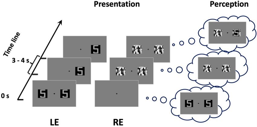
www.nature.com/scientificreports/
the attentive blink. In another study using the same attentional blink design, however, some synesthetic partici-
pants (5 out of 10) were able to perceive synesthetic colours, even when the synesthetic primes fell within the
attentional blink temporal w indow14. As such, an interference effect in a colour naming task was still noticed
when participants were unable to overtly identify the synesthetic prime in the visual sequence. In line with this
finding, it has been reported in four grapheme-colour synesthetes that conscious letter recognition is not required
to perceive the colour of hidden l etters15.
The aforementioned studies are not conclusive with regard to whether synesthesia can occur without visual
awareness9,13–15. To further investigate this question, we looked at the flash suppression paradigm, which has been
used extensively to study visual processing in the absence of awareness16,17. The flash suppression phenomenon
is typically induced by two different monocular images presented asynchronously; an image is first presented to
one eye for a while (while a blank field is presented to the other eye), after which the second image is abruptly
shown, i.e., flashed, to the other eye at the corresponding retinal points. Unlike the masking and attentional
blink paradigms, flash suppression allows a stimulus to be rendered invisible temporarily, even though it remains
physically present for the observer. Thus, it allows manipulation of the onset of interocular suppression, i.e., the
awareness of the suppressed stimulus, before binocular rivalry between the competing images occurs16. Unlike
binocular rivalry, which involves alternation of perceptual dominance and complex neural dynamics at several
brain levels18, flash suppression offers better control of the monocular s uppression19. Previous studies using
flash suppression have shown that even if subjects are not aware of the presence of a stimulus in one eye, visual
processing of that stimulus may still occur16,20–22.
It was previously reported in normal observers that the flash suppression of a coloured Gabor grating is
shorter than that of an achromatic Gabor grating17, and colour can break suppression more efficiently than
other visual f eatures23. Thus the present study tested the hypothesis that the effect of flash suppression on the
synesthetic (subjectively colored) stimulus is weaker than that of the non-synesthetic stimulus. By measuring
the time the hidden stimulus takes to break through to awareness, we aimed to determine the effect of synaes-
thesia on perceptual suppression. If synesthesia occurs when the participants are not aware of the synesthetic
achromatic grapheme, the prediction is that they will exhibit a shorter duration of suppression in comparison
to non-synesthetic conditions.
Results
One synesthete (participant 6) consistently reported longer reaction times (RTs) for the non-flash-suppression
trials, significantly longer than those obtained from the rest of the participants (Z-scores ≤ − 3.5). This participant
was thus excluded from the data analysis.
The analysis of variance (ANOVA) conducted on the perceptual flash suppression trials showed no
main group effect [F(1,19) = 0.523, p = 0.478, η2p 0.027] or interaction of the group with the other factors:
condition*group [F(1,19) = 0.280, p = 0.603, η2p 0.015], stimulus*group [F(1,19) = 0.716, p = 0.408, η2p 0.036] and
the condition*stimulus*group [F(1,19) = 0.001, p = 0.975, η2p 0.00005]. However, a main effect of the condition
[F(1,19) = 63.833, p < 0.001, η2p = 0.771], the stimulus [F(1,19) = 21.591, p < 0.001, η2p = 0.532] as well as the interaction
of condition*stimulus [F(1,19) = 36.989, p < 0.001, η2p = 0.661] was found to be statistically significant. As is depicted
in Fig. 1, all participants exhibited a significantly shorter duration of suppression in the chromatic condition
than in the achromatic condition, and this effect was stronger for alphanumeric stimuli.
Two sensitivity analyses were also performed. First, an additional ANOVA was conducted on the perceptual
suppression duration while excluding the participant reporting a “d” synesthetic stimulus, instead of the "5"
observed by the other synesthetes. Second, a similar approach was conducted by excluding the synesthete par-
ticipant who showed a projector synesthesia profile, as revealed by the online synesthesia battery test. Results
from these two ANOVA remained the same (data not shown). It should be noted that Z-score tests revealed that
the performance of the projector synesthete participant was not significantly different from the other synesthete
participants, or from the control participants.
For non-flash-suppression trials, the ANOVA revealed robust significant effects of the condition
[F(1,19) = 21.111, p < 0.001, η2p = 0.526], the stimulus [F(1,19) = 23.970, p < 0.001, η2p = 0.558], and the group
[F(1,19) = 5.651, p = 0.028, η2p = 0.229]. No interaction effects between factors were found to be statistically sig-
nificant: condition*stimulus*group [F(1,19) = 2.268, p = 0.146, η2p = 0.107], condition*stimulus [F(1,19) = 0.586,
p = 0.453, η2p = 0.030], condition*group [F(1,19) = 2.644, p = 0.120, η2p = 0.122], stimulus*group [F(1,19) = 2.877,
p = 0.106, η2p = 0.132]. Thus, participants perceived chromatic stimuli faster than achromatic stimuli (Fig. 2).
In addition, alphanumeric stimuli were more rapidly reported than abstract stimuli. In comparison to controls,
target detection in all conditions was in general faster in synesthete participants. Of note, the performance of
the synesthetes in the achromatic condition was not statistically different (t(9) = 1.231, p = 0.249) between the
alphanumeric (M = 1.001, SD = 0.164) and abstract stimuli (M = 1.077 SD = 0.216).
To confirm these findings with a maximum of statistical power, data were also analyzed using mixed effects
modeling to treat subjects as a random factor, and the condition, stimulus and group as fixed factors. This
approach was applied separately for flash suppression trials and non-flash-suppression trials. Results (see Sup-
plementary Tables S1 and S2) were in line with those obtained with the aforementioned ANOVAs. However,
the mixed model in the non-flash suppression condition (Table S2) revealed a trend toward significance for the
interaction between group and condition (p = 0.059). Exploratory follow-up analysis indicated that synesthetes
were actually faster than the controls in the achromatic condition (M = 1.039, SD = 0.080 vs. M = 1.347, SD = 0.077,
p = 0.011).
Bayesian repeated measures ANOVAs were also conducted to quantify the plausibility of both the null and
alternative hypotheses, permitting interpretation of null findings. In Bayesian inference, the likelihood of the data
is considered under both hypotheses, and these probabilities are compared via the Bayes factor (BF). According to
Scientific Reports | (2021) 11:7566 | https://doi.org/10.1038/s41598-021-87223-w 2
Vol:.(1234567890)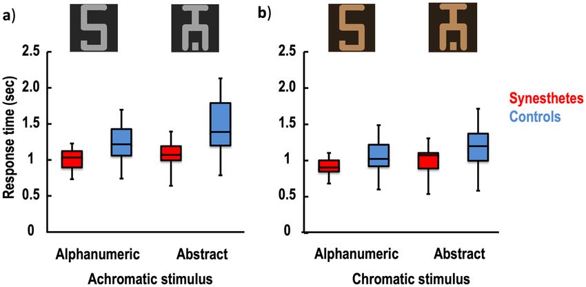
www.nature.com/scientificreports/
Figure 1. Perceptual stimulus suppression. The flash suppression duration is shown for the achromatic
(a) and chromatic (b) stimulus conditions in the synesthetic participants (red) and the control participants
(blue). Considering that the physical achromatic grapheme stimulus is associated with a colored percept in the
synesthetes, a significant difference between the alphanumeric and abstract stimuli in the achromatic condition
was expected in the synesthetes, but not in the controls. The chromatic condition is illustrated by the orange
colour as an example. The whiskers indicate the minimum and maximum range of the distributions; the top and
bottom of the boxes show the first and third quartiles (the 25th and 75th percentiles), and the horizontal bars
inside the boxes represent the medians.
Figure 2. Stimulus detection in the absence of flash suppression. Response time is shown for the achromatic
(a) and chromatic (b) stimulus conditions in the synesthetic participants (red) and the control participants
(blue). Considering that the physical achromatic grapheme stimulus is associated with a colored percept in the
synesthetes, a significant difference between the alphanumeric and abstract stimuli in the achromatic condition
was expected in the synesthetes, but not in the controls. The chromatic condition is illustrated by the orange
colour as an example. The whiskers indicate the minimum and maximum range of the distributions, the top and
bottom of the boxes show the first and third quartiles (the 25th and 75th percentiles), and the horizontal bars
inside the boxes represent the medians.
Lee and Wagenmakers’ classification24, the level of evidence was deemed inconclusive/anecdotal for BF between
0.33 and 3, moderate for BF < 0.33 or > 3, and strong for BF < 0.1 or > 10. Following the JASP guidelines25, BF
comparing the null model against all other models were computed and each experimental effects was obtained
by calculating the inclusion BF across matched models. Results revealed overwhelming evidence in favor of a
main effect of condition (BF of 1.887e+8 and 8.465e+3 for flash and non-flash trials, respectively), a main effect
Scientific Reports | (2021) 11:7566 | https://doi.org/10.1038/s41598-021-87223-w 3
Vol.:(0123456789)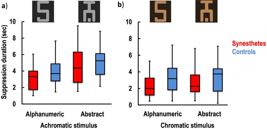
www.nature.com/scientificreports/
of stimulus (BF of 8892.142 and 3226.375 for flash and non-flash trials, respectively) as well as their interaction
(BF = 17.523, for flash trials). However, inconclusive evidence was found for or against of a main or interaction
effect of group (all BF values ranged between 0.33 and 3). Details are presented in Supplementary Tables S3 and
S4.
Discussion
The present study investigated whether synesthesia shortens the duration of interocular suppression. Results
from the flash suppression trials revealed no evidence of suppression modulation from synesthetic stimuli,
and no significant differences between the synesthete and non-synesthete groups. However, chromatic stimuli
exhibited shorter suppression latencies, as they emerge more quickly, than achromatic stimuli. We also found
in both synesthetes and controls that, in comparison to the abstract stimuli, the alphanumeric stimuli reduced
suppression durations. In the non-flash-suppression trials, all participants detected coloured stimuli faster than
achromatic stimuli. They also showed shorter reaction times for the detection of alphanumeric stimuli than for
abstract stimuli. In the non-flash-suppression trials, we found that the synesthete participants exhibited a better
performance than the controls, regardless of the type (synesthetic or not) and the colour (chromatic or achro-
matic) of the stimuli used. The small sample size is a major limitation of the study, which might be particularly
challenging with between-subject designs and non-significant findings. We statistically addressed this issue
by re-analyzing the data using mixed effects models, with the subject level variability as a random effect. The
results remained the same. Bayesian analyses support the presence of strong effects of stimulus chromaticity and
stimulus type on performance, but did not provide sufficient evidence for or against an effect of synesthesia, i.e.,
BF > 0.33 and < 3. Thus, given the modest magnitude of the Bayes factors, it appears that the observed data were
insensitive to detect an effect of synesthesia, suggesting that our study was underpowered (i.e., more participants
might be required).
The stimulus features, i.e., the colour and familiarity, was found to influence the performance in all partici-
pants, whether under interocular suppression viewing condition or not. Some data from normal observers in the
seminal study of Tsuchiya and K och17 on the continuous flash suppression (CFS) suggest a break through effect
of chromaticity that favor stimulus predominance. This observation is in agreement with the notion that the
interocular suppression of colour is weaker than on form23, and with the general consensus that colour improves
signal detection by enhancing the saliency of the s timulus26,27. Regarding the effect of the alphanumeric stimuli,
it is possible that response time in detection (with or without interocular suppression) was shorter because of
higher familiarity and/or meaning of those stimuli, in comparison to the abstract and unusual symbols. Although
it is debated whether semantic processing under continuous flash suppression occurs at all (see28,29), Gobbini
and coworkers30 reported that processing of familiar stimuli (faces) is more likely to resist flash suppression than
non-familiar ones. This result is in line with the fact that, under explicit viewing conditions, familiar stimuli are
more efficiently processed and detected than meaningless or unfamiliar t argets31. Interestingly, our results also
showed that the effect of stimulus colour and familiarity on reducing interocular suppression are not independ-
ent, but interact. Thus, coloured stimuli overcame suppression faster when they were also familiar.
The current study failed to demonstrate that synesthetic stimuli biased the performance of the synesthete, sug-
gesting that synesthesia does not bias interocular suppression or modulate stimuli that are rendered temporarily
invisible by flash suppression. This interpretation is in agreement with the notion that conscious recognition is
required in order to elicit synesthetic p ercepts9, including the study of Rich and M attingley13 using attentional
blink, in which no synesthetic effect (i.e., interference stroop-like effect of T2 on subsequent colour naming) was
observed when the synesthetic prime (T2) was not consciously perceived. However, some methodological aspects
of our study may have prevented any breakthrough effects of synesthesia. Indeed, small target size (1° × 0.6°) was
used to minimize piecemeal percepts (and thus perceptual ambiguity32). Based on our pilot experiments that
confirmed that targets break through into awareness as an exclusive percept most of time, we asked the partici-
pants to respond when the stimulus re-appeared in its entirety. However, we cannot exclude the possibility that
some piecemeal perception may have occurred. Thus, a more liberal instruction (e.g., respond as soon as you
feel you can identify the stimulus) could have yielded different results. In line with Hong and Blake23, one can
speculate that some synesthetic percepts would have been reported in a piecemeal and unbound fashion before
explicit detection of the stimulus.
The absence of bias to synesthetic stimuli was also observed for the viewing condition exempted from
interocular suppression. Indeed, response time for synesthetes was not statistically faster for synesthetic stimuli
than non-synesthetic stimuli in the achromatic condition (although the difference was trending). Based on the
facilitator influence of colour on target detection27, we expected the achromatic synesthetic stimuli to evoke
colour sensation, as opposed to non-synesthetic stimuli, to improve explicit visual detection. In line with our
finding, some studies, under the explicit viewing condition, have reported that achromatic synesthetic stimuli
do not always show a significant advantage in reaction time over achromatic non-synesthetic symbols33,34. Other
studies have failed to find perceptual differences between synesthetes and non-synesthetes. For example, no
advantage for synesthetes has been found in some visual search t asks35,36, or in the identification of embedded
figures37. Furthermore, the putative brain atypicalities in synesthetes have been recently c hallenged38–40, and many
brain mechanisms observed in synesthetes are likely to follow the same rules as those found in non-synesthete
individuals41,42.
In the explicit detection task (Fig. 5), synesthetes were faster than controls, regardless of whether the stimuli
were coloured and/or synesthetic. One parsimonious explanation is that synesthetes are better at detecting the
types of visual stimulus. There is indeed some experimental evidence suggesting that individuals experiencing
coloured synesthesia show atypical visual processing for non-synesthetic stimuli. For instance, synesthetes show
superior colour perception compared to controls, not only for hue d iscrimination43 but also for luminance and
Scientific Reports | (2021) 11:7566 | https://doi.org/10.1038/s41598-021-87223-w 4
Vol:.(1234567890)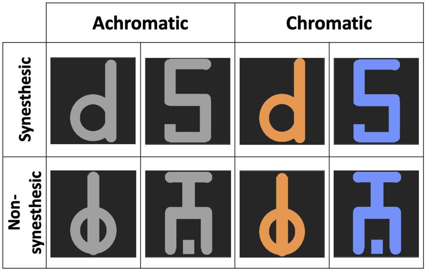
www.nature.com/scientificreports/
Demographic characteristics Synesthesia characteristics
Projector/associator
Synesthetes Age (year: month) Sex Consistency score score Alphanumeric stimulus
1 32:11 F 0.91 − 0.60 Associator 5 (red)
2 32:06 F 0.73 0.09 Projector 5 (blue)
3 23:11 F 0.43 − 0.17 Associator 5 (red)
4 24:10 F 0.65 − 1.75 Associator 5 (red)
5 22:03 M 0.49 − 1.92 Associator 5 (fuchsia)
6 25:00 F 0.53 − 1.83 Associator 5 (orange)
7 27:00 M 0.85 − 2.33 Associator 5 (red)
8 21:04 F 0.80 − 1.42 Associator 5 (green)
9 28:10 F 0.51 − 0.83 Associator 5 (green)
10 31:07 M 0.73 − 2.75 Associator d (blue)
11 23:07 M 0.42 − 0.57 Associator 5 (orange)
Table 1. Demographic and synesthesia characteristics of the synesthetic participants. Consistency and
projector/associator scores were obtained from the Online Synesthesia Battery.
chroma44. Moreover, lower contrast discrimination thresholds and enhanced performance in colour and shape/
curvature discrimination tasks have been r eported45,46. Other factors may also explain the performance of syn-
esthetes. It has been suggested that synesthetes might exhibit some specific personality traits such as openness
or disposition to get involved in new experiences, and that they are even more sensitive to mental i magery47,48.
One can reasonably speculate that such subjective particularities might influence perception. Thus, synesthetes
might differ in how willing they are to affirm the existence of a stimulus, which corresponds with the decision
criterion in the signal detection theory, and impacts the observers’ discrimination responses. The faster target
detection may therefore be the result of a criterion shift (response bias) rather than a true effect of discriminabil-
ity. While our study was not designed to verify this possibility, it appears unlikely because the performance of
the synesthetes was not different from the controls in the flash suppression condition. In addition, many studies
using a signal detection theory design failed to reveal a significant difference in response bias between synesthete
and non-synesthete p articipants49–51.
Further individual differences in experiencing synesthesia may have influenced the findings of the present
study. It is well known that synesthetic experiences differ qualitatively between individuals. For instance, some
synesthetes perceive colours as being “outside” of their body, while others perceive them internally, i.e., in the
“mind’s eye”. These phenomenological distinctions in synesthetic percepts are known as the projector type and the
associator type, r espectively10, although this classification is still under d
ebate35,52. The enhanced performance in
synesthesia reported by most studies is best shown for projector s ynesthetes10,53,54, including for brain a ctivity55,56.
For example, the study conducted by Ramachandran et al.15 suggested that conscious letter recognition is not
required for synesthetic perception, but only projector synesthetes were examined. By contrast, all synesthetes
in the present study were classified as associator types, based on the self-report in the Synesthesia Battery, with
the exception of one participant (ID2 in Table 1) with a score of 0.09, i.e., theoretically within the projector zone.
Considering that this value was very close to zero (i.e., the associator/projector cutoff) and that her performance
in all tasks was not significantly different from the other synesthetes, it is impossible to ascertain whether or not
this participant was a true projector. For now, the role of such individual differences in the synesthetic experi-
ence remain poorly understood, as there is currently no reliable tool to distinguish projector from associator.
In fact, most studies have failed to systematically evaluate and compare participants’ performance with regard
to their synesthetic p rofiles9,13–15.
In summary, our results showed that synesthesia does not bias perceptual flash suppression, indicating that
synesthesia is less likely to manifest under implicit conditions; however, a more comprehensive assessment of
synesthesia in relation to visual awareness is necessary to support this interpretation. Further studies using differ-
ent designs such as the continuous flash suppression paradigm, which does not require pre-adaptation to achieve
reliable disappearance, are needed to directly test whether synesthesic processing can occur without conscious
awareness of the stimulus. Some key individual difference variables also needed to be considered, such as atypical
visual functioning or discrimination thresholds, synesthesia type (associator vs. projector), and subject’s criterion.
Methods
Participants. Eleven grapheme-colour type synesthetes (Table 1) and 11 control participants were recruited
to take part in this experiment. Synesthetes were matched with controls based on age (21–32 years old) and
sex (4 men, 7 women). Grapheme-colour associations in the synesthetes were assessed qualitatively during a
semi-structured interview and quantitatively using the grapheme-colour consistency test from the online Syn-
esthesia Battery, developed by Eagleman53. The synesthete participants were assessed twice using this battery,
with a minimum lapse of 2 months between each testing session. Consistency test scores (see Table 1) below 1.0
are indicators of synesthetic associations. Scores between 1.0 and 2.0 are not sufficiently conclusive to consider
the presence of synesthetic associations, while scores higher than 2.0 rule out the possibility altogether. In our
study, the average consistency scores for synesthetic participants between the initial testing and the re-testing
Scientific Reports | (2021) 11:7566 | https://doi.org/10.1038/s41598-021-87223-w 5
Vol.:(0123456789)www.nature.com/scientificreports/
Figure 3. Stimuli. The synesthetic alphanumeric (first row) and the non-synesthetic abstract stimuli (second
row) were randomly presented. The achromatic alphanumeric stimuli were experienced as colored for the
synesthetes.
were within the range of synesthetic associations. More precisely, the minimal average score for consistency was
0.43, while the maximum average score for consistency was 0.91. These values confirmed that the synesthetic
associations reported by the participants were highly consistent over time. A projector-associator score to char-
acterize the type of synesthetic association was also obtained from the Synesthesia Battery (Table 1). Negative
scores indicate that the synesthesia percept is in the "mind’s eye" while positive scores indicate an “out of mind”
synesthetic perception.
None of the participants had a history of neurological or psychiatric disorders, and reported normal or
corrected-to-normal vision. Visual acuity was measured using the Snellen acuity chart and the contrast sensitiv-
ity FACT test (Stereo Optical Company Inc., Chicago, IL, United States). Stereoscopic vision was assessed using
the Randot Test (Stereo Optical Co., Inc., Chicago, IL). All participants gave written informed consent before
participating in this study. They also received a financial compensation ($20 CAN). The experimental procedure
conformed to the World Medical Association’s Declaration of Helsinki and was approved by the Research Ethics
Committee of the Université du Québec à Montréal (FSH-2013-92).
Stimuli and design. Three types of stimulus were used: a synesthetic stimulus, a non-synesthetic stimu-
lus, and a suppressor stimulus. The synesthetic stimulus was a number “5” for all participants except one (see
Table 1), while the non-synesthetic stimulus was a symbol created from the trait features of the respective alpha-
numeric synesthetic stimulus (see Fig. 3). The number “5” was chosen because it evokes a vivid synesthetic
sensation of colour in all participants. For synesthete participant #10, the synesthetic stimuli were replaced by a
letter “d”, as this participant experienced no synesthesia with digits. The size of the stimuli was 1° × 0.6°. Stimuli
were presented on a black square. The suppressor stimulus (1° × 1°) was a visual noise composed of random
grains, ranging from black to white. These stimuli were presented side by side at a distance of 3° from the central
fixation point of the screen (see Fig. 4).
Stimuli were generated and controlled with Psykinematix sofware, version 1.5 (KyberVision, Sendai, Japan).
They were presented dichoptically using 3D glasses (head-mounted virtual-reality display model Z800 3D-Visor;
eMagin Corp, Bellevue, WA) driven by a MAC G4 Desktop with an NVIDIA graphics card (GeForce 9400M,
Santa Clara, CA). The resolution of each monocular, organic light emitting diode (OLED) screen was 800 by 600
pixels. In each OLED, the refresh rate was 60 Hz and the visual field was 32° by 23°. The size of a pixel subtended
an angle of 144 arc/s (0.04°).
Synesthetic and non-synesthetic stimuli, that is, the alphanumeric and abstract stimuli, respectively, were
displayed in two experimental conditions. In the achromatic condition, stimuli were presented in grey scale
(RGB values = 160). In the chromatic condition, stimuli were displayed with the colour that corresponded to
the personal synesthetic perception of each synesthete. The RGB values of the images were then adjusted in
order to reach physical equiluminance with the achromatic stimuli (~ 49 cd/m2). All stimuli in all conditions
were presented on a black background (0.01 cd/m2). The stimuli covered approximately 70% of the surface of
the background frame. The contrast level was 50%. Participants in the control group were tested with the same
stimuli as their corresponding synesthete participant.
Four stimulus sets were generated according to the condition (achromatic and chromatic) and stimulus type
(synesthetic and non-synesthetic): alphanumeric achromatic stimulus, achromatic abstract stimulus, alphanu-
meric chromatic stimulus, and abstract chromatic stimulus. Stimuli were displayed randomly in 3 blocks, each
Scientific Reports | (2021) 11:7566 | https://doi.org/10.1038/s41598-021-87223-w 6
Vol:.(1234567890)www.nature.com/scientificreports/
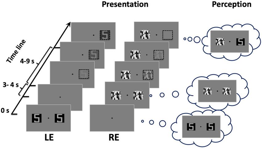
Figure 4. An example of a flash suppression trial. Following the noise patch stimulation in one eye, the initial
pair of images in the opposite eye disappeared and one of the two images (either on the right or left side)
reappeared after some time. LE: left eye; RE: right eye.
Figure 5. An example of a non-flash-suppression trial. The stimulus presentation was controlled (left side) to
mimic the subjective perception (right side) so that one of the two images initially presented reappeared after
being suppressed by the sudden presentation of a flash. Dashed-line squares (on the left) illustrate the fade-in/
fade-out interplay of the images that were used to mimic the breaking of the flash suppression. LE: left eye; RE:
right eye.
comprising 28 flash suppression trials. In the flash suppression trials, the presentation of the stimuli was dichoptic
(see Fig. 4). A pair of suppressor stimuli (visual noise patches) was abruptly presented to the previously-unstim-
ulated eye 3 to 4 s after the stimulus onset (time 0). As a result, the initial pair of target stimuli disappeared from
awareness. Thus, the presentation of the visual noise was perceived by participants as a “flash,” which masked
the test stimulus, even though it was physically present on the screen.
In addition to the flash suppression trials, “non-flash-suppression” trials (12 trials per block) were embed-
ded in the task to mimic a stimulus presentation similar to the experimental trials. In the non-flash-suppression
trials (see Fig. 5), stimuli were presented in such a way that flash suppression did not occur; the initial stimuli
displayed to one eye disappeared after 3–4 s. At the same time the two noise patches were presented to the other
eye. After a while, the stimulus target reappeared slowly in one eye (fade-in, from 0 to 50% contrast) while the
corresponding noise patch in the other eye disappeared slowly (fade-out, from 50 to 0%). The flash suppression
(Fig. 4) and the non-flash-suppression trials (Fig. 5) were randomly presented in each testing block. In addition,
stimulus presentation was counterbalanced between the left and the right eyes.
Scientific Reports | (2021) 11:7566 | https://doi.org/10.1038/s41598-021-87223-w 7
Vol.:(0123456789)www.nature.com/scientificreports/
Procedure. Participants were comfortably seated in a dimly lit room. They were instructed to adjust the
alignment of the 3D glasses by moving the lenses sideways and to adjust their proximity. Eight practice trials
were performed. The participants were instructed to maintain their gaze on the central fixation dot while the
images were presented.
The task method consisted of a spatial two-alternative forced choice. The participants were instructed to
press the left or the right arrow key when one stimulus re-appeared in its entirety, either on the left or the right
side of the screen. The time required for a participant to report the reappearance of the hidden stimulus was
calculated as the duration of suppression in the flash suppression trials. For the non-flash-suppression trials, the
time required to detect the fake suppressed stimulus was measured. Both types of trials (flash suppression and
non-flash-suppression) ended when participants responded, or after 15 s. Each trial was launched by pressing
the “enter” key. Participants were allowed to take breaks between blocks if they desired.
Data analyses. Intra- and inter-group differences were assessed using analyses of variance (ANOVA) with
the condition (achromatic and chromatic), the stimulus (alphanumeric and abstract stimuli), and the group
(synesthetes and controls) as the main factors. Separate ANOVA were conducted for the flash suppression and
the non-flash-suppression trials. Data were also analyzed using mixed effects modeling to treat subjects as a
random factor, while the condition, stimulus and group were fixed. For the flash suppression trials, the duration
of suppression was calculated by subtracting the onset of the suppressor stimulus presentation (noisy patches)
from the participants’ reaction time. A lower value meant a faster time for the target to reach visual awareness.
For the non-flash-suppression trials, the time required to detect the fake suppressed stimulus was calculated
from the fade-in onset of the stimulus target to the reaction time of the participant. A lower value meant that the
stimulus was rapidly detected. In all analyses, the p values were set to be significant at an α level of < 0.05. Bonfer-
roni corrections were applied to detect the significance in post-hoc pairwise comparisons. ANOVA and mixed
effects modeling were analyzed using IBM SPSS Statistics (version 24.0). Additional Bayesian statistical analyses
were conducted with JASP package (version 0.14.1)25 to weigh evidence for null versus alternative hypotheses.
Bayesian ANOVAs were conducted using JASP default priors, and effects are reported as the Bayes factor for the
inclusion of a particular effect, calculated as the ratio between the likelihood of the data given the model with vs
the next simpler model without that effect.
Received: 10 April 2020; Accepted: 23 March 2021
References
1. Simner, J. et al. Synaesthesia: The prevalence of atypical cross-modal experiences. Perception 35, 1024–1033 (2006).
2. Dovern, A. et al. Intrinsic network connectivity reflects consistency of synesthetic experiences. J. Neurosci. 32, 7614–7621 (2012).
3. Dixon, M. J., Smilek, D., Cudahy, C. & Merikle, P. M. Five plus two equals yellow. Nature 406, 365 (2000).
4. Eagleman, D. M. & Goodale, M. A. Why color synesthesia involves more than color. Trends Cogn. Sci. 13, 288–292 (2009).
5. Lupiáñez, J. & Callejas, A. Automatic perception and synaesthesia: Evidence from colour and photism naming in a stroop-negative
priming task. Cortex 42, 204–212 (2006).
6. Mills, C. B. Digit synaesthesia: A case study using a Stroop-type test. Cogn. Neuropsychol. 16, 181–191 (1999).
7. Odgaard, E. C., Flowers, J. H. & Bradman, H. L. An investigation of the cognitive and perceptual dynamics of a colour–digit
synaesthete. Perception 28, 651–664 (1999).
8. Smilek, D., Dixon, M. J., Cudahy, C. & Merikle, P. M. Synaesthetic Photisms Influence Visual Perception Identification of Masked
Digits. 930–936 (2001).
9. Mattingley, J. B., Rich, A. N., Yelland, G. & Bradshaw, J. L. Unconscious priming eliminates automatic binding of colour and
alphanumeric form in synaesthesia. Nature 410, 580–582. https://doi.org/10.1038/35069062 (2001).
10. Dixon, M. J., Smilek, D. & Merikle, P. M. Not all synaesthetes are created equal: Projector versus associator synaesthetes. Cogn.
Affect. Behav. Neurosci. 4, 335–343 (2004).
11. Kim, C.-Y. & Blake, R. Psychophysical magic: Rendering the visible ‘invisible’. Trends Cogn. Sci. 9, 381–388 (2005).
12. Shapiro, K. L., Raymond, J. E. & Arnell, K. M. The attentional blink. Trends Cogn. Sci. 1, 291–296. https://doi.org/10.1016/S1364-
6613(97)01094-2 (1997).
13. Rich, A. N. & Mattingley, J. B. Out of sight, out of mind: The attentional blink can eliminate synaesthetic colours. Cognition 114,
320–328 (2010).
14. Johnson, A., Jepma, M. & de Jong, R. Colours sometimes count: Awareness and bidirectionality in grapheme-colour synaesthesia.
Quart. J. Exp. Psychol. 2006(60), 1406–1422. https://doi.org/10.1080/17470210601063597 (2007).
15. Ramachandran, V. S. & Seckel, E. Synesthetic colors induced by graphemes that have not been consciously perceived. Neurocase
21, 216–219. https://doi.org/10.1080/13554794.2014.890728 (2015).
16. Yang, E., Brascamp, J., Kang, M.-S. & Blake, R. On the use of continuous flash suppression for the study of visual processing outside
of awareness. Front. Psychol. 5, 724 (2014).
17. Tsuchiya, N. & Koch, C. Continuous flash suppression reduces negative afterimages. Nat. Neurosci. 8, 1096–1101. https://doi.org/
10.1038/nn1500 (2005).
18. Blake, R. & Logothetis, N. K. Visual competition. Nat. Rev. Neurosci. 3, 13 (2002).
19. Wolfe, J. M. Reversing ocular dominance and suppression in a single flash. Vision. Res. 24, 471–478 (1984).
20. Jiang, Y., Costello, P. & He, S. Processing of invisible stimuli: Advantage of upright faces and recognizable words in overcoming
interocular suppression. Psychol. Sci. 18, 349–355 (2007).
21. Lupyan, G. & Ward, E. J. Language can boost otherwise unseen objects into visual awareness. Proc. Natl. Acad. Sci. USA. 110,
14196–14201. https://doi.org/10.1073/pnas.1303312110 (2013).
22. Mudrik, L., Breska, A., Lamy, D. & Deouell, L. Y. Integration without awareness: Expanding the limits of unconscious processing.
Psychol. Sci. 22, 764–770. https://doi.org/10.1177/0956797611408736 (2011).
23. Hong, S. W. & Blake, R. Interocular suppression differentially affects achromatic and chromatic mechanisms. Atten. Percept.
Psychophys. 71, 403–411. https://doi.org/10.3758/APP.71.2.403 (2009).
24. Lee, M. D. & Wagenmakers, E.-J. (Cambridge University Press, 2013).
Scientific Reports | (2021) 11:7566 | https://doi.org/10.1038/s41598-021-87223-w 8
Vol:.(1234567890)www.nature.com/scientificreports/
25. JASP Team. JASP (Version 0.14.1) [Computer software] (https://jasp-stats.org, 2020).
26. Treisman, A. Preattentive processing in vision. Comput. Vis. Graph. Image Process. 31, 156–177 (1985).
27. Wolfe, J. M. Guided search 2.0 a revised model of visual search. Psychon. Bull. Rev. 1, 202–238 (1994).
28. Gayet, S., Van der Stigchel, S. & Paffen, C. L. Breaking continuous flash suppression: Competing for consciousness on the pre-
semantic battlefield. Front. Psychol. 5, 460 (2014).
29. Moors, P., Hesselmann, G., Wagemans, J. & van Ee, R. Continuous flash suppression: Stimulus fractionation rather than integra-
tion. Trends Cogn. Sci. 21, 719–721 (2017).
30. Gobbini, M. I. et al. Prioritized detection of personally familiar faces. PLoS ONE 8, e66620. https://doi.org/10.1371/journal.pone.
0066620 (2013).
31. Krueger, L. E. Familiarity effects in visual information processing. Psychol. Bull. 82, 949 (1975).
32. Blake, R., O’Shea, R. P. & Mueller, T. Spatial zones of binocular rivalry in central and peripheral vision. Vis. Neurosci. 8, 469–478
(1992).
33. Palmeri, T. J., Blake, R., Marois, R., Flanery, M. A. & Whetsell, W. The perceptual reality of synesthetic colors. Proc. Natl. Acad.
Sci. 99, 4127–4131 (2002).
34. Sagiv, N., Heer, J. & Robertson, L. Does binding of synesthetic color to the evoking grapheme require attention?. Cortex 42, 232–242
(2006).
35. Edquist, J., Rich, A. N., Brinkman, C. & Mattingley, J. B. Do synaesthetic colours act as unique features in visual search?. Cortex
42, 222–231. https://doi.org/10.1016/S0010-9452(08)70347-2 (2006).
36. Nijboer, T. C., Satris, G. & Van der Stigchel, S. The influence of synesthesia on eye movements: No synesthetic pop-out in an
oculomotor target selection task. Conscious. Cogn. 20, 1193–1200. https://doi.org/10.1016/j.concog.2011.03.017 (2011).
37. Rothen, N. & Meier, B. Do synesthetes have a general advantage in visual search and episodic memory? A case for group studies.
PLoS ONE 4, e5037 (2009).
38. Dojat, M., Pizzagalli, F. & Hupe, J. M. Magnetic resonance imaging does not reveal structural alterations in the brain of grapheme-
color synesthetes. PLoS ONE 13, e0194422. https://doi.org/10.1371/journal.pone.0194422 (2018).
39. Hupé, J.-M., Bordier, C. & Dojat, M. The neural bases of grapheme-color synesthesia are not localized in real color-sensitive areas.
Cereb. Cortex 22, 1622–1633. https://doi.org/10.1093/cercor/bhr236 (2012).
40. Weiss, F., Greenlee, M. W. & Volberg, G. No atypical white-matter structures in grapheme-or color-sensitive areas in synesthetes.
bioRxiv 618611 (2019).
41. Arias, D. J., Hosein, A. & Saint-Amour, D. Assessing lateral interaction in the synesthetic visual brain. Vision 3, 7 (2019).
42. Sagiv, N. & Ward, J. Crossmodal interactions: Lessons from synesthesia. Prog. Brain Res. 155, 259–271. https://doi.org/10.1016/
S0079-6123(06)55015-0 (2006).
43. Banissy, M. J., Walsh, V. & Ward, J. Enhanced sensory perception in synaesthesia. Exp. Brain Res. 196, 565–571 (2009).
44. Banissy, M. J. et al. Synesthesia for color is linked to improved color perception but reduced motion perception. Psychol. Sci. 24,
2390–2397. https://doi.org/10.1177/0956797613492424 (2013).
45. Terhune, D. B., Song, S. M., Duta, M. D. & Cohen Kadosh, R. Probing the neurochemical basis of synaesthesia using psychophysics.
Front. Hum. Neurosci. 8, 89–89. https://doi.org/10.3389/fnhum.2014.00089 (2014).
46. Ward, J., Rothen, N., Chang, A. & Kanai, R. The structure of inter-individual differences in visual ability: Evidence from the general
population and synaesthesia. Vision. Res. 141, 293–302. https://doi.org/10.1016/j.visres.2016.06.009 (2017).
47. Banissy, M. J. et al. Personality traits in people with synaesthesia: Do synaesthetes have an atypical personality profile?. Personal.
Individ. Differ. 54, 828–831. https://doi.org/10.1016/j.paid.2012.12.018 (2013).
48. Chun, C. A. & Hupe, J. M. Are synesthetes exceptional beyond their synesthetic associations? A systematic comparison of creativity,
personality, cognition, and mental imagery in synesthetes and controls. Br. J. Psychol. 107, 397–418. https://doi.org/10.1111/bjop.
12146 (2016).
49. Amsel, B. D., Kutas, M. & Coulson, S. (PubMed Abstract Publisher Full Text).
50. Lunke, K. & Meier, B. New insights into mechanisms of enhanced synaesthetic memory: Benefits are synaesthesia-type-specific.
PLoS ONE 13, e0203055 (2018).
51. Whittingham, K. M., McDonald, J. S. & Clifford, C. W. G. Synesthetes show normal sound-induced flash fission and fusion illu-
sions. Vis. Res. 105, 1–9 (2014).
52. Yokosawa, K. & Asano, M. Relation between synesthetic grapheme-color associations and the sub-types of synesthesia. J. Vis. 15,
132–132 (2015).
53. Eagleman, D. M., Kagan, A. D., Nelson, S. S., Sagaram, D. & Sarma, A. K. A standardized test battery for the study of synesthesia.
J. Neurosci. Methods 159, 139–145 (2007).
54. Ward, J., Li, R., Salih, S. & Sagiv, N. Varieties of grapheme-colour synaesthesia: A new theory of phenomenological and behavioural
differences. Conscious. Cogn. 16, 913–931. https://doi.org/10.1016/j.concog.2006.09.012 (2007).
55. Cohen, M. X., Weidacker, K., Tankink, J., Scholte, H. S. & Rouw, R. Grapheme-color synesthesia subtypes: Stable individual dif-
ferences reflected in posterior alpha-band oscillations. Cogn. Neurosci. 6, 56 (2015).
56. van Leeuwen, T. M., den Ouden, H. E. M. & Hagoort, P. Effective connectivity determines the nature of subjective experience in
grapheme-color synesthesia. J. Neurosci. 31, 9879–9884. https://doi.org/10.1523/JNEUROSCI.0569-11.2011 (2011).
Acknowledgements
We thank Anthony Hosein for his very helpful technical assistance.
Author contributions
The experiments were conceived and designed by D.J.A. and D.S.-A. and they were conducted by D.J.A. The
Data was collected by D.J.A. Data was analyzed and interpreted by D.J.A. and D.S.-A. Writing-Original Draft was
prepared by D.J.A. and edited by D.S.-A. All authors have seen and approved the final version of the manuscript.
Competing interests
The authors declare no competing interests.
Additional information
Supplementary Information The online version contains supplementary material available at https://doi.org/
10.1038/s41598-021-87223-w.
Correspondence and requests for materials should be addressed to D.S.-A.
Reprints and permissions information is available at www.nature.com/reprints.
Scientific Reports | (2021) 11:7566 | https://doi.org/10.1038/s41598-021-87223-w 9
Vol.:(0123456789)www.nature.com/scientificreports/
Publisher’s note Springer Nature remains neutral with regard to jurisdictional claims in published maps and
institutional affiliations.
Open Access This article is licensed under a Creative Commons Attribution 4.0 International
License, which permits use, sharing, adaptation, distribution and reproduction in any medium or
format, as long as you give appropriate credit to the original author(s) and the source, provide a link to the
Creative Commons licence, and indicate if changes were made. The images or other third party material in this
article are included in the article’s Creative Commons licence, unless indicated otherwise in a credit line to the
material. If material is not included in the article’s Creative Commons licence and your intended use is not
permitted by statutory regulation or exceeds the permitted use, you will need to obtain permission directly from
the copyright holder. To view a copy of this licence, visit http://creativecommons.org/licenses/by/4.0/.
© The Author(s) 2021
Scientific Reports | (2021) 11:7566 | https://doi.org/10.1038/s41598-021-87223-w 10
Vol:.(1234567890)You can also read