Effect of the acid suppressor omeprazole on the proliferation, migration, invasion and cell cycle of esophageal squamous cell carcinoma cells via ...
←
→
Page content transcription
If your browser does not render page correctly, please read the page content below
EXPERIMENTAL AND THERAPEUTIC MEDICINE 22: 1187, 2021

    Effect of the acid suppressor omeprazole on the proliferation,
    migration, invasion and cell cycle of esophageal squamous cell
     carcinoma cells via the aryl hydrocarbon receptor pathway
                              YU BAI, PEIYAO ZHU, KUN ZHOU and SHU‑GUANG ZHANG

  Department of Thoracic Surgery, The First Hospital of China Medical University, Shenyang, Liaoning 110001, P.R. China

                                        Received February 4, 2021; Accepted July 14, 2021

                                                   DOI: 10.3892/etm.2021.10621

Abstract. Esophageal cancer is a malignant tumor type with           results of the wound‑healing and Transwell assays respectively
one of the highest mortality rates worldwide. The aryl hydro‑        suggested that cell migration and invasion were reduced. In
carbon receptor (AHR), which has been investigated in recent         conclusion, OME inhibited the proliferation, migration and
years, has been confirmed to be associated with the occurrence       invasion of ESCC cells and blocked the cell cycle via the AHR
and development of esophageal cancer. AHR has a variety of           pathway, which may provide a therapeutic effect on esophageal
different ligands, which regulate its activity following binding.    squamous cell cancer.
The widely known acid inhibitor omeprazole (OME) also affects
AHR and its downstream proteins (such as the cytochrome              Introduction
P450 family) by non‑ligand binding; however, the mechanisms
have remained to be fully elucidated. Therefore, the aim of the      According to the Global Cancer Observatory, 400,000 deaths
present study was to investigate the role of OME in esophageal       from esophageal cancer occurred in 2012 and it ranks 6th
squamous cell carcinoma (ESCC), whether the mechanism                among all cancer types worldwide (1‑3). In addition, among
proceeds via the AHR pathway and how OME regulates AHR               the top 10 malignant tumor types in China, esophageal
to affect the occurrence and development of esophageal carci‑        cancer ranks 4th in males and 8th in females (4). The major
noma. The AHR‑selective regulator OME was used to treat the          pathological subtypes of esophageal cancer are squamous
ESCC cell lines TE1 and KYSE150. Western blot analysis was           cell carcinoma, adenocarcinoma and small cell carcinoma,
used to verify the effect of OME on AHR and proliferating            and squamous cell carcinoma is the major type in China,
cell nuclear antigen (PCNA) protein expression levels, while         accounting for >90% of cases, while adenocarcinoma is the
Cell Counting Kit (CCK)‑8, wound‑healing and Transwell               major type in European countries and in both North and South
assays were used to determine the proliferation, migration           America (5). The risk factors of the different pathological
and invasion of the ESCCs, respectively, following treatment         types of esophageal squamous cell carcinoma (ESCC) are
with OME. In addition, flow cytometry was used to investigate        also different and include sex, ethnicity, smoking, alcohol
the cell cycle distribution of the ESCCs following incubation        consumption, diet, nutritional status and hereditary factors (6).
with OME. AHR was highly expressed in the ESCCs and                      The aryl hydrocarbon receptor (AHR) is in the basic
following treatment with OME, the protein expression levels          helix‑loop‑helix/Per‑ARNT‑SIM (bHLH/PAS) subgroup
of AHR and PCNA were downregulated. The CCK‑8 assay                  in the bHLH transcription factor superfamily. The gene is
indicated that the proliferation of the ESCCs was also reduced       ~60 Kbp long and has 22 exons. An unusual exon/intron junc‑
following treatment with OME. Furthermore, flow cytometry            tion sequence was detected in the 11th intron of the gene, which
revealed a notable block of the cells in G1/G0 phase, while the      begins at the 5' end of the GC region. The exon/intron orga‑
                                                                     nization of the mouse AhR nuclear translocator (mArnt) gene
                                                                     is different from the other members in the same bHLH/PAS
                                                                     family, as it does not contain a TATA box and has several
                                                                     transcriptional initiation sites (7). The promoter region of the
Correspondence to: Professor Shu‑Guang Zhang, Department of          mArnt gene is rich in GC and contains numerous hypoth‑
Thoracic Surgery, The First Hospital of China Medical University,
                                                                     esized regulatory DNA sequences, such as two GC‑boxes,
155 North Nanjing Street, Heping, Shenyang, Liaoning 110001,
P.R. China
                                                                     a cyclic adenosine monophosphate response element, an
E‑mail: shgzhang@cmu.edu.cn                                          E‑box, an activator protein‑1 locus and a CAAT‑box. The
                                                                     AHR is the only member of the family, which is known to
Key words: omeprazole, esophageal squamous cell carcinoma,           be activated by a ligand (8). The AHR is a ligand‑activated
aryl hydrocarbon receptor, ligand, malignant tumor, proliferation,   transcription factor, located in the cytoplasm and combines
migration, invasion                                                  with the heat shock protein (HSP)90, the AHR‑interacting
                                                                     protein and the HSP90‑interacting protein P23 (9). After the
                                                                     ligands are combined, the AHR is transported into the nucleus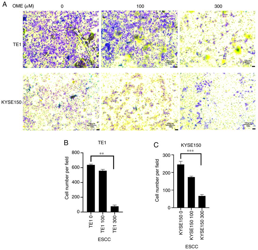
2 BAI et al: OMEPRAZOLE CHEMOTHERAPY FOR ESOPHAGEAL CANCER THROUGH AHR
and dimerizes with the aryl receptor nuclear translocator.
Subsequently, it combines with xenobiotic response elements
to regulate and participate in the downstream signaling
pathway (9,10).
Omeprazole (OME; Fig. 1), a well‑known acid suppressant,
is used to inhibit the proton pump in gastric wall cells (H+/K+
enzyme). In addition, animal experiments have demonstrated
that OME had a protective effect on gastric mucosa damage,
and was able to increase gastric mucosa blood flow due to an
association with a variety of different factors, such as glyco‑
proteins, nitric oxide and TNF‑ α (11). Furthermore, OME Figure 1. Molecular structure of omeprazole.
has an anti‑Helicobacter pylori (H. pylori) effect (12). Due to
its mode of action, OME is widely used in the treatment of
peptic ulcers, reflux esophagitis, upper gastrointestinal hemor‑
rhage, H. pylori infection, Zole‑Aids syndrome and gastric and blots were visualized using enhanced chemiluminescence
ulcer caused by non‑steroidal anti‑inflammatory drugs (13). (Beyotime Institute of Biotechnology) solution. The grayscale
Following increases in AHR research, OME has also been values of the resulting bands were measured using ImageJ
indicated to be a selective regulator of AHR (14). Therefore, software (version, 1.46r; National Institutes of Health). Each
the present study aimed to investigate whether OME regu‑ experiment was performed in triplicate.
lates the expression level of AHR in ESCC cells to affect the
proliferation, cell cycle, migration and invasion, to provide a Cell proliferation assay. The TE1 and KYSE150 cells were
novel treatment for ESCC. cultured, collected and then counted. Subsequently, the cells
were seeded in a 96‑well plate (~2,000 cells/well; 200 µl/well).
Materials and methods After the cells had attached, they were treated with 0, 100
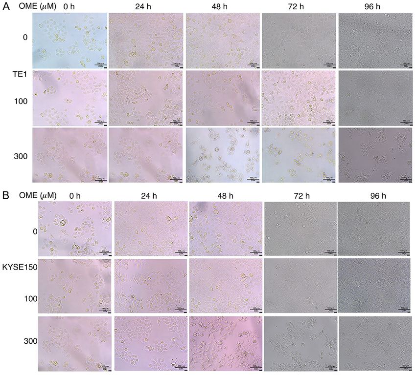
and 300 µM OME for at 24, 48, 72 or 96 h. Cell Counting
Cell culture. The human ESCC cell lines TE1 and KYSE150 Kit‑8 solution (MedChemExpress) was added for 2 h and
were purchased from the Chinese Academy of Sciences. The cell proliferation was measured at 560 nm using a microplate
cells were cultured in RPMI‑1640 medium (Beijing Solarbio reader (Thermo Fisher Scientific, Inc.). At the same time, the
Science & Technology Co., Ltd.) containing 10% FBS photomicrographs of the ESCCs were captured at 0, 24, 48, 72
(Cellmax Nutrients B.V.) at 37˚C in a humidified incubator and 96 h with an inverted microscope (magnification, x200).
with 5% CO2. Each experiment was performed in triplicate.
Western blot analysis. OME was first dissolved in DMSO Wound‑healing assay. The TE1 and KYSE150 cells were
at a high concentration to produce a stock solution and then cultured and seeded in a six‑well plate. After the cells grew to
dissolved in the corresponding medium during later use (the 90% confluence (~1x10⁶ cells per well), they were scratched
concentration of DMSO in the final solution did not exceed with a 200‑µl pipette tip and the serum‑free medium was
0.1%). TE1 and KYSE150 cells were treated with 0, 100 and replaced with serum‑free OME at 0, 100 and 300 µM. Images
300 µM OME for 48 h. The cells were then lysed with RIPA of the wounds were captured at 0, 24 and 48 h with an
buffer (Beyotime Institute of Biotechnology) for 15 min and inverted microscope (magnification, x50). Wound areas were
the protein was extracted. The concentration of total protein measured using ImageJ (version 1.46r; National Institutes of
was measured using the bicinchoninic acid assay (Thermo Health). The migration rate was then calculated as follows:
Fisher Scientific, Inc.). Protein aliquots (30 µg) were loaded (The difference of the wound area between 24, 48 and
with SDS buffer (Beyotime Institute of Biotechnology) and 0 h)/(wound area at 0 h). Each experiment was performed in
boiled at 99˚C for 15 min. Equal amounts of protein samples triplicate.
per lane were separated using SDS‑PAGE on a 10% gel.
Following electrophoresis, the cells were transferred to a Transwell assay. The TE1 and KYSE150 cells were collected
PVDF membrane (EMD Millipore). Fast blocking solution and then seeded in a six‑well plate. After the cells had
(Beyotime Institute of Biotechnology) was used for blocking attached, they were treated with OME at 0, 100 and 300 µM
for 15 min at room temperature and the membrane was incu‑ for 48 h. The cells were then collected and ~5,000 cells/well in
bated with the following primary antibodies on a shaking table 200 µl/well were seeded into the upper chamber of a Transwell
overnight at 4˚C: Anti‑GAPDH (1:1,000; cat. no. ab181602; insert (Corning, Inc.) that had been pre‑coated with Matrigel™
Abcam), anti‑AHR (1:500; cat. no. ab182642; Abcam), (1:8; BD Biosciences) for 30 min at 37˚C for the invasion
anti‑proliferating cell nuclear antigen (PCNA; 1:2,000; assay. RPMI‑1640 medium containing 20% FBS (500 µl/well)
cat. no. 2586; Cell Signaling Technology, Inc.) and anti‑MMP9 was added to the lower chamber. The cells were fixed with
(1:1,000; cat. no. 3582; Cell Signaling Technology, Inc.). methanol for 10 min and stained with crystal violet for 20 min
Subsequently, samples were incubated with the corresponding at room temperature. After 48 h of incubation, cells on the
secondary antibodies: Goat anti‑Rabbit secondary antibody upper surface were scraped off and cells on the lower surface
(1:10,000; cat. no. 31460; Thermo Fisher Scientific, Inc.) and were viewed under a light microscope (Leica Microsystems;
Goat anti‑mouse secondary antibody (1:10,000; cat. no. 31430; magnification, x100). Images were captured using ImageJ
Thermo Fisher Scientific, Inc.) for 2 h at room temperature, software.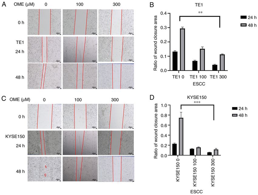
EXPERIMENTAL AND THERAPEUTIC MEDICINE 22: 1187, 2021                                                             3

Figure 2. OME inhibits the protein expression levels of AHR. (A) The TE1 and KYSE150 cell lines were treated with 0, 100 and 300 µM OME and the protein
expression level of AHR was determined by western blot analysis. (B) The results were semi‑quantitatively analyzed using gray‑scale analysis. ***P
4                          BAI et al: OMEPRAZOLE CHEMOTHERAPY FOR ESOPHAGEAL CANCER THROUGH AHR

Figure 3. OME decreases the protein expression levels of the proliferation‑related gene PCNA. (A) The TE1 and KYSE150 cell lines were treated with 0, 100
and 300 µM OME and the protein expression levels of PCNA were determined by western blot analysis. (B) The results were semi‑quantitatively analyzed
using gray‑scale analysis **P
EXPERIMENTAL AND THERAPEUTIC MEDICINE 22: 1187, 2021 5
Figure 5. Photomicrographs of the (A) TE1 and (B) KYSE150 cells following treatment with 0, 100 and 300 µM OME for 24, 48 and 72 h. OME, omeprazole.
was reduced and the high concentration of OME inhibited skin (30), prostate (31) and pancreatic cancers (32,33). High
migration to the greatest extent (Fig. 8). In addition, as protein expression levels were confirmed in the present
expected, the results of the Transwell assay revealed that the study and promoted the process of tumor development. AHR
invasive ability of the TE1 and KYSE150 cells treated with has a variety of endogenous and exogenous ligands, and in
OME was also reduced and this effect was observed to be previous studies, OME, a widely known acid inhibitor, has
dose‑dependent (Fig. 9). also been determined to be a selective regulator of AHR.
OME is a proton pump inhibitor, which is widely used in
Discussion the treatment of various diseases, including digestive tract
ulcers and reflux esophagitis. In addition, OME was indicated
Esophageal cancer is one of the most common types of cancer to regulate the expression level of AHR in a non‑ligand
in the Western world, with high aggressiveness and a low 5‑year manner, thereby affecting the occurrence and development
survival rate. Despite advances in diagnosis and treatment, the of tumors (14,16,29). However, the underlying mechanism
overall 5‑year survival rate for patients with esophageal cancer has remained elusive, and to the best of our knowledge, no
is only 15‑20% in the US (21). There are two major subtypes previous studies have investigated the effect of the interaction
of esophageal cancer: Squamous cell carcinoma and adeno‑ between OME and AHR on tumorigenesis and progression
carcinoma, and both account for >95% of cases of esophageal of ESCC.
cancer. Clinical studies have indicated that the combination of From the results of the present study, several conclusions
OME and aspirin reduced the mortality rate in patients with may be drawn. First, western blot analysis revealed that the
esophageal adenocarcinoma (22). Therefore, the present study TE1 and KYSE150 cell lines treated with 0, 100 and 300 µM
focused on ESCC and OME. OME for 48 h exhibited decreased AHR protein expression
AHR, as a ligand‑activated transcription factor, has levels and this effect was dose‑dependent. Combined with
been investigated in numerous types of tumor, such as the results of previous studies by our group (34,35), it may be
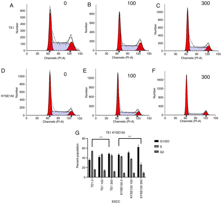
colorectal (23), breast (24,25), lung (26‑28), stomach (29), preliminarily suggested that OME affects the proliferation,6 BAI et al: OMEPRAZOLE CHEMOTHERAPY FOR ESOPHAGEAL CANCER THROUGH AHR Figure 6. OME induces G1 phase arrest in esophageal squamous cell carcinoma cells. Cell cycle distribution of the TE1 cell line treated with (A) 0, (B) 100 and (C) 300 µM OME for 48 h. Cell cycle distribution of the KYSE150 cell line treated with (D) 0, (E) 100 and (F) 300 µM OME. (G) Proportion of cells in G1/G 0, S and G2/M phases according to quantitative analysis of the TE1 and KYSE150 cell lines (G1/G0 phase; **P
EXPERIMENTAL AND THERAPEUTIC MEDICINE 22: 1187, 2021                                                           7

Figure 7. OME inhibits the protein expression of MMP9. (A) The protein expression of MMP9 in the TE1 and KYSE150 cell lines treated with 0, 100 and
300 µM OME were determined by western blot analysis. (B) The results were quantitatively analyzed using gray‑scale analysis. **P
8                          BAI et al: OMEPRAZOLE CHEMOTHERAPY FOR ESOPHAGEAL CANCER THROUGH AHR

Figure 9. OME reduces the invasive capacity of ESCC cells. (A) The invasive ability of the TE1 and KYSE150 cell lines treated with 0, 100 and 300 µM OME
was assessed in a Transwell assay and representative images of membranes with stained invaded cells are provided (magnification, x100; scale bar, 150 µm).
(B and C) Invasion of (B) TE1 and (C) KYSE150 cells was quantitatively analyzed. **P
EXPERIMENTAL AND THERAPEUTIC MEDICINE 22: 1187, 2021 9
Patient consent for publication 18. Patrizi B and Siciliani de Cumis M: TCDD toxicity mediated by
epigenetic mechanisms. Int J Mol Sci 19: 4101, 2018.
19. Opitz C, Litzenburger U, Sahm F, Ott M, Tritschler I, Trump S,
Not applicable. Schumacher T, Jestaedt L, Schrenk D, Weller M, et al: An endog‑
enous tumour‑promoting ligand of the human aryl hydrocarbon
receptor. Nature 478: 197‑203, 2011.
Competing interests 20. Huang H: Matrix metalloproteinase‑9 (MMP‑9) as a cancer
biomarker and MMP‑9 biosensors: Recent advances. Sensors
The authors declare that they have no competing interests. (Basel) 18: 3249, 2018.
21. Pennathur A, Gibson MK, Jobe BA and Luketich JD: Oesophageal
carcinoma. Lancet 381: 400‑412, 2013.
References 22. Jankowski JAZ, de Caestecker J, Love SB, Reilly G, Watson P,
Sanders S, Ang Y, Morris D, Bhandari P, Brooks C, et al:
1. Ilic M, Kocic S, Radovanovic D, Macuzic IZ and Ilic I: Trend in Esomeprazole and aspirin in Barrett's oesophagus (AspECT): A
esophageal cancer mortality in Serbia, 1991‑2015 (a population‑ randomised factorial trial. Lancet 392: 400‑408, 2018.
based study): An age‑period‑cohort analysis and a joinpoint 23. Shiizaki K, Kido K and Mizuta Y: Insight into the relationship
regression analysis. J BUON 24: 1233‑1239, 2019. between aryl‑hydrocarbon receptor and β ‑catenin in human
2. Ferlay J, Soerjomataram I, Dikshit R, Eser S, Mathers C, colon cancer cells. PLoS One 14: e0224613, 2019.
Rebelo M, Parkin DM, Forman D and Bray F: Cancer incidence 24. Tomblin JK, Arthur S, Primerano DA, Chaudhry AR, Fan J,
and mortality worldwide: Sources, methods and major patterns Denvir J and Salisbury TB: Aryl hydrocarbon receptor (AHR)
in GLOBOCAN 2012. Int J Cancer 136: E359‑E386, 2015. regulation of L‑type amino acid transporter 1 (LAT‑1) expres‑
3. Malhotra G, Yanala U, Ravipati A, Follet M, Vijayakumar M and sion in MCF‑7 and MDA‑MB‑231 breast cancer cells. Biochem
Are C: Global trends in esophageal cancer. J Surg Oncol 115: Pharmacol 106: 94‑103, 2016.
564‑579, 2017. 25. Donovan M, Selmin O and Romagnolo D: Aryl hydrocarbon
4. Lin Y, Totsuka Y, He Y, Kikuchi S, Qiao Y, Ueda J, Wei W, receptor diet and breast cancer risk. Yale J Biol Med 91: 105‑127,
Inoue M and Tanaka H: Epidemiology of esophageal cancer in 2018.
Japan and China. J Epidemiol 23: 233‑242, 2013. 26. Terashima J, Jimma Y, Jimma K, Hakata S, Yachi M, Habano W
5. Smyth E, Lagergren J, Fitzgerald R, Lordick F, Shah M, and Ozawa S: The regulation mechanism of AhR activated
Lagergren P and Cunningham D: Oesophageal cancer. Nat Rev by benzo[a]pyrene for CYP expression are different between
Dis primers 3: 17048, 2017. 2D and 3D culture of human lung cancer cells. Drug Metab
6. Arnal MJ, Arenas ÁA and Arbeloa ÁL: Esophageal cancer: Pharmacokinet 33: 211‑214, 2018.
Risk factors, screening and endoscopic treatment in Western 27. Gao H, Ye G, Lin Y, Chi Y and Dong S: Benzo[a]pyrene at
and Eastern countries. World J Gastroenterol 21: 7933‑7943, human blood equivalent level induces human lung epithelial cell
2015. invasion and migration via aryl hydrocarbon receptor signaling.
7. Schulte K, Green E, Wilz A, Platten M and Daumke O: Structural J Appl Toxicol 40: 1087‑1098, 2020.
basis for aryl hydrocarbon receptor‑mediated gene activation. 28. Dong S, Zhu P and Zhang S: Expression of collagen type 1
Structure 25: 1025‑1033, 2017. alpha 1 indicates lymph node metastasis and poor outcomes in
8. Wang F, Gao J, Mimura J, Kobayashi A, Sogawa K and squamous cell carcinomas of the lung. PeerJ 8: e10089, 2020.
29. Yoshinari K, Ueda R, Kusano K, Yoshimura T, Nagata K and
Fujii‑Kuriyama Y: Structure and expression of the mouse Yamazoe Y: Omeprazole transactivates human CYP1A1 and
AhR nuclear translocator (mArnt) gene. J Biol Chem 273: CYP1A2 expression through the common regulatory region
24867‑24873, 1998. containing multiple xenobiotic‑responsive elements. Biochem
9. Nebert D: Aryl hydrocarbon receptor (AHR): ‘Pioneer member’ Pharmacol 76: 139‑145, 2008.
of the basic‑helix/loop/helix per‑Arnt‑sim (bHLH/PAS) family 30. Vogeley C, Esser C, Tüting T, Krutmann J and Haarmann-
of ‘sensors’ of foreign and endogenous signals. Prog Lipid Stemmann T: Role of the aryl hydrocarbon receptor in
Res 67: 38‑57, 2017. environmentally induced skin aging and skin carcinogenesis. Int
10. Shiizaki K, Ohsako S, Kawanishi M and Yagi T: Omeprazole J Mol Sci 20: 6005, 2019.
alleviates benzo[a]pyrene cytotoxicity by inhibition of CYP1A1 31. Chen Z, Cai A, Zheng H, Huang H, Sun R, Cui X, Ye W, Yao Q,
activity in human and mouse hepatoma cells. Basic Clin Chen R and Kou L: Carbidopa suppresses prostate cancer via
Pharmacol Toxicol 103: 468‑475, 2008. aryl hydrocarbon receptor‑mediated ubiquitination and degrada‑
11. Gao W, Li HY, Wang LX, Hao LJ, Gao JL, Zheng RJ, Cai CJ tion of androgen receptor. Oncogenesis 9: 49, 2020.
and Si YL: Protective effect of omeprazole on gastric mucosal 32. Wang L, Tang W, Yang S, He P, Wang J, Gaedcke J, Ströbel P,
of cirrhotic portal hypertension rats. Asian Pac J Trop Med 7: Azizian A, Ried T, Gaida MM, et al: NO /RUNX3/kynurenine
402‑406, 2014. metabolic signaling enhances disease aggressiveness in pancre‑
12. Weil J, Bell GD, Powell K, Morden A, Harrison G, Gant PW, atic cancer. Int J Cancer 146: 3160‑3169, 2020.
Jones PH and Trowell JE: Omeprazole and helicobacter pylori: 33. Masoudi S, Nemati AH, Fazli HR, Beygi S, Moradzadeh M,
Temporary suppression rather than true eradication. Aliment Pourshams A and Mohamadkhani A: An increased level of aryl
Pharmacol Ther 5: 309‑313, 1991. hydrocarbon receptor in patients with pancreatic cancer. Middle
13. Blum R: Lansoprazole and omeprazole in the treatment of East J Dig Dis 11: 38‑44, 2019.
acid peptic disorders. Am J Health Syst Pharm 53: 1401‑1415, 34. Zhu P, Yu H, Zhou K, Bai Y, Qi R and Zhang S:
1996. 3,3'‑Diindolylmethane modulates aryl hydrocarbon receptor of
14. Jin UH, Lee SO and Safe S: Aryl hydrocarbon receptor esophageal squamous cell carcinoma to reverse epithelial‑mesen‑
(AHR)‑active pharmaceuticals are selective AHR modulators in chymal transition through repressing RhoA/ROCK1‑mediated
MDA‑MB‑468 and BT474 breast cancer cells. J Pharmacol Exp COX2/PGE2 pathway. J Exp Clin Cancer Res 39: 113, 2020.
Ther 343: 333‑341, 2012. 35. Zhu P, Zhou K, Lu S, Bai Y, Qi R and Zhang S: Modulation of
15. Nguyen LP and Bradfield CA: The search for endogenous aryl hydrocarbon receptor inhibits esophageal squamous cell
activators of the aryl hydrocarbon receptor. Chem Res Toxicol 21: carcinoma progression by repressing COX2/PGE2/STAT3 axis.
102‑116, 2008. J Cell Commun Signal 14: 175‑192, 2020.
16. Jin UH, Kim SB and Safe S: Omeprazole inhibits pancreatic
cancer cell invasion through a nongenomic aryl hydrocarbon This work is licensed under a Creative Commons
receptor pathway. Chem Res Toxicol 28: 907‑918, 2015. Attribution-NonCommercial-NoDerivatives 4.0
17. Murray IA, Patterson AD and Perdew GH: Aryl hydrocarbon
International (CC BY-NC-ND 4.0) License.
receptor ligands in cancer: Friend and foe. Nat Rev Cancer 14:
801‑814, 2014.You can also read