Minimal oxidative load: a prerequisite for thyroid cell function
←
→
Page content transcription
If your browser does not render page correctly, please read the page content below
161

Minimal oxidative load: a prerequisite for thyroid cell function
Sylvie Poncin, Ides M Colin and Anne-Catherine Gérard
Unité de Morphologie Expérimentale, Université Catholique de Louvain, UCL-5251, 52 Avenue E. Mounier, B-1200 Bruxelles, Belgium
(Correspondence should be addressed to A-C Gérard; Email: anne-catherine.gerard@uclouvain.be)

Abstract
In addition to reactive oxygen species (ROS) produced by                      expression was unaffected. Among antioxidant systems,
mitochondria during aerobic respiration, thyrocytes are                       peroxiredoxin (PRDX) five expression was reduced by NAC,
continuously producing H2O2, a key element for hormono-                       whereas peroxiredoxin three increased and catalase remained
genesis. Because nothing is known about ROS implication in                    unchanged. In vivo, the expression of both dual oxidases and
normal non-stimulated cells, we studied their possible                        peroxiredoxin five proteins was also decreased by NAC. In
involvement in thyrocytes incubated with a potent antioxidant,                conclusion, when intracellular ROS levels drop below a basal
N-acetylcysteine (NAC). NAC, which blocked the production                     threshold, the expression of proteins involved in thyroid cell
of intracellular ROS, also decreased dual oxidases, thyroper-                 function is hampered. This suggests that keeping ROS at a
oxidase, pendrin, and thyroglobulin protein and/or gene                       minimal level is required for safeguarding thyrocyte function.
expression. By contrast, NaC/IK symporter mRNA                                Journal of Endocrinology (2009) 201, 161–167

Introduction                                                                  (OS) in strict limits to avoid OS becoming eventually
                                                                              harmful. Among them, selenium, which is required for
Reactive species include both reactive oxygen species                         glutathione peroxidase activity, has been shown to exert
(ROS) and reactive nitrogen species (RNS). In all cell                        protective effects in various situations where OS is elevated,
types, ATP generation by mitochondrial aerobic respiration                    while its defect is deleterious (Contempre et al. 1993, 2004,
generates ROS as by-products of oxidative phosphorylations.                   Schomburg & Kohrle 2008, Schweizer et al. 2008).
In physiological conditions, ROS in excess must be constantly                    Up to now, nothing has been reported about the physio-
detoxified by antioxidant systems to avoid cell damage.                       logical implication of ROS in normal resting thyrocytes. To
In thyrocytes, H2O2 is a ROS that is required for                             answer this question, we analyzed the in vitro and in vivo
hormonogenesis. It is produced from dual oxidases                             expression of proteins involved in thyroid cell function in the
(DUOX1/2) (Dupuy et al. 1999, De Deken et al. 2002) at                        presence of N-acetylcysteine (NAC), an agent supposed to
the apical pole of the cell, where it oxidizes iodide (actively               deprive cells from intracellular ROS, because of potent
transported across the basal membrane by NaC/IK symporter                     antioxidant properties. We also investigated the regulation of
(NIS, product of SLC5A5) (Dai et al. 1996)) into iodine in a                  antioxidant systems when the cell oxidative load is reduced.
reaction catalyzed by thyroperoxidase (TPO). Iodine is then
incorporated into tyrosine residues of thyroglobulin (TG),
again by TPO. Hence, H 2O2 and free radicals are                              Materials and Methods
continuously produced in thyroid cells, even in physiological
conditions (Denef et al. 1996, De Deken et al. 2002,                          Cell cultures
Schweizer et al. 2008). Obviously, when ROS are over-                         PCCL3 cells (a continuous line of non-transformed rat
produced in pathological situations, they may exert a wide                    thyroid follicular cells, Fusco et al. 1987) were a gift from
range of actions, usually deleterious (Adler et al. 1999,                     Dr F. Miot (Université Libre de Bruxelles, IRIBHN, Brussels,
Blokhina et al. 2003). To preserve cell integrity, several                    Belgium), and human thyroids from multinodular goiters
protective systems against ROS, such as peroxiredoxins                        were obtained at surgery after patients gave their informed
(PRDX), catalase, and glutathione peroxidases are heavily                     consent. The study was performed after approval from the
expressed and active in thyrocytes. Thus, PRDX5 and                           ethical committee had been received. Cells were cultured as
glutathione peroxidase are increased in goitrous thyroids,                    previously reported (Poncin et al. 2008b). NAC (5, 2.5, 1, or
both in human and rodents (Mano et al. 1997, Mutaku et al.                    0.5 mM; Sigma–Aldrich) was added for 3 days in the cell
2002, Gerard et al. 2005, Poncin et al. 2008a). Cells can rely                medium containing 0.5% newborn calf serum and 1 mU/ml
on various antioxidant tools to maintain the oxidative stress                 TSH. All experiments were repeated at least twice.

Journal of Endocrinology (2009) 201, 161–167                                                                                  DOI: 10.1677/JOE-08-0470
0022–0795/09/0201–161 q 2009 Society for Endocrinology       Printed in Great Britain            Online version via http://www.endocrinology-journals.org

                                                                                                           Downloaded from Bioscientifica.com at 08/02/2021 08:41:51AM
                                                                                                                                                         via free access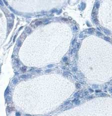
162   S PONCIN   and others . ROS and thyroid function

      ROS production                                                        Quantitative PCR
      Thyrocytes were incubated in multichamber glass slides in             For each condition, cells from six individual wells were
      appropriate medium. ROS production was measured using a               suspended in TriPure isolation reagent (Roche Diagnostics
      fluorescent dye, 2 0 , 7 0 dichlorofluorescein diacetate (DCFH-       GmbH). Total RNA purification and reverse transcription
      DA, Molecular Probes, Paisley, UK). Phosphate buffer saline           were performed as previously described (Gerard et al. 2008).
      (PBS, pH 7.4)-washed thyroid cells were incubated in Krebs–           Quantitative PCR was performed in an iCycler apparatus
      Ringer–HEPES (KRH) medium (pH 7.4) containing                         (IQ5, Bio-Rad). cDNAs (2 ml) were mixed with 500 nM of
      DCFH-DA (25 mM) at 37 8C for 1 h. The excess of dye                   each selected primers (Table 1) and SYBR green reaction
      was removed by two washes with PBS. Cells were stained                mix (Bio-Rad) in a final volume of 25 ml. Reactions were
      with Hoechst for 20 min and rinsed in PBS. Cover slides were          performed as follows: 95 8C/1.5 min, followed by 40 cycles
      mounted in fluorescent mounting medium (DakoCytoma-                   of 958C/15 s, annealing temperature (Table 1)/45 s, and
      tion, Carpinteria, CA, USA) for microscopic observation.              81 8C/15 s. Amplification levels were normalized to those of
      ROS production was visualized on a fluorescent microscope             b-actin. All melting curves were analyzed for each PCR to
      equipped with a digital camera (Zeiss, Zaventem, Belgium).            avoid genomic DNA amplification.

      Viability assay                                                       Western blotting
      Cell viability was assessed using the Alamar blue assay               Thyrocytes were suspended in Laemmli buffer (50 mM Tris–
      (Biosource International, Camarillo, CA, USA), as previously          HCl, pH 6.8, 2% SDS, 10% glycerol), containing a protease
      described (Gerard et al. 2006).                                       inhibitor cocktail (Sigma), and were sonicated during 30 s.
                                                                            Protein concentration was determined using a BCA protein
                                                                            assay kit (Pierce, Rockfort, IL, USA). DUOXs (antibody
      Apoptosis detection
                                                                            provided by F. Miot, IRIBHN), TPO (antibody provided by
      Caspase activity was measured by using a CaspACE FITC-                J. Ruf, Université de la Méditerranée, Marseille, France),
      VAD-fmk in situ marker (Promega) that binds activated                 PRDX5 (antibody provided by B. Knoops, UCL, Louvain La
      caspases, according to the manufacturer’s instructions. Briefly,      Neuve), and b-actin (Sigma) western blottings were
      cells were incubated with 20 mM FITC-VAD-fmk at 37 8C                 performed as previously described (Gerard et al. 2006). Mem-
      for 20 min. Cells were then washed twice with PBS, dyed               branes were blocked for 1 h at room temperature (RT) in PBS
      with Hoechst for 20 min, and rinsed in PBS. Cells were fixed          (pH 7.4), 5% non-fat dry milk, 0.1% Tween, and incubated
      in 10% buffered formalin for 30 min and rinsed with PBS.              overnight at 4 8C with the primary antibody at a dilution of
      Cover slides were mounted in fluorescent mounting medium              1/4000 (DUOXs, TPO), 1/10 000 (PRDX5), or 1/2000
      for microscopic observation. Cells treated with staurosporin          (b-actin). Membranes were incubated for 1 h at RT with
      (5 mM, Sigma) were used as positive controls.                         EnVision (1/200, DakoCytomation) peroxidase-labeled
                                                                            secondary antibody, and visualized with enhanced chemi-
      Nitrite assay                                                         luminescence (SuperSignal West Pico, Pierce) on CLXposure
                                                                            TM films (Pierce). Western blots were scanned and quantified
      Nitrite accumulation in the medium of human thyrocytes was            by densitometry using the NIH Scion Image Analysis Soft-
      measured by the Griess reaction using a commercially                  ware (National Institutes of Health, Bethesda, MA, USA).
      available kit (Promega).                                              Densitometric values were normalized to b-actin expression.

      Table 1 Forward and reverse primers and annealing temperatures

                      Primer forward                                   Primer reverse                                             Annealing T (8C)

      Actin           5 0 -CATCCTGCGTCTGGACCT-3 0                      5 0 -AGGAGGAGCAATGATCTTGAT-3 0                             62
      r DUOX1/2       5 0 -GTGGCTGGAGGGAGCCAT-3 0                      5 0 -CCGTGAACAGACTCCTGT-3 0                                60
      r TPO           5 0 -CAGGTTTTGGTGGGAGAA-3 0                      5 0 -CTGCACACTCATTAACATCTT-3 0                             58
      r NIS           5 0 -GCGCTGCGACTCTCCCACTGAC-3 0                  5 0 -GGCGGTAGAAGATCGGCAAGAAGA-3 0                          60
      r Pendrin       5 0 -CATCAAGACACATCTCCGTTGGCCCT-3 0              5 0 -GGTACTTCCGTTACCACTGGGC-3 0                            60
      r TG            5 0 -GCAGAACAACCACCATCACTGGAGC-3 0               5 0 -TGGCACTGGGGACTCTGGACTTGAC-3 0                         59
      h DUOX1/2       5 0 -GTGGCTGGCTGACATCAT-3 0                      5 0 -TGCAGGGAGTTGAAGAA-3 0                                 58
      h TPO           5 0 -CACGATGCAGAGAAACCTCAA-3 0                   5 0 -ATAGACTGGAGGGAGCCAT-3 0                               60
      h NIS           5 0 -ACCGCGCCCCACCTCTTTCTTATT-3 0                5 0 -CCCCCTCCTGATTCTGGTTGTTG-3 0                           62
      h Pendrin       5 0 -TGGAACATCAAGACATATCTCAGTTG-3 0              5 0 -TGCTGCTGGATACGAGAAAGTG-3 0                            60
      h TG            5 0 -CGCCTGGCGGCTCAGTCTACCTT-3 0                 5 0 -AGCAGTTTCTGCGTGGGAG-3 0                               60

      Journal of Endocrinology (2009) 201, 161–167                                                                www.endocrinology-journals.org

                                                                                                Downloaded from Bioscientifica.com at 08/02/2021 08:41:51AM
                                                                                                                                              via free access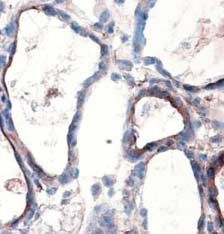
ROS and thyroid function . S PONCIN and others 163
PRDX5 immunofluorescence granules. NAC (1 mM)-treated cells showed a marked
decrease in fluorescence. Identical results were obtained in
Thyrocytes were cultured in multichamber glass slides in
PCCL3 cells (data not shown).
appropriate medium. They were fixed for 30 min in 4%
In human thyrocytes, nitrite levels (Fig. 1B, left panel), the
paraformaldehyde, rinsed once with PBS, permeabilized for
stable end product of NO generation, were low in media from
15 min in a PBS–Triton 1% solution at RT, and washed with
PBS supplemented with 1% BSA (PBS–BSA). Cells were then control cells and slightly increased in NAC (1 mM)-treated
incubated for 1 h with PRDX5 primary antibody (1/75) at cells (1.6-fold, P!0.05). Nos2 mRNA expression was not
RT. After being washed in PBS, a FITC-conjugated secondary influenced by NAC (Fig. 1B, right panel).
antibody was added for 1 h at RT at a dilution of 1/30 In staurosporin-treated cells, used as positive control for
(anti-rabbit; DakoCytomation). Cover slides were mounted in apoptosis, all nuclei were labeled with CaspACE FITC-VAD-
fluorescent-mounting medium for microscopic observation. fmk marker (Fig. 1C). By contrast, apoptosis was detected
neither in control, nor in NAC (1 mM)-treated cells. Cell
Animals and treatments viability was therefore unaffected by NAC (Fig. 1D).
NMRI mice of 2-months-old received a standard diet.
Animals were given i.p. injected of saline solution of NAC NAC reduces the expression of proteins involved in thyroid cell
(100 mg/kg per day) for 4 days. Mice were housed and handled function
according to Belgian regulation of Laboratory Animal Welfare. In human thyrocytes, DUOXs and TPO proteins were
detected by Western blot (Table 2). NAC at concentrations
Preparation of tissue samples for microscopy ranging from 0 . 5 to 5 mM strongly decreased the
expression of both proteins. As no difference was observed
Five animals of each group (control and NAC) were from one concentration to another, 1 mM was selected as
anesthetized with pentothal and thyroid lobes were dissected. reference concentration in all experiments. NAC also
One thyroid lobe was fixed in paraformaldehyde (4% in PBS)
decreased Duoxs and Tpo mRNA expressions, both in
for 24 h and embedded in paraffin. Thick sections (5 mm)
human and PCCL3 cells (Table 3). By contrast, NAC had
were used for PRDX5 immunohistochemistry. The second
no effect on Nis mRNA expression. Tg and pendrin mRNAs
lobe was frozen and cryostat sections (5 mm) were used for
were also downregulated in PCCL3 cells, but not in human
DUOXs immunohistochemistry.
cells. Thus, except NIS, all thyroid differentiation genes
DUOXs and PRDX5 immunohistochemistry studied in this work were downregulated in ROS-deprived
cells.
Tissue sections were washed with PBS–BSA and incubated at
RT for 30 min with normal goat serum (1/50, Vector Labs,
Burlingham, CA, USA) in PBS–BSA. Slides were then incub- NAC differentially affects the expression of PRDX5 and
ated at RT for 1 h with DUOXs (1/75) or PRDX5 (1/250) PRDX3
primary antibodies. The antibody was detected using Envision Both in human and PCCL3 cells, NAC decreased PRDX5
(DakoCytomation) or ABC kit (Vector labs) for DUOXs and protein expression by 2.6-folds (P!0.05, Fig. 2A). The
PRDX5 detection respectively. The peroxidase activity was analysis of PRDX5 protein expression by immunofluores-
revealed using AEC (DakoCytomation) as substrate. Sections cence (Fig. 2B) showed a cytoplasmic and granular pattern,
were counterstained with Mayer’s hematoxylin. suggesting a mitochondrial localization of the protein
(Banmeyer et al. 2005). By contrast, NAC induced a
Data analysis and statistics twofold increase in PRDX3 protein expression (P!0.05,
Data were expressed as mean GS.E.M., nZ6 for all assays. All Fig. 2C). Catalase protein expression remained unchanged
experiments were repeated at least twice. Statistical analyses (Fig. 2D).
were performed using ANOVA followed by Tukey–Kramer
multiple comparison test (GraphPad InStat, San Diego, CA, NAC decreases DUOXs and PRDX5 expressions in mouse
USA), or by unpaired t-test. P!0.05 was considered as thyroids
statistically significant.
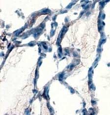
In mouse thyroids, DUOXs was detected at the apical pole
of thyrocytes (Fig. 3A). In NAC- treated mice, DUOXs
Results immunostaining was more faint and detected in few cells
(Fig. 3B). A PRDX5 signal (Fig. 3C) was present in the
NAC reduces intracellular ROS production without inducing cytosol and in nuclei, as previously reported (Poncin et al.
apoptosis or affecting cell viability 2008a). In NAC-treated mice (Fig. 3D), the signal was
In human thyrocytes, ROS, as detected by DCFH-DA strongly reduced, which is in accordance with the above
fluorescence (Fig. 1A), were mainly observed in cytoplasm reported in vitro observations.
www.endocrinology-journals.org Journal of Endocrinology (2009) 201, 161–167
Downloaded from Bioscientifica.com at 08/02/2021 08:41:51AM
via free access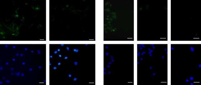
164 S PONCIN and others . ROS and thyroid function
A C
Control NAC Staurosporin Control NAC
D
expression Nos2/β-actin
B
1·0 * Relative mRNA 0·004 100
Cell viability %
0·8 80
Nitrite (µM)
0·003
0·6 60
0·002
0·4 40
0·2 0·001 20
0·0 0·000 0
Control NAC Control NAC
AC M
M
M
AC mM
l
tro
5m
5m
5m
on
1
2·
0·
AC
C
AC
N
N
N
N
Figure 1 NAC reduces intracellular ROS production without inducing apoptosis or affecting
cell viability. ROS production was assessed in control- and NAC-treated cells using DCFH-
DA fluorescence (A, upper panel). Nuclei were stained with Hoechst (A, lower panel). Scale
bars, 20 mm. Nitrite accumulation in the culture medium of human thyrocytes (B, left panel)
was assessed using Griess reaction. Results are expressed as meanGS.E.M. of six (nZ6)
individual wells of one representative experiment. *P!0.05 versus control cells. Nos2 mRNA
expression in human thyrocytes was analyzed by qPCR (B, right panel). Values, adjusted to
the b-actin signal, are expressed as mean GS.E.M. of one representative experiment (nZ6).
Apoptosis was detected using a CaspACE FITC-VAD-fmk in situ marker (C) in staurosporin
(5 mM)-treated cells used as positive control, in control cells, and in NAC-treated cells.
Nuclei were stained with Hoechst (lower panel). Scale bars, 25 mm. Cell viability
was assessed using Alamar Blue assay (D). Full colour version of this figure available via
http://dx.doi.org/10.1677/JOE-08-0470.
Discussion other tissues (Krohn et al. 2007, Maier et al. 2007). H2O2 is
likely one of these ROS as theDCFH-DA probe mainly reacts
This study shows that PCCL3 and human thyroid cells with H2O2. Besides DUOXs produced H2O2, other possible
produce a given amount of intracellular ROS in basal sources of intracellular ROS include mitochondria and
conditions, likely as a result of physiological aerobic peroxisomes (Bedard & Krause 2007). It is likely that ROS
metabolism. But thyrocytes are somewhat unique in that other than H2O2, such as hydroxyl radicals and anion
they produce H2O2 that, along with iodine, is a key element superoxide, are produced in physiological conditions, but
for TH synthesis. Thus, ROS in the thyroid are definitively
more than just by-products of aerobic respiration. They are in Table 2 DUOXs and TPO protein expressions in human thyrocytes.
fact intrinsically involved in endocrine function. H2O2 is DUOXs and TPO protein expressions in human thyrocytes were
produced outside the cell by NADPH oxidases, DUOXs analyzed by western blot
localized at the apical pole of the thyrocyte (De Deken et al.
Human
2002). The hormone synthesis then occurs in the follicular
lumen, close to the apical pole. Until now, H2O2 has always Control NAC 1 mM
been detected in culture media (Bjorkman & Ekholm 1992,
DUOXs 2.39G0.38 1.17G0.38*
Fortemaison et al. 2005), never inside the cell. Here, we show TPO 3.69G0.72 0.98G0.82*
for the first time that ROS are physiologically produced into
cells without provoking significant cell damage, despite the Values, normalized to b-actin, are expressed as mean GS.E.M. of one
fact that basal DNA damage in thyrocytes is higher than in representative experiment (nZ6). *P!0.05 versus control cells.
Journal of Endocrinology (2009) 201, 161–167 www.endocrinology-journals.org
Downloaded from Bioscientifica.com at 08/02/2021 08:41:51AM
via free access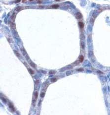
ROS and thyroid function .            S PONCIN    and others 165

Table 3 Relative mRNA expression of thyroid genes. Duoxs, Tpo,              between protein cysteines and glutathione, as recently
Nis, Tg, and pendrin mRNA levels were measured by qRT-PCR in                suggested (Cao et al. 2005, le-Donne et al. 2007). Although,
human thyrocytes and PCCL3 cells                                            ROS are sometimes known to act as transduction signals, for
          Human                                 PCCL3                       instance in cells stimulated by ligands (cytokines, growth
                                                                            factors), or in pathological situations (Lander 1997, Finkel
          Control       NAC 1 mM                Control      NAC 1 mM       1998, Adler et al. 1999), it is the first time that the
Duoxs     1.61G0.44     0.93G0.27*              1.09G0.13    0.62G0.14*
                                                                            maintenance of a minimal intracellular oxidative load is
Tpo       0.72G0.11     0.15G0.05*              0.81G0.19    0.34G0.08*     shown to be required for safeguarding endocrine functions.
Nis       1.61G0.53     1.59G0.38               0.89G0.17    0.79G0.14      This has already been suggested in pancreatic cells or in the
Tg        0.88G0.26     0.85G0.11               0.63G0.16    0.33G0.1*      lung (Lenzen 2008), but without any convincing demon-
Pendrin   0.83G0.18     0.46G0.08               0.74G0.09    0.35G0.05*
                                                                            stration. Thus, when the oxidative load in thyrocytes drops
                                                                            below a minimal threshold, various actors involved in the TH
Values, normalized to b-actin, are expressed as mean GS.E.M. of one
representative experiment (nZ6). *P!0.05 versus control cells.              synthesis are also downregulated. This might eventually be
                                                                            relevant in terms of thyroid function in patients treated with
likely in very low amounts, as suggested by previous models                 NAC, but as far as we know, there is no robust data yet
(Denef et al. 1996). Further studies are required to find out the           reporting such interactions in the literature.
source and the type of intracellular ROS, and to sort out those                 In addition to ROS, thyrocytes also produce RNS in low
resulting from aerobic metabolism as in every other organs,                 amounts (basal nitrite release) associated with NOS2 and
and those specifically involved in hormone synthesis.                       NOS3 expressions (Colin et al. 1995), suggesting a role for NO
   NAC may decrease cell OS, either directly, as a source of                in normal thyroids. In contrast with ROS, NAC increased
sulfhydryl groups that neutralize ROS, or indirectly as                     NO production without affecting Nos2 mRNA expression.
glutathione precursor by restoring glutathione levels (GSH)                 The interpretation of this result is somewhat difficult as NAC-
(Gillissen & Nowak 1998). The present study shows that                      induced effects on NOS2 expression remains a matter of
NAC decreases intracellular ROS to very low levels without                  debate. It may either increase, or decrease, or have no effect on
inducing apoptosis or affecting cell viability. This marked                 NOS2 expression and NO release (Ramasamy et al. 1999, Vos
decrease in intracellular ROS levels is associated with                     et al. 1999, Chen et al. 2000, Zafarullah et al. 2003).
decreased DUOXs, TPO, TG, and pendrin expressions, but                      Nevertheless, NO levels are particularly low when compared
not Nis mRNA expression. Such effects on DUOXs                              with those produced for instance in cells incubated with Th1
expression were also observed in vivo. As NAC may also                      cytokines (Kasai et al. 1995, van den Hove et al. 2002, Gerard
influence GSH intracellular levels, it is possible that some                et al. 2006). Although this slight increase in NO production
specific biochemical processes might be altered by glutathio-               may appear odd in cells treated with NAC, one should keep in
nylation, i.e., the reversible formation of disulfide bonds                 mind that superoxide anions, which are constantly produced

                                                                          B                  Control              NAC
                          A               1·2
                          PRDX5/β-actin
                           expression

                                          0·8
                             Protein

                                                                *
                                          0·4

                                          0·0
                                                   Control    NAC

                         C                2·0                             D                  2·0
                                                                    *
                                                                          catalase/β-actin
                          PRDX3/β-actin

                                          1·5                                                1·5
                           expression

                                                                            expression
                             Protein

                                                                              Protein

                                          1·0                                                1·0

                                          0·5                                                0·5

                                          0·0                                                0·0
                                                   Control      NAC                                    Control           NAC
                      Figure 2 NAC differentially regulates the expression of antioxidant enzymes in human
                      thyrocytes. PRDX5 (A), PRDX3 (C), and catalase (D) protein expressions were analyzed by
                      western blot. Densitometric values normalized to b-actin are expressed as mean GS.E.M. of one
                      representative experiment (nZ6). *P!0.05 versus control cells. PRDX5 protein (B) localization
                      was analyzed by immunofluorescence in control- and NAC-treated cells. Scale bars: 20 mm. Full
                      colour version of this figure available via http://dx.doi.org/10.1677/JOE-08-0470.

www.endocrinology-journals.org                                                                                   Journal of Endocrinology (2009) 201, 161–167

                                                                                                                 Downloaded from Bioscientifica.com at 08/02/2021 08:41:51AM
                                                                                                                                                               via free access
166 S PONCIN and others . ROS and thyroid function
Control NAC In conclusion, our results showed for the first time that in
A B physiological conditions, thyroid cells produce intracytoplas-
mic ROS. When their intracellular levels drop very low, as
in NAC-treated cells, the expression of important proteins
involved in TH synthesis is hampered. This strongly suggests
that the maintenance of the oxidative load above a minimum
threshold is required to safeguard the function of thyroid cells.
Declaration of interest
The authors declare that there is no conflict of interest that could be perceived
as prejudicing the impartiality of the research reported.
C D
Funding
This work was supported by the grant no. 3.4552.08 (Fonds National de la
Recherche Scientifique (FNRS-FRSM)). A-C Gérard is a postdoctoral
researcher (Fonds National de la Recherche Scientifique).
References
Adler V, Yin Z, Tew KD & Ronai Z 1999 Role of redox potential and reactive
oxygen species in stress signaling. Oncogene 18 6104–6111.
Figure 3 NAC decreases DUOX and PRDX5 expressions in mouse Banmeyer I, Marchand C, Clippe A & Knoops B 2005 Human mitochondrial
thyroids. DUOXs protein expression was detected by immunohis- peroxiredoxin 5 protects from mitochondrial DNA damages induced by
tochemistry on frozen sections. In control thyroids (A), DUOXs was hydrogen peroxide. FEBS Letter 579 2327–2333.
detected at the apical pole of the cells (arrow). In NAC-treated mice Bast A, Wolf G, Oberbaumer I & Walther R 2002 Oxidative and nitrosative
(B), DUOXs was barely detected only in few cells. PRDX5 protein stress induces peroxiredoxins in pancreatic beta cells. Diabetologia 45
expression was detected by immunohistochemistry on paraffin 867–876.
sections. In control thyroids (C), PRDX5 was detected in the Bedard K & Krause KH 2007 The NOX, family of ROS-generating NADPH
cytoplasm and in nuclei. In NAC-treated cells (D), the staining was oxidases: physiology and pathophysiology. Physiological Reviews 87 245–313.
greatly decreased. Scale bars, 10 mm. Full colour version of this Bjorkman U & Ekholm R 1992 Hydrogen peroxide generation and its
figure available via http://dx.doi.org/10.1677/JOE-08-0470. regulation in FRTL-5 and porcine thyroid cells. Endocrinology 130 393–399.
Blokhina O, Virolainen E & Fagerstedt KV 2003 Antioxidants, oxidative
damage and oxygen deprivation stress: a review. Annals of Botany 91 179–194.
by mitochondria during aerobic respiration (Bedard & Krause Cao X, Kambe F, Lu X, Kobayashi N, Ohmori S & Seo H 2005
2007), react with NO in normal cells to form low amounts of Glutathionylation of two cysteine residues in paired domain regulates DNA
nitrotyrosines. Although this remains to be proven, it could be binding activity of Pax-8. Journal of Biolgical Chemistry 280 25901–25906.
Chen G, Wang SH & Warner TD 2000 Regulation of iNOS mRNA levels in
speculated that in case of NAC-induced ROS suppression, endothelial cells by glutathione, a double-edged sword. Free Radical Research
superoxide anions are no more available in sufficient amounts 32 223–234.
to react with NO. Consequently, NO might then be released Colin IM, Nava E, Toussaint D, Maiter DM, vanDenhove MF, Luscher TF,
in culture media in slightly greater amounts. Ketelslegers JM, Denef JF & Jameson JL 1995 Expression of nitric oxide
As the oxidative load was deeply affected by NAC, it made synthase isoforms in the thyroid gland: evidence for a role of nitric oxide in
vascular control during goiter formation. Endocrinology 136 5283–5290.
sense to analyze how antioxidant systems react in such Contempre B, Denef JF, Dumont JE & Many MC 1993 Selenium deficiency
conditions. Our results indicate that they are actually aggravates the necrotizing effects of a high iodide dose in iodine deficient
influenced by intracellular ROS, especially PRDXs whose rats. Endocrinology 132 1866–1868.
expression is known to be regulated by OS (Bast et al. 2002). Contempre B, de Escobar GM, Denef JF, Dumont JE & Many MC 2004
Thiocyanate induces cell necrosis and fibrosis in selenium- and iodine-
In our study, PRDX5 expression decreased, while PRDX3 deficient rat thyroids: a potential experimental model for myxedematous
increased in NAC-treated cells, indicating that both proteins endemic cretinism in central Africa. Endocrinology 145 994–1002.
are differently regulated in conditions of ROS depletion. By Dai G, Levy O & Carrasco N 1996 Cloning and characterization of the
contrast, catalase protein expression remained unchanged, thyroid iodide transporter. Nature 379 458–460.
De Deken X, Wang D, Dumont JE & Miot F 2002 Characterization of
which is in line with the low enzymatic activity previously
ThOX proteins as components of the thyroid H(2)O(2)-generating system.
reported in the thyroid (Nadolnik & Valentyukevich 2007). Experimental Cell Research 273 187–196.
Thus, in accordance with previous results (Gerard et al. 2005, Denef JF, Many MC & van den Hove MF 1996 Iodine-induced thyroid
Poncin et al. 2008a), our present data confirm that PRDXs 5 inhibition and cell necrosis: two consequences of the same free-radical
and 3 are in fact highly regulated in the thyroid, suggesting mediated mechanism? Molecular Cellular Endocrinology 121 101–103.
Dupuy C, Ohayon R, Valent A, Noel-Hudson MS, Deme D & Virion A 1999
that they may play important roles in detoxification processes Purification of a novel flavoprotein involved in the thyroid NADPH
along side the glutathione peroxidase system (Nadolnik & oxidase. Cloning of the porcine and human cdnas. Journal of Biolgical
Valentyukevich 2007). Chemistry 274 37265–37269.
Journal of Endocrinology (2009) 201, 161–167 www.endocrinology-journals.org
Downloaded from Bioscientifica.com at 08/02/2021 08:41:51AM
via free accessROS and thyroid function . S PONCIN and others 167
le-Donne I, Rossi R, Giustarini D, Colombo R & Milzani A 2007 Mano T, Shinohara R, Iwase K, Kotake M, Hamada M, Uchimuro K,
S-glutathionylation in protein redox regulation. Free Radical Biology and Hayakawa N, Hayashi R, Nakai A, Ishizuki Y et al. 1997 Changes in free
Medicine 43 883–898. radical scavengers and lipid peroxide in thyroid glands of various thyroid
Finkel T 1998 Oxygen radicals and signaling. Current Opinion in Cell Biology disorders. Hormone and Metabolic Research 29 351–354.
10 248–253. Mutaku JF, Poma JF, Many MC, Denef JF & van den Hove MF 2002
Fortemaison N, Miot F, Dumont JE & Dremier S 2005 Regulation of H2O2 Cell necrosis and apoptosis are differentially regulated during goitre
generation in thyroid cells does not involve Rac1 activation. European development and iodine-induced involution. Journal of Endocrinology 172
Journal Endocrinology 152 127–133. 375–386.
Fusco A, Berlingieri MT, Di Fiore PP, Portella G, Grieco M & Vecchio G Nadolnik LI & Valentyukevich OI 2007 Peculiarities of the antioxidant
1987 One- and two-step transformations of rat thyroid epithelial cells by status of the thyroid gland. Bulletin of Experimental Biology and Medicine 144
retroviral oncogenes. Molecular and Cellular Biology 7 3365–3370. 529–531.
Gerard AC, Many MC, Daumerie C, Knoops B & Colin IM 2005 Poncin S, Gerard AC, Boucquey M, Senou M, Calderon PB, Knoops B,
Peroxiredoxin 5 expression in the human thyroid gland. Thyroid 15 Lengele B, Many MC & Colin IM 2008a Oxidative stress in the thyroid
205–209. gland: from harmlessness to hazard depending on the iodine content.
Gerard AC, Boucquey M, van den Hove MF & Colin IM 2006 Endocrinology 149 424–433.
Expression of TPO and ThOXs in human thyrocytes is downregulated Poncin S, Lengele B, Colin IM & Gerard AC 2008b Differential interactions
by IL-1a/IFN-g, an effect partially mediated by nitric oxide. American between Th1/Th2, Th1/Th3, and Th2/Th3 cytokines in the regulation of
Journal of Physiology-Endocrinology and Metabolism 291 E242–E253. TPO and DUOX expression, and of thyroglobulin secretion in thyrocytes
Gerard AC, Poncin S, Caetano B, Sonveaux P, Audinot JN, Feron O, Colin in vitro. Endocrinology 149 1534–1542.
IM & Soncin F 2008 Iodine deficiency induces a thyroid stimulating Ramasamy S, Drummond GR, Ahn J, Storek M, Pohl J, Parthasarathy S &
hormone-independent early phase of microvascular reshaping in the Harrison DG 1999 Modulation of expression of endothelial nitric oxide
thyroid. American Journal of Pathology 172 748–760. synthase by nordihydroguaiaretic acid, a phenolic antioxidant in cultured
Gillissen A & Nowak D 1998 Characterization of N-acetylcysteine and endothelial cells. Molecular Pharmacology 56 116–123.
ambroxol in anti-oxidant therapy. Respiratory Medicine 92 609–623. Schomburg L & Kohrle J 2008 On the importance of selenium and iodine
van den Hove MF, Stoenoiu MS, Croizet K, Couvreur M, Courtoy PJ, metabolism for thyroid hormone biosynthesis and human health. Molecular
Devuyst O & Colin IM 2002 Nitric oxide is involved in interleukin- Nutrition & Food Research 52 1235–1246.
1alpha-induced cytotoxicity in polarised human thyrocytes. Journal of Schweizer U, Chiu J & Kohrle J 2008 Peroxides and peroxide-degrading
Endocrinology 173 177–185. enzymes in the thyroid. Antioxidants & Redox Signaling 10 1577–1592.
Kasai K, Hattori Y, Nakanishi N, Manaka K, Banba N, Motohashi S & Vos TA, Van GH, Tuyt L, De Jager-Krikken A, Leuvenink R, Kuipers F,
Shimoda S 1995 Regulation of inducible nitric oxide production by Jansen PL & Moshage H 1999 Expression of inducible nitric oxide synthase
cytokines in human thyrocytes in culture. Endocrinology 136 4261–4270. in endotoxemic rat hepatocytes is dependent on the cellular glutathione
Krohn K, Maier J & Paschke R 2007 Mechanisms of disease: hydrogen status. Hepatology 29 421–426.
peroxide, DNA damage and mutagenesis in the development of thyroid Zafarullah M, Li WQ, Sylvester J & Ahmad M 2003 Molecular mechanisms of
tumors. Nature Clinical Practice. Endocrinology & Metabolism 3 713–720. N-acetylcysteine actions. Cellular and Molecular Life Sciences 60 6–20.
Lander HM 1997 An essential role for free radicals and derived species in signal
transduction. FASEB Journal 11 118–124.
Lenzen S 2008 Oxidative stress: the vulnerable beta-cell. Biochemical Society Received in final form 9 January 2009
Transactions 36 343–347.
Maier J, van SH, van OC, Paschke R, Weiss RE & Krohn K 2007 Iodine
Accepted 23 January 2009
deficiency activates antioxidant genes and causes DNA damage in the Made available online as an Accepted Preprint
thyroid gland of rats and mice. Biochemica and Biophysica Acta 1773 990–999. 23 January 2009
www.endocrinology-journals.org Journal of Endocrinology (2009) 201, 161–167
Downloaded from Bioscientifica.com at 08/02/2021 08:41:51AM
via free accessYou can also read


