ORIGINAL ARTICLE MIR-126 ENHANCES VEGF EXPRESSION IN INDUCED PLURIPOTENT STEM CELL-DERIVED RETINAL NEURAL STEM CELLS BY TARGETING SPRED-1
←
→
Page content transcription
If your browser does not render page correctly, please read the page content below
Int J Clin Exp Pathol 2018;11(2):1023-1030
www.ijcep.com /ISSN:1936-2625/IJCEP0064048

Original Article
MiR-126 enhances VEGF expression in induced
pluripotent stem cell-derived retinal neural
stem cells by targeting spred-1
Lin Ye, Yun Peng, Jinsong Mo, Yuanyuan Yao

Shenzhen Key Laboratory of Ophthalmology, Shenzhen Eye Hospital, Jinan University, Shenzhen, Guangdong,
China
Received August 20, 2017; Accepted December 7, 2017; Epub February 1, 2018; Published February 15, 2018

Abstract: Pathological retinal neovascularization (RNV) is a leading cause of vision loss in several ocular diseases;
however, the underlying molecular mechanisms involved in the development of RNV remain unclear. It has been
shown that microRNAs contribute to the process of angiogenesis, which has received greater attention by investiga-
tors who study the progression of RNV. In the present study, we investigated the function of miR-126 expression in
retinal neural stem cells derived from induced pluripotent stem cell (IPSs) obtained from patients with RNV. Dur-
ing the induction process, the levels of both miR126 and vascular endothelial growth factor C (VEGF-C) gradually
decreased, while the levels of spred-1 significantly increased. The existence of conserved miR-126-binding sites in
spred-1 mRNA was predicted by computational algorithms, and verified by the luciferase reporter assay. The use of
miR-126 mimics revealed dramatically reduced levels of spred-1, and increased levels of VEGF. When using shRNA
to target spred-1, the resultant decreased levels of spred-1 were associated with significantly enhanced levels of
VEGF expression. Our results demonstrate that miR-126 promotes VEGF expression in IPS cells by suppressing
spred-1 expression, which contributes to angiogenesis during the progression of RNV. These findings suggest that
miR-126 and spred-1 might serve as novel molecular targets for treating RNV-related ocular diseases.

Keywords: Pathological retinal neovascularization, induced pluripotent stem cell, micro-126, VEGF

Introduction                                                   clear. Recent studies have shown that microR-
                                                               NAs contribute to the process of angiogenesis,
Pathological retinal neovascularization (RNV) is               which has gained more attention from investi-
a leading cause of vision loss in several ocular               gators who study RNV progression.
diseases, including diabetic retinopathy, retinal
vein occlusion, retinopathy of prematurity, and                MicroRNAs (miRNAs or miRs) comprise a group
age-related macular degeneration. As RNV pro-                  of small (19-25 nucleotides) and ubiquitously
gresses, new capillary sprouts from the retinal                distributed endogenous non-coding single-
veins grow out of the vitreous surface of the                  stranded RNAs. MiRNAs post-transcriptionally
                                                               modulate gene expression by binding to the 3’
retina, causing vitreous hemorrhage and retinal
                                                               untranslated regions of their target mRNA mol-
detachment that eventually lead to vision loss
                                                               ecules. This binding causes degradation of the
and even blindness [1-4]. As the population
                                                               target mRNAs and blocks production of their
ages, the incidence rates of ocular diseases                   encoded proteins [11]. MiRNAs are implicated
caused by RNV have increased [5]. Several dif-                 in various physiological processes and patho-
ferent mediators, including ADAM8, ROCK,                       logical conditions, including cell proliferation,
Dll4, and VEGF contribute to the development                   differentiation, apoptosis, inflammatory resp-
of RNV [6-9]. While various therapeutic                        onses, angiogenesis, cardiovascular diseases,
approaches have been developed and used to                     and cancer [11-15].
treat pathologic angiogenesis and RNV in the
clinic [10], the exact underlying molecular                    Several recent studies have identified some
mechanisms for these disorders remain un-                      miRNAs that mediate angiogenesis and play a
MiR-126 promotes RNV by targeting spred-1

crucial role in RNV progression. These miRNAs       instructions. Biopsy results were interpreted
are termed “angiomiRs”, and include miR-23,         according to the Updated Banff 07 criteria by
miR-21, and miR-24. Fish et al and Wang et al       H.R. The protocols used in the present study
showed that miR-126 is involved in both angio-      were approved by Shenzhen Eye Hospital, and
genesis and maintenance of vascular integrity       a signed written informed consent was obtained
[16-18]. Therefore, it is important to gain a       from each enrolled subject.
greater understanding of how microRNAs help
regulate pathogenic angiogenesis and RNV, if        Cell culture and induction
new therapeutic approaches are to be devel-
                                                    CD34+ cells were isolated using an EasySep™
oped for these disorders.
                                                    Human CD34 Positive Selection Kit according
MiR-126 is a recently identified novel microRNA     to the manufacturer’s protocol. The cells were
that is expressed by endothelial cells found in     cultured in StemPro™34 SFM supplemented
all blood vessels, and has been shown to play       with SCF (100 ng/mL), FLT3 (100 ng/mL), IL-3
functional roles in cancer, diabetes, allergic      (20 ng/mL), and IL-6 (20 ng/mL) for 4 days
asthma, and cystic fibrosis [19-22]. MiR-126        after isolation. The cells were incubated in a 5%
mainly functions in the regulation and control of   CO2 incubator at 37°C, and then transduced
angiogenesis by targeting critical angiogenetic     using a CytoTune™-iPS 2.0 Sendai Rep-
factors such as EGFL7 and CRK [22, 23].             rogramming Kit. Virus was removed after 24
However, the mechanism by which miR-126             hours of transduction, and the cells were plat-
                                                    ed onto rh-Vitronectin (VTN) matrix at 3 days
mediates the angiogenesis process during the
                                                    after transduction. At 7 days after transduc-
development of RNV is remains under investi-
                                                    tion, the medium was changed to Essential 8™
gation. In the present study, we used pluripo-
                                                    Medium. To induce neural differentiation, cell
tent stem cells (IPSs) induced from peripheral
                                                    differentiation was induced by switching the
blood mononuclear cells obtained from patients
                                                    cell culture medium from mTESR1 to retinal
with RNV as an in vitro model. Our results
                                                    induction medium (RIM) containing DMEM/
revealed that the levels of miR126 and VEGF-C
                                                    F12, N2 and B27 serum-free supplements,
expression gradually decreased, while the level
                                                    100 units/mL penicillin, 100 μg/mL strepto-
of spred-1 expression significantly increased       mycin (Life Technology, Carlsbad, CA, USA),
during the induction process. MiR-126-binding       0.45% glucose (Sigma, St. Louis, MO, USA), 20
sites in spred-1 mRNA were predicted by com-        μg/mL human insulin (Roche, Basal Sw-
putational algorithms and verified by the lucif-    itzerland), and 50 ng/mL human Noggin
erase reporter assay. The use of miR-126 mim-       (Peprotech, Rocky Hill, NJ. USA). On day 5, the
ics revealed reduced spred-1 levels and             RIM medium was switched to neural differen-
increased VEGF levels. When using spred-1           tiation medium plus (NDM+), containing
3’UTR miRNA, the resultant reduced levels of        Neurobasal, N2 and B27 serum-free supple-
spred-1 significantly enhanced VEGF expres-         ments, 100 units/mL penicillin, 100 μg/mL
sion. This study showed that miR126 enhances        streptomycin, Glutamax, MEM non-essential
VEGF expression in IPS cells by inhibiting spred-   amino acids (Life Technology), 0.45% glucose,
1 expression, which contributes to angiogene-       and 50 ng/mL human Noggin. Cells were har-
sis during the progression of RNV. Our results      vested for use in analytical procedures at each
suggest that spred-1, the target gene of            of the following 10 time points: 1) PBMCs were
miR126, might serve as a novel target for treat-    isolated from peripheral blood; 2) CD34+ cells
ing RNV-related ocular diseases.                    were isolated from PBMCs; 3) two weeks the
                                                    induction of iPS cells; 4) iPS cells were gener-
Materials and methods                               ated; 5) second passage of the iPS cells; 6)
                                                    third passage of the iPS cells; 7, 8, 9, 10) day 1,
Patients and sample collections
                                                    day 3, day 5, and day 7 of the retinal neural
                                                    stem cell induction process.
Twenty patients with RNV were included in this
study, and provided the blood samples used for      Immunocytochemical staining
analysis. Peripheral blood mononuclear cells
(PBMCs) were isolated using Histopaque 1077         Cells were fixed by incubation with 4% parafor-
reagents kits according to the manufacturer’s       maldehyde for 2 minutes, and then washed

1024                                                   Int J Clin Exp Pathol 2018;11(2):1023-1030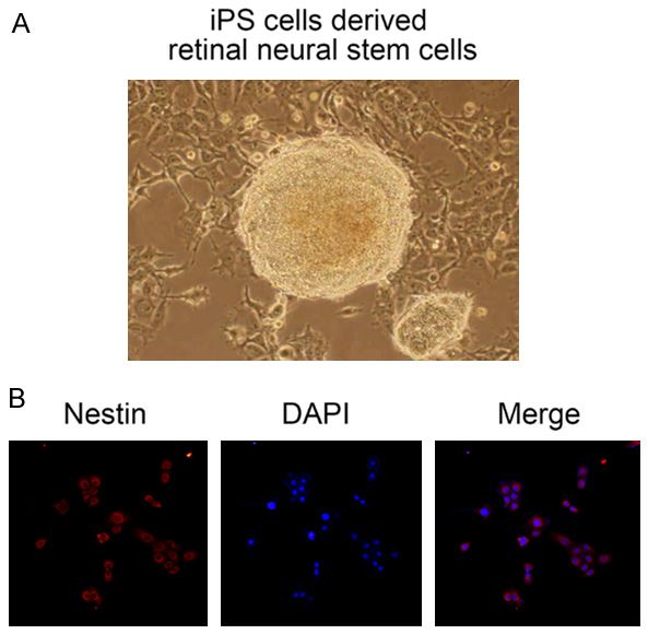
MiR-126 promotes RNV by targeting spred-1

                                                                              Transfection with miR-126
                                                                              mimics

                                                                              MiR-126 mimics and negative
                                                                              controls were chemically syn-
                                                                              thesized by Suzhou Jima. Cells
                                                                              were seeded into 6-well or
                                                                              24-well cell culture plates that
                                                                              contained fresh medium with-
                                                                              out antibiotics. When the cells
                                                                              reached 80% confluence, they
                                                                              were transfected with oligonu-
                                                                              cleotides. All transfections
                                                                              were performed using Lipo-
                                                                              fectamine 3000 reagent (In-
                                                                              vitrogen, Carlsbad, CA, USA)
                                                                              according to the manufactur-
                                                                              er’s instructions.

                                                                              RNA extraction and real-time
                                                                              PCR

                                                                                Total cellular RNA was extract-
                                                                                ed with Trizol reagent. First-
                                                                                strand cDNA was synthesized
                                                                                from 1 μg of total RNA by using
                                                                                a Reverse Transcription Sy-
                                                                                stem Bestar qPCR RT Kit
                                                                                according to the manufactur-
Figure 1. Relative expression of miR-126 and spred-1 during the induction       er’s instructions. Real-time
of iPSs. A. Expression of miR-126 at 10 time points during induction of iPSs;   PCR was performed on an ABI
B. Expression of spred-1 at 10 time points during induction of iPSs. *P <
0.05, **P < 0.01, ***P < 0.001, NS, not significant.                            7500 Real-Time PCR System.
                                                                                Each assay was performed in
                                                                                triplicate, and β-actin served
three times with PBS. The cells were then per-                 as an endogenous control gene. The primer
meabilized by incubation with 0.1% Triton X-100                sequences used were as follows: spred-1,
for 5 minutes, and washed with PBS. Next, the                  5’-CAGCCAGGCTTGGACATTCA-3’ (forward) and
cells were treated for 30 minutes with a solu-                 5’-TGGGACTTTAGGCTTCCACAT-3’ (reverse); miR-
tion composed of 5% bovine serum albumin                       126, 5’-ACACTCCAGCTGGGTCGTACCGTGAGTA-
and 5% normal goat serum, and incubated                        ATAA-3’ (forward) and 5’-CTCAACTGGTGTCGT-
overnight at 4°C with the primary antibody:                    GGAGTCGGCAATTCAGTTGAGCGCATT-3’ (reve-
mouse anti-human nestin (1:100 dilution;                       rse); GAPDH, 5’-TGTTCGTCATGGGTGTGAAC-3’
Santa Cruz Biotechnology, Santa Cruz, CA,                      (forward) and 5’-ATGGCATGGACTGTGGTCAT-3’
USA). Following incubation, excess primary anti-               (reverse) (Sangon Biotech, Shanghai, China).
body was removed by washing the cells five                     The relative amounts of miR-126 and BCL11B
times with PBS; after which, the cells were incu-              synthesized were calculated using the 2-ΔΔCt
bated with secondary antibodies (fluorescein-                  method and normalized by using β-actin cDNA
conjugated goat anti-mouse IgG and rhoda-                      as an internal control.
mine-conjugated goat anti-mouse IgG, [Santa
Cruz Biotechnology]) at 37°C for 1 hour. After                 Target prediction and luciferase assay
washing with PBS, the cell nuclei were stained
with 4’,6-diamidoino-2-phenylindole. Finally,                  The putative targets of miR-126 were predicted
the cells were overlaid with glycerol, visualized,             by Target Scan Release 6.2 software. The
and photographed with a laser scanning micro-                  human spred-1 wild-type and mutant 3’UTR
scope (Nikon, Japan).                                          reporter vectors were constructed by inserting

1025                                                           Int J Clin Exp Pathol 2018;11(2):1023-1030
MiR-126 promotes RNV by targeting spred-1
126 levels in the stem cells from RNV subjects
showed a gradual significant decrease during
the induction period when compared to the
miR-126 levels in stem cells from the control
subjects (Figure 1A). Spred-1 expression levels
were also evaluated by qPCR. We found that
spred-1 levels exhibited an opposite tendency
by showing an increasing trend throughout the
induction period (Figure 1B).
IPS cells differentiated into retinal neural stem
cells via induction
In order to verify the successful induced differ-
entiation of retinal neural stem cells, we tested
the newly formed neural spheres (Figure 2A)
for their expression of nestin (a retinal stem cell
Figure 2. Verification of iPS-derived retinal neural marker) by immunofluorescence (Figure 2B).
stem cells. A. Neural spheres were formed after in- We found that the neural spheres had the mor-
duction of IPSs. B. Positive results for nestin expres- phological appearance of retinal neural stem
sion after induction. Editor Note: Please change the
label of 2A to read: IPS cell-derived neural stem cells. cells, and displayed positive immunostaining
for nestin. These findings indicated that retinal
neural stem cells had been successfully
annealed oligonucleotides with tow flanking induced from iPS cells (Figure 2).
restriction sites into a pmirGLO vector. Firefly
luciferase and Renilla luciferase signals were MiR-126 mimics enhanced VEGF expression
measured using a Dual-luciferase assay report- by downregulating spred-1
er kit and quantitated by a Lumat LB 9501
luminator. To explore the functional role of miR-126 in iPS
cell-derived retinal neural stem cells, miR-126
Statistical analysis mimics or a negative control were transfected
into human iPS cell-derived retinal neural stem
All statistical calculations were performed cells obtained from the healthy subjects. We
using Prism 6 software (GraphPad, San Diego, then used qPCR to evaluate the levels of miR-
California, USA). Results are presented as the 126, spred-1, and VEGF expression. Tran-
mean ± SEM. Student’s t test was used for sfection with the miR-126 mimics significantly
comparisons between 2 groups, and 1-way increased the levels of miR-126 and VEGF when
ANOVA followed by the Tukey test was used for compared to transfection with the negative
comparisons among multiple groups. A P-val control; however, spred-1 levels dramatically
ue < 0.05 was considered statistically sign- decreased (Figure 3). GAPDH RNA was used as
ificant. an internal control for all qPCR analyses of
miRNA expression.
Results
Spred-1 as a target of miR-126
MiR-126 levels decreased during IPS cell
induction We used computational prediction (miRBase,
TargetScan, PicTar, and MiRanda) to identify
Quantitative real-time PCR (qPCR) was used to potential binding sites for spred-1 mRNA on
evaluate the level of miR-126 expression miR-126. We found that the 3’UTR of spred-1
throughout the iPS cell induction period. MiR- mRNA bears a miR-126-binding site that is con-
126 expression in retinal neural stem cells served in mammals (Figure 4A). The effect of
derived from subjects with RNV was compared miR-126 on translation of spred-1 mRNA into
with that in neural stem cells derived from protein was evaluated by the luciferase report-
healthy control subjects. We found that miR- er assay. Transfection with the miR-126 mimics
1026 Int J Clin Exp Pathol 2018;11(2):1023-1030MiR-126 promotes RNV by targeting spred-1
healthy patients, the inhibition
of spred-1 expression by
spred-1-specific shRNA was
evaluated by qPCR. Expression
of spred-1 in cells transfected
with shRNA was significantly
reduced when compared with
spred-1 expression in the neg-
ative control cells, while the
levels of VEGF-C increased
after inhibition of spred-1
expression (Figure 5).
Discussion
The development of retinal
neovascularization (RNV) in
the eye is a leading cause of
vision loss among people of
all ages. RNV underlies the
pathology of conditions such
as diabetic retinopathy, age-
related macular degeneration,
retinal vein occlusion, and
external exudative retinopa-
thy, which all have a negative
impact on quality of life. The
lack of an effective treatment
for RNV has led to increased
incidence rates of ocular dis-
eases caused by RNV, and
novel alternative therapeutic
approaches are urgently need-
ed. During the progression of
RNV, the abnormal growth of
new blood vessels within the
choroid extends to the retina,
and VEGF plays a critical role
in this process. New treat-
Figure 3. Relative expression of miR-126, spred-1, and VEGF-C during iPS ments are available that tar-
induction relative to controls (PBMCs from healthy individuals). get VEGF, and anti-VEGF
agents such as Macugen,
Eylea, and Lucentis are cur-
significantly decreased the luciferase activity of rently being used to treat ocular diseases
the reporter gene with the wild-type spred-1 caused by RNV [24-26]. However, anti-VEGF
3’UTR when compared to transfection with the agents are only able to control RNV progres-
negative control. However, this regulatory effect sion, and are not very efficient at improving
of miR-126 was suppressed when the predict- vision. Therefore, the need for combination
ed miR-126-binding site in spred-1 mRNA was therapies is becoming increasingly imperative.
mutated (Figure 4B).
Studies have reported that a group of miRNAs
Inhibition of spred-1 increased VEGF expres- contribute to angiogenesis; these include miR-
sion 23, miR-21, and miR-24. Therefore, the identifi-
cation of miRNAs that serve as vital mediators
To further explore the relationship between of RNV progression might be on the critical
spred-1 and VEGF-C in PBMCs obtained from path for developing novel therapeutic strate-
1027 Int J Clin Exp Pathol 2018;11(2):1023-1030MiR-126 promotes RNV by targeting spred-1
study, we showed that a novel
miRNA, miR-126, helps medi-
ate RNV progression via its
ability to regulate VEGF ex-
pression, suggesting that miR-
126 might play a role in modu-
lating RNV. We found that
transfection of miR-126 mim-
ics increased VEGF levels, and
that the levels of a neurofibro-
min recruitment factor (spred-
1) decreased significantly
after transfection. Conserved
miR-126-binding sites were
identified in spred-1 mRNA by
Figure 4. MiR-126 inhibited spred-1 expression by binding to the 3’UTR computationalalgorithmsandve-
of spred-1 mRNA. A. MiR-126 targeted spred-1 by binding to the 3’UTR of rified by the luciferase report-
spred-1 mRNA; B. Double luciferase assays showed that miR-126 could er assay, suggesting that miR-
directly target spred-1 mRNA. C. MiR-126 inhibited spred-1 expression by 126 had increased the levels
direct targeting. *P < 0.05.
of VEGF by inhibiting spred-1
expression.
Recently, spred-1 has been
studied in several pathological
conditions, including cancer
and RNV, due to its sprouty-
related function in angiogene-
sis. It has been shown that
spred-1 is involved in regulat-
ing hepatocellular carcinoma
and age-related macular de-
generation [28, 29]. Moreover,
several studies have identified
spred-1 mRNA as a binding
target for various miRNAs
(miR-132, miR212, miR-206
miR-21, and miR-126) [30-
32]. Our results demonstrated
that inhibition of spred-1 ex-
pression in IPS cells remark-
ably increased the levels of
VEGF in those cells, suggest-
ing that spred-1 might down-
regulate VEGF expression.
Moreover, our results also pro-
vide evidence that miR-126
mediates both angiogenesis
and RNV by targeting spred-1.
Figure 5. Spred-1 shRNA enhanced expression of VEGF-C.
Our study used pluripotent
stem cells (IPSs) that had
gies. Zhuang and Qin et al [27] showed that been induced from the peripheral blood mono-
downregulation of miRNA-155 attenuates RNV nuclear cells of patients with RNV as in vitro
via the PI3K/Akt pathway [27]; Kong and Sun et model. This in vitro model represents an inno-
al suggested that miR-155 regulates RNV via vative approach for studying human genetic
the Slit-Robo signaling pathway. In the present diseases and various pathological conditions
1028 Int J Clin Exp Pathol 2018;11(2):1023-1030MiR-126 promotes RNV by targeting spred-1
such as neurogenetic diseases, muscular dys- occlusion. Acta Ophthalmol Scand 1997; 75:
trophies, and cardiovascular diseases [33, 34]. 441-442.
IPS cells generated from primary patient sam- [3] Moravski CJ, Kelly DJ, Cooper ME, Gilbert RE,
Bertram JF, Shahinfar S, Skinner SL and
ples can be used to provide information rele-
Wilkinson-Berka JL. Retinal neovascularization
vant to genomics, proteomics, and metabolo-
is prevented by blockade of the renin-angioten-
mics, and support the development of novel sin system. Hypertension 2000; 36: 1099-
therapeutic approaches. In our study, we suc- 1104.
cessfully induced IPS cells from patient sam- [4] Campochiaro PA. Ocular neovascularization. J
ples to differentiate into retinal neural stem Mol Med (Berl) 2013; 91: 311-321.
cells, which were used to gain new insights into [5] Das A and McGuire PG. Retinal and choroidal
the underlying molecular mechanism of RNV angiogenesis: pathophysiology and strategies
progression. for inhibition. Prog Retin Eye Res 2003; 22:
721-748.
In summary, this is the first study to use IPS [6] Guaiquil VH, Swendeman S, Zhou W, Guaiquil
cells from patients with RNV as in vitro model, P, Weskamp G, Bartsch JW and Blobel CP.
ADAM8 is a negative regulator of retinal neo-
and then show they could be induced to form
vascularization and of the growth of hetero-
retinal neural stem cells. We evaluated the miR- topically injected tumor cells in mice. J Mol
126 levels in our model cells, and identified Med (Berl) 2010; 88: 497-505.
spred-1 as a target for miR-126. This finding [7] Kroll J, Epting D, Kern K, Dietz CT, Feng Y,
suggests that miR-126 enhanced VEGF expres- Hammes HP, Wieland T and Augustin HG. Inhi-
sion in our induced pluripotent stem cell model bition of Rho-dependent kinases ROCK I/II ac-
by targeting spred-1. Our results provide a new tivates VEGF-driven retinal neovascularization
insight into the mechanism of RNV progression, and sprouting angiogenesis. Am J Physiol
and can be used to develop new strategies for Heart Circ Physiol 2009; 296: H893-899.
[8] Lobov IB, Renard RA, Papadopoulos N, Gale
treating RNV-related ocular diseases.
NW, Thurston G, Yancopoulos GD and Wiegand
SJ. Delta-like ligand 4 (Dll4) is induced by VEGF
Acknowledgements
as a negative regulator of angiogenic sprout-
ing. Proc Natl Acad Sci U S A 2007; 104: 3219-
This research was supported by the Shenzhen
3224.
Science and Technology Commission Interna- [9] Ferrara N and Bunting S. Vascular endothelial
tional Cooperation Project (GJHZ20150316- growth factor, a specific regulator of angiogen-
150106271) and Shenzhen Sanming Project of esis. Curr Opin Nephrol Hypertens 1996; 5:
Medicine in Shenzhen (SZSM201512039). 35-44.
[10] Tah V, Orlans HO, Hyer J, Casswell E, Din N, Sri
Disclosure of conflict of interest Shanmuganathan V, Ramskold L and Pasu S.
Anti-VEGF therapy and the retina: an update. J
None. Ophthalmol 2015; 2015: 627674.
[11] Kloosterman WP and Plasterk RH. The diverse
Address correspondence to: Lin Ye, Shenzhen Key functions of microRNAs in animal develop-
Laboratory of Ophthalmology, Shenzhen Eye Hospi- ment and disease. Dev Cell 2006; 11: 441-
tal of Jinan University, 18 Zetian Road, Futian 450.
District, Shenzhen 518040, Guangdong, China. [12] Calin GA and Croce CM. MicroRNA signatures
E-mail: yelin0711@126.com in human cancers. Nat Rev Cancer 2006; 6:
857-866.
References [13] Krutzfeldt J and Stoffel M. MicroRNAs: a new
class of regulatory genes affecting metabo-
[1] Ishida S, Usui T, Yamashiro K, Kaji Y, Amano S, lism. Cell Metab 2006; 4: 9-12.
Ogura Y, Hida T, Oguchi Y, Ambati J, Miller JW, [14] Small EM and Olson EN. Pervasive roles of mi-
Gragoudas ES, Ng YS, D’Amore PA, Shima DT croRNAs in cardiovascular biology. Nature
and Adamis AP. VEGF164-mediated inflamma- 2011; 469: 336-342.
tion is required for pathological, but not physi- [15] Garofalo M and Croce CM. microRNAs: Master
ological, ischemia-induced retinal neovascu- regulators as potential therapeutics in cancer.
larization. J Exp Med 2003; 198: 483-489. Annu Rev Pharmacol Toxicol 2011; 51: 25-43.
[2] Avunduk AM, Cetinkaya K, Kapicioglu Z and [16] Fish JE, Santoro MM, Morton SU, Yu S, Yeh RF,
Kaya C. The effect of posterior vitreous detach- Wythe JD, Ivey KN, Bruneau BG, Stainier DY
ment on the prognosis of branch retinal vein and Srivastava D. miR-126 regulates angio-
1029 Int J Clin Exp Pathol 2018;11(2):1023-1030MiR-126 promotes RNV by targeting spred-1
genic signaling and vascular integrity. Dev Cell [27] Zhuang Z, Xiao Q, Hu H, Tian SY, Lu ZJ, Zhang
2008; 15: 272-284. TZ and Bai YL. Down-regulation of microR-
[17] Kuhnert F, Mancuso MR, Hampton J, Stanku- NA-155 attenuates retinal neovascularization
nas K, Asano T, Chen CZ and Kuo CJ. Attribu- via the PI3K/Akt pathway. Mol Vis 2015; 21:
tion of vascular phenotypes of the murine 1173-1184.
Egfl7 locus to the microRNA miR-126. Develop- [28] Ji JS, Xu M, Song JJ, Zhao ZW, Chen MJ, Chen
ment 2008; 135: 3989-3993. WQ, Tu JF and Yang XM. Inhibition of microR-
[18] Wang S, Aurora AB, Johnson BA, Qi X, McAnally NA-126 promotes the expression of Spred1 to
J, Hill JA, Richardson JA, Bassel-Duby R and Ol- inhibit angiogenesis in hepatocellular carcino-
son EN. The endothelial-specific microRNA ma after transcatheter arterial chemoemboli-
miR-126 governs vascular integrity and angio- zation: in vivo study. Onco Targets Ther 2016;
genesis. Dev Cell 2008; 15: 261-271. 9: 4357-4367.
[19] Liu B, Peng XC, Zheng XL, Wang J, Qin YW. MiR- [29] Zhou Q, Anderson C, Hanus J, Zhao F, Ma J,
126 restoration down-regulate VEGF and in- Yoshimura A and Wang S. Strand and cell type-
hibit the growth of lung cancer cell lines in vitro specific function of microRNA-126 in Angio-
and in vivo. Lung cancer 2009; 66: 169-175. genesis. Mol Ther 2016; 24: 1823-1835.
[20] Zampetaki A, Kiechl S, Drozdov I, Willeit P, [30] Lei Z, van Mil A, Brandt MM, Grundmann S,
Mayr U, Prokopi M, Mayr A, Weger S, Oberhol- Hoefer I, Smits M, El Azzouzi H, Fukao T, Cheng
lenzer F, Bonora E, Shah A, Willeit J and Mayr C, Doevendans PA and Sluijter JP. MicroR-
M. Plasma microRNA profiling reveals loss of NA-132/212 family enhances arteriogenesis
endothelial miR-126 and other microRNAs in after hindlimb ischaemia through modulation
type 2 diabetes. Circ Res 2010; 107: 810-817. of the Ras-MAPK pathway. J Cell Mol Med
[21] Mattes J, Collison A, Plank M, Phipps S and 2015; 19: 1994-2005.
Foster PS. Antagonism of microRNA-126 sup- [31] Pitzler L, Auler M, Probst K, Frie C, Bergmeier V,
presses the effector function of TH2 cells and Holzer T, Belluoccio D, van den Bergen J, Etich
the development of allergic airways disease. J, Ehlen H, Zhou Z, Bielke W, Poschl E, Pauls-
Proc Natl Acad Sci U S A 2009; 106: 18704- son M and Brachvogel B. miR-126-3p pro-
18709. motes matrix-dependent perivascular cell at-
[22] Oglesby IK, Bray IM, Chotirmall SH, Stallings tachment, migration and intercellular in-
RL, O’Neill SJ, McElvaney NG and Greene CM. teraction. Stem Cells 2016; 34: 1297-1309.
miR-126 is downregulated in cystic fibrosis air- [32] Sharma SB, Lin CC, Farrugia MK, McLaughlin
way epithelial cells and regulates TOM1 ex- SL, Ellis EJ, Brundage KM, Salkeni MA and
pression. J Immunol 2010; 184: 1702-1709. Ruppert JM. MicroRNAs 206 and 21 cooperate
[23] Feng R, Chen X, Yu Y, Su L, Yu B, Li J, Cai Q, Yan to promote RAS-extracellular signal-regulated
M, Liu B and Zhu Z. miR-126 functions as a kinase signaling by suppressing the transla-
tumour suppressor in human gastric cancer. tion of RASA1 and SPRED1. Mol Cell Biol 2014;
Cancer Lett 2010; 298: 50-63. 34: 4143-4164.
[24] Brown DM, Kaiser PK, Michels M, Soubrane G, [33] Salani S, Donadoni C, Rizzo F, Bresolin N, Comi
Heier JS, Kim RY, Sy JP and Schneider S. Ra- GP and Corti S. Generation of skeletal muscle
nibizumab versus verteporfin for neovascular cells from embryonic and induced pluripotent
age-related macular degeneration. N Engl J stem cells as an in vitro model and for therapy
Med 2006; 355: 1432-1444. of muscular dystrophies. J Cell Mol Med 2012;
[25] Rosenfeld PJ, Brown DM, Heier JS, Boyer DS, 16: 1353-1364.
Kaiser PK, Chung CY and Kim RY. Ranibizumab [34] Ebert AD, Liang P and Wu JC. Induced pluripo-
for neovascular age-related macular degenera- tent stem cells as a disease modeling and drug
tion. N Engl J Med 2006; 355: 1419-1431. screening platform. J Cardiovasc Pharmacol
[26] Zampros I, Praidou A, Brazitikos P, Ekonomidis 2012; 60: 408-416.
P and Androudi S. Antivascular endothelial
growth factor agents for neovascular age-relat-
ed macular degeneration. J Ophthalmol 2012;
2012: 319728.
1030 Int J Clin Exp Pathol 2018;11(2):1023-1030You can also read