VceC Mediated IRE1 Pathway and Inhibited CHOP-induced Apoptosis to Support Brucella Replication in Goat Trophoblast Cells - MDPI
←
→
Page content transcription
If your browser does not render page correctly, please read the page content below
International Journal of
            Molecular Sciences

Article
VceC Mediated IRE1 Pathway and Inhibited
CHOP-induced Apoptosis to Support Brucella
Replication in Goat Trophoblast Cells
Feijie Zhi 1,† , Dong Zhou 1,2,† , Furong Bai 1 , Junmei Li 1 , Caixia Xiang 1 , Guangdong Zhang 1 ,
Yaping Jin 1,2 and Aihua Wang 1, *
 1    Key Laboratory of Animal Biotechnology of the Ministry of Agriculture, Northwest A&F University,
      Yangling 712100, China
 2    College of Veterinary Medicine, Northwest A&F University, Yangling 712100, China
 *    Correspondence: aihuawang@126.com
 †    These authors contributed equally to this work.

 Received: 22 July 2019; Accepted: 20 August 2019; Published: 22 August 2019

 Abstract: The effectors of the type IV secretion system (T4SS) of bacteria play important roles in
 mediating bacterial intracellular proliferation and manipulating host-related pathway responses to
 bacterial infection. Brucella Spp. inhibit the apoptosis of host cells to benefit their own intracellular
 proliferation. However, the underlying mechanisms between T4SS effectors and Brucella-inhibited
 apoptosis in goat trophoblast cells remain unclear. Here, based on Brucella suis vaccine strain 2,
 the VceC was deleted by allelic exchange. We show that ∆VceC was able to infect and proliferate to
 high titers in goat trophoblast cells (GTCs) and increase C/EBP-homologous protein (CHOP)-mediated
 apoptosis. GRP78 expression decreased upon ∆VceC infection. In addition, we discovered that the
 inositolrequiring enzyme 1 (IRE1) pathway was inhibited in this process. Changing endoplasmic
 reticulum (ER) stress affected Brucella intracellular replication in GTCs. The replication of ∆VceC
 was more sensitive under the different ERstress conditions in the GTC line after treatment with ER
 stress inhibitors 4 phenyl butyric acid (4-PBA) or ER stress activator Tm. Together, our findings show
 that VceC has a protective effect on the intracellular persistence of Brucella infection, and inhibits
 ER stress-induced apoptosis in the CHOP pathway. The present work provides new insights for
 understanding the mechanism of VceC in the establishment of chronic Brucella infection.

 Keywords: Brucella suis S2; type IV secretion system; VceC; goat trophoblast cells; apoptosis;
 endoplasmic reticulum stress; unfold protein response

1. Introduction
     Brucellosis is a zoonotic infectious disease caused by bacteria of the genus Brucella. The infection
affects more than 500,000 people across the world, a number that may be higher in agricultural
communities worldwide [1,2]. Brucella, a Gram-negative and facultative intracellular bacterium, mainly
parasitizes in phagocytic cells, such as macrophages and trophoblast cells [3,4]. The pathogenicity of
Brucella is due to its ability to adapt to the environmental conditions encountered in its intracellular
replicative niche including low levels of nutrients and oxygen, acidic pH, and reactive oxygen
intermediates [5]. To date, no vaccine can be safely and effectively used to prevent human brucellosis,
and the disease in human is difficult to treat with antibiotics [6]. Because of the Brucella characteristics,
it could be used as a bioweapon [7]. The Brucella vaccines, strain 19 and RB51, are effective in
controlling brucellosis in animal [8]. Brucella suis vaccine strain 2 (B. suis S2), a naturally attenuated
variant in china, was isolated from the embryo of an aborted sow in 1952 by the researchers of

Int. J. Mol. Sci. 2019, 20, 4104; doi:10.3390/ijms20174104                         www.mdpi.com/journal/ijms
Int. J. Mol. Sci. 2019, 20, 4104                                                                      2 of 12

China Institute of Veterinary Drug Control and is most extensively used for the prevention and
control of brucellosis in sheep, goats, cattle, and other domestic animals [9]. However, these vaccines
have numerous drawbacks, including interference with diagnostic tests, pathogenicity for humans,
potential to cause abortion in pregnant animals, and so on. Therefore, it is critical to understand
the molecular mechanisms of Brucella intracellular survival and proliferation during infection for
preventing brucellosis and developing vaccines.
      The type IV secretion system (T4SS) is essential for persistent Brucella infection, since T4SS
mutants are incapable of intracellular survival and replication in phagocytic cells and attenuated in
a mouse model of infection [10]. T4SS injects Brucella effector proteins from the bacterium into the
host-cell cytosol to impact cellular homeostasis and normal physiology. De Jong et al. [11] provide
first direct evidence that effector protein VceC is conserved in all sequenced Brucella genomes and
is translocated into cells by the Brucella T4SS. The translocated VceC results in a cytotoxic effect on
macrophages. VceC translocates to the endoplasmic reticulum (ER) where it binds the GRP78 and
induces an IRE1α-dependent inflammation [12]. Keestra-Gounder et al. [13] suggest that VceC can
trigger ER stress, contributing to abortion during B. abortus infection in mice.
      In animal primary hosts, Brucella have a particular tropism for the reproductive system,
often leading to abortion in pregnant female animals. Because of the presence of high Brucella
loads within placental trophoblast cells, the infection ultimately results in disruption of the placenta
and infection of the fetus [14]. Therefore, trophoblast cells are a primary cellular target for the efficient
survival and proliferation of Brucella in the natural host. However, the molecular mechanisms of the
Brucella infectious process in goat trophoblast cells (GTCs) remain unclear. Once inside the host cells,
Brucella in turn interact with the early and late endosomes, ER, and autophagy-like vacuoles, resulting
in the completion of the intracellular lifecycle of Brucella and cell-to-cell spreading [4,5]. Brucella
require fusion with the ER for survival, establishing a proliferation niche, and multiplication within
host cells [15]. The ER fusion dramatically restructures the ER and disrupts ER homeostasis, leading
to a condition known as ER stress [16]. To maintain ER homeostasis, the unfolded protein response
(UPR) is induced, especially by the inositol-requiring enzyme 1 (IRE1) pathway, which promotes
Brucella intracellular survival and proliferation in macrophages or GTCs [13,17]. In response to ER
stress, the binding immunoglobulin protein (Bip, also known as GRP78) is recruited away from three
sensors that are located in the ER membrane to assist in refolding proteins within the ER, resulting
in activation of the UPR signaling pathway [18]. However, when persistent or excessive ER stress
exceeds the ability of the UPR to manage misfolded and unfolded proteins, the UPR switches from an
adaptive pathway to one that induces cell death [18]. UPR-mediated apoptosis is a new apoptosis
signaling pathway, and one of the most significant activations of this pathway is induced by CHOP [19].
The manipulation of host cell death is a critical strategy of Brucella to maintain dissemination and
intracellular persistence. The VceC also mediates the cytotoxicity effect by translocation of this effector
protein into macrophages resulting in lysis of the host cells [11]. However, the interaction of VceC and
B. suis S2-induced apoptosis mediated by ER stress in GTCs has scarcely been studied.
      Here, we constructed a B. suis S2 T4SS effector protein VceC deletion mutant and investigated
the mechanisms of VceC on B. suis S2-induced apoptosis in GTCs. Our results showed that the VceC
mutant increased CHOP expression, interacted with GRP78, and mediated IER1 pathway of UPR
to promoted apoptosis in GTCs. The replication of the VceC mutant was more sensitive under the
different ER stress conditions after treatment with ER stress inhibitors or inducers in the GTC line.
These findings demonstrate that VceC is a vital Brucella virulence by activating ER stress and further
manipulating UPR to inhibit GTC apoptosis during Brucella infection.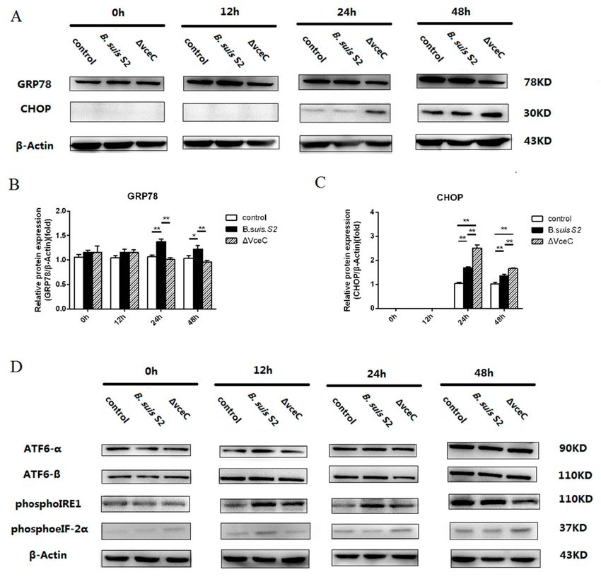
Int. J. Mol. Sci. 2019, 20, 4104                                                                        3 of 12

2. Results

2.1. Compare to VceC Amino Acid Sequences Derived from Different Brucella Strains
     VceC is present in all sequenced Brucella strains, including B. suis S2 (BSS2_I1011), B. suis 1330
(BR1038), B. abortus 2308 (BAB1_1058), B. melitensis 16M (BEMI0948), and B. cains GB1 (C6Y57_05925)
(Figure 1). VceC amino acid sequences were compared and analyzed based on the above strains.
The VceC protein of B. suis S2 contained 410 amino acids, with a proline rich central domain.
An N-terminal region of approximately 260 amino acids was conserved in proteins of the above strains.
A 1 bp missed in B. suis and B. cains GB1 vceC led to a frameshift in the C-termini of B. suis and B. canis
GB1. VceC protein had one amino acid change (Ser-275) in B. abortus 2308, one amino acid change
(Asn-343) in B. cains GB1, and two amino acid changes (Phe-264 and Ser-275) in B. melitensis 16M,
when compared with B. suis S2.

      Figure 1. Alignment of VceC nucleotide sequences (A) and amino acid sequences (B) for Brucella suis
      S2 (BSS2_I1011), B. suis 1330 (BR1038), B. abortus 2308 (BAB1_1058), B. melitensis 16M (BEMI0948),
      and B. cains GB1 (C6Y57_05925). Amino acid differences are shaded in red and green.

2.2. Mutant Strains ∆VceC Intracellular Survival in GTCs
      The VceC gene deletion mutant based on B. suis S2 was successfully constructed. Trophoblast
cells were a primary cellular target for Brucella in the natural host. Our previous study demonstrated
B. suis S2 was able to infect GTCs cultured in vitro [17] To evaluate the bacterial adherence and
intracellular survival in GTCs, the number of CFUs was counted following infection with B. suis S2 and
the mutant strains ∆VceC at 100 multiplicity of infection (MOI). The bacterial adherence of ∆VceC was
not significantly different compared to B. suis S2 strains (data not shown). We demonstrated B. suis S2
(Figure 2A) and ∆VceC (Figure 2B) were able to infect GTCs cultured in vitro. To determine whether
deletion of VceC affected the intracellular survival of Brucella, the number of CFUs was counted
following infection with B. suis S2 and ∆VceC at different times. The results indicated the intracellular
survival of B. suis S2 and ∆VceC occurred in a time-dependent manner. However, the bacterial
intracellular survival of ∆VceC was not significantly different (Figure 2C).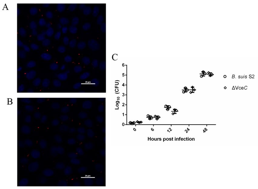
Int. J. Mol. Sci. 2019, 20, 4104                                                                                         4 of 12

      Figure 2. Infection and proliferation of B. suis S2 and ∆VceC in goat trophoblast cells (GTCs). (A,B) Representative
      confocal micrographs of GTCs infected with B. suis S2 (A, red) and ∆VceC (B, red) at 24 h post infection. The data
      shown are representative of three independent experiments. (C) Intracellular survival in GTCs of wild type
      (B. suis S2, circle) and the mutant obtained (∆VceC, square). The results are expressed as the means ± standard
      deviation from three independent experiments at each time point.

2.3. Effect of ∆VceC on GTC Apoptosis
     To evaluate whether the deletion of VceC affected the apoptosis level of GTCs infected by B. suis S2,
we assessed the effect of apoptosis by flow cytometry in combination with Annexin V/PI double staining.
When the cells were infected with ∆VceC for 12 h and 48 h, the average apoptosis of only Annexin
V-positive cells (early apoptosis cells) increased significantly, reaching approximately 3.04 ± 0.10%
and 8.96 ± 0.99%, respectively (Tables 1 and 2 and Supplementary Figure S1). However, the average
apoptosis of early apoptosis cells to B. suis S2 was approximately 1.77 ± 0.23% and 5.15 ± 2.46%,
respectively (Tables 1 and 2 and Supplementary Figure S1). The results demonstrated that ∆VceC
increased the proportion of early apoptosis in GTCs at 12 h and 48 h post infection.
     Because ∆VceC infection increased cells’ early apoptosis, we measured cleaved caspase-3
(the marker of apoptosis), procaspase-9 (the marker of mitochondrion-induced cell death), and CHOP
(the marker of ER stress-induced cell death) expression by Western blot at different times (Figure 3). At
48 h, cleaved caspase-3 expression was enhanced after ∆VceC infection compared with the B. suis S2
infected group. However, cleaved caspase-3 expression was not significantly different at 0, 12, and
24 h among the uninfected group, ∆VceC-infected group, and B. suis S2-infected group. Caspase-9
expression was also not significantly different at 0, 12, 24, and 48 h among the uninfected group,
∆VceC-infected group, and B. suis S2 infection group. Furthermore, CHOP expression was increased
at 24 and 48 h after ∆VceC infection compared with the B. suis S2 infection group, but no significant
difference was found at 0 and 12 h. According to our Western blot analysis, cleaved caspase-3 expression
at 48 h, as well as CHOP expression at 24 h and 48 h, was more strongly induced in the ∆VceC
infection group than in the B. suis S2 infection group. The results suggested VceC is involved in
Brucella-mediated apoptosis through the UPR signaling pathway.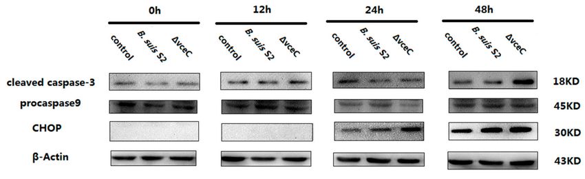
Int. J. Mol. Sci. 2019, 20, 4104                                                                                              5 of 12

      Table 1. Results of Annexin V-FITC/PI staining for cell apoptosis after B. suis S2 and ∆VceC infection at 12 h.

                                                        The Early Apoptotic Cells           The Late Apoptotic Cells
                 Group              Normal Cells
                                                                   (%)                                (%)
                Control              85.05 ± 0.63                 1.77 ± 0.23                        3.59 ± 0.25
          B. suis S2-infected        84.45 ± 4.03                 1.23 ± 0.34                        3.96 ± 0.49
           ∆VceC-infected            84.55 ± 1.06                3.04 ± 0.10 *                       4.08 ± 0.74
      Data represent the means ± standard deviations from three replicates. The asterisks (*) represent significant differences
      (p < 0.05) of the cell cycle distribution in GTCs infected by ∆VceC compared to that in B. suis S2-infected cells.

      Table 2. Results of Annexin V-FITC/PI staining for cell apoptosis after B. suis S2 and ∆VceC infection at 48 h.

                                                        The Early Apoptotic Cells           The Late Apoptotic Cells
                 Group              Normal Cells
                                                                   (%)                                (%)
                Control              75.20 ± 1.41                 5.15 ± 2.46                        8.01 ± 4.51
          B. suis S2-infected        79.50 ± 3.68                 5.25 ± 0.71                        6.18 ± 0.51
           ∆VceC-infected            72.85 ± 2.47                8.96 ± 0.99 *                       6.37 ± 0.98
      Data represent the means ± standard deviations from three replicates. The asterisks (*) represent significant differences
      (p < 0.05) of the cell cycle distribution in GTCs infected by ∆VceC compared to that in B. suis S2-infected cells.

      Figure 3. Effect of deletion of VceC on apoptosis of Brucella following infection of GTCs. GTCs were
      infected with 100 MOI of B. suis S2 and ∆VceC for 0 h, 12 h, 24 h, and 48 h, lysed, and subjected
      to Western blot analysis to detect the expression of apoptosis-related cleaved caspase-3, caspase-9,
      and CHOP proteins. The data shown are representative of three independent experiments.

2.4. Deletion of VceC Decreases Brucella-Mediated ER Stress
     UPR is a cytoprotective response that is aimed at monitoring the survival and proliferation
of intracellular pathogens. To more directly examine ER stress after ∆VceC infection in GTCs, we
examined the expression of GRP78 in ∆VceC infection GTCs. GRP78 expression was increased at 24
and 48 h in the Brucella-infected group compared to the uninfected group and was decreased at 24 and
48 h in the ∆VceC-infected group compared to the Brucella-infected group, but no significant difference
was found at 0 and 12 h among all groups (Figure 4A–C). The results indicate the deletion of VceC
changes Brucella-mediated ER stress.
     To investigate UPR induction during ∆VceC infection, GTCs were infected with B. suis S2 or
∆VceC, and the activation of three UPR sensors was analyzed by Western blot. ATF6-α, ATF6-β,
and phosphoeIF-2α (downstream protein of PERK branch) were not significantly different in all groups
(Figure 4D). However, PhosphoIRE1 expression was decreased at 12, 24, and 48 h in the ∆VceC infection
group compared to the Brucella infection group (Figure 4D). We conclude that VceC is involved with
Brucella-mediated ER stress by IRE1 branch activation of UPR.
Int. J. Mol. Sci. 2019, 20, 4104 6 of 12
Figure 4. The unfolded protein response (UPR) pathway is induced by B. suis S2 and ∆VceC. (A) GTCs
were infected with 100 MOI of B. suis S2 and ∆VceC for 0 h, 12 h, 24 h, and 48 h, and the untreated
group was used as a negative control, followed by lysis and detection of GRP78 and CHOP protein
expression by Western blot. The data shown are representative of three independent experiments.
(B,C) Quantification of band intensities from three independent results was determined by densitometric
analysis. Data represent the mean ± standard deviation from three independent experiments at each
time point.* p < 0.05; ** p < 0.01 (D) GTCs were infected with 100 MOI of B. suis S2 and ∆VceC for 0, 12,
24, and 48 h, and the untreated group was used as a negative control, followed by lysis and detection
of ATF6-α, ATF6-β, phosphoIRE1, and phosphoeIF-2α protein expression by Western blot. The data
shown are representative of three independent experiments.
2.5. The Replication of ∆VceC Was More Sensitive after Changing ER Stress in GTCs
To more directly assess the role of ER stress in Brucella survival and proliferation in GTCs,
we explored whether ER stress altered by Tm or 4-PBA affects B. suis S2 or ∆VceC in GTCs. Increasing
ER stress with 0.5 µg/mL Tm significantly increased ER stress marker GRP78 and CHOP expression
(Figure 5A,B). In contrast, decreasing ER stress with 1 µM 4-PBA (Figure 5A,B) significantly inhibited
GRP78 and CHOP protein expression. In addition, increasing ER stress with Tm significantly inhibited
the proliferation of B. suis S2 or ∆VceC, and decreasing ER stress with 4-PBA enhanced the proliferation
of B. suis S2 or ∆VceC (Figure 5B–E). This observation is consistent with other reports that Brucella
proliferation in host cells is affected by apoptosis in infected cells. Furthermore, the proliferation of
∆VceC was significantly different compared with that of the B. suis S2 infection group under changing
ER stress. The results reconfirmed that the deletion of VceC changes Brucella-mediated ER stress.Int. J. Mol. Sci. 2019, 20, 4104 7 of 12
Figure 5. Intracellular survival of B. suis S2 and ∆VceC by changing endoplasmic reticulum (ER)
stress. (A) The UPR marker GRP78 and CHOP were analyzed using Western blot by treating with
0.5 µg/mL Tm or 1 µM 4-PBA in GTCs to establish an ER stress activated and inhibited model. The data
shown are representative of three independent experiments. (B) (bar = 20 µm) Representative confocal
micrographs of GRP78 protein (green) infected with B. suis S2 (red) and ∆VceC (red) by added 0.5 µg/mL
Tm or 1 µM 4-PBA before infection in GTCs. The blue represents cell nucleus. (D–E) CFUs of B. suis S2
(C) and ∆VceC (D) were determined in GTCs at 24 h post infection. The 0.5 µg/mL Tm or 1 µM 4-PBA
was added before infection. Data represent the mean ± standard deviation from three independent
experiments. * p < 0.05
3. Discussion
The ER mediates biosynthesis, folding, and modification of secretory and transmembrane
proteins as well as maintains of calcium homeostasis. Elevated physiological demand for protein
folding can disrupt the ability of ER, leading to misfolded or unfolded protein accumulation in thisInt. J. Mol. Sci. 2019, 20, 4104 8 of 12
organelle, a condition called ER stress. The unfolded protein response (UPR) is induced to restore ER
homeostasis [18]. However, excessive stress to the ER triggers CHOP-induced apoptosis [20].
Our results indicated that the B. suis S2 T4SS-secreted protein VceC can trigger ER stress but
inhibit CHOP-induced apoptosis. The manipulation of host cell death is a critical strategy of Brucella to
maintain dissemination or long-term intracellular persistence [5,21]. Macrophage/monocytes infected
with Brucella suis or Brucella melitensis stains inhibit the apoptosis pathways, whereas GTCs infected with
B. suis S2 undergo apoptotic cell death mediated by ER stress [17,22,23]. T4SS is one of the key factors
for Brucella intracellular survival and plays an essential role in the inhibition of host-cell death [24].
Inactivation of virB, which encodes the T4SS, in smooth B. melitensis prevented the cytotoxicity of
Brucella for macrophages; in contrast, overexpression of virB enhances cytotoxic effects [24]. The VceC
also mediates the cytotoxicity effect by translocation of this effector protein into macrophages, resulting
in lysis of the host cells [11]. However, the cytotoxicity or lysis of Brucella for host cells resembles
oncosis and nerosis, but not apoptosis [11]. In our studies, B. suis S2 infection induced caspase-9
protein expression in GTCs, but, interestingly, VceC mutants enhanced cleaved caspase-3 and CHOP
protein expression and induced an increase in apoptosis in trophoblast cells. During ER stress, CHOP
expression increases to activate downstream genes, leading to ER stress-induced apoptosis [25,26].
Martinon et al. [27] suggested that Brucella lipopolysaccharide (LPS) may sufficiently temper CHOP
induction to avert apoptosis. These findings suggested that VceC may synergize with LPS to inhibit
CHOP-induced apoptosis. Further work is needed to verify the interaction between VceC and LPS
during Brucella infection. Therefore, our results demonstrated that VceC promoted intracellular
persistence of Brucella infection, which may be related to decreased CHOP expression and inhibition of
the ERS-induced apoptosis pathway.
UPR is known to support the Brucella intracellular life cycle within the host cell. First, the UPR
mobilizes amino acid transport and supports lipid biogenesis. Second, the UPR regulates autophagy
to providing more nutrients. Third, the UPR enhances the protein-folding capacity to suppress
downstream apoptosis. Fourth, the UPR allows cells to cope with oxidative stresses. Finally, the UPR
enables host cells to survive the disruption of the ER structure and function [16]. During B. abortus
infection, the effector protein VceC interacts with the ER chaperone GRP78 and localizes to the ER,
then disrupts the ER structure, and results in ER stress in HeLa cells [12]. Our previous study shows
that inhibiting ER stress with 4-PBA increased the number of B. suis S2 CFUs in GTCs. Enhancing
ER stress with Tm inhibited the proliferation of B. suis S2 in GTCs [17]. GRP78 plays an essential
role in supporting Brucella replication in GTCs. Decreasing GRP78 expression inhibited B. suis S2
proliferation in GTCs by promoting ER stress-induced apoptosis [17]. Those findings are consistent
with our result that the VceC mutant reduced the GRP78 expression and enhanced that of CHOP to
promote ER stress-induced apoptosis. The replication of the VceC mutant was more sensitive under
the different ER stress conditions in the GTC line after treatment with ER stress inhibitors 4-PBA or ER
stress inducer Tm. Our studies indicate that effector protein VceC is essential for Brucella replication by
interaction with GRP78 to inhibit the apoptosis of B. suis S2-infected GTCs.
ER stress transmembrane sensor IRE1 plays a pivotal role in Brucella replication. Previous studies
suggested that B. abortus infection of macrophages or HeLa cells activates IRE1α pathway [16,28].
Yip1A, a host protein, is required for rBCV biogenesis and intracellular B. abortus replication through
mediated IRE1 activation in HeLa cells [28]. De Jong et al. [12] demonstrated that IRE1-mediated UPR
activation is activated by VceC. In our study, we confirmed that VceC mutant infection did not activate
the IRE1 pathway in GTCs. VceC mutant infection did not affect the B. suis S2 CFU in GTCs. Our results
are consistent with the study that IRE1 knockdown in bone marrow-derived macrophages (BMDM) or
decreasing phosphoIRE1α and IRE1α in GTCs did not affect the number of Brucella bacteria at 24 h
post infection [17,28]. An in vivo study suggested that infection with the B. abortus wild type and the
VceC mutant resulted in similar numbers in the placenta of mice [13]. Taken together, we speculated
that more effector protein synergies exist with VceC to activate the IRE1 pathway of UPR to supportInt. J. Mol. Sci. 2019, 20, 4104 9 of 12
the Brucella replication in host cells, since inactivation of VceC alone did not affect the replication
competence of Brucella [12].
In summary, the results demonstrated that B. suis S2 T4SS effector protein VceC decreased CHOP
expression, interacted with GRP78 to inhibit apoptosis, and mediated the IER1 pathway of UPR to
support B. suis S2 replication in GTCs. Our findings also suggested that more effector proteins should
exist in synergy with VceC to support persistent Brucella intracellular infection. The present work
provides new insights for understanding the mechanism of VceC in the establishment of chronic
Brucella infection.
4. Materials and Methods
4.1. Bacterial Strains
The bacterial strains used in this study were smooth attenuated virulent Brucella suis vaccine strain
2 (B. suis S2, CVCC70502), and they were obtained from the Chinese veterinary culture collection center
(Beijing, China). The B. suis S2 were grown in tryptic soy broth (TSB; Takana) and tryptic soy ager (TSA;
Takana). Goat trophoblast cells (GTCs) were immortalized by transfection with human telomerase
reverse transcriptase (hTERT); these cells were provided by Dewen Tong (Northwest A&F University,
Yangling, Shaanxi, China). For infection of GTCs, the bacteria were collected by centrifugation at
6000× g for 10 min at 4 ◦ C and washed three times with 15 mL of phosphate-buffered saline (PBS).
The number of B. suis S2 were counted by plating on TSA. Escherichia coli strain DH5α (takana) was
cultured in Luria-Bertani (LB) medium. When appropriate, 50 µg/mL gentamicin or ampicillin were
respectively added. Plasmid PUC19 was purchased from Takana.
4.2. Construction of the Mutant Strain ∆VceC
∆VceC was constructed as described previously [29]. Briefly, primers were designed using the sequence
B. suis S2 genome and plasmid PBBR1MCS-5. The 896 bp VceC upstream fragment, 964 bp VceC downstream
fragment, and 751 bp gentamicin fragment were obtained in three independent PCR reactions using primer
STAR Max Mix with primer pairs VceC-UF: ctgcag (Pst1) TCGGAAGCGAGCACCTGA; VceC-UR: tctaga (XbaI)
GCGGATACCCTCTTACACTATAAAC; VceC-DF: gagctc (SacI) CCAAGGGAGAAACCCGCA; VceC-DR:
gaattc (EcoRI) CGTGTTCACAACCGATAAGG; G-F tctaga (XbaI) TTGACATAAGCCTGTTCGGTTCGTA;
G-R: gagctc (SacI) TTAGGTGGCGGTACTTGGGTCGATA. After purification by gel extraction, the three
fragments were cloned into PMD19T-simple, then digested with PstI and XbaI; SacI and EcoRI; and XbaI and
SacI sequentially, and then subcloned into the PstI- and EcoRI-digested PUC19 plasmid. The recombinant
plasmid with the correct sequence was designated PUC19-VceC. Then this plasmid was electroporated
into B. suis S2, where it is incapable of autonomous replication. The potential GntR deletion mutant was
selected by plating on TSA-containing gentamicin, which was then verified by PCR with primer pairs
VceC-F: CTTCTCATTGGCAAGCACTTC and VceC-R: GCATCATTCGCCGTTTCA. The mutant strain was
called ∆VceC.
4.3. Cell Infection Assay
The process of the B. suis S2 infection assays was carried out as described previously. Briefly,
GTCs were seeded in 6-well plates (5 × 10⁵ cells per well) or in 24-well plates (1 × 10⁵ cells per well)
and were infected with B. suis S2 or ∆VceC at a multiplicity of infection of 100:1. After 4 h of incubation
at 37 ◦ C with 5% CO2 atmosphere, GTCs were washed three times with PBS and then further cultured
with cell culture medium containing 50 µg/mL kanamycin to eliminate B. suis S2 or ∆VceC adhering
to the GTCs and in the culture medium. After 1 h, the GTCs were washed three times with PBS and
were further cultured with cell culture medium containing 25 µg/mL kanamycin to avert continuous
infection. This point in time was considered 0 h and the time point of treatment with Tm (ER stress
activator) and 4-PBA (ER stress antagonist). The cells were collected, and relevant experiments were
performed at specific times (0, 6, 12, 24, and 48 h).Int. J. Mol. Sci. 2019, 20, 4104 10 of 12
For intracellular survival assays, cells were seeded in 24-well plates prior to infection. Then, cells
were infected with B. suis S2 or ∆VceC as described. At different times following infection, wells of
infected cells were washed three times with PBS and lysed with 0.5% Triton X-100 in PBS for 10 min.
The lysates were serial diluted in PBS and plated onto TSA for 72 h to determine the colony-forming
units (CFUs).
For adherence, cells were seeded in 24-well plates prior to infection. Then, cells were infected
with B. suis S2 or ∆VceC as described. After 1 h of incubation at 37 ◦ C with 5% CO2 atmosphere,
wells of infected cells were washed three times with PBS and lysed with 0.5% Triton X-100 in PBS for
10 min. Next, the lysates were serial diluted in PBS and plated onto TSA for 72 h to determine the
colony-forming units (CFUs).
4.4. Western Blot Analysis
GTCs were harvested in a tube after infection at 0, 12, 24, and 48 h, and then lysed on ice for
30–45 min in lysis buffer. The supernatant was obtained by centrifugation for 15 min at 14,000 rpm at 4 ◦ C.
The protein concentration was determined by the Bicinchoninic acid (BCA) assay. Total cellular protein
was extracted with 5 × sodium dodecyl sulfate polyacrylamide gel electrophoresis (SDS-PAGE) loading
buffer after boiling for 5 min in water. Samples were electrophoresed on a 12% polyacrylamide gel for
SDS-PAGE. The gels were then electro-transferred onto polyvinylidene fluoride (PVDF) membranes.
The membranes were blocked for 1 h in Tris-buffered saline containing 0.5% Tween-20 (TBST) with
5%–10% skimmed milk at room temperature and then incubated overnight at 4 ◦ C in blocking
solution containing GRP78 (Abcam, 1:1000 dilution), CHOP (Abcam, 1:1000 dilution), ATF-6 (Abcam,
1:1000 dilution), phosphoIRE1 (Abcam, 1:1000 dilution), phosphoeIF2α (Abcam, 1:1000 dilution),
caspase-3 (proteintech, 1:1000 dilution), caspase-9 (proteintech, 1:1000 dilution), and anti-β-actin
(Tianjin Sungene Biotech Co, 1:1000 dilution). The membranes were washed five times with TBST for
5 min and then incubated for 1 h with the corresponding secondary antibody conjugated to horse
radish peroxidase (HRP) (1:5000, Zhongshan Golden Bridge Biotechnology, Nanjing, China). Finally,
the membranes were washed five times in TBST for 5 min. The blots were visualized using the Gel
Image System (Tannon, Biotech, Shanghai, China).
4.5. Immunofluorescence Assay
GTCs were seeded in 24-well plates and were infected with B. suis S2 and ∆VceC at 100 MOI.
At 0 and 24 h post infection, infected cells were washed twice with PBS and then fixed with 4%
paraformaldehyde at room temperature for 30 min. After three washes with PBS, cells were incubated
with PBS containing 0.25% Triton X-100 at room temperature for 20 min. After three washes with
PBS, goat anti-brucella polyclonal antibody (1:100 dilution), rabbit anti-GRP78 monoclonal antibody
(1:200 dilution), and mouse anti-LAMP-1 monoclonal antibody (1:200 dilution) were used as the
primary antibody. Donkey anti-goat alexa fluor 555, donkey anti-mouse alexa fluor 488, and donkey
anti-rabbit alexa fluor 488 were used as the secondary antibody at 1:200 dilutions. Next, coverslips
were mounted on glass slides, and cells were observed under a microscope. Assays were performed
in triplicate.
4.6. Statistical Analysis
Statistical analysis was performed using Graphpad Prism software 6 (GraphPad software Inc.,
La Jolla, CA, USA). Statistical significance was determined using two-way ANOVA or one-way ANOVA.
P values less than 0.05 were considered statistically significant.
Supplementary Materials: Supplementary materials can be found at http://www.mdpi.com/1422-0067/20/17/
4104/s1. Supplementary Figure 1: Results of cell apoptosis after B. suis S2 and ∆VceC infection at 12 h and 48 h.
Author Contributions: Formal analysis, F.B., J.L., C.X. and G.Z.; Funding acquisition, D.Z. and A.W.; Investigation,
F.Z. and D.Z.; Writing—original draft, F.Z. and D.Z.; Writing—review & editing, Y.J. and A.W.Int. J. Mol. Sci. 2019, 20, 4104 11 of 12
Acknowledgments: This research was funded by National Key R&D Program of China (2018YFD0500900);
National Natural Science Foundation of China (31672584, 31702310); China Postdoctoral Science Foundation
(2016M602883); Natural Science Basic Research Plan in Shaanxi Province of China (2017JQ30100).
Conflicts of Interest: The authors declare that they have no competing interests.
References
1. Pappas, G.; Papadimitriou, P.; Akritidis, N.; Christou, L.; Tsianos, E.V. The new global map of human
brucellosis. Lancet Infect. Dis. 2006, 6, 91–99. [CrossRef]
2. Byndloss, M.X.; Tsolis, R.M. Brucella spp. Virulence Factors and Immunity. Annu. Rev. Anim. Biosci. 2016, 4,
111–127. [CrossRef] [PubMed]
3. Samartino, L.E.; Enright, F.M. Pathogenesis of abortion of bovine brucellosis. Comp. Immunol. Microbiol. Infect. Dis.
1993, 16, 95–101. [CrossRef]
4. Celli, J. The changing nature of the Brucella-containing vacuole. Cell. Microbiol. 2015, 17, 951–958. [CrossRef]
[PubMed]
5. Ahmed, W.; Zheng, K.; Liu, Z.F. Establishment of Chronic Infection: Brucella’s Stealth Strategy. Front. Cell. Infect.
Microbiol. 2016, 6, 30. [CrossRef] [PubMed]
6. Pascual, D.W.; Yang, X.; Wang, H.; Goodwin, Z.; Hoffman, C.; Clapp, B. Alternative strategies for vaccination
to brucellosis. Microbes Infect. 2018, 20, 599–605. [CrossRef] [PubMed]
7. Jaiswal, V.; Chauhan, R.S.; Rout, C. Common antigens prediction in bacterial bioweapons: A perspective for
vaccine design. Infect. Genet. Evol. 2014, 21, 315–319. [CrossRef] [PubMed]
8. Caporale, V.; Bonfini, B.; Di Giannatale, E.; Di Provvido, A.; Forcella, S.; Giovannini, A.; Tittarelli, M.;
Scacchia, M. Efficacy of Brucella abortus vaccine strain RB51 compared to the reference vaccine Brucella abortus
strain 19 in water buffalo. Vet. Ital. 2010, 46, 5–11.
9. Zhu, L.; Feng, Y.; Zhang, G.; Jiang, H.; Zhang, Z.; Wang, N.; Ding, J.; Suo, X. Brucella suis strain 2 vaccine is
safe and protective against heterologous Brucella spp. infections. Vaccine 2016, 34, 395–400. [CrossRef]
10. Myeni, S.; Child, R.; Ng, T.W.; Kupko, J.J., 3rd; Wehrly, T.D.; Porcella, S.F.; Knodler, L.A.; Celli, J. Brucella
modulates secretory trafficking via multiple type IV secretion effector proteins. PLoS Pathog. 2013, 9, e1003556.
[CrossRef] [PubMed]
11. De Jong, M.F.; Sun, Y.H.; den Hartigh, A.B.; van Dijl, J.M.; Tsolis, R.M. Identification of VceA and VceC,
two members of the VjbR regulon that are translocated into macrophages by the Brucella type IV secretion
system. Mol. Microbiol. 2008, 70, 1378–1396. [CrossRef] [PubMed]
12. De Jong, M.F.; Starr, T.; Winter, M.G.; den Hartigh, A.B.; Child, R.; Knodler, L.A.; van Dijl, J.M.; Celli, J.;
Tsolis, R.M. Sensing of Bacterial Type IV Secretion via the Unfolded Protein Response. mBio 2013, 4, e00418-12.
[CrossRef] [PubMed]
13. Keestra-Gounder, A.M.; Byndloss, M.X.; Seyffert, N.; Young, B.M.; Chavez-Arroyo, A.; Tsai, A.Y.; Cevallos, S.A.;
Winter, M.G.; Pham, O.H.; Tiffany, C.R.; et al. NOD1 and NOD2 signalling links ER stress with inflammation.
Nature 2016, 532, 394. [CrossRef] [PubMed]
14. Von Bargen, K.; Gorvel, J.P.; Salcedo, S.P. Internal affairs: Investigating the Brucella intracellular lifestyle.
FEMS Microbiol. Rev. 2012, 36, 533–562. [CrossRef] [PubMed]
15. Celli, J.; de Chastellier, C.; Franchini, D.M.; Pizarro-Cerda, J.; Moreno, E.; Gorvel, J.P. Brucella evades
macrophage killing via VirB-dependent sustained interactions with the endoplasmic reticulum. J. Exp. Med.
2003, 198, 545–556. [CrossRef] [PubMed]
16. Smith, J.A.; Khan, M.; Magnani, D.D.; Harms, J.S.; Durward, M.; Radhakrishnan, G.K.; Liu, Y.P.; Splitter, G.A.
Brucella induces an unfolded protein response via TcpB that supports intracellular replication in macrophages.
PLoS Pathog. 2013, 9, e1003785. [CrossRef] [PubMed]
17. Wang, X.; Lin, P.; Li, Y.; Xiang, C.; Yin, Y.; Chen, Z.; Du, Y.; Zhou, D.; Jin, Y.; Wang, A. Brucella suis Vaccine
Strain 2 Induces Endoplasmic Reticulum Stress that Affects Intracellular Replication in Goat Trophoblast
Cells In vitro. Front. Cell. Infect. Microbiol. 2016, 6, 19. [CrossRef]
18. Cao, S.S.; Kaufman, R.J. Unfolded protein response. Curr. Biol. 2012, 22, R622-6. [CrossRef]
19. Lee, W.-S.; Yoo, W.-H.; Chae, H.-J. ER stress and autophagy. Curr. Mol. Med. 2015, 15, 735–745. [CrossRef]
20. Szegezdi, E.; Logue, S.E.; Gorman, A.M.; Samali, A. Mediators of endoplasmic reticulum stress-induced
apoptosis. EMBO Rep. 2006, 7, 880–885. [CrossRef]Int. J. Mol. Sci. 2019, 20, 4104 12 of 12
21. Atluri, V.L.; Xavier, M.N.; de Jong, M.F.; den Hartigh, A.B.; Tsolis, R.M. Interactions of the human pathogenic
Brucella species with their hosts. Annu. Rev. Microbiol. 2011, 65, 523–541. [CrossRef] [PubMed]
22. Gross, A.; Terraza, A.; Ouahrani-Bettache, S.; Liautard, J.P.; Dornand, J. In vitro Brucella suis infection prevents
the programmed cell death of human monocytic cells. Infect. Immun. 2000, 68, 342–351. [CrossRef] [PubMed]
23. Tolomeo, M.; Di Carlo, P.; Abbadessa, V.; Titone, L.; Miceli, S.; Barbusca, E.; Cannizzo, G.; Mancuso, S.;
Arista, S.; Scarlata, F. Monocyte and lymphocyte apoptosis resistance in acute and chronic brucellosis and
its possible implications in clinical management. Clin. Infect. Dis. Off. Publ. Infect. Dis. Soc. Am. 2003, 36,
1533–1538. [CrossRef] [PubMed]
24. Zhong, Z.; Wang, Y.; Qiao, F.; Wang, Z.; Du, X.; Xu, J.; Zhao, J.; Qu, Q.; Dong, S.; Sun, Y.; et al. Cytotoxicity of
Brucella smooth strains for macrophages is mediated by increased secretion of the type IV secretion system.
Microbiology 2009, 155 Pt 10, 3392–3402. [CrossRef]
25. Yao, Y.; Lu, Q.; Hu, Z.; Yu, Y.; Chen, Q.; Wang, Q.K. A non-canonical pathway regulates ER stress signaling
and blocks ER stress-induced apoptosis and heart failure. Nat. Commun. 2017, 8, 133. [CrossRef]
26. Masciarelli, S.; Fra, A.M.; Pengo, N.; Bertolotti, M.; Cenci, S.; Fagioli, C.; Ron, D.; Hendershot, L.M.; Sitia, R.
CHOP-independent apoptosis and pathway-selective induction of the UPR in developing plasma cells.
Mol. Immunol. 2010, 47, 1356–1365. [CrossRef] [PubMed]
27. Martinon, F.; Chen, X.; Lee, A.H.; Glimcher, L.H. TLR activation of the transcription factor XBP1 regulates
innate immune responses in macrophages. Nat. Immunol. 2010, 11, 411–418. [CrossRef]
28. Taguchi, Y.; Imaoka, K.; Kataoka, M.; Uda, A.; Nakatsu, D.; Horii-Okazaki, S.; Kunishige, R.; Kano, F.;
Murata, M. Yip1A, a novel host factor for the activation of the IRE1 pathway of the unfolded protein response
during Brucella infection. PLoS Pathog. 2015, 11, e1004747. [CrossRef]
29. Zhou, D.; Zhi, F.J.; Qi, M.Z.; Bai, F.R.; Zhang, G.D.; Li, J.M.; Liu, H.; Chen, H.T.; Lin, P.F.; Tang, K.Q.; et al.
Brucella induces unfolded protein response and inflammatory response via GntR in alveolar macrophages.
Oncotarget 2018, 9, 5184–5196. [CrossRef]
© 2019 by the authors. Licensee MDPI, Basel, Switzerland. This article is an open access
article distributed under the terms and conditions of the Creative Commons Attribution
(CC BY) license (http://creativecommons.org/licenses/by/4.0/).You can also read