B7 costimulation and intracellular indoleamine-2, 3-dioxygenase (IDO) expression in peripheral blood of healthy pregnant and non-pregnant women
←
→
Page content transcription
If your browser does not render page correctly, please read the page content below
Grozdics et al. BMC Pregnancy and Childbirth 2014, 14:306
http://www.biomedcentral.com/1471-2393/14/306

 RESEARCH ARTICLE                                                                                                                                 Open Access

B7 costimulation and intracellular indoleamine-2,
3-dioxygenase (IDO) expression in peripheral blood
of healthy pregnant and non-pregnant women
Enikő Grozdics1†, László Berta1†, Anna Bajnok1, Gábor Veres2, István Ilisz3, Péter Klivényi2, János Rigó Jr4,
László Vécsei2,5, Tivadar Tulassay1,6 and Gergely Toldi1,4,6*

  Abstract
  Background: B7 costimulatory molecules are expressed on antigen presenting cells (APCs) and are important
  regulators of T cell activation. We investigated the role of the B7 family of costimulatory molecules in the
  development of the systemic maternal immune tolerance during healthy pregnancy (HP). We also aimed to
  investigate the intracellular expression of indoleamine-2,3-dioxygenase (IDO) and plasma levels of tryptophane
  (TRP), kynurenine (KYN) and kynurenic acid (KYNA), important molecules with immunoregulatory properties, in order
  to describe their potential contribution to the pregnancy-specific maternal immune tolerance.
  Methods: We determined the frequency of activated (CD11b+) monocytes expressing B7-1, B7-2, B7-H1, and B7-H2,
  and that of T cells and CD4+ T helper cells expressing CD28, CTLA-4, PD-1, and ICOS in peripheral blood samples of
  healthy pregnant (HP) and non-pregnant (NP) women using flow cytometry. We also examined the intracellular
  expression of IDO applying flow cytometry and plasma levels of TRP, KYN and KYNA using high-performance liquid
  chromatography.
  Results: A significant increase in the prevalence of CD28+ T cells was observed in HP compared to NP women. At the
  same time a decrease was shown in the expression of CTLA-4 on these cells. The frequency of CD80+ monocytes was
  lower in HP women. The prevalence of IDO-expressing T cells and monocytes was higher in HP compared to NP
  women. Plasma KYN, KYNA and TRP levels were lower, while at the same time, the KYN/TRP ratio was higher in HP than
  in NP women.
  Conclusions: Costimulation via CD28 may not contribute to the immunosuppressive environment, at least in the
  third trimester of pregnancy. The development of the pregnancy-specific immune tolerance in the mechanism of
  B7 costimulation may be more related to the altered expression of B7 proteins on APCs rather than that of their
  receptors on T cells. The elevated intracellular IDO expression in monocytes and T cells, as well as higher plasma
  enzymatic IDO activity are likely to contribute to the systemic immunosuppressive environment in the third
  trimester characteristic for healthy gestation.
  Keywords: B7, CD28, CTLA-4, Indoleamine 2,3-dioxygenase, Kynurenine, Monocyte, T cell, Tryptophan

* Correspondence: toldigergely@yahoo.com
†
 Equal contributors
1
 First Department of Pediatrics, Semmelweis University, Bókay u. 53-54,
Budapest H-1083, Hungary
4
 First Department of Obstetrics and Gynecology, Semmelweis University,
Baross u. 27, Budapest H-1088, Hungary
Full list of author information is available at the end of the article

                                        © 2014 Grozdics et al.; licensee BioMed Central Ltd. This is an Open Access article distributed under the terms of the Creative
                                        Commons Attribution License (http://creativecommons.org/licenses/by/4.0), which permits unrestricted use, distribution, and
                                        reproduction in any medium, provided the original work is properly credited. The Creative Commons Public Domain
                                        Dedication waiver (http://creativecommons.org/publicdomain/zero/1.0/) applies to the data made available in this article,
                                        unless otherwise stated.
Grozdics et al. BMC Pregnancy and Childbirth 2014, 14:306 Page 2 of 9 http://www.biomedcentral.com/1471-2393/14/306 Background surface of T cells, thus competing with CD28 for B7 bind- Since the conceptus is half of foreign origins, presenting pa- ing and blocking the costimulatory signal. The affinity of ternal antigens, it is considered a semi-allograft to maternal the inhibitory receptor, CTLA-4 is higher than that of immunity. Therefore, a maternal immune tolerance must CD28 for B7-1 (CD80) and B7-2 (CD86). Besides its com- develop to avoid immunological rejection of the fetus. The petitive role, CTLA-4 also prevents T cell activation via the alterations contributing to maternal tolerance are present emission of direct inhibitory signals [7]. not only at the maternal-fetal interface, but also at the sys- Another B7 family member, B7-H1 (CD274) possesses temic level. Several components of this pregnancy-specific mostly inhibitory properties on T cells. This inhibitory func- immune tolerance have been described over the recent tion is mediated via programmed death-1 receptor (PD-1, years [1]. One of the most important factors is the CD279), which induces apoptosis or anergy of self-reactive decreased level of activation of T cells compared to the T cells. B7-H2 (CD275) serves as the ligand for inducible non-pregnant state. The second and third trimesters of costimulator of T cells (ICOS, CD278), and promotes T cell pregnancy are characterized by a shift of the inflammatory activation, differentiation, and effector responses [8]. In con- balance towards the anti-inflammatory direction via the trast to the costimulatory effect of CD28, ICOS most effect- upregulation of Th2 cells [2] and a decrease in the Th17/ ively induces IL-10 instead of IL-2 production [9]. regulatory T cell (Treg) ratio [3]. The kinetics of calcium in- Besides initiating signal transduction in T lymphocytes, flux upon stimulation via the T cell receptor (TCR) is B7-1 and B7-2 may back-signal into the APC and influ- decreased in Th1 and CD8 cells compared to lymphocytes ence the local immune environment through induced ex- isolated from non-pregnant women [4]. Several other pression of immunosuppressive factors independently of factors may account for the decreased level of peripheral their effects on T cells [5]. Reverse signalling through B7-1 T lymphocyte activation in healthy pregnancy (HP). and B7-2 was shown to upregulate the tryptophan (TRP) B7 costimulatory molecules are expressed on antigen pre- catabolic enzyme, indoleamine 2,3-dioxygenase (IDO) senting cells (APCs) and are important regulators of T cell [10]. The potent immunosuppressive activity of IDO was activation (Figure 1). Besides the ligation of the T cell recep- first identified in pregnancy, when it was demonstrated tor (TCR) by the antigen associated with major histocom- that inhibition of IDO abolished allogenic gestation in patibility complex (MHC) molecules, a costimulatory signal mice [11]. In the first steps of the kynurenine (KYN) path- occurs through CD28, inducing the production of IL-2 in T way, TRP is transformed into KYN by IDO. KYN is then cells, thus protecting them from apoptosis and anergy [5]. further metabolized by different enzymes. One of them is Without costimulation, the signal from the TCR induces kynurenine aminotransferase, leading to the production the tolerance of T cells to their cognate antigen instead of of kynurenic acid (KYNA), a broad-spectrum endogenous being activated [6]. antagonist of excitatory amino acid receptors [12,13] On the other hand, B7 proteins mediate not only stimula- with emerging recent implications in immunomodulation tory, but also inhibitory effects on T cells, thus potentially [14,15]. The rate of TRP degradation, represented by the contributing to the lower reactivity of T lymphocytes in HP K/T (KYN to TRP) ratio, allows a good estimate of IDO [5]. Upon the stimulation of the TCR, cytotoxic T lympho- activity. The local depletion of TRP and the production of cyte antigen 4 (CTLA-4, CD154) becomes stabilized on the pro-apoptotic TRP metabolites of the kynurenine pathway, Figure 1 B7 family proteins on antigen-presenting cells (APCs) and their cognate receptors on T cells.
Grozdics et al. BMC Pregnancy and Childbirth 2014, 14:306 Page 3 of 9
http://www.biomedcentral.com/1471-2393/14/306
such as 3-hydroxyanthranilic acid and quinolinic acid are Flow cytometry
among the mechanisms potentially responsible for the im- PBMCs were stained for 30 min at room temperature in the
munosuppressive effects related to IDO [14]. dark with PerCP-conjugated CD3, PE Cy7-conjugated CD4,
In this study, we aimed to determine the frequency of PE-conjugated CD28, APC-conjugated CD152 (CTLA-4),
activated monocytes expressing B7-1, B7-2, B7-H1 and B- FITC-conjugated CD278 (ICOS) and APC-Cy7-conjugated
7H2 costimulatory molecules, as well as that of T cells and CD279 (PD-1) mAbs, or PerCP-conjugated CD3, PE Cy7-
T helper cells expressing CD28, CTLA-4, PD-1 and ICOS conjugated CD11b, APC-conjugated CD80 (B7-1) and PE-
in peripheral blood samples of HP compared to non- conjugated CD275 (B7-H2) mAbs, or PerCP-conjugated
pregnant (NP) women. We also investigated the intracel- CD3, PE Cy7-conjugated CD11b, APC-conjugated CD86
lular expression of IDO in activated monocytes and T (B7-2) and PE-conjugated CD274 (B7-H1) mAbs in separate
cells, as well as plasma levels of TRP, KYN and KYNA. tubes, respectively (BioLegend, San Diego, CA, USA). After
washing, cells were fixed with Fixation/Permeabilization so-
Methods lution and treated with Permeabilization Buffer according to
Sample collection the manufacturer’s instructions (eBioscience, San Diego,
Peripheral blood samples were taken from 20 healthy CA, USA). They were then stained with a mouse anti-
pregnant (HP) women in the third trimester and 14 age- human IDO monoclonal antibody (Millipore, USA) for
matched, healthy non-pregnant (NP) women between 30 min at 4°C in the dark. After washing, cells were stained
01/2012 and 08/2012. The latter group was synchro- with FITC-labelled goat anti-mouse antibody (Millipore,
nized in terms of menstrual cycle for the luteal phase. USA) for 15 min at 4°C in the dark. After washing, cells
Clinical characteristics of participants are summarized were analyzed on a BD FACSAria flow cytometer (BD Bio-
in Table 1. Informed consent was obtained from all sub- sciences) equipped with 488 nm and 633 nm excitation la-
jects, and our study was reviewed and approved by an sers. Data were processed using the FACSDiVa software.
independent ethical committee of the institution (Scien- 100000 cells were recorded. The populations of lymphocytes
tific and Research Ethics Committee, Semmelweis Uni- and monocytes were gated from PBMCs according to For-
versity, Budapest, Hungary). The study was adhered to ward Scatter Characteristics and Side Scatter Characteristics.
the tenets of the most recent revision of the Declaration As control of FITC-labelled goat anti-mouse specificity
of Helsinki and to the STROBE guidelines for observa- staining, PBMCs were incubated with surface antibodies
tional studies (checklist included as Additional file 1). and FITC-labelled goat anti-mouse antibody in the absence
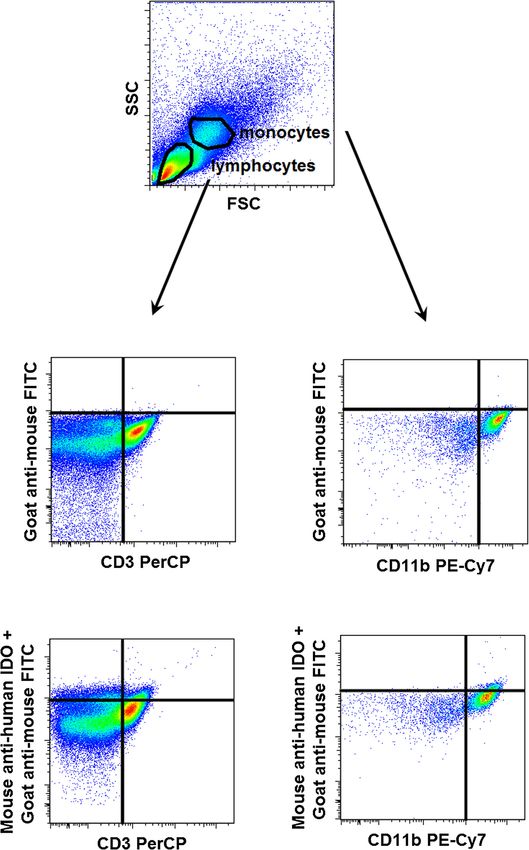
of mouse anti-human IDO monoclonal antibody. Figure 2
PBMC isolation represents the measurement of intracellular IDO expression
Peripheral blood mononuclear cells (PBMCs) were sepa- by flow cytometry.
rated by a standard density gradient centrifugation (Ficoll
Paque, Amersham Biosciences AB, Uppsala, Sweden, 25 mi- High-performance liquid chromatography (HPLC)
nutes, 400 g, 22°C) from freshly drawn blood collected in The investigated reference compounds (L-TRP, L-KYN
lithium heparin-treated tubes (BD Vacutainer, BD Biosci- sulfate salt, KYNA) and zinc acetate dihydrate were pur-
ences, San Jose, CA, USA). Cells were kept at −80°C in chased from Sigma-Aldrich (Saint Louis, MO, USA),
Fetal Bovine Serum containing 10% DMSO until analysis. acetonitrile and perchloric acid (PCA) were purchased
After thawing, cells were washed twice in phosphate- from Scharlau (Barcelona, Spain) and acetic acid was
buffered saline and their viability was assessed by trypan purchased from VWR International (Radnar, PA, USA).
blue exclusion (consistently > 90%). Plasma samples were stored at −80°C until analysis.
Before analysis, the samples were thawed and after a
Table 1 Clinical characteristics of non-pregnant and brief vortex 300 μl of plasma sample was ‘shot’ onto
healthy pregnant women 700 μl precipitation solvent (containing 3.57 w/w% PCA
Non-pregnant Healthy pregnant and 2.857 μM 3-nitro-L-tyrosine as internal standard).
women (n = 14) women (n = 20) Following that the samples were centrifuged at 13000 g
Age (years) 32 (27–34) 33.5 (30–36) for 10 min at 4°C, and the supernatant was collected.
No. of primiparas - 12 (60%) The KYN, KYNA and TRP concentrations of the sam-
Gestational age at - 36 (34–37)
ples were quantified based on the slightly modified
blood collection (weeks) method of Herve et al. [16], with an Agilent 1100 HPLC
Gestational age at - 39 (38–40) system (Agilent Technologies, Santa Clara, CA, USA).
delivery (weeks) The system was equipped with a fluorescent and a UV
Fetal birth weight (grams) - 3180 (2915–3725) detector, the former was applied for the determination
Data are presented as median (interquartile range) for continuous variables
of KYNA and TRP, and the latter for the determination
and as number (percentage) for categorical variables. of KYN and the internal standard. ChromatographicGrozdics et al. BMC Pregnancy and Childbirth 2014, 14:306 Page 4 of 9 http://www.biomedcentral.com/1471-2393/14/306 Figure 2 Indoleamine-2,3-dioxygenase (IDO) enzyme expression in CD3+ lymphocytes and CD11b + monocytes measured by flow cytometry. FSC – forward scatter characteristics, SSC – side scatter characteristics. separations were performed on an Onyx Monolithic C18 size (Agilent Technologies, Santa Clara, CA, USA) with a column, 100 mm × 4.6 mm I.D. (Phenomenex Inc., mobile phase composition of 0.2 M zinc acetate/ACN = Torrance, CA, USA) after passage through a Hypersil 95/5 (v/v%) with a pH adjusted to 6.2 with glacial acetic ODS pre-column, 20 mm × 2.1 mm I.D., 5 μm particle acid, applying isocratic elution. The flow rate and the
Grozdics et al. BMC Pregnancy and Childbirth 2014, 14:306                                                        Page 5 of 9
http://www.biomedcentral.com/1471-2393/14/306

injection volume were 1.5 ml/min and 20 μl, respectively.       with 0.45 effect size to detect differences between sam-
The fluorescent detector was set at excitation and emis-        ple populations. Comparisons between sample popula-
sion wavelengths of 344 nm and 398 nm, and after                tions were made with Mann–Whitney tests. Correlation
3.5 min of each run the wavelengths were changed to             analyses were performed using Spearman tests. p-values
254 nm and 398 nm. The UV detector was set at a wave-           less than 0.05 were considered significant. Statistics were
length of 365 nm. Figure 3 shows representative chro-           calculated using the STATISTICA software (version 8.0;
matograms of the measured metabolites.                          StatSoft, Inc., Tulsa, Oklahoma, USA).

HPLC method validation                                          Results
Calibration curve and linearity                                 Our results are detailed in Tables 2 and 3 and Figure 4. A
Calibrants were prepared at 6 different concentration levels,   significant increase in the prevalence of CD28+ T cells
from 1 to 100 nM, 0.1 to 5 μM, 5 to 50 μM and 0.5 to            was observed in HP compared to NP women. At the same
7.5 μM for KYNA, KYN, TRP and the internal standard, re-        time a decrease was shown in the expression of CD152 on
spectively. Three parallel injections of each solution were     these cells. The prevalence of both CD278+ and CD279+
made under the chromatographic conditions described             T cells was higher in HP than in NP women.
above. The peak area responses were plotted against the           Within the CD4 subset, the ratio of CD28+, CD28+
corresponding concentration, and the linear regression          CD152+ and CD279+ cells was comparable in HP and
computations were carried out by the least square method        NP women, while that of CD278+ cells was higher in
with the R software [17]. Very good linearity was observed      HP than in NP individuals.
throughout the investigated concentration ranges for KYN,         The frequency of both CD80+ and CD275+ monocytes
KYNA, TRP and the internal standard when either fluores-        was lower in HP women, however, no difference was ob-
cence or UV detection was applied.                              served regarding CD86+ and CD274+ monocytes.
                                                                  The prevalence of IDO-expressing T cells and monocytes
Selectivity                                                     was higher in HP compared to NP women. At the same
The selectivity of the method was checked by comparing          time, the mean fluorescence intensity (MFI) values for IDO
the chromatograms of KYN, KYNA, TRP and the in-                 were also significantly higher in both cell subsets in HP.
ternal standard for a blank plasma sample and those for           Plasma KYN, KYNA and TRP levels were lower, while
a spiked plasma sample. All compounds could be de-              at the same time, the K/T ratio was higher in HP than in
tected in their own selected chromatograms without any          NP women.
significant interference.                                         In order to explore whether reverse signalling via CD80
                                                                and CD86 is present in monocytes, correlation analyses
LOD and LLOQ                                                    were performed. However, we could not detect a correlation
Limit of detection (LOD) and lower limit of quantitation        between the frequency of CD80+ or CD86+ monocytes and
(LLOQ) was determined via signal-to-noise ratio with            the frequency of IDO-expressing T cells or monocytes or
threshold 3 and 10, according to the ICH guidelines [18].       the MFI of IDO in the investigated study groups.
The LOD was 100, 1 and 15 nM, while LLOQ was 275,
3.75 and 35 nM for KYN, KYNA and TRP, respectively.             Discussion
                                                                In this study, we aimed to characterize the prevalence of
Precision                                                       B7 costimulatory molecules on monocytes and their corre-
Replicate HPLC analysis showed that the relative stand-         sponding receptors on T lymphocytes in HP compared to
ard deviation was ≤ 2.2% for the peak area response             NP women, as well as the intracellular expression of IDO
and ≤ 0.1% for the retention time.                              and plasma levels of TRP, KYN and KYNA, important
                                                                molecules with immunoregulatory properties, in order to
Recovery                                                        describe their potential contribution to the pregnancy-
The relative recoveries were estimated by measuring spiked      specific maternal immune tolerance. Pregnancy is an im-
samples of KYN, KYNA and TRP at 2 different concentra-          munosuppressive state, with well known alterations in the
tions with 3 replicates of each. No significant differences     prevalence and function of T lymphocytes [1]. We as-
were observed for the lower and higher concentrations.          sumed that alterations in costimulation mechanisms via
The recoveries ranged from 108 to 110%, 86 to 91% and 85        B7 proteins might contribute to the lower level of T
to 89% for KYN, KYNA and TRP, respectively.                     lymphocyte activation compared to NP women.
                                                                  However, surprisingly, the expression of CD28 was in-
Statistics                                                      creased, while that of CTLA-4 was decreased on T lym-
Data are expressed as median and interquartile range.           phocytes isolated from HP women. This finding indicates
The sample size was estimated to achieve 80% power              that costimulation via CD28 is of great importance also
Grozdics et al. BMC Pregnancy and Childbirth 2014, 14:306 Page 6 of 9 http://www.biomedcentral.com/1471-2393/14/306 Figure 3 Chromatograms of the investigated molecules. a. represents the chromatogram of kynurenine (KYN) and the internal standard 3-nitro-L-tyrosine (3-NLT), made by UV detector (x: time (min), y: miliAbsorbanceUnit (mAU)). b and c. shows the chromatogram of kynurenic acid (KYNA) and tryptophan (TRP), respectively, made by fluorescent detector (x: time (min), y: Luminescence Unit (LU)). during pregnancy in T cells, and it may not contribute to decreased on HP monocytes, while that of B7-2 was un- the immunosuppressive environment characteristic for altered, which might reduce the intensity of costimulation gestation. At the same time, the expression of B7-1 was via CD28.
Grozdics et al. BMC Pregnancy and Childbirth 2014, 14:306                                                                                        Page 7 of 9
http://www.biomedcentral.com/1471-2393/14/306

Table 2 Frequency of the investigated cell surface and intracellular markers
                                                                               Non-pregnant women (n = 14)             Healthy pregnant women (n = 20)
CD3+ CD28+ cells/CD3+ lymphocytes                                              76.0 (64.7-82.9)%                       88.4* (81.8-90.6)%
CD3+ CD28+ CD152+ cells/CD3+ CD28+ lymphocytes                                 8.90 (7.57-11.4)%                       6.64* (5.07-9.89)%
CD3+ CD278+ cells/CD3+ lymphocytes                                             55.7 (49.5-56.7)%                       89.9* (75.7-91.6)%
CD3+ CD279+ cells/CD3+ lymphocytes                                             46.3 (39.3-51.2)%                       51.9* (47.9-67.7)%
CD4+ CD28+ cells/CD4+ lymphocytes                                              97.8 (96.7-98.8)%                       97.3 (93.0-99.2)%
CD4+ CD28+ CD152+ cells/CD4+ CD28+ lymphocytes                                 6.92 (4.93-8.63)%                       5.61 (3.65-9.09)%
CD4+ CD278+ cells/CD4+ lymphocytes                                             56.7 (52.8-59.5)%                       87.7* (76.3-92.2)%
CD4+ CD279+ cells/CD4+ lymphocytes                                             49.4 (42.1-54.5)%                       44.2 (37.5-65.2)%
CD11b + CD80+ cells/CD11b + monocytes                                          55.5 (17.3-69.8)%                       17.6* (13.6-25.4)%
CD11b + CD86+ cells/CD11b + monocytes                                          23.8 (17.3-29.5)%                       20.7 (14.9-31.9)%
CD11b + CD274+ cells/CD11b + monocytes                                         78.4 (70.3-85.9)%                       80.7 (77.2-87.8)%
CD11b + CD275+ cells/CD11b + monocytes                                         63.8 (59.0-72.1)%                       17.3* (14.1-27.0)%
CD3+ IDO + cells/CD3+ lymphocytes                                              5.90 (2.65-16.9)%                       24.1* (13.3-56.4)%
IDO mean fluorescence intensity in CD3+ IDO + cells (arbitrary unit)           9888 (7482–11475)                       62500* (19800–84475)
CD11b + IDO + cells/CD11b + monocytes                                          2.59 (1.99-11.7)%                       22.5* (13.6-50.5)%
IDO mean fluorescence intensity in CD11b + IDO + cells (arbitrary unit)        21400 (19075–23875)                     70450* (38775–114000)
*p < 0.05 versus non-pregnant women. Data are presented as median (interquartile range). IDO – indoleamine-2,3-dioxygenase, MFI – mean fluorescence intensity.

  In cases of miscarriage, the expression of B7-2 was                            expressing T lymphocytes was also elevated in HP. The
found to be highly upregulated at the fetomaternal inter-                        inhibitory effect of this receptor may play a role in inhi-
face and this was associated with high levels of Th1 cy-                         biting the activation of T cells during gestation. Interest-
tokines (IL-2 and IFN-gamma) and low levels of Th2                               ingly, Taglauer et al. demonstrated that the expression of
cytokines (IL-4 and IL-10) [19]. Furthermore, it was re-                         PD-1 expression on CD3 cells was low in non-pregnant
ported that in vivo blockade of B7-2 costimulation                               endometrium but increased in first-trimester decidua
shifted the cytokine balance from a Th1 to a Th2 pre-                            and remained elevated in term decidua. Additionally,
dominance at the fetomaternal interface, and expanded                            higher relative proportions of term decidual CD8bright,
peripheral CD4+ CD25+ regulatory T cells. Thus, reduc-                           CD4, and Treg cells expressed PD-1 in comparison to
tion in the level or function of B7-2 appears to be advan-                       autologous peripheral blood, further strengthening the
tageous to HP in the first half of pregnancy. We could                           role of this molecule in the development of maternal im-
not, however, demonstrate this reduction in third tri-                           mune tolerance [20].
mester peripheral blood HP samples.                                                 The prevalence of IDO-producing T cells and mono-
  The expression of ICOS, a stimulator of T cell activa-                         cytes was elevated in HP compared to NP samples. The
tion was strongly elevated in HP, while that of its corre-                       well-known immunosuppressive activity of this enzyme
sponding costimulatory molecule, B7-H2 was strongly                              may play an important role in the development of
decreased on HP monocytes. Since ICOS most effect-                               pregnancy-specific immune tolerance towards the de-
ively induces IL-10 instead of IL-2 production [9], its                          veloping fetus. IDO is a key enzyme in the catabolism
higher level may contribute to the Th2 shift observed in                         of tryptophan and initiates the production of kynure-
the third trimester of HP [1]. The frequency of PD-1                             nines. These metabolites have several immunological
                                                                                 and non-immunological regulatory functions. By lo-
Table 3 Plasma levels of kynurenine (KYN), kynurenic                             cally depleting TRP and increasing the levels of KYN
acid (KYNA) and tryptophan (TRP)                                                 and its metabolites, IDO provides a suppression of T
                        Non-pregnant                    Healthy pregnant         cell-mediated immune response via inhibiting the pro-
                        women (n = 14)                  women (n = 20)
                                                                                 liferation and inducing the apoptosis of activated T
KYN (uM)                1.80 (1.70-2.08)                1.55* (1.31-1.83)        cells, as well as promoting the development of regula-
KYNA (nM)               29.8 (25.6-47.7)                18.8* (15.0-23.0)        tory T cells and tolerogenic DCs [21]. Furthermore, we
TRP (uM)                54.1 (49.0-59.3)                38.6* (33.9-42.5)        found that not only the prevalence of IDO-producing
K/T ratio               0.037 (0.032-0.040)             0.041* (0.038-0.045)     cells, but also the intracellular amount of IDO is ele-
*p < 0.05 versus non-pregnant women. Data are presented as median
                                                                                 vated in HP (represented by the higher MFI values
(interquartile range).                                                           compared to NP women).
Grozdics et al. BMC Pregnancy and Childbirth 2014, 14:306 Page 8 of 9
http://www.biomedcentral.com/1471-2393/14/306
Figure 4 Frequency of the investigated cell surface markers on CD3+ lymphocytes and CD11b + monocytes. Horizontal line – median,
box – interquartile range, whisker – range. NP – non-pregnant women, HP – healthy pregnant women. *p < 0.05 vs NP.
KYN, KYNA, and especially TRP levels were decreased different roles: first, at the level of T cells where CTLA-4
in HP, resulting in an elevated K/T ratio compared to NP. as a negative receptor regulates TCR signal transduction;
As noted above, the depletion of TRP may directly con- second, at the level of APCs where CTLA-4 as a ligand
tribute to the immunosuppressive environment in HP. signals to the APC to induce IDO expression [23]. We
The increase in IDO activity in HP (represented by the el- hypothesized that reverse signalling may play a role in
evated K/T ratio) corresponds well with our finding of the higher IDO activity and expression observed in HP.
higher IDO-expressing T cell and monocyte numbers However, correlation analyses with B7-1 and B7-2 ex-
in HP. Interestingly, although the prevalence of IDO- pression did not support this notion.
producing T cells was decreased in our earlier study in PE
compared to HP [22], KYN, KYNA and TRP levels, as well Conclusions
as the K/T ratio were comparable in HP and PE (1.55 In conclusion, costimulation via CD28 may not contribute
(1.31-1.83) vs. 1.67 (1.33-1.93) uM, 18.8 (15.0-23.0) vs. to the immunosuppressive environment, at least in the last
22.3 (16.7-28.5) nM, 38.6 (33.9-42.5) vs. 37.1 (30.0-41.3) stage of pregnancy. Based on our findings, the develop-
uM, 0.041 (0.038-0.045) vs. 0.044 (0.036-0.049), respect- ment of the pregnancy-specific immune tolerance in the
ively). Hence, the alterations observed in the frequency of mechanism of B7 costimulation may be more related to
IDO-producing cells are not reflected by the enzymatic ac- the altered expression of B7 proteins on APCs rather than
tivity of IDO in PE, in contrast to HP. that of their receptors on T cells. The elevated intracellular
Another reason for decreased KYN, KYNA and TRP IDO expression in monocytes and T cells, as well as higher
levels in HP compared to NP may be the fact that these plasma enzymatic IDO activity are likely to contribute to
molecules cross the placenta via yet partly unidentified the systemic immunosuppressive environment in the third
mechanisms and transporters, and contribute to the devel- trimester characteristic for healthy gestation.
opment of the decreased immune responsiveness of the
fetus. Our unpublished recent data, indicating elevated Additional file
KYN, KYNA and TRP levels in cord blood of healthy term
neonates compared to adult peripheral blood may support Additional file 1: STROBE Statement—checklist of items that
this assumption. However, further experimental data are should be included in reports of observational studies.
needed to confirm or refute this hypothesis.
The engagement of B7-1 and B7-2 by CTLA-4 induces Abbreviations
back signalling into the monocyte and promotes the pro- APC: Antigen presenting cell; CTLA-4: Cytotoxic T lymphocyte antigen 4;
HP: Healthy pregnancy; HPLC: High-performance liquid chromatography;
duction of IFN-gamma, which acts in an autocrine or ICOS: Inducible costimulator of T cells; IDO: Indoleamine-2,3-dioxygenase;
paracrine manner to upregulate IDO expression, thereby KYN: Kynurenine; KYNA: Kynurenic acid; LOD: Limit of detection; LLOQ: Lower
initiating the degradation of TRP and resulting in an im- limit of quantitation; MFI: Mean fluorescence intensity; MHC: Major
histocompatibility complex; NP: Non-pregnant; PBMC: Peripheral blood
munosuppressive effect as described above. Thus, the mononuclear cell; PCA: Perchloric acid; PD-1: Programmed death-1 receptor;
interaction between CTLA-4 and B7 proteins plays two TCR: T cell receptor; Treg: Regulatory T cell; TRP: Tryptophane.Grozdics et al. BMC Pregnancy and Childbirth 2014, 14:306 Page 9 of 9
http://www.biomedcentral.com/1471-2393/14/306
Competing interests 10. Grohmann U, Orabona C, Fallarino F, Vacca C, Calcinaro F, Falorni A,
The authors declare that they have no competing interests. Candeloro P, Belladonna ML, Bianchi R, Fioretti MC, Puccetti P: CTLA-4-Ig
regulates tryptophan catabolism in vivo. Nat Immunol 2002, 3:1097–1101.
Authors’ contributions 11. Munn DH, Zhou M, Attwood JT, Bondarev I, Conway SJ, Marshall B, Brown C,
EG performed flow cytometry, designed study and drafted manuscript, LB Mellor AL: Prevention of allogeneic fetal rejection by tryptophan catabolism.
performed flow cytometry and analyzed results, AB performed flow Science 1998, 281:1191–1193.
cytometry and interpreted data, GV performed HPLC and analyzed results, II 12. Swartz KJ, During MJ, Freese A, Beal MF: Cerebral synthesis and release of
designed study and interpreted data, PK analyzed results and interpreted kynurenic acid: an endogenous antagonist of excitatory amino acid
data, JR Jr collected samples and interpreted data, LV designed study and receptors. J Neurosci 1990, 10:2965–2973.
drafted manuscript, TT interpreted data and critically read manuscript, GT 13. Vecsei L, Miller J, MacGarvey U, Beal MF: Kynurenine and probenecid
designed study, interpreted data and drafted manuscript. All Authors read inhibit pentylenetetrazol-induced and NMDA-induced seizures and
and approved the final manuscript. increase kynurenic acid concentrations in the brain. Brain Res Bull 1992,
28:233–238.
14. Mándi Y, Vécsei L: The kynurenine system and immunoregulation.
Acknowledgements
J Neural Transm 2012, 119:197–209.
We are grateful to Levente Szalárdy and Dénes Zádori for their valuable
15. Vécsei L, Szalárdy L, Fülöp F, Toldi J: Kynurenines in the CNS: recent
technical advice. This work was supported by the Hungarian Scientific
advances and new questions. Nat Rev Drug Discov 2013, 12:64–82.
Research Fund (OTKA; grant numbers 109451, K 75628), the National Brain
16. Herve C, Beyne P, Jamault H, Delacoux E: Determination of tryptophan and
Research Program (NAP; grant number KTIA_NAP_13), the European Union
its kynurenine pathway metabolites in human serum by high-performance
and the State of Hungary, co-financed by the European Social Fund in the
liquid chromatography with simultaneous ultraviolet and fluorimetric
framework of TÁMOP 4.2.4. A/2-11-1-2012-0001 ‘National Excellence Program –
detection. J Chromatogr B 1996, 675:157–161.
Elaborating and operating an inland student and researcher personal support
17. R Development Core Team: R: a language and environment for statistical
system convergence program’, TÁMOP-4.2.2/B-10/1-2010-0012, and TÁMOP-4.2.2.
computing. [http://www.R-project.org]
A-11/1/KONV-2012-0052. G.T. is an International Society for the Advancement of
18. Validation of Analytical Procedures: Text and Methodology Q2 (R1):
Cytometry (ISAC) Scholar.
Validation of analytical procedures: text and methodology Q2 (R1).
[http://www.ich.org/fileadmin/Public_Web_Site/ICH_Products/Guidelines/
Author details
1 Quality/Q2_R1/Step4/Q2_R1__Guideline.pdf]
First Department of Pediatrics, Semmelweis University, Bókay u. 53-54,
19. Jin LP, Fan DX, Zhang T, Guo PF, Li DJ: The costimulatory signal
Budapest H-1083, Hungary. 2Department of Neurology, Faculty of Medicine,
upregulation is associated with Th1 bias at the maternal–fetal interface
Albert Szent-Györgyi Clinical Center, University of Szeged, Semmelweis u. 6,
in human miscarriage. Am J Reprod Immunol 2011, 66:270–278.
Szeged H-6725, Hungary. 3Department of Inorganic and Analytical Chemistry,
20. Taglauer ES, Trikhacheva AS, Slusser JG, Petroff MG: Expression and
University of Szeged, Dóm tér 7, H-6720 Szeged, Hungary. 4First Department
function of PDCD1 at the human maternal-fetal interface. Biol Reprod
of Obstetrics and Gynecology, Semmelweis University, Baross u. 27, Budapest
2008, 79:562–569.
H-1088, Hungary. 5MTA-SZTE Neuroscience Research Group, Semmelweis u.
21. Grohmann U, Fallarino F, Puccetti P: Tolerance, DCs and tryptophan: much
6, Szeged H-6725, Hungary. 6MTA-SE Pediatrics and Nephrology Research
ado about IDO. Trends Immunol 2003, 24:242–248.
Group, Bókay u. 53-54, Budapest H-1083, Hungary.
22. Toldi G, Vásárhelyi B, Biró E, Fügedi G, Rigó J Jr, Molvarec A: B7
costimulation and intracellular indoleamine-2,3-dioxygenase (IDO)
Received: 7 May 2014 Accepted: 2 September 2014
expression in peripheral blood of healthy pregnant and preeclamptic
Published: 4 September 2014
women. Am J Reprod Immunol 2013, 69:264–271.
23. Wang S, Chen L: T lymphocyte co-signaling pathways of the B7-CD28
References family. Cell Mol Immunol 2004, 1:37–42.
1. Toldi G, Treszl A, Vásárhelyi B: T Lymphocyte Characteristics and Immune
Tolerance During Human Pregnancy. In Autoimmune Disorders –
doi:10.1186/1471-2393-14-306
Pathogenetic Aspects. Edited by Mavragani C. Rijeka: Intech; 2011:463–486.
Cite this article as: Grozdics et al.: B7 costimulation and intracellular
2. Saito S, Sakai M, Sasaki Y, Tanebe K, Tsuda H, Michimata T: Quantitative indoleamine-2,3-dioxygenase (IDO) expression in peripheral blood of
analysis of peripheral blood Th0, Th1, Th2 and the Th1:Th2 cell ratio healthy pregnant and non-pregnant women. BMC Pregnancy and Childbirth
during normal human pregnancy and preeclampsia. Clin Exp Immunol 2014 14:306.
1999, 117:550–555.
3. Santner-Nanan B, Peek MJ, Khanam R, Richarts L, Zhu E, Fazekas de St Groth B,
Nanan R: Systemic increase in the ratio between Foxp3+ and IL-17-producing
CD4+ T cells in healthy pregnancy but not in preeclampsia. J Immunol 2009,
183:7023–7030.
4. Toldi G, Stenczer B, Treszl A, Kollár S, Molvarec A, Tulassay T, Rigó J Jr,
Vásárhelyi B: Lymphocyte calcium influx characteristics and their
modulation by Kv1.3 and IKCa1 channel inhibitors in healthy pregnancy
and preeclampsia. Am J Reprod Immunol 2011, 65:154–163.
5. Petroff MG, Perchellet A: B7 family molecules as regulators of the
maternal immune system in pregnancy. Am J Reprod Immunol 2010,
63:506–519. Submit your next manuscript to BioMed Central
6. Gimmi CD, Freeman GJ, Gribben JG, Gray G, Nadler LM: Human T-cell and take full advantage of:
clonal anergy is induced by antigen presentation in the absence of B7
costimulation. Proc Natl Acad Sci U S A 1993, 90:6586–6590.
• Convenient online submission
7. Chambers CA, Kuhns MS, Egen JG, Allison JP: CTLA-4-mediated inhibition
in regulation of T cell responses: mechanisms and manipulation in • Thorough peer review
tumor immunotherapy. Annu Rev Immunol 2001, 19:565–594. • No space constraints or color figure charges
8. Coyle AJ, Lehar S, Lloyd C, Tian J, Delaney T, Manning S, Nguyen T, Burwell T,
• Immediate publication on acceptance
Schneider H, Gonzalo JA, Gosselin M, Owen LR, Rudd CE, Gutierrez-Ramos JC:
The CD28-related molecule ICOS is required for effective T cell-dependent • Inclusion in PubMed, CAS, Scopus and Google Scholar
immune responses. Immunity 2000, 13:95–105. • Research which is freely available for redistribution
9. Hutloff A, Dittrich AM, Beier KC, Eljaschewitsch B, Kraft R, Anagnostopoulos I,
Kroczek RA: ICOS is an inducible T-cell co-stimulator structurally and
functionally related to CD28. Nature 1999, 397:263–266. Submit your manuscript at
www.biomedcentral.com/submitYou can also read