Haplopelma hainanum venom induces inflammatory skin lesions - PeerJ
←
→
Page content transcription
If your browser does not render page correctly, please read the page content below
Haplopelma hainanum venom induces
                                      inflammatory skin lesions
                                      Zhili Deng1 ,2 ,3 ,4 , Yaling Wang1 , Wei Shi1 ,4 , Lei Zhou1 , San Xu1 ,2 ,3 ,4 ,
                                      Ji Li1 ,2 ,3 ,4 and Yiya Zhang1 ,2 ,4
                                  1
                                    Department of Dermatology, Xiangya Hospital, Central South University, Changsha, China
                                  2
                                    Key Laboratory of Organ Injury, Aging and Regenerative Medicine of Hunan Province, Central South
                                    University, Changsha, China
                                  3
                                    Center for Molecular Medicine, Xiangya Hospital, Central South University, Changsha, China
                                  4
                                    National Clinical Research Center for Geriatric Disorders, Xiangya Hospital, Central South University,
                                    Changsha, China

                                         ABSTRACT
                                         The Haplopelma hainanum is a species of theraphosid spider from China. Its large size
                                         and charming appearance make this species a popular pet. According to a previous
                                         study, theraphosid spider bites can induce pain, erythema, and edema in humans
                                         and can present more severely in domestic animals. The pathological consequences
                                         of envenomation by H. hainanum remain unclear. In this study, we investigated the
                                         effects and mechanisms of H. hainanum envenomation in mice. We showed that the
                                         venom induced slight swelling, intense inflammatory response, and increased the
                                         microvascular density in mice skin. Moreover, we found that 50 µg/ml of the spider’s
                                         venom induced IL-1β expression in both HaCaT cells and fibroblast cells, but repressed
                                         CXCL10 expression in fibroblasts. The venom significantly induced cell senescence
                                         and repressed cell proliferation and migration in both HaCaT cells and fibroblast
                                         cells. Finally, we examined the expression of Nav channel in HaCaT and fibroblast
                                         cells and found that H. hainanum venom effectively inhibited Na+ currents in HaCaT
                                         cells. Our study calls for further investigation of the pathological consequences and
                                         potential mechanisms of H. hainanum envenomation. This information might assist in
                                         the development of suitable therapy.

Submitted 16 July 2019
Accepted 21 November 2019             Subjects Toxicology, Dermatology
Published 10 January 2020             Keywords H. hainanum venom, Envenomation, Inflammation, Revascularization
Corresponding author
Yiya Zhang, yiya0108@csu.edu.cn
Academic editor
                                      INTRODUCTION
Bruno Lomonte                         Spiders are one of the oldest and most abundant venomous animals, with a fossil history
Additional Information and            spanning more than 300 million years and over 40,000 species (Deng et al., 2016). Every
Declarations can be found on
page 12
                                      year, approximately 10,000 spider bites are reported in Brazil and nearly 3,000 bites in
                                      America (Braitberg & Segal, 2009).
DOI 10.7717/peerj.8264
                                         The venom of most spiders causes only minor discomfort including edema, hemorrhage,
    Copyright                         and sometimes subsequent ulceration (Dunbar et al., 2018; Isbister & Fan, 2011). Though
2020 Deng et al.
                                      relatively rare, spider envenomation also can cause severe reactions such as systemic
Distributed under                     loxoscelism, which can progress to acute renal failure and even death (Manzoni-de Almeida
Creative Commons CC-BY 4.0
                                      et al., 2018; Okamoto et al., 2017). Most studies on spider envenomation focus on one of the
OPEN ACCESS                           most venomous spiders, the Loxosceles. Studies have shown that histopathologic alterations

                                      How to cite this article Deng Z, Wang Y, Shi W, Zhou L, Xu S, Li J, Zhang Y. 2020. Haplopelma hainanum venom induces inflammatory
                                      skin lesions. PeerJ 8:e8264 http://doi.org/10.7717/peerj.8264
induced by Loxosceles envenomation include edema, vasodilatation, and hemorrhage
                                   in dermis–epidermis dissociation (Van den Berg et al., 2007). Also, the complement
                                   system plays an important role in envenomation-induced inflammation (Patel et al.,
                                   1994; Ribeiro et al., 2015; Tambourgi et al., 2005). An increasing number of studies have
                                   revealed the important role of fibroblasts and keratinocytes in spider venom-induced
                                   pathological alterations in the skin. Loxosceles envenomation partly induces dermonecrosis
                                   by upregulating proinflammatory cytokine expression in fibroblasts (Dragulev et al.,
                                   2007; Rojas et al., 2017). Another study showed that keratinocyte-secreted matrix
                                   metalloproteinase contributed to the induction of dermonecrosis by both Loxoscles laeta
                                   and Loxosceles intermedia venom (Correa et al., 2016). Moreover, Loxosceles venom triggers
                                   cell death by apoptosis in human skin fibroblasts (Dantas et al., 2014) and keratinocytes,
                                   contributing to the pathogenesis of cutaneous loxoscelism (Paixao-Cavalcante & Van den
                                   Berg, 2006).
                                       Theraphosid spiders, also called bird spiders, are increasingly being kept as pets due
                                   to their size and beautiful coloring (Fuchs et al., 2014). Although theraphosid spiders are
                                   considered harmless, their venom has been proven to cause localized pain, erythema, and
                                   edema in humans, with more severe symptoms in canines, including death (Isbister et
                                   al., 2003; Rocha et al., 2016). Haplopelma hainanum is a venomous species of theraphosid
                                   spider from the Hainan province in southern China (Xiao & Liang, 2003). Previous studies
                                   have focused on the peptides in H. hainanum venom that directly regulate the activation
                                   of ion channels, producing analgesic effects (Zhang et al., 2015). The histopathologic
                                   alterations caused by H. hainanum envenomation, however, are virtually unknown.
                                       In this study, we examined the pathological alterations induced by H. hainanum venom
                                   in mice, and discovered the mechanism that potentially contributes to lesion development
                                   in HaCaT and fibroblast cells. We developed an understanding of the action of the
                                   molecular mechanisms of H. hainanum venom, which may assist in the development of
                                   various treatments aimed at ameliorating the symptoms of spider envenomation.

                                   MATERIALS & METHODS
                                   Animals
                                   Twenty female C57BL/6 mice (8 weeks old) were used in this study. All mice received food
                                   and water prior to the experiment with a 12 h/12 h day/night cycle. All animal experiments
                                   were approved by the Animal Care and Use Committee of the Xiangya Hospital of Central
                                   South University (201703211).

                                   Spider venom and treatment
                                   The venom was collected from adult female H. hainanum using an electro-pulse stimulator
                                   as described previously (Hu et al., 2014; Yan et al., 2018). Expelled venom was collected
                                   from the fang tips with a tube, pooled, and freeze-dried. The freeze-dried crude venom
                                   was stored at −20 ◦ C prior to analysis. H. hainanum venom (0, 1, 3, 10 and 30 µg/site)
                                   was injected into the ear in a fixed volume of 25 µl in PBS. 24 h after the intradermal (i.d.)
                                   injection of venom, the skin was collected and stored at −80 ◦ C.

Deng et al. (2020), PeerJ, DOI 10.7717/peerj.8264                                                                           2/17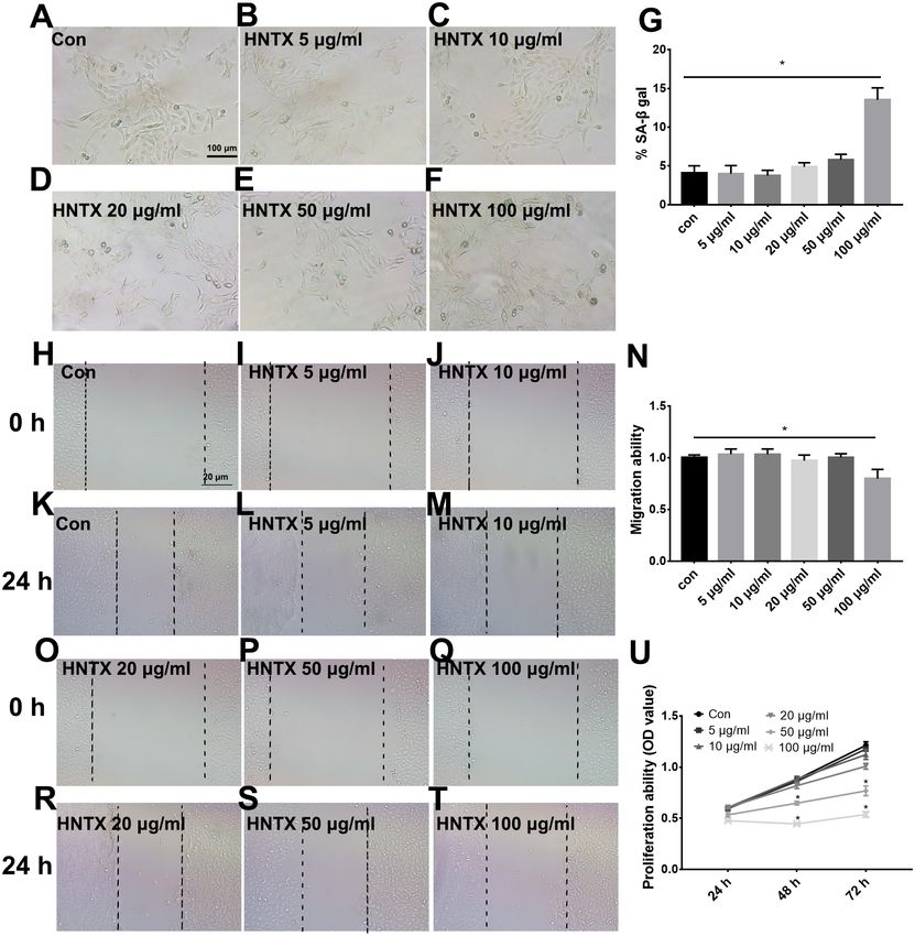
Table 1 Primers for qRT-PCR.

                                     Primier           Forward (50 to 30 )                   Reverse (50 to 30 )
                                     IL-1β             AGCTACGAATCTCCGACCAC                  CGTTATCCCATGTGTCGAAGAA
                                     CXCL10            GTGGCATTCAAGGAGTACCTC                 TGATGGCCTTCGATTCTGGATT
                                     TNFα              CCTCTCTCTAATCAGCCCTCTG                GAGGACCTGGGAGTAGATGAG
                                     IL-6              CCTGAACCTTCCAAAGATGGC                 TTCACCAGGCAAGTCTCCTCA
                                     IL-17             TCCCACGAAATCCAGGATGC                  GGATGTTCAGGTTGACCATCAC
                                     CCL2              CAGCCAGATGCAATCAATGCC                 TGGAATCCTGAACCCACTTCT

                                   Cell culture and treatment
                                   Human keratinocyte HaCaT cells were purchased from the Cell Bank of Chinese Academy
                                   of Sciences (Shanghai, China). The primary human skin fibroblast cells were cultured
                                   by digesting human skin with type II collagenase (Sigma, Aldrich, St. Louis, MO, USA)
                                   to isolate human keratinocytes as described previously (Xie et al., 2013). Fibroblast cells
                                   were cultured in Dulbecco’s modified Eagle’s medium (DMEM, Hyclone, Logan, USA)
                                   with 10% fetal bovine serum (Hyclone, Logan, UT, USA) as previously described (Li et al.,
                                   2010). The human keratinocyte HaCaT cells were cultured in free-calcium basal medium
                                   (DMEM; Gibco, USA) with 10% fetal bovine serum (Hyclone, Logan, USA) as previously
                                   described (Li et al., 2019) in an incubator at 37 ◦ C, 5% CO2 .

                                   Histologic analysis
                                   Mice ears were sectioned at 4 µm thickness and then stained with hematoxylin and eosin
                                   (H&E) (four ears per group). The histological alterations were detected by microscopy
                                   (OLYMPUS, Japan).

                                   Immunofluorescence
                                   The ears were sectioned at 8 µm thickness and incubated with anti-CD4, anti-CD31, and
                                   anti-MHCII antibodies (4 ears per group), and then stained with anti-goat IgG antibodies
                                   (Alexa Fluor 488) as previously described (Su et al., 2018). All antibodies were purchased
                                   from ebioscience (San Diego, USA).

                                   Real-time PCR analysis
                                   A TRIzol reagent (Invitrogen Life Technologies) was used to derive the total RNA from
                                   HaCaT and fibroblast cells. Two µg of RNA was reverse transcribed to cDNA. We then
                                   performed qPCR to obtain mRNA expression as previously described (Li et al., 2019).
                                   The primers of IL-1β, CXCL10, TNF-α, IL-6, IL-17, and CCL2 are listed in Table 1. The
                                   real-time PCR analysis was repeated in three independent experiments.

                                   Cell migration
                                   The cell migration ability was detected using a scratch-wound assay. Cells were seeded and
                                   cultured in a 6-well plate. When cells reached ∼80% confluence, a 100 µl tip was used for
                                   scratching. The cells were treated with 0, 5, 10, 20, 50, and 100 µg/ml venom in 5% FBS
                                   DMEM for 12 h. The cell wound conditions were then photographed using the Zeiss Axio
                                   Scope A1 microscope (Zeiss, Oberkochen, Germany). This was repeated 4 times.

Deng et al. (2020), PeerJ, DOI 10.7717/peerj.8264                                                                        3/17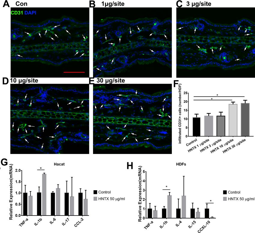
Cell senescence
                                   Senescence was detected using SA-β-gal staining as previously described (Xie et al., 2017).
                                   When cells reached 70% confluence, SA-β-gal solution was used for staining. β-Gal-
                                   positive cells were detected using microscopy (OLYMPUS, Japan). This was repeated in
                                   three independent experiments.

                                   Cell proliferation
                                   The cell proliferation ability was assessed using an MTT assay. 1 × 10⁴ cells were seeded in
                                   96-well plates and cultured for 24 h, 48 h, and 72 h. MTT (Sigma-Aldrich) and dimethyl
                                   sulfoxide (DMSO) (Sigma-Aldrich) were added to the 96-well plates and the absorbance
                                   was measured at 490 nm. This was repeated in 5 replicates per experiment and in 3
                                   independent experiments.

                                   Patch clamp
                                   The solidum currents in HaCaT and fibroblast cells were detected using the whole-cell
                                   patch-clamp technique (Axon 700B patch-clamp, Irvine, CA, USA) as previously described
                                   (Yan et al., 2018). To detect voltage-gated Nav currents, we used an extracellular solution
                                   containing (in mM): 145 NaCl, 1.5 CaCl2 , 2 MgCl2 , 2.5 KCl, 10 D-glucose and 10 HEPES,
                                   (pH 7.4). The pipette solution contained (in mM): 5 NaCl, 135 CsCl, 5 MgATP, 10
                                   D-glucose, 10 HEPES and 10 EGTA (PH 7.2). The current was elicited by -10 mV from a
                                   holding potential of −40 mV. This was repeated for 3 different experiments.

                                   Statistical analysis
                                   GraphPad Prism 6 (La Jolla, CA) was used for statistical analysis. Data were presented as
                                   means ± SEM. Statistical comparisons of the two groups were analyzed by the Student’s
                                   t -test. P < 0.05 was considered significant as compared to the control group.

                                   RESULTS
                                   Histological assessment and inflammatory cell infiltration of skin
                                   damage
                                   Histopathological analysis of the mice’s ear skin 24 h after i.d. H. hainanum venom injection
                                   showed histological alterations including slight swelling and an intense leukocyte infiltrate
                                   in which neutrophils were the predominant cell type deep in the dermis (Figs. 1A–1F).
                                   We also detected the infiltration of CD4+ T cells (CD4+ ) and APCs (MHCII+ ) using
                                   immunofluorescence staining. As shown in Figs. 1G–1L, the number of CD4+ T cells
                                   significantly increased in the lesions where H. hainanum venom was applied at 10 µg per
                                   site and 30 µg per site. Moreover, the number of MHCII+ cells also increased at the 10 µg
                                   per site and 30 µg per site where venom was applied (Figs. 1M–1R). Together, these results
                                   indicate that H. hainanum venom induces inflammatory cell infiltration in mice.

                                   Alterations in the microvascular density
                                   Previous studies have shown alterations in microvascular density in mice treated with snake
                                   venom treated (Jimenez et al., 2008) Here, we demonstrated the role of H. hainanum venom
                                   on microvascular density by detecting CD31+ cell using immunofluorescence. As shown

Deng et al. (2020), PeerJ, DOI 10.7717/peerj.8264                                                                          4/17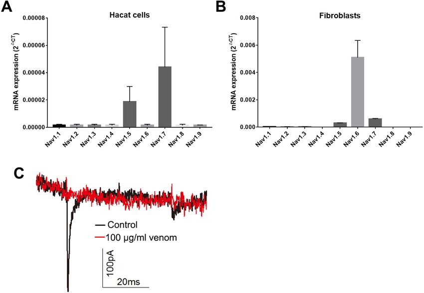
Figure 1 Histological alterations in H. hainanum. venom induced skin lesion. (A–F) H&E stain re-
                                     vealed the histological alterations. The arrows are inflammatory cells. (G–L) Immunofluorescence re-
                                     vealed the CD4+ T cells infiltration. The arrows are CD4+ cells. Scale bar: 100 µm. (M–R) Immunoflu-
                                     orescence revealed the MHCII+ cells infiltration. The arrows are MHCII+ cells. Scale bar: 100 µm. Data
                                     represent the means Âś SEM.*P < 0.05, compared to control group.
                                                                                                       Full-size DOI: 10.7717/peerj.8264/fig-1

Deng et al. (2020), PeerJ, DOI 10.7717/peerj.8264                                                                                         5/17
Figure 2 The effects of H. hainanum venom on the alterations in the microvascular density in mice
and inflammatory cytokines in keratinocytes and fibroblasts. (A–F) CD31+ cells were detected using
immunofluorescence. Scale bar: 100 µm. The arrows are CD31+ cells. Data represent the means ± SEM.
*P < 0.05, compared to control group. (G) qPCR revealed the expression of inflammatory cytokines in
keratinocytes. (H) qPCR revealed the expression of inflammatory cytokines in fibroblasts. Data represent
the means ± SEM. *P < 0.05, compared to control group.
Full-size DOI: 10.7717/peerj.8264/fig-2
in Figs. 2A–2F, at 10 µg per site and 30 µg per site venom evidently increased the number
of CD31+ cells. These results indicate that H. hainanum venom induces revascularization.
Venom induces the production of inflammatory cytokines
Keratinocytes, when provoked by environmental stimuli, can produce inflammatory
cytokines and chemokine (Hermann et al., 2017; Lowes et al., 2013; Zhang et al., 2016).
Moreover, dermal fibroblasts produce cytokines and antimicrobial peptides to defend
against pathogens (Hesse-Macabata et al., 2019). In this study, we assessed the effects of H.
hainanum venom on inflammatory cytokine expression in HaCaT cells and fibroblast cells.
As shown in Fig. 2G, 50 µg/ml venom evidently induced IL-1β expression in HaCaT cells.
Moreover, 50 µg/ml venom induced IL-1β expression and repressed CXCL10 expression
in fibroblasts (Fig. 2H). TNF-α, IL-6, IL-17, and CCL2, however, were not affected by H.
Deng et al. (2020), PeerJ, DOI 10.7717/peerj.8264 6/17hainanum venom. Together, these results indicate that venom partly induces inflammatory
infiltration by upregulating IL-1β expression in both HaCaT cells and fibroblast cells.
The effects of venom on the proliferation, migration, and senescence
of keratinocytes
Keratinocytes are the major component of the epidermis, which plays a central role in
maintaining the barrier function of the skin. In this study, we treated HaCaT cells with 0,
5, 10, 20, 50, and 100 µg/ml H. hainanum venom and then determined cell proliferation,
migration, and senescence using MTT, wound healing, and SA-β-gal staining, respectively.
As shown in Figs. 3A–3G and 3H–3T, 5, 10, 20, and 50 µg/ml H. hainanum venom
did not significantly affect cell senescence or repressed cell migration ability. 100 µg/ml
H. hainanum venom evidently induced cell senescence and repressed the migration
ability of HaCaT cells. As shown in Fig. 3U, 50 and 100 µg/ml H. hainanum venom
significantly repressed the cell proliferation ability of HaCaT cells. Five, 10, and 20 µg/ml
H. hainanum venom did not seem to impact HaCaT cell proliferation. In conclusion, the
data demonstrate that a high enough concentration of H. hainanum venom directly affects
the proliferation, migration, and senescence of keratinocytes.
The effects of venom on the proliferation, migration, and senescence
of fibroblasts
Next, we explored the potential role of H. hainanum venom on fibroblast cell proliferation,
migration, and senescence using MTT, wound healing, and SA-β-gal staining, respectively.
As shown in Figs. 4A–4U, 50 µg/ml and 100 µg/ml H. hainanum venom evidently induced
cell senescence and repressed the cell migration and proliferation ability of fibroblast cells.
Five, 10, and 20 µg/ml H. hainanum venom did not significantly affect cell proliferation,
migration, or senescence (Fig. 4). In conclusion, these data demonstrate that a high enough
concentration of H. hainanum venom directly affects the proliferation, migration, and
senescence of fibroblasts.
Venom inhibits the currents of voltage-gated sodium channels in
HaCaT and fibroblast cells
Studies have shown that keratinocytes and fibroblasts have sodium channels expression
(Zhao et al., 2008). In this study, we determined the expression levels of sodium channels in
HaCaT and fibroblasts cells. In HaCaT cells, the Nav1.5 and Nav1.7 expression levels were
higher than those of other sodium channels (Fig. 5A). In fibroblast cells, the Nav1.5, Nav1.6
and Nav1.7 expression levels were higher than those of other sodium channels (Fig. 5B).
We next detected the sodium currents of HaCaT and fibroblast cells using a patch clamp.
As shown in Fig. 5C, voltage-gated sodium currents were detected in HaCaT cells, as 100
µg/ml venom evidently repressed the sodium currents. Unfortunately, no sodium currents
were detected in fibroblasts.
DISCUSSION
In humans, envenomation by theraphosid spiders can result in painful skin lesions that
include erythema and edema (Rocha et al., 2016). The object of this study was to investigate
Deng et al. (2020), PeerJ, DOI 10.7717/peerj.8264 7/17Figure 3 The H. hainanum venom affects proliferation, migration and senescence of keratinocytes.
Cells were treated with 0, 5, 10, 20, 50 and 100 µg/ml venom. (A–G) SA-β-gal staining was used to deter-
minate cell senescence of keratinocytes. (H–T) wound healing was used to determinate cell migration of
keratinocytes. U, MTT was used to determinate cell proliferation of keratinocytes. Data was represented as
the means ± SEM. *P < 0.05, compared to control group.
Full-size DOI: 10.7717/peerj.8264/fig-3
the potential role of H. hainanum venom in the pathology of such skin lesions in vivo
and in vitro. We found that H. hainanum venom induces effects that closely mimic the
envenomation-induced lesions in humans. The histological alterations include slight
swelling, inflammatory cell infiltration, and revascularization. Moreover, H. hainanum
venom evidently induced the production of proinflammatory cytokines and significantly
affected the cell proliferation, migration, and senescence in keratinocytes and fibroblasts.
Spider venom is a complex cocktail of proteins, neurotoxic peptides, and small molecules.
It is used to capture prey and defend against predators (Windley et al., 2012). Although the
bite of most spiders has little or no effect on mammalian tissue, envenomation by Loxosceles,
Deng et al. (2020), PeerJ, DOI 10.7717/peerj.8264 8/17Figure 4 The H. hainanum. venom affect proliferation, migration and senescence of fibroblasts. (A–
G) SA-β-gal staining was used to determinate cell senescence of fibroblasts. (H–T) wound healing was
used to determinate cell migration of fibroblasts. (U) MTT was used to determinate cell proliferation of fi-
broblasts. Data represent the means ± SEM. *P < 0.05, compared to control group.
Full-size DOI: 10.7717/peerj.8264/fig-4
for example, can induce localized pain, erythema, edema and dermonecrosis in skin. Spider
envenomation can also induce severe systemic reactions, even death (Manzoni-de Almeida
et al., 2018; Okamoto et al., 2017). For example, Loxosceles venom can induce edema,
vascular anomalies, hemorrhaging, and dermis–epidermis dissociation (Mackinnon &
Witkind, 1954). Spider venom can also induce a severe inflammatory response, including
an increase of proinflammatory cytokines and chemokines and leukocyte infiltration
(Dunbar, Sulpice & Dugon, 2019; Griesbacher et al., 1998; Rojas et al., 2017; Tambourgi et
al., 2005). In this study, we examined the envenomation of mice with H. hainanum venom.
Deng et al. (2020), PeerJ, DOI 10.7717/peerj.8264 9/17Figure 5 Venom inhibits the currents of voltage gated sodium channels in Hacat cells. (A) The expres-
sion levels of sodium channels in Hacat. (B) The expression levels of sodium channels in fibroblasts. (C)
The sodium currents on Hacat using patch clamp. Data represent the means ± SEM.
Full-size DOI: 10.7717/peerj.8264/fig-5
Consistent with the clinical presentation of theraphosid spider bites in human (Rocha
et al., 2016), H. hainanum venom injection caused histological alterations including
slight swelling, an intense leukocyte infiltrate, and increased microvascular density in
mice (Figs. 1 and 2). Studies have shown that phospholipases D is an important active
constituent of venom in spiders from the Sicariidae family (Lopes et al., 2013). It plays a
proinflammatory role by activating leukocytes (Manzoni-de Almeida et al., 2018) in the
progression of the dermonecrotic lesion (Paixao-Cavalcante et al., 2007). There was no
phospholipases D, however, detected in the venom of theraphosid spiders. Moreover,
LmTX-I (a basic phospholipase A2) is considered an important component of Lachesis
muta muta venom that induces microvascular permeability in skin (Ferreira et al., 2009).
Kinin-related peptides were reported to play a key role in Vespula vulgaris venom, inducing
paw edema in rats (Griesbacher et al., 1998). Various neurotoxic peptides in theraphosid
spider venom attracts enormous interest, given their potential for pharmacological use in
regulating the activation of various ion channels (Zhang et al., 2015). Our previous studies
demonstrated that hainantoxin-I, a peptide toxin in H. hainanum venom, is an activator of
the KCa3.1 channel (Huang et al., 2014). The channel modulates Ca(2+ ) influx and plays a
key role in the activity of various immune cells, including mast cells, inflammatory CD4+
T, and antigen-specific memory T cells (Chiang et al., 2017; Duffy et al., 2015; Matsui et
al., 2018). These results indicate that hainantoxin-I may be involved in the inflammation
induced by H. hainanum venom.
Deng et al. (2020), PeerJ, DOI 10.7717/peerj.8264 10/17Keratinocytes are not merely a major component of the skin’s physical barrier.
They are also critical for innate immunity, producing a variety of proinflammatory
cytokines that play a key role in the activation and recruitment of immune cells in the
skin (Kabashima et al., 2019). Fibroblasts, which are the major cells for the production
of collagen and the most abundant component of the ECM in the dermis, are also
reported to contribute to immune defense by producing proinflammatory cytokines and
antimicrobial peptides (Hesse-Macabata et al., 2019). Previous studies have shown that
Loxosceles venom induced the production of proinflammatory cytokines and chemokines
in endothelial cells and fibroblasts (Desai et al., 1999; Gomez et al., 1999; Rojas et al., 2017).
Our work has demonstrated that H. hainanum venom evidently induced IL-1β expression
in both HaCaT cells and fibroblast cells. The expression of TNF-α, IL-6, IL-17, and CCL2,
however, was not affected by H. hainanum venom. In conclusion, these results indicated
that H. hainanum venom-induced inflammation was partly caused by the production
of cytokines in HaCaT and fibroblast cells. Additionally, H. hainanum venom evidently
affected biological functions including cell senescence, migration, and proliferation in both
HaCaT cells and fibroblasts. Fibroblasts showed more sensitivity to H. hainanum venom
than HaCaT cells did. It has previously been shown that Loxosceles venom induced cell
apoptosis of human keratinocytes, which is consistent with our results for H. hainanum
venom (Paixao-Cavalcante & Van den Berg, 2006). Other studies have shown that that
keratinocytes and fibroblasts have sodium channel expression (Zhao et al., 2008). In this
study, we showed that Nav1.5 and Nav1.7 were highly expressed in HaCaT cells, while
Nav1.5, Nav1.6 and Nav1.7 were highly expressed in fibroblasts. 100 µg/ml H. hainanum
venom evidently repressed the sodium currents in HaCaT cells. These results indicate
that H. hainanum venom could affect the function of HaCaT cells partly by inhibiting
voltage-gated sodium channels. Moreover, previous studies have also indicated that spider
venom can influence the function of immune cells. For example, Loxosceles venom induced
the activation of blood leukocytes (Manzoni-de Almeida et al., 2018). Our previous studies
have demonstrated that H. hainanum venom can activate KCa3.1 channels (Huang et al.,
2014), which play a key role in the activity of various immune cells (Chiang et al., 2017;
Duffy et al., 2015; Matsui et al., 2018). Therefore, we speculated that H. hainanum venom
may also affect the activities of immune cells.
CONCLUSIONS
In conclusion, these data show that H. hainanum envenomation induces edema,
inflammation, and hemorrhage. The histological alterations include slight swelling, an
intense leukocyte infiltration, and increased microvascular density in vivo. Furthermore,
venom-induced IL-1β expression and the alteration of cell proliferation, migration, and
senescence in HaCaT and fibroblast cells are possible factors that are involved in the
pathogenesis of venom-induced inflammatory lesions. These results provide new insights
into the mechanisms of the pathology induced by H. hainanum venom, contributing to
the development of a suitable therapy.
Deng et al. (2020), PeerJ, DOI 10.7717/peerj.8264 11/17ADDITIONAL INFORMATION AND DECLARATIONS
Funding
This work was supported by the National Natural Science Foundation of China (81703149
and 81874251) and the Key Technology R&D Program of Hunan Provincial Project of
Hunan (2018SK2087). The funders had no role in study design, data collection and analysis,
decision to publish, or preparation of the manuscript.
Grant Disclosures
The following grant information was disclosed by the authors:
National Natural Science Foundation of China: 81703149, 81874251.
Key Technology R&D Program of Hunan Provincial Project of Hunan: 2018SK2087.
Competing Interests
The authors declare there are no competing interests.
Author Contributions
• Zhili Deng conceived and designed the experiments, performed the experiments,
prepared figures and/or tables, and approved the final draft.
• Yaling Wang performed the experiments, prepared figures and/or tables, and approved
the final draft.
• Wei Shi analyzed the data, prepared figures and/or tables, and approved the final draft.
• Lei Zhou and San Xu performed the experiments, prepared figures and/or tables, and
approved the final draft.
• Ji Li analyzed the data, authored or reviewed drafts of the paper, and approved the final
draft.
• Yiya Zhang analyzed the data, conceived and designed the experiments, prepared figures
and/or tables, authored or reviewed drafts of the paper, and approved the final draft.
Animal Ethics
The following information was supplied relating to ethical approvals (i.e., approving body
and any reference numbers):
All animal experiments were approved by the Animal Care and Use Committee of the
Xiangya Hospital of Central South University (201703211).
Data Availability
The following information was supplied regarding data availability:
Raw data is available at Figshare: Zhang (2019): 2019.7.16.zip. figshare. Figure.
10.6084/m9.figshare.8872733.v1.
REFERENCES
Braitberg G, Segal L. 2009. Spider bites—assessment and management. Australian Family
Physician 38:862–867.
Deng et al. (2020), PeerJ, DOI 10.7717/peerj.8264 12/17Chiang EY, Li T, Jeet S, Peng I, Zhang J, Lee WP, DeVoss J, Caplazi P, Chen J, Warming
S, Hackos DH, Mukund S, Koth CM, Grogan JL. 2017. Potassium channels Kv1.3
and KCa3.1 cooperatively and compensatorily regulate antigen-specific memory T
cell functions. Nature Communications 8:14644 DOI 10.1038/ncomms14644.
Correa MA, Okamoto CK, Goncalves-de Andrade RM, Van den Berg CW, Tambourgi
DV. 2016. Sphingomyelinase D from Loxosceles laeta venom induces the expression
of MMP7 in Human Keratinocytes: contribution to Dermonecrosis. PLOS ONE
11:e0153090 DOI 10.1371/journal.pone.0153090.
Dantas AE, Horta CC, Martins TM, Do Carmo AO, Mendes BB, Goes AM, Kalapothakis
E, Gomes DA. 2014. Whole venom of Loxosceles similis activates caspases-3, -6, -7,
and -9 in human primary skin fibroblasts. Toxicon 84:56–64
DOI 10.1016/j.toxicon.2014.04.002.
Deng M, Hu Z, Cai T, Liu K, Wu W, Luo X, Jiang L, Wang M, Yang J, Xiao Y, Liang
S. 2016. Characterization of ion channels on subesophageal ganglion neurons
from Chinese tarantula Ornithoctonus huwena: exploring the myth of the spider
insensitive to its venom. Toxicon 120:61–68 DOI 10.1016/j.toxicon.2016.07.011.
Desai A, Miller MJ, Gomez HF, Warren JS. 1999. Loxosceles deserta spider venom
induces NF-kappaB-dependent chemokine production by endothelial cells. Journal
of Toxicology. Clinical Toxicology 37:447–456 DOI 10.1081/CLT-100102435.
Dragulev B, Bao Y, Ramos-Cerrillo B, Vazquez H, Olvera A, Stock R, Algaron A,
Fox JW. 2007. Upregulation of IL-6, IL-8, CXCL1, and CXCL2 dominates gene
expression in human fibroblast cells exposed to Loxosceles reclusa sphingomyelinase
D: insights into spider venom dermonecrosis. Journal of Investigative Dermatology
127:1264–1266 DOI 10.1038/sj.jid.5700644.
Duffy SM, Ashmole I, Smallwood DT, Leyland ML, Bradding P. 2015. Orai/CRACM1
and KCa3.1 ion channels interact in the human lung mast cell plasma membrane.
Cell Communication and Signaling 13:32 DOI 10.1186/s12964-015-0112-z.
Dunbar JP, Afoullouss S, Sulpice R, Dugon MM. 2018. Envenomation by the
noble false widow spider Steatoda nobilis (Thorell, 1875)—five new cases of
steatodism from Ireland and Great Britain. Clinical Toxicology 56:433–435
DOI 10.1080/15563650.2017.1393084.
Dunbar JP, Sulpice R, Dugon MM. 2019. The kiss of (cell) death: can venom-induced
immune response contribute to dermal necrosis following arthropod envenoma-
tions? Clinical Toxicology 57:677–685 DOI 10.1080/15563650.2019.1578367.
Ferreira T, Camargo EA, Ribela MT, Damico DC, Marangoni S, Antunes E, De Nucci
G, Landucci EC. 2009. Inflammatory oedema induced by Lachesis muta muta
(Surucucu) venom and LmTX-I in the rat paw and dorsal skin. Toxicon 53:69–77
DOI 10.1016/j.toxicon.2008.10.016.
Fuchs J, Von Dechend M, Mordasini R, Ceschi A, Nentwig W. 2014. A verified spider
bite and a review of the literature confirm Indian ornamental tree spiders (Poe-
cilotheria species) as underestimated theraphosids of medical importance. Toxicon
77:73–77 DOI 10.1016/j.toxicon.2013.10.032.
Deng et al. (2020), PeerJ, DOI 10.7717/peerj.8264 13/17Gomez HF, Miller MJ, Desai A, Warren JS. 1999. Loxosceles spider venom induces
the production of alpha and beta chemokines: implications for the pathogenesis of
dermonecrotic arachnidism. Inflammation 23:207–215.
Griesbacher T, Althuber P, Zenz M, Rainer I, Griengl S, Lembeck F. 1998. Vespula
vulgaris venom: role of kinins and release of 5-hydroxytryptamine from skin mast
cells. European Journal of Pharmacology 351:95–104
DOI 10.1016/S0014-2999(98)00276-3.
Hermann H, Runnel T, Aab A, Baurecht H, Rodriguez E, Magilnick N, Urgard E,
Sahmatova L, Prans E, Maslovskaja J, Abram K, Karelson M, Kaldvee B, Reemann
P, Haljasorg U, Ruckert B, Wawrzyniak P, Weichenthal M, Mrowietz U, Franke
A, Gieger C, Barker J, Trembath R, Tsoi LC, Elder JT, Tkaczyk ER, Kisand K,
Peterson P, Kingo K, Boldin M, Weidinger S, Akdis CA, Rebane A. 2017. miR-
146b probably assists miRNA-146a in the suppression of keratinocyte proliferation
and inflammatory responses in psoriasis. Journal of Investigative Dermatology
137:1945–1954 DOI 10.1016/j.jid.2017.05.012.
Hesse-Macabata J, Morgner B, Morgenstern S, Grimm MO, Elsner P, Hipler UC,
Wiegand C. 2019. Innate immune response of human epidermal keratinocytes and
dermal fibroblast to in vitro incubation of Trichophyton benhamiae DSM 6916. The
Journal of the European Academy of Dermatology and Venereology 33(6):1177–1188
DOI 10.1111/jdv.15472.
Hu Z, Zhou X, Chen J, Tang C, Xiao Z, Ying D, Liu Z, Liang S. 2014. The venom of
the spider Selenocosmia jiafu contains various neurotoxins acting on voltage-
gated ion channels in rat dorsal root ganglion neurons. Toxins 6:988–1001
DOI 10.3390/toxins6030988.
Huang P, Zhang Y, Chen X, Zhu L, Yin D, Zeng X, Liang S. 2014. The activation effect
of hainantoxin-I, a peptide toxin from the Chinese spider, Ornithoctonus hainana,
on intermediate-conductance Ca2+-activated K+ channels. Toxins 6:2568–2579
DOI 10.3390/toxins6082568.
Isbister GK, Fan HW. 2011. Spider bite. Lancet 378:2039–2047
DOI 10.1016/s0140-6736(10)62230-1.
Isbister GK, Seymour JE, Gray MR, Raven RJ. 2003. Bites by spiders of the family
Theraphosidae in humans and canines. Toxicon 41:519–524
DOI 10.1016/S0041-0101(02)00395-1.
Jimenez N, Escalante T, Gutierrez JM, Rucavado A. 2008. Skin pathology in-
duced by snake venom metalloproteinase: acute damage, revascularization, and
re-epithelization in a mouse ear model. Journal of Investigative Dermatology
128:2421–2428 DOI 10.1038/jid.2008.118.
Kabashima K, Honda T, Ginhoux F, Egawa G. 2019. The immunological anatomy of the
skin. Nature Reviews Immunology 19:19–30 DOI 10.1038/s41577-018-0084-5.
Li J, Tang H, Hu X, Chen M, Xie H. 2010. Aquaporin-3 gene and protein expression
in sun-protected human skin decreases with skin ageing. Australasian Journal of
Dermatology 51:106–112 DOI 10.1111/j.1440-0960.2010.00629.x.
Deng et al. (2020), PeerJ, DOI 10.7717/peerj.8264 14/17Li Y, Xie H, Deng Z, Wang B, Tang Y, Zhao Z, Yuan X, Zuo Z, Xu S, Zhang Y, Li J.
2019. Tranexamic acid ameliorates rosacea symptoms through regulating immune
response and angiogenesis. International Immunopharmacology 67:326–334
DOI 10.1016/j.intimp.2018.12.031.
Lopes PH, Bertani R, Goncalves-de Andrade RM, Nagahama RH, Van den Berg CW,
Tambourgi DV. 2013. Venom of the Brazilian spider Sicarius ornatus (Araneae,
Sicariidae) contains active sphingomyelinase D: potential for toxicity after enveno-
mation. PLOS Neglected Tropical Diseases 7:e2394
DOI 10.1371/journal.pntd.0002394.
Lowes MA, Russell CB, Martin DA, Towne JE, Krueger JG. 2013. The IL-23/T17
pathogenic axis in psoriasis is amplified by keratinocyte responses. Trends in
Immunology 34:174–181 DOI 10.1016/j.it.2012.11.005.
Mackinnon JE, Witkind J. 1954. Pathogenic action and specificity of venom from the
spider Loxosceles laeta (Nicolet). Bulletin de la Societe de Pathologie Exotique et de ses
Filiales 47:101–104.
Manzoni-de Almeida D, Squaiella-Baptistao CC, Lopes PH, Van den Berg CW,
Tambourgi DV. 2018. Loxosceles venom Sphingomyelinase D activates human
blood leukocytes: Role of the complement system. Molecular Immunology 94:45–53
DOI 10.1016/j.molimm.2017.12.009.
Matsui M, Terasawa K, Kajikuri J, Kito H, Endo K, Jaikhan P, Suzuki T, Ohya S. 2018.
Histone deacetylases enhance Ca(2+)-activated K(+) channel KCa31 expression in
murine inflammatory CD4(+) T cells. International Journal of Molecular Sciences
19(10) DOI 10.3390/ijms19102942.
Okamoto CK, Van den Berg CW, Masashi M, Goncalves-de Andrade RM, Tambourgi
DV. 2017. Tetracycline reduces kidney damage induced by loxosceles spider venom.
Toxins 9(3) DOI 10.3390/toxins9030090.
Paixao-Cavalcante D, Van den Berg CW. 2006. Role of matrix metalloproteinases in
HaCaT keratinocytes apoptosis induced by loxosceles venom sphingomyelinase D.
Journal of Investigative Dermatology 126:61–68 DOI 10.1038/sj.jid.5700049.
Paixao-Cavalcante D, Van den Berg CW, Goncalves-de Andrade RM, Fernandes-
Pedrosa Mde F, Okamoto CK, Tambourgi DV. 2007. Tetracycline protects against
dermonecrosis induced by Loxosceles spider venom. Journal of Investigative Derma-
tology 127:1410–1418 DOI 10.1038/sj.jid.5700688.
Patel KD, Modur V, Zimmerman GA, Prescott SM, McIntyre TM. 1994. The necrotic
venom of the brown recluse spider induces dysregulated endothelial cell-dependent
neutrophil activation. Differential induction of GM-CSF, IL-8, and E-selectin
expression. Journal of Clinical Investigation 94:631–642 DOI 10.1172/jci117379.
Ribeiro MF, Oliveira FL, Monteiro-Machado M, Cardoso PF, Guilarducci-Ferraz
VV, Melo PA, Souza CM, Calil-Elias S. 2015. Pattern of inflammatory response
to Loxosceles intermedia venom in distinct mouse strains: a key element to
understand skin lesions and dermonecrosis by poisoning. Toxicon 96:10–23
DOI 10.1016/j.toxicon.2015.01.008.
Deng et al. (2020), PeerJ, DOI 10.7717/peerj.8264 15/17Rocha ESTA, Linardi A, Antunes E, Hyslop S. 2016. Pharmacological characterization
of the edema caused by vitalius dubius (theraphosidae, mygalomorphae) spider
venom in rats. Journal of Pharmacology and Experimental Therapeutics 356:13–19
DOI 10.1124/jpet.115.226787.
Rojas JM, Aran-Sekul T, Cortes E, Jaldin R, Ordenes K, Orrego PR, Gonzalez J, Araya
JE, Catalan A. 2017. Phospholipase D from loxosceles laeta spider venom induces IL-
6, IL-8, CXCL1/GRO-alpha, and CCL2/MCP-1 production in human skin fibroblasts
and stimulates monocytes migration. Toxins 9(4) DOI 10.3390/toxins9040125.
Su Y, Deng MF, Xiong W, Xie AJ, Guo J, Liang ZH, Hu B, Chen JG, Zhu X, Man
HY, Lu Y, Liu D, Tang B, Zhu LQ. 2018. MicroRNA-26a/death-associated
protein kinase 1 signaling induces synucleinopathy and dopaminergic neu-
ron degeneration in Parkinson’s disease. Biological Psychiatry 85(9):769–781
DOI 10.1016/j.biopsych.2018.12.008.
Tambourgi DV, Paixao-Cavalcante D, Goncalves de Andrade RM, Fernandes-Pedrosa
Mde F, Magnoli FC, Paul Morgan B, Van den Berg CW. 2005. Loxosceles sphin-
gomyelinase induces complement-dependent dermonecrosis, neutrophil infiltra-
tion, and endogenous gelatinase expression. Journal of Investigative Dermatology
124:725–731 DOI 10.1111/j.0022-202X.2005.23654.x.
Van den Berg CW, Goncalves-de Andrade RM, Magnoli FC, Tambourgi DV. 2007.
Loxosceles spider venom induces the release of thrombomodulin and endothelial
protein C receptor: implications for the pathogenesis of intravascular coagulation
as observed in loxoscelism. Journal of Thrombosis and Haemostasis 5:989–995
DOI 10.1111/j.1538-7836.2007.02382.x.
Windley MJ, Herzig V, Dziemborowicz SA, Hardy MC, King GF, Nicholson
GM. 2012. Spider-venom peptides as bioinsecticides. Toxins 4:191–227
DOI 10.3390/toxins4030191.
Xiao Y, Liang S. 2003. Inhibition of neuronal tetrodotoxin-sensitive Na+ channels
by two spider toxins: hainantoxin-III and hainantoxin-IV. European Journal of
Pharmacology 477:1–7 DOI 10.1016/S0014-2999(03)02190-3.
Xie H, Liu F, Liu L, Dan J, Luo Y, Yi Y, Chen X, Li J. 2013. Protective role of AQP3 in
UVA-induced NHSFs apoptosis via Bcl2 up-regulation. Archives of Dermatological
Research 305:397–406 DOI 10.1007/s00403-013-1324-y.
Xie HF, Liu YZ, Du R, Wang B, Chen MT, Zhang YY, Deng ZL, Li J. 2017. miR-377
induces senescence in human skin fibroblasts by targeting DNA methyltransferase
1. Cell Death & Disease 8:e2663 DOI 10.1038/cddis.2017.75.
Yan S, Huang P, Wang Y, Zeng X, Zhang Y. 2018. The Venom of Ornithoctonus
huwena affect the electrophysiological stability of neonatal rat ventricular myocytes
by inhibiting sodium, potassium and calcium current. Channels 12:109–118
DOI 10.1080/19336950.2018.1449497.
Zhang LJ, Sen GL, Ward NL, Johnston A, Chun K, Chen Y, Adase C, Sanford JA,
Gao N, Chensee M, Sato E, Fritz Y, Baliwag J, Williams MR, Hata T, Gallo RL.
2016. Antimicrobial peptide LL37 and MAVS signaling drive interferon-beta
Deng et al. (2020), PeerJ, DOI 10.7717/peerj.8264 16/17production by epidermal keratinocytes during skin injury. Immunity 45:119–130
DOI 10.1016/j.immuni.2016.06.021.
Zhang YY, Huang Y, He QZ, Luo J, Zhu L, Lu SS, Liu JY, Huang PF, Zeng XZ, Liang
SP. 2015. Structural and functional diversity of peptide toxins from tarantula
haplopelma hainanum (ornithoctonus hainana) venom revealed by transcrip-
tomic, peptidomic, and patch clamp approaches. Journal of Biological Chemistry
290:14192–14207 DOI 10.1074/jbc.M114.635458.
Zhao P, Barr TP, Hou Q, Dib-Hajj SD, Black JA, Albrecht PJ, Petersen K, Eisenberg E,
Wymer JP, Rice FL, Waxman SG. 2008. Voltage-gated sodium channel expression in
rat and human epidermal keratinocytes: evidence for a role in pain. Pain 139:90–105
DOI 10.1016/j.pain.2008.03.016.
Deng et al. (2020), PeerJ, DOI 10.7717/peerj.8264 17/17You can also read