A clock and wavefront mechanism for somite formation
←
→
Page content transcription
If your browser does not render page correctly, please read the page content below
Developmental Biology 293 (2006) 116 – 126
                                                                                                                                   www.elsevier.com/locate/ydbio

                    A clock and wavefront mechanism for somite formation
                                              R.E. Baker a,⁎, S. Schnell a,b,1 , P.K. Maini a
                               a
                                   Centre for Mathematical Biology, Mathematical Institute, 24-29 St. Giles', Oxford OX1 3LB, UK
                                                             b
                                                               Christ Church, Oxford OX1 1DP, UK

                                    Received for publication 29 March 2005; revised 9 January 2006; accepted 23 January 2006
                                                                 Available online 20 March 2006

Abstract

   Somitogenesis, the sequential formation of a periodic pattern along the antero-posterior axis of vertebrate embryos, is one of the most obvious
examples of the segmental patterning processes that take place during embryogenesis and also one of the major unresolved events in
developmental biology. In this article, we develop a mathematical formulation of a new version of the Clock and Wavefront model proposed by
Pourquié and co-workers (Dubrulle, J., McGrew, M.J., Pourquié, O., 2001. FGF signalling controls somite boundary position and regulates
segmentation clock control of spatiotemporal Hox gene activation. Cell 106, 219–232). Dynamic expression of FGF8 in the presomitic mesoderm
constitutes the wavefront of determination which sweeps along the body axis interacting as it moves with the segmentation clock to gate cells into
somites. We also show that the model can mimic the anomalies formed when progression of the wavefront is disturbed and make some
experimental predictions that can be used to test the hypotheses underlying the model.
© 2006 Elsevier Inc. All rights reserved.

Keywords: Somitogenesis; Clock and Wavefront model; FGF8; Segmentation clock; Mathematical formulation

Introduction                                                                           Somites are derived from two parallel bands of tissue
                                                                                   known as the presomitic mesoderm (PSM) that lie alongside
    Somites are formed as the result of a complex interaction of                   the notochord. At regular time intervals (every 90 min in the
processes that take place in the early vertebrate embryo: a                        chick), a group of cells at the anterior end of the PSM undergo
seemingly uniform field of cells is organised into discrete                        changes in their adhesive and migratory properties and
blocks via a mechanism which is tightly regulated both in space                    coalesce together to form an epithelial block of cells known
and time (Pourquié, 2003). Further differentiation of the cells                    as a somite. Somites form in a strict AP sequence (Gossler and
within these somitic segments leads to the formation of the                        Hrabě de Angelis, 1998; Stickney et al., 2000; Stockdale et al.,
vertebrae, ribs and other associated features of the vertebrate                    2000), and budding of cells from the anterior part of the PSM
musculature. Somitogenesis is one of the most well-studied                         compensates for the addition of cells at the posterior end of
examples of pattern formation in the developing embryo and is                      the PSM as the body axis lengthens. In this way, the PSM
becoming, more and more, a leading candidate in developmen-                        travels down the AP axis, remaining approximately constant in
tal biology for a study that aims to couple findings at a                          length throughout the process of segmentation and a wave of
molecular level with those at a cell and tissue level and lends                    cell determination appears to sweep along the AP axis behind
itself openly to investigation from a more theoretical viewpoint                   the PSM, leaving somites in its wake (Collier et al., 2000;
(Schnell et al., 2002; Baker et al., 2003).                                        Schnell et al., 2002).
                                                                                       Several genes are expressed dynamically in the PSM with
                                                                                   cycling times equal to the time taken to form one somite
 ⁎ Corresponding author. Fax: +44 1865 270515.                                     (McGrew and Pourquié, 1998; Palmeirim et al., 1997). For
    E-mail addresses: ruth.baker@maths.ox.ac.uk (R.E. Baker),                      example, during the formation of one somite, gene expression
schnell@indiana.edu (S. Schnell), maini@maths.ox.ac.uk (P.K. Maini).
  1
    Present address: Indiana University, School of Informatics and Biocom-
                                                                                   bands of c-hairy-1 and l-fng sweep along the PSM: expression
plexity Institute, Eigemann Hall 906, 1900 East 10th Street, Bloomington, IN       is considered to arise as a result of a segmentation clock acting
47406, USA.                                                                        within cells of the PSM.
0012-1606/$ - see front matter © 2006 Elsevier Inc. All rights reserved.
doi:10.1016/j.ydbio.2006.01.018
R.E. Baker et al. / Developmental Biology 293 (2006) 116–126                                                  117

    Another gene with dynamic expression in the PSM is
fgf8. High transcription levels of fgf8 occur in the posterior-
most part of the PSM (Dubrulle and Pourquié, 2004) which
generates an FGF8 gradient with elevated signalling levels in
the posterior PSM decreasing in a graded fashion with
movement in the anterior direction (Dubrulle and Pourquié,
2002, 2004). As the axis elongates, the wavefront of FGF8
moves in a posterior direction so that signalling levels
remain constant relative to the moving PSM. Cells move up
through the PSM as development proceeds so that cells are
initially part of the region where FGF8 signalling prevails
and begin to experience lower levels of FGF8 as the gradient
recedes.
    Dubrulle and co-workers have shown that the different levels
of FGF8 in the PSM coincide with regions of differing structure:
in the posterior-most two-thirds of the PSM where FGF8
signalling is high, cells are arranged in a loose mesenchymal
manner, whereas in the anterior-most third where FGF8
signalling is low, cell arrangement has become more compact
and the epithelialisation process underlying somite formation
has already begun (Dubrulle et al., 2001).
    Besides the obvious structural differences between tissue
found in the different regions of the PSM, there are also
disparities between levels of segmental determination (Dubrulle
et al., 2001). Determination refers to the irreversible commit-
ment of a cell to a particular developmental pathway.
    The border which separates the two regions of FGF8
signalling is known as the determination front. It has been found         Fig. 1. An illustration of the vertebrate body plan during somite formation. The
that “FGF8 is sufficient to maintain the caudal identity of               red blocks denote individual somites, the yellow blocks denote the determined
presomitic mesoderm cells and that down-regulation of FGF8                region of cells with its pre-pattern of somites and the yellow bands represent the
signalling at the level of the determination front is required to         undetermined regions of the PSM. There is a determination front at the level of
enable cells to proceed further with the segmentation process”            somite -V which divides the PSM into two distinct regions; the pre-patterned
                                                                          region where the epithelialisation process has begun (low FGF8 signalling) and
(Dubrulle et al., 2001). Fig. 1 is an illustration of the vertebrate      the spatially homogeneous region where the cells are still in an immature state
body plan during somite formation with the determined and                 (high FGF8 signalling).
undetermined regions clearly marked.
    Perturbation of the FGF8 signalling wavefront has been
investigated experimentally by implanting a heparin bead                  Cell Cycle model
soaked in FGF8 alongside the PSM. FGF8 diffuses out from                      The Cell Cycle model of Stern and co-workers (Stern et
the bead along the PSM, and the effect of this is to displace the         al., 1988; Primmett et al., 1988, 1989) assumes that cells are
determination front from its normal position. The result is the           arranged along the AP axis such that their cell cycles are in
formation of abnormally small somites on the side of the embryo           synchrony. Cells in the anterior PSM being further advanced
on which the bead is grafted, extending for a distance of up to 6–        through the cycle than cells in the posterior PSM.
7 somites rostral to the bead with one abnormally large somite            Segmentation occurs when cells reach a certain time
forming caudal to the bead such that the sequence of affected             window in their cell cycle. A mathematical formulation of
somites falls back into register with the control side (Dubrulle et       the Cell Cycle model was first proposed by Collier et al.
al., 2001).                                                               (2000).
                                                                              The hypotheses underlying the Cell Cycle model are now
Previous models                                                           widely disputed. Palmeirim and co-workers have argued
                                                                          against the role of the cell cycle as a segmentation clock
   Several models have already been suggested for somitogen-              based on the cycling times of c-hairy-1 in the PSM
esis: Cooke and Zeeman's original Clock and Wavefront model               (Palmeirim et al., 1997) and the model cannot explain the
(Cooke and Zeeman, 1976); Meinhardt's Reaction-Diffusion                  effects of local application of FGF8 (Dubrulle et al., 2001).
model (Meinhardt, 1986); and Stern's Cell Cycle model (Stern              However, the mathematical basis of the model is that of a
et al., 1988; Primmett et al., 1988; Primmett et al., 1989), to           signalling process, with control of the signal and its
name but a few. The models named above have been reviewed                 subsequent actions being determined by external factors:
by the authors in Baker et al. (2003) and therefore we only               with suitable modifications, it can still be applied to study
discuss them briefly here.                                                somitogenesis.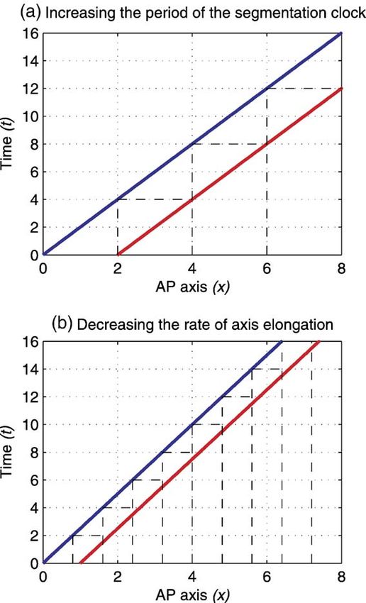
118                                     R.E. Baker et al. / Developmental Biology 293 (2006) 116–126

Reaction-Diffusion model                                                We then extend our model to include the effects of local
   Meinhardt's Reaction-Diffusion model assumes that cells              application of FGF8, presenting both a schematic view of the
can be in one of two possible states, a or p, which correspond          resulting anomalies and numerical simulation of the accompa-
to the anterior and posterior phenotypes of the somite. The a           nying mathematical model. Finally, we present some experi-
and p states are such that they locally exclude each other but          mentally testable predictions that may be used to verify the
stimulate each other over a long range. Cells switch from one           validity of our model and discuss the need for estimation of the
state to another until they reach a stable state. In this way, a        parameter values involved in the model.
pattern of stable apap… stripes is formed (Meinhardt, 1986),
corresponding to the observed pattern of division of somites            A new Clock and Wavefront model for somite formation
into anterior and posterior halves: each ap segment constitutes
a somite.                                                                  The Clock and Wavefront model proposed by Pourquié and
   The wavefront of FGF8 provides the positional information            co-workers (Dubrulle and Pourquié, 2002; Dubrulle et al., 2001;
gradient needed to generate the pattern, and further evidence for       Pourquié, 2004a) hypothesises that there is some interaction
Meinhardt's model comes from the confined expression of c-              between the wavefront of FGF8 and the segmentation clock in
hairy-1 to the posterior half of the somite (Dale and Pourquié,         the PSM that acts to gate cells into potential somites. For a cell
1997). In fact, the Reaction-Diffusion model is the only one to         at a particular point, they assume that competence to segment
address anterior/posterior somite subdivision. However, in its          will only be achieved once FGF8 signalling has decreased
present form, the Reaction-Diffusion model cannot explain the           below a certain threshold. The threshold level of FGF8 is the
effects of local FGF8 application.                                      level expressed at the determination front (Dubrulle and
                                                                        Pourquié, 2002).
Clock and Wavefront model                                                  To develop a mathematical formulation of Pourquié's Clock
   The Clock and Wavefront model was first proposed by                  and Wavefront model, we constructed a system of equations in
Cooke and Zeeman (1976). The model postulates the existence             which the segmentation clock controls when the boundaries of
of a longitudinal positional information gradient down the AP           the somites will form and the determination front controls where
axis of vertebrate embryos, which interacts with a smooth               they form. This is in agreement with Dubrulle and co-workers'
cellular oscillator (the clock), to set the time in each cell at        observations (Dubrulle et al., 2001; Tabin and Johnson, 2001).
which it will undergo a catastrophe. By catastrophe, they mean          In addition, we introduce the following assumptions:
a rapid change of state, which could possibly be the rapid
change in locomotory and adhesive behaviour of cells when               1. Once cells have reached the determination front, they
they form somites.                                                         become competent to segment by gaining the ability to
   The wavefront of FGF8 provides a likely candidate for the               respond to a chemical signal and produce a somitic factor.
longitudinal positional information gradient, and there is wide         2. At a certain time ts later, they become able to signal. ts is
evidence for the segmentation clock. However, the model has                equal to the period of the segmentation clock, which is
not been formulated mathematically. In this article, we employ a           coincident with the period of the cycling genes.
signalling process similar to that developed for the Cell Cycle         3. Once a cell has reached the determination front, it will
model as a rigorous framework for the Clock and Wavefront                  change its response to FGF8; essentially, we will assume that
model.                                                                     it becomes refractory to FGF8 signalling.

Aims and outline                                                            This model is based on the signalling model presented by
                                                                        Maini and co-workers (Collier et al., 2000; Schnell et al., 2002;
   The specific version of the Clock and Wavefront model                McInerney et al., 2004): at a certain time, a small fraction of
considered in this article is that proposed by Pourquié and co-         cells at the anterior-most end of the PSM will gain the ability to
workers (Dubrulle and Pourquié, 2002; Pourquié, 2004a). Our             signal, that is, they would have reached the determination front
goal is to investigate the consequences of the interaction              at a time ts previously. These pioneer cells will produce and emit
between the clock and the wavefront in response to experi-              a signal which will diffuse along the PSM. Any cell which has
mental perturbations. We do not aim to model specifically the           reached the determination front, and therefore becomes
mechanisms underlying the clock nor the wavefront. However,             competent to segment, would respond to such a signal by
we note that the FGF8 signalling gradient along the PSM                 increasing its adhesion to neighbouring cells which are
provides a biological basis for a wavefront and the periodic,           responding in a similar manner, thereby forming a potential
dynamic expression of genes such as c-hairy-1 and l-fng                 somite. At this point, a cell has been specified as somitic, and it
provides evidence for the presence of a segmentation clock.             will go on to segment and form part of a somite during
   In the following section, we detail a “word” model which is          subsequent oscillations of the segmentation clock. The process
accompanied by a mathematical formulation (see Appendix A)              begins again once the new pioneer cells at the anterior edge of
based on the previous model of Maini and co-workers (Collier            the PSM become competent to signal. Emission of the signal is
et al., 2000; Schnell et al., 2002; McInerney et al., 2004). We         transient due to negative feedback on signal production by the
demonstrate that this new model can produce a coherent pattern          cells which react to the signal. This feedback loop results in
of somites which are regulated both temporally and spatially.           periodic pulses in the signal and hence the specification of
R.E. Baker et al. / Developmental Biology 293 (2006) 116–126                                        119

somites at regular time intervals. Fig. 2 illustrates the vertebrate                  along the PSM and undergoes linear decay (see Baker et al. (in
body plan during somite formation as envisaged by the new                             press) for more details). The result is the formation of an FGF8
Clock and Wavefront model. The interaction of the segmenta-                           gradient similar to that shown in Fig. 3(a). The FGF8 gradient
tion clock with the determination front is clearly shown.                             moves in a posterior direction along the PSM and confers the
    It should be noted at this point that the clock used in our                       ability upon cells to produce a somitic factor; at time ts later, they
model does not have an immediate correspondence with outputs                          gain the ability to signal. Somitic factor production is activated
of the segmentation clock observed experimentally by Pourquié                         in response to a pulse in the signal emitted from the pioneer cells
and others (Dale et al., 2001; McGrew et al., 1998; Palmeirim et                      at the anterior end of the PSM. Rapid inhibition of signal
al., 1997; Saga and Takeda, 2001). We do not doubt this large                         production by the somitic factor ensures that peaks in signal
body of evidence regarding gene oscillation in the PSM, and we                        concentration are transient and produced at regular intervals (see
explore this point more thoroughly in the Discussion.                                 McInerney et al., 2004 for further details).
    The mathematical model constructed from Pourquié's                                    We can anticipate the pattern of somites formed using our
descriptive Clock and Wavefront model consists of a coupled                           model by considering the progress of the points Pu and Pv which
system of three non-linear partial differential equations. The                        measure the times at which cells become competent to produce
state variables which the system describes will be a somitic                          somitic factor and signalling molecule (respectively). In a
factor which is integral in determining the fate of cells (a cell                     control case, the determination front remains at a constant axial
will only go on to form part of a somite once it has produced a                       position relative to the PSM and hence moves down the AP axis
high enough level of somitic factor), a diffusive signalling                          at a constant speed. Cells gain the ability to produce signalling
molecule which is produced by the pioneer cells at the anterior-                      molecule a time ts after they become competent to produce
most end of the PSM and finally a generic FGF8 molecule which                         somitic factor. Therefore, the number of cells with the ability to
is able to confer the ability of cells to produce somitic factor and                  segment and the number of cells able to produce signalling
signal (according to their level of expression of FGF8). The                          molecule increase at a constant rate. Fig. 4 illustrates the
complete system of equations is outlined in Appendix A.                               progress of Pu and Pv along the AP axis and also the expected
    We know that whilst Hensen's node is regressing along the                         pattern of somites formed. In this example, at t = 0, cells at x = 1
AP axis, cells are left behind and incorporated into the PSM with                     become the next set of pioneer cells and also become competent
a certain level of fgf8 which decays over time. FGF8 is translated                    to produce somitic factor. At time t = 2, these pioneer cells
by these cells and this creates the signalling gradient. We choose                    become competent to produce the diffusive signalling molecule,
to model this complex phenomenon by assuming that FGF8 is                             and they send out a signal. All cells between x = 1 and x = 2
produced only in the tail and that it diffuses out from the tail                      have now reached the determination front and are able to
                                                                                      respond to this signal by producing somitic factor. These cells
                                                                                      between x = 1 and x = 2 produce somitic factor in a coherent
                                                                                      manner, thereby committing themselves to form a somite
                                                                                      together. It is encouraging to note from Fig. 4 that somites are
                                                                                      regular in size and form at regular intervals.
                                                                                          We also solved our mathematical formulation of the model
                                                                                      (see Appendix A) numerically using the NAG library routine
                                                                                      D03PCF (see Appendix B for more details). Fig. 5 shows the
                                                                                      results of the numerical computation: the top panel shows the
                                                                                      dynamics of the somitic factor, the middle panel shows the
                                                                                      dynamics of the signalling molecule and the bottom panel
                                                                                      shows the dynamics of FGF8. We see that the region of high
                                                                                      FGF8 expression moves in a posterior direction along the AP
                                                                                      axis with constant speed. A sequence of successive signals,
                                                                                      moving in a posterior direction, produces a series of coherent
                                                                                      rises in the level of somitic factor which then enables cells to
                                                                                      progress to form discrete somites. We note, once again, that our
                                                                                      model predicts a spatially uniform series of somites forming at
Fig. 2. Diagrammatic representation of the vertebrate body plan during somite         regular time intervals.
formation within Pourquié's Clock and Wavefront model. In the top part of the
diagram, the FGF8 wavefront is illustrated together with the position of the
determination front. The middle section of the diagram shows the AP axis of the
                                                                                      Local application of FGF8
embryo with the somites (red blocks), determined region and its pre-pattern
(yellow blocks) and the undetermined PSM (yellow band) clearly marked. The               Earlier in this work, we detailed the experiments of Dubrulle
bottom part of the diagram shows the segmentation clock with the time t at            and co-workers regarding local perturbation of FGF8 in the
which a cell reaches the determination front and the time ts later at which it        PSM (Dubrulle et al., 2001). A source of FGF8 implanted in the
becomes competent to signal. The hollow yellow block marks the position of the
next somite to be specified: the posterior boundary is fixed by the position of the   PSM caused somite anomalies as a result of a disturbance in the
determination front at the time at which pioneer cells at the anterior boundary       progression of the determination front. We incorporate this into
produce a signal.                                                                     our model by assuming that FGF8 is not only produced in the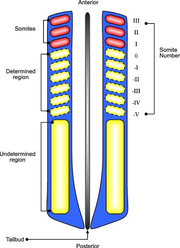
120 R.E. Baker et al. / Developmental Biology 293 (2006) 116–126
Fig. 3. Numerical solution for the FGF8 profile along the AP axis. In each graph, the solution is plotted for t = 4, 8, 12, 16, 20, 24, 28, 32, 36 and 40 (indicated by
arrows) and the hypothetical level of FGF8 marking the determination front is indicated by the dashed line. (a) In a control embryo, the position of the determination
front moves with constant speed down the AP axis. (b) A bead soaked in FGF8 is implanted at x = 10.0 resulting in perturbation of the determination front: in a region
ahead of the bead, progression of the determination front slows, whilst behind the bead the rate of progression of the determination front increases. The anterior end of
the PSM lies on the left-hand side (LHS) of the figure and the posterior end on the right-hand side (RHS). Parameters are as follows: η = 1.0, Dw = 50, ξ = 0.5, xb = 0
and ϕ = 5.0.
tail region of the embryo, but also via a small source of FGF8 progression slows ahead of the bead whilst it increases behind
implanted at a constant axial level (see Appendix C for details the bead.
of the extended mathematical model). Solving numerically the In Fig. 6, we use the same method as before to plot the
new equations for the FGF8 signalling dynamics results in the patterns of somites that could arise as a result of local application
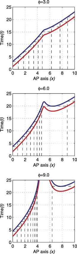
FGF8 profile shown in Fig. 3(b). The dashed line indicates the of FGF8. The top panel shows the results when a weak source of
progression of the determination front, and we see that its FGF8 (ϕ = 3.0) is implanted alongside the PSM: the lines
representing Pu and Pv deviate slightly from their control paths
and the result is a series of slightly smaller somites forming
anterior to the bead where the progression of the determination
front is slowed and a large somite posterior to the bead where the
progression of the determination front has increased. We note
that somite formation falls back in line with the control case
within one or two somites distance posterior to the bead.
The middle panel shows the results of our model when a
slightly stronger source of FGF8 (ϕ = 6.0) is implanted
alongside the PSM: in this example, the anomalies produced are
more marked, and we see a sequence of 6–7 small somites
forming anterior to the bead and a very large somite forming
posterior to the bead. Once again, somite formation falls back
into register both spatially and temporally with the control
embryo pictured in Fig. 4.
Finally, the bottom panel of Fig. 6 shows the results of local
application of a very strong source of FGF8 (ϕ = 9.0). This could
correspond to the source being implanted inside the PSM rather
than alongside it. This case differs from the others shown in this
figure as it results in a region of the PSM that will never segment:
Fig. 4. Illustration of the manner in which somites are formed in the new the level of FGF8 produced in a region surrounding the bead is
Clock and Wavefront model. In the top diagram, the positions of successive
high enough to ensure that cells in this region will never have
somites are found by tracing the lines as shown. The diagonal lines indicate
the positions of the points Pu (red) and Pv (blue). Pioneer cells, at x = 0, 1, sufficiently low levels of FGF8 to reach the determination front
2,…, send out signals, at times t = 0, 2, 4,… respectively. Cells which have and hence never be able to produce somitic factor. We see a
reached the determination front are able to produce somitic factor (those sequence of small somites in a region anterior to the bead
between x = 0 and x = 1, between x = 1 and x = 2, etc.) and they do so in a followed by a region where the cells remain unsegmented
coherent manner, thereby forming a somite together. The bottom diagram
followed by a large somite posterior to the bead and then normal
illustrates the relative somite sizes. The reciprocal of the gradient of the lines
representing Pu and Pv is a measure of the speed at which somites are formed segmentation.
and the period of the clock is ts = 2.0. The anterior end of the PSM lies on It is encouraging to note that all three of the cases pictured
the LHS of the figure and the posterior end on the RHS. in Fig. 6 have been observed experimentally (Dubrulle et al.,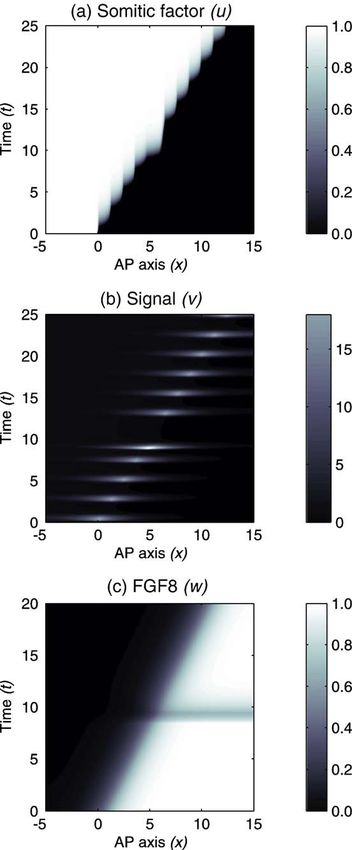
R.E. Baker et al. / Developmental Biology 293 (2006) 116–126                                                       121

                                                                                     formation: perturbing the speed at which the determination front
                                                                                     progresses along the AP axis and perturbing the period of the
                                                                                     segmentation clock. Using our model, we would expect to find
                                                                                     results in vivo similar to those seen in Fig. 8.
                                                                                        Increasing the period of the segmentation clock (whilst
                                                                                     keeping regression of the determination front constant) would
                                                                                     result in increased somite size: when the pioneer cells at the

Fig. 5. Numerical solution of the new mathematical formulation of the Clock
and Wavefront model for somite formation showing the spatio-temporal
dynamics of the somitic factor (a), the signalling molecule (b) and FGF8 (c). The
anterior end of the PSM lies on the LHS of the figure and the posterior end on the
RHS. Parameters are as follows: μ = 10−4, γ = 10−3, κ = 10, ε = 10−3, η = 1.0,
Dv = 50, Dw = 20, xn = 0.0, and cn = 0.5.

2001). Fig. 7 shows a typical numerical simulation of the
mathematical model corresponding to the middle panel of Fig.
                                                                                     Fig. 6. The progress of Pu and Pv as ϕ, the strength of the bead source, is varied.
6. Note the extra large somite.                                                      The red line depicts the progress of Pu and therefore the time at which cells reach
                                                                                     the determination front and become able to produce somitic factor. The blue line
Experimental predictions                                                             depicts the progress of Pv and shows the time at which cells become able to send
                                                                                     out a signal. The boundaries of the presumptive somites are marked by the
   In order to test the validity of Pourquié's Clock and                             dashed lines. As previously, the positions of the somite boundaries are found by
                                                                                     tracing between the two lines. The anterior end of the PSM lies on the LHS of
Wavefront model, it is necessary to make some experimentally                         the figure and the posterior end on the RHS. Parameters are as follows: η = 1.0,
testable predictions. First, we investigate two of the more                          Dw = 10, xn = 0.0, cn = 0.5, xb = 5.0, ξ = 0.2, ts = 2.0 and F = 0.5. See Baker et al.
(mathematically) simple ways in which to perturb somite                              (in press) for more details.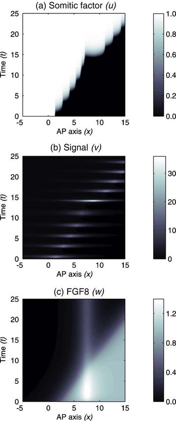
122                                                 R.E. Baker et al. / Developmental Biology 293 (2006) 116–126

                                                                                      be less cells gaining the ability to segment and hence smaller
                                                                                      somites. This is depicted in Fig. 8(b).
                                                                                          The third prediction we make is regarding inhibition of FGF8
                                                                                      in the PSM. There have been some preliminary experiments to
                                                                                      investigate the effect of inhibiting the FGF8 signalling gradient
                                                                                      in the PSM (Dubrulle et al., 2001). Upon treatment with
                                                                                      SU5402, a drug known to specifically block the kinase activity
                                                                                      of FGF8 receptors, embryos formed with a large pair of somites
                                                                                      at the level of somite -IV at the time of application of SU5402.
                                                                                      We can explain the formation of this anomalous somite by
                                                                                      reasoning that the SU5402 treatment decreases the level of FGF8
                                                                                      signalling throughout the PSM, resulting in a posterior shift of
                                                                                      the determination front and an anomalous somite. SU5402 is
                                                                                      rapidly degraded, and it is expected that FGF8 signalling is only
                                                                                      affected during a time frame approximately equal to the time
                                                                                      taken to form one somite (Pourquie, 2004b). Degradation of
                                                                                      SU5402 would result in an increase in FGF8 signalling activity,
                                                                                      back to the original undisturbed level. We model this

Fig. 7. Numerical solution of the new Clock and Wavefront model for somite
formation showing the spatio-temporal dynamics of the somitic factor (a), the
signalling molecule (b) and FGF8 (c). With a source of FGF8 implanted in the
PSM, the somite anomalies are obvious. The anterior end of the PSM lies on the
LHS of the figure and the posterior end on the RHS. Parameters are as follows:
μ = 10−4, γ = 10−3, κ = 10, ε = 10−3, η = 1.0, ϕ = 1.5, Dv = 50, Dw = 20, xn = 0.0,
cn = 0.5, xb = 50 and ξ = 0.5.

anterior end of the PSM become competent to produce a
                                                                                      Fig. 8. (a) Increasing the period of the segmentation clock (ts = 4.0) results in
signal, more cells would have reached the determination front                         larger somites. (b) Decreasing the rate of determination front progression (whilst
(consider Fig. 2) and become able to segment. This is shown                           keeping the period of the segmentation clock constant, ts = 2.0) results in smaller
in Fig. 8(a).                                                                         somites. The red line depicts the progress of Pu and therefore the time at which
   Decreasing the rate at which the determination front is                            cells reach the determination front and become able to produce somitic factor.
regressing (whilst keeping the period of the segmentation clock                       The blue line depicts the progress of Pv and shows the time at which cells
                                                                                      become able to send out a signal. The boundaries of the presumptive somites are
constant) would result in the determination front moving a                            marked by the dashed lines. As previously, the positions of the somite
shorter distance during one oscillation of the segmentation                           boundaries are found by tracing between the two lines. The anterior end of the
clock than it would in the control case. The result of this would                     PSM lies on the LHS of the figure and the posterior end on the RHS.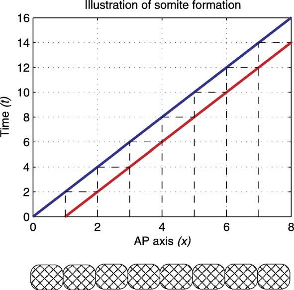
R.E. Baker et al. / Developmental Biology 293 (2006) 116–126                                                      123

mathematically by introducing a sink term into our equation for
FGF8 which is active for a certain period and proportional to the
amount of FGF8 present in the PSM.
    We can use similar methods to those used throughout the
rest of this paper to estimate the anomalies formed when
SU5402 is used to inhibit FGF8 signalling. Fig. 9(a) shows the
anomalies produced when FGF8 signalling is inhibited for a
brief period: a large somite forms at t = 6.0 and normal
segmentation is seen thereafter. Fig. 9(b) shows the anomalies
produced when FGF8 signalling is inhibited for a longer
period: a smaller somite forms posterior to the larger somite.
We note that, should FGF8 be inhibited for an even longer
period, our model would predict the formation of a large
somite, followed by a series of normal somites, followed by a
small somite and then normal segmentation.
    The results generated by numerically solving the mathemat-
ical model are shown in Fig. 10. SU5402 is injected at time
t = 8.5, and we assume that the effects last until time t = 9.5. We

                                                                                     Fig. 10. Numerical solution of the new mathematical formulation of the Clock
                                                                                     and Wavefront model for somite formation showing the spatio-temporal
                                                                                     dynamics of the somitic factor (a), the signalling molecule (b) and FGF8, (c).
                                                                                     With inhibition of FGF8 signalling using SU5402, we see the formation of a
                                                                                     large somite at the level of the determination front at the time the drug is applied
                                                                                     (t = 8.5 until t = 9.5). The anterior end of the PSM lies on the LHS of the figure
                                                                                     and the posterior end on the RHS. Parameters are as follows: μ = 10−4, γ = 10−3,
                                                                                     κ = 10, ε = 10−3, η = 1.0, Dv = 50, Dw = 20, xn = 0.0, cn = 0.5, xb = 5.0 and
Fig. 9. Inhibition of FGF8 signalling results in somite anomalies. (a) Case in       ξ = 0.5.
which SU5402 is assumed to act for a brief period (less than the period of the
segmentation clock). (b) Case in which FGF8 is assumed to be inhibited for a
longer period. The red line depicts the progress of Pu and therefore the time at     see the corresponding decrease in FGF8 signalling throughout
which cells reach the determination front and become able to produce somitic         the PSM in Fig. 10(c). The posterior shift in the determination
factor. The blue line depicts the progress of Pv and shows the time at which cells   front as a result of SU5402 application causes the fifth signal
become able to send out a signal. The boundaries of the presumptive somites are      and fifth somite to be generated early, and the fifth somite is
marked by the dashed lines. As previously, the positions of the somite
boundaries are found by tracing between the two lines. The anterior end of the       larger than normal. Rapid degradation of SU5402 results in the
PSM lies on the LHS of the figure and the posterior end on the RHS. In both          determination front re-assuming its normal position and
cases, the period of the clock is ts = 2.0.                                          segmentation patterns returning to normal.
124                                       R.E. Baker et al. / Developmental Biology 293 (2006) 116–126

Parameter estimation                                                         The results of this work clearly show that perturbation of
                                                                          somitogenesis leads to the incorporation of certain cells into
   In order to make quantitative predictions for the somite               differently numbered somites than their control counterparts. It
anomalies produced when FGF8 expression is perturbed, it will             can be seen that such cells will go on to segment within a
be necessary to have accurate estimates of the parameters                 different time step and will therefore experience a different
involved, more specifically, the rate at which FGF8 is able to            number of clock oscillations before segmenting (Tabin and
diffuse along the PSM and its rate of decay in relation to the            Johnson, 2001). Dubrulle et al. (2001) demonstrate that “FGF8
speed of axis elongation and, correspondingly, somite formation.          treatment can increase the number of clock oscillations
   In experiments in which FGF8 is perturbed locally by                   experienced by PSM cells without altering their absolute axial
implantation of a bead alongside the PSM, it is uncertain how             position in tissue. Cells which experience an extra oscillation
much of the diffusing FGF8 is actually able to enter the PSM              become incorporated into a differently numbered somite and
and affect somite formation. Our model does not take this into            exhibit Hox expression indicative of a more posterior fate when
account, but it may be possible to match the anomalies seen               compared with contralateral control cells.” Our model clearly
experimentally under different concentrations of bead source              accounts for this result (compare Figs. 4 and 6).
with the anomalies produced by the mathematical model. In this               Lastly, we have chosen to simplify the clock: removing its
way, it may be possible to estimate the amount of FGF8 entering           complexity by modelling it as a signalling process. We note that
the PSM from the severity of the anomalies produced. Before               the clock present in our model does not correspond to the most
this can happen however, we need accurate estimation of the               commonly observed somitogenesis cycling genes (Dale et al.,
rate at which FGF8 is able to diffuse along the PSM and its rate          2001; McGrew et al., 1998; Palmeirim et al., 1997; Saga and
of decay.                                                                 Takeda, 2001), which have been widely accepted as constituting
                                                                          the segmentation clock. Expression of such genes begins as a
Discussion                                                                wide stripe in the posterior PSM: the stripe travels in an anterior
                                                                          direction, narrowing as it moves, until it comes to rest in the
    In this paper, we have presented a revised version of                 newly forming somite. However, genes such as l-fng and c-
Pourquié's Clock and Wavefront model for somitogenesis with               hairy-1 are readouts from the segmentation clock, and although
a mathematical basis derived from the models by Maini and co-             these genes and their role in somitogenesis have been widely
workers (Collier et al., 2000; Schnell et al., 2002; McInerney et         documented, the mechanisms underlying the segmentation
al., 2004). We combine outputs from the FGF8 wavefront and                clock are still far from being completely understood. There has
the segmentation clock to control the signalling process                  been some progress in modelling aspects of the clock (see for
described by the mathematical model.                                      example Lewis, 2003; Monk, 2003), but these are early models
    We next moved to extend this mathematical model to include            and we do not feel that incorporation of the mechanisms
the effects of local perturbation of FGF8. We represented a               considered by these authors into our model would allow us to
heparin-soaked bead implanted alongside the PSM by a                      shed any further light on the aspects of somitogenesis
constant production term for FGF8, which is confined to a                 considered here. It is for these reasons that we choose to
small region of the AP axis. In a similar manner, we solved the           simplify the clock, removing its enormous complexity by
revised equation for FGF8 expression numerically and                      modelling it as a signalling process, with control of the somite
demonstrated the displacement of the determination front                  pattern held by a combination of this process and the FGF8
from its conventional path.                                               wavefront. It is interesting to note, however, that recent
    In both the control case and the perturbed cases, we were             experiments of Ishikawa et al. (2005) have identified a gene,
able to demonstrate the ability of our model to produce the               nkd1, which could be linked to our signalling molecule, v. nkd1
results seen in vivo; progression of the determination front              oscillates in the PSM with the same period as the other cycling
along the AP axis conferred upon cells the ability to produce a           genes, but it is only expressed in anterior regions of the PSM.
series of signals with the centre of the pulse travelling                    In a sister paper to this (Baker et al., in press), we study the
posteriorly along the AP axis, leaving a series of coherent               mathematical basis for our model in more detail. In particular,
somites in their wake. The control case produced a series of              we explain our reasons for modelling a generic fgf8 factor, and
uniform somites and the perturbed cases a series of abnormal              we detail the simplifying approximations applied to the model
somites with the intensity of the somite anomalies produced               used here that allowed us to predict the sequences of somites
mirroring the severity of the disturbance in the FGF8 profile.            formed under local application of FGF8.
    We also note at this stage that it is likely that the effects of         Future avenues of exploration lie in several areas. Firstly, in
local application of FGF8 via a heparin-soaked bead will wear             trying to rework the model to use a mechanism better related to
off before all the PSM that would otherwise be affected by this           the segmentation clock: the somitic factor could be linked to a
perturbation can be gated into somites (Pourquie, 2004b).                 number of genes, for example, Mesp2, but as yet there is no
Decaying effects of local application of FGF8 could result in             well-established biological basis for the signal. One possible
pronounced “large” somite anomalies as decay of the source                alteration would be to allow the periodic expression of the
would confer the potential to become somitic to very many cells           cycling genes to activate somitic factor production, but only in
at the same time. The effects of a temporally varying local               cells that have reached the determination front and therefore
source of FGF8 is something that remains to be investigated.              become competent to form somites.
R.E. Baker et al. / Developmental Biology 293 (2006) 116–126 125
The FGF8 wavefront provides the second avenue for where μ, γ, κ, ε, η, Dv, Dw, w*, ts, xn and cn are positive
future studies. It has recently been shown that retinoic acid is constants. Production of u, v and w are controlled by the
expressed along the AP axis in a manner opposite to FGF8: respective Heaviside functions
high in the somites (anterior) and low in the posterior part of
the PSM (Diez del Corral et al., 2003). Findings suggest that vu ¼ Hðw* wÞ; ð4Þ
the FGF and retinoid pathways are mutually inhibitory and
vv ¼ Hðt tw ðw*; xÞ ts Þ; ð5Þ
act to control somite formation (Diez del Corral and Storey,
2004). vw ¼ Hðx xn cn tÞ; ð6Þ
Lastly, it should be noted that Wnt3a has been postulated to
play a major role in somitogenesis: in control of the where w* is the level of FGF8 at the determination front, tw
segmentation clock via Notch signalling; and in control of the (w*, x) is the time at which a cell at x reaches the
determination front via FGF8 expression (Aulehla et al., 2003). determination front (i.e. w(x, tw) = w*), ts is the period of the
This finding suggests a link between the segmentation clock and segmentation clock, xn represents the initial position of the
the FGF8 wavefront and deserves further investigation. tail and cn represents the rate at which the AP axis is
extending. The Heaviside function works like a switch: it is
Acknowledgments equal to unity when the bracketed expression is positive, and
zero otherwise.
REB would like to thank EPSRC for a Doctoral Training The dynamics of the spatially homogeneous equations
Award and Wadham College, Oxford for a Senior Scholarship. have been analysed thoroughly in previous work (McInerney
SS has been funded by the Research Training Fellowship et al., 2004). Within a certain parameter regime (which is
programme in Mathematical Biology (Grant No. 069155) of the detailed in the article), the system displays periodic pulses in
Wellcome Trust (London). PKM thanks the Biocomplexity the signalling molecule as a result of non-linear interactions
Institute and the School of Informatics (Indiana University, between the somitic factor u, and the signalling molecule v.
Bloomington) for support and hospitality during a visit in the These periodic pulses in v lead to coherent rises in the level
Spring 2005. of somitic factor and the generation of a regular array of
The authors would also like to express their kind thanks to somites.
Paul Kulesa and Olivier Pourquié for their kind hospitality at the
Stowers Institute and to Olivier Pourquié once more for helpful Appendix B. Numerical solution of the model
comments on the manuscript.
We solved the above system numerically using the NAG
Appendix A. Mathematical formulation of the new Clock library routine D03PCF, which is designed for non-linear
and Wavefront model parabolic (including some elliptic) partial differential equa-
tions (PDEs) in one spatial variable. The routine is based on
Letting u denote the concentration of somitic factor, v the method of lines, using a finite difference approximation to
denote the concentration of the diffusive signalling molecule reduce the system of PDEs to a system of ordinary
and w denote the concentration of FGF8, we choose to differential equations in the time variable. The resulting
model somite formation using the following non-dimensional system is solved using an implementation of the Backward
model: Differentiation Formula method. The independent variables
form a grid: in general, the mesh consisted of 2001 × 2001
Bu ðu þ lvÞ2 points, the output of which was plotted using the Matlab
¼ v u ; ð1Þ function imagesc. The accuracy of the numerical method was
Bt g þ u2 u
f
tested by both varying the mesh resolution and the error
f
Linear decay
Activation by v and regulated activation by u parameter of the routine (which controls integration in the
0 1 time direction).
B C
Bv B vv C B2 v Appendix C. Mathematical formulation of the perturbed
B C
¼ jB v C þ Dv 2 ; ð2Þ Clock and Wavefront model
Bt B eþu C Bx
f
@ Linear decayA
f
f
Inhibition by u Diffusion The modified non-dimensional equations for somite forma-
tion in the presence of a local source of FGF8 are:
Bw
¼ vw gw ð3Þ
Bt
f
Bu ðu þ lvÞ2
f
Production of w in the node Linear decay ¼ v u; ð7Þ
Bt g þ u2 u
B2 w
þ Dw ;
Bx2
Bv vv B2 v
f
¼j v þ Dv 2 ; ð8Þ
Diffusion Bt eþu Bx126 R.E. Baker et al. / Developmental Biology 293 (2006) 116–126
Bw B2 w Gossler, A., Hrabě de Angelis, M., 1998. Somitogenesis. Curr. Top. Dev. Biol.
¼ vw þ /vb gw þ Dw 2 ; ð9Þ
Bt Bx 38, 225–287.
Ishikawa, A., Kitajima, S., Takahashi, Y., Kokubo, H., Kanno, J., Inoue, T.,
Saga, Y., 2005. Mouse Nkd1, a Wnt antagonist, exhibits oscillatory gene
where χu, χv and χw are as in Appendix A and χb = H(ξ − xb + x) expression in the PSM under the control of Notch signalling. Mech. Dev.
H(ξ + xb − x) represents a source of FGF8 from a bead. xb is the 121, 1443–1453.
position of the midpoint of the bead implant and ξ is a measure Lewis, J., 2003. Autoinhibition with transcriptional delay: a simple mechanism
of the width of the bead (χb is non-zero over a region of width for the zebrafish somitogenesis oscillator. Curr. Biol. 13, 1398–1408.
2ξ, centred at xb). McGrew, M.J., Pourquié, O., 1998. Somitogenesis: segmenting a vertebrate.
Curr. Opin. Genet. Dev. 8, 487–493.
McGrew, M.J., Dale, J.K., Fraboulet, S., Pourquié, O., 1998. The lunatic Fringe
References gene is a target of the molecular clock linked to somite segmentation in avian
embryos. Curr. Biol. 8, 979–982.
Aulehla, A., Wehrle, C., Brand-Saberi, B., Kemler, R., Gossler, A., Kanzier, B., McInerney, D., Schnell, S., Baker, R.E., Maini, P.K., 2004. A mathematical
Herrman, B.G., 2003. Wnt3a plays a major role in the segmentation clock formulation for the cell cycle model in somitogenesis: parameter constraints
controlling somitogenesis. Dev. Cell 4, 395–406. and numerical solutions. IMA J. Math. Appl. Med. Biol. 21, 85–113.
Baker, R.E., Schnell, S., Maini, P.K., 2003. Formation of vertebral precursors: Meinhardt, H., 1986. Models of segmentation. In: Bellairs, R., Ede, D.A.,
past models and future predictions. J. theor. Med. 5, 23–35. Lash, J.W. (Eds.), Somites in Developing Embryos. Plenum Press, New
Baker, R.E., Schnell, S., Maini, P.K., in press. A mathematical investigation of a York, pp. 179–189.
Clock and Wavefront model for somitogenesis. J. Math. Biol. (doi:10.1007/ Monk, N.A.M., 2003. Oscillatory expression of Hes1, p53 and NF-κB driven by
S00285-005-0362-2). transcriptional time delays. Curr. Biol. 13, 1409–1413.
Collier, J.R., McInerney, D., Schnell, S., Maini, P.K., Gavaghan, D.J., Houston, Palmeirim, I., Henrique, D., Ish-Horowicz, D., Pourquié, O., 1997. Avian hairy
P., Stern, C.D., 2000. A cell cycle model for somitogenesis: mathematical gene expression identifies a molecular clock linked to vertebrate
formulation and numerical solution. J. Theor. Biol. 207, 305–316. segmentation and somitogenesis. Cell 91, 639–648.
Cooke, J., Zeeman, E.C., 1976. A Clock and Wavefront model for control of the Pourquié, O., 2003. The segmentation clock: converting embryonic time into
number of repeated structures during animal morphogenesis. J. Theor. Biol. spatial pattern. Science 301, 328–330.
58, 455–476. Pourquié, O., 2004a. The chick embryo: a leading model for model in
Dale, K.J., Pourquié, O., 1997. A clock-work somite. BioEssays 22, 83. somitogenesis studies. Mech. Dev. 121, 1069–1079.
Dale, J.K., Maroto, M., Dequeant, M.-L., Malapert, P., McGrew, M., 2001. Pourquié, O., Pers. Comm. 2004b.
Periodic notch inhibition by lunatic fringe underlies the chick segmentation Primmett, D.R.N., Stern, C.D., Keynes, R.J., 1988. Heat shock causes repeated
clock. Nature 421, 275–278. segmental anomalies in the chick embryo. Development 104, 331–339.
Diez del Corral, R., Olivera-Martinez, I., Goriely, A., Gale, E., Maden, M., Primmett, D.R.N., Norris, W.E., Carlson, G.J., Keynes, R.J., Stern, C.J., 1989.
Storey, K., 2003. Opposing FGF and retinoid pathways control ventral Periodic segmental anomalies induced by heat shock in the chick embryo are
neural pattern, neuronal differentiation and segmentation during body axis associated with the cell cycle. Development 105, 119–130.
extension. Neuron 40, 65–79. Saga, Y., Takeda, H., 2001. The making of the somite: molecular events in
Diez del Corral, R., Storey, K., 2004. Opposing FGF and retinoid pathways: a vertebrate segmentation. Nat. Rev., Genet. 2, 835–845.
signalling switch that controls differentiation and patterning onset in the Schnell, S., Maini, P.K., McInerney, D., Gavaghan, D.J., Houston, P., 2002.
extending vertebrate body axis. BioEssays 26, 857–869. Models for pattern formation in somitogenesis: a marriage of cellular and
Dubrulle, J., Pourquié, O., 2002. From head to tail: links between the molecular biology. C.R. Biol. 325, 179–189.
segmentation clock and antero-posterior patterning of the embryo. Curr. Stern, C.D., Fraser, S.E., Keynes, R.J., Primmett, D.R.N., 1988. A cell
Opin. Genet. Dev. 5, 519–523. lineage analysis of segmentation in the chick embryo. Development
Dubrulle, J., Pourquié, O., 2004. fgf8 mRNA decay establishes a gradient that 104S, 231–244.
couples axial elongation to pattering in the vertebrate embryo. Nature 427, Stickney, H.L., Barresi, M.S.J., Devoto, S.H., 2000. Somite development in
419–422. zebrafish. Dev. Dyn. 219, 287–303.
Dubrulle, J., McGrew, M.J., Pourquié, O., 2001. FGF signalling controls somite Stockdale, F.E., Nikovits Jr., W., Christ, B., 2000. Molecular and cellular
boundary position and regulates segmentation clock control of spatiotem- biology of avian somite development. Dev. Dyn. 219, 304–321.
poral Hox gene activation. Cell 106, 219–232. Tabin, C.J., Johnson, R.L., 2001. Clocks and Hox. Nature 412, 780–781.You can also read


