Vitamin C affects G0/G1 cell cycle and autophagy by downregulating of cyclin D1 in gastric carcinoma cells
←
→
Page content transcription
If your browser does not render page correctly, please read the page content below
Bioscience, Biotechnology, and Biochemistry, 2021, Vol. 0, No. 0, 1-9

                                                                         doi: 10.1093/bbb/zbaa040
                                                                         Advance access publication date: 20 January 2021
                                                                         REGULAR PAPER

R E G U L A R PA P E R

                                                                                                                                                 Downloaded from https://academic.oup.com/bbb/advance-article/doi/10.1093/bbb/zbaa040/6104873 by guest on 13 February 2021
Vitamin C affects G0/G1 cell cycle and autophagy by
downregulating of cyclin D1 in gastric carcinoma cells
Chenxia Ren,1,† Cuiling Wu,2,† Changqing Yang                                               ,3,∗ and Changhong Lian4
1
 Central Laboratory, Changzhi Medical College, Changzhi, China; 2 Faculty of Basic Medicine, Changzhi Medical
College, Changzhi, China; 3 Department of Gastroenterology, Heping Hospital Affiliated to Changzhi Medical
College, Changzhi, China; and 4 Department of General Surgery, Heping Hospital Affiliated to Changzhi
Medical College, Changzhi, China
∗
    Correspondence: Changqing Yang, Young@czmc.edu.cn
†
    These authors contributed equally to this work.

ABSTRACT
Vitamin C has re-emerged as a promising anticancer agent. This study attempts to analyze the differential gene
expression of profiles GSE11919 to look for some clues, and the most significant cell cycle pathway caused by vitamin C
was identified by integrated bioinformatics analysis. Inspired by this, we investigated the effect of vitamin C treatment
on gastric carcinoma cells by detection of cell cycle, apoptosis, and autophagy. Vitamin C significantly elevated the
percentage of cells at G0/G1 phase, whereas the percentage of S phase cells was decreased. Meanwhile, vitamin C
treatment resulted in downregulation of cell cycle-related protein Cyclin D1. We deduced that the downregulation of
Cyclin D1 by vitamin C accompanied by significantly increased 5 AMP-activated protein kinase and induced autophagy
in MKN45 cells. These results suggest that vitamin C has the antiproliferation effect on gastric carcinoma cells via the
regulation of cell cycle and autophagy by Cyclin D1.

Graphical Abstract

A schematic diagram of the targets of vitamin C on cells analyzed by integrated bioinformatics and experimental
methods. Vitamin C affects the cell cycle pathway in 2 different types of cells and autophagy through downregulating of
Cyclin D1 and pAMPK in gastric cancer cells.

Keywords: autophagy, cell cycle, cyclin D1, gastric carcinoma cells, vitamin C

Received: 25 August 2020; Accepted: 16 October 2020
© The Author(s) 2021. Published by Oxford University Press on behalf of Japan Society for Bioscience, Biotechnology, and Agrochemistry.
All rights reserved. For permissions, please e-mail: journals.permissions@oup.com

                                                                                                                                          1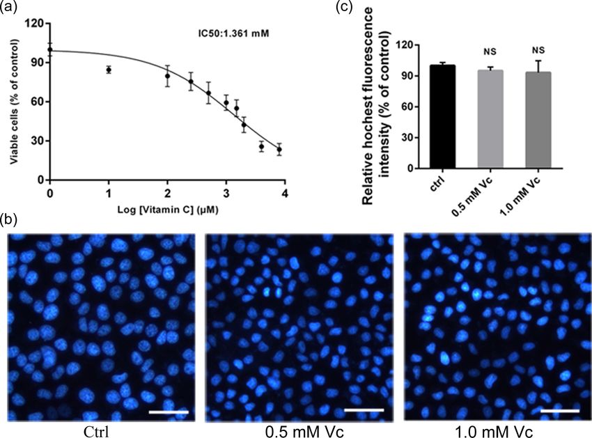
2     Bioscience, Biotechnology, and Biochemistry, 2021, Vol. 0, No. 0

Gastric cancer is predicted to be the third leading cause of                potential differentially expressed genes (DEGs) between the vi-
cancer-related death worldwide, with the mortality rate as high             tamin C treated group and the control group. We set the adj.
as 8.2% (Bray et al. 2018; Van Cutsem et al. 2016). Therefore, signif-      P < .05 and |log FC| >2 as the cut-off criterion. The protein-
icant attention should be paid to the treatment of gastric cancer.          protein interaction (PPI) network was further analyzed with the
Studies of vitamin C on tumor treatment have been continuing                STRING online database ( http://string-db.org) and visualized by
for quite a long time since the 1950s (Cameron and Pauling 1976;            Cytoscape software. Gene Ontology (GO) enrichment of DEGs
Shenoy et al. 2018). The obvious cytotoxicity of vitamin C was ob-          analysis was performed using DAVID (http://david.ncifcrf.gov)
served in cancer cell lines due to extracellular H2 O2 generation           and visualized by EasyChart (http://www.ehbio.com/ImageGP/
via ascorbate radical as the electron donor (Chen et al. 2005). In          index.php/Home/Index/index.html). The enriched Kyoto Ency-
addition, several other studies also confirmed the antitumor ef-            clopedia of Genes and Genomes (KEGG) pathway of DEGs were

                                                                                                                                                  Downloaded from https://academic.oup.com/bbb/advance-article/doi/10.1093/bbb/zbaa040/6104873 by guest on 13 February 2021
fect of vitamin C in subcutaneous tumor-bearing glioblastoma,               analyzed and visualized with the ClueGO tool in Cytoscape. The
pancreatic cancer, ovarian cancer, colon cancer, and breast can-            top 10 pathways with an adjusted P-value < .01 were selected.
cer models (Deubzer et al. 2010; Du et al. 2010; Ullah et al. 2011;
Kim et al. 2012). However, the sensitivity and mechanism of vita-
min C treatment in different tumors were discrepant according               Cell culture and vitamin C treatment
to previous reports (Seo et al. 2015).                                      The human gastric carcinoma cell lineswas obtained from ATCC
    Increasing evidence suggest that an increased intake of vi-             (Manassas, VA, USA). The cells were maintained in RPMI-1640
tamin C may reduce gastric cancer risk, and the total consump-              medium (#11 875 093, Invitrogen, Carlsbad, CA, USA) with 10% fe-
tion of fruit and vitamin C-contributing foods showed a negative            tal bovine serum (FBS, #04-001-1A/B, Biological Industries, Israel)
correlation with gastric cancer. The mean levels of vitamin C in            and 1% penicillin/streptomycin mixture (#PS2004HY, Institute of
plasma and gastric fluid were significantly lower in patients with          Biomedical Engineering, Chinese Academy of Medical Sciences,
gastric disease, such as gastritis, pernicious anemia, peptic ul-           Shanghai, China) at 37 °C in a humidified atmosphere of 5% CO2
cer disease, and gastric cancer (Hoang et al. 2016; Lavecchia et al.        and 95% air.
1994; Botterweck et al. 2000; Goodman et al. 1997). The inverse re-             Vitamin C powder (CAS NO. 795 437, HPLC, purity ≥ 99%) was
lationship between vitamin C and the risk of gastric cancer was             obtained from Sigma–Aldrich. In the cell viability assay, vitamin
so far based on case-control studies and cohort studies, and the            C was added to the cultured gastric carcinoma cells with final
inhibitory effect and related mechanism of vitamin C in gastric             concentrations of 10, 100, 250, 500, 1000, 1500, 2000, 4000, and
cancer cells need to be further studied.                                    8000 μM for 24 h. For other experiments, assays were performed
    Gene chips can be used to quickly detect all the gene ex-               after 24 h in incubation with 0.5 mM and 1 mM vitamin C.
pression information before and after the treatment and dosage,
which is particularly suitable for differentially expressed gene
screening. We searched on the gene expression omnibus (GEO)                 Cell viability assay by cell counting kit-8 assay
database and found the GSE11919 dataset, which is on the in-
                                                                            The cell counting kit-8 (CCK-8) was performed according to
fluence of vitamin C on skin fibroblasts (Duarte et al. 2009). We
                                                                            the manufacturer’s protocols (#CK04, Dojindo Laboratories, Ku-
used GEO2R to filter differentially expressed genes and found
                                                                            mamoto, Japan). The absorbance of each well at 450 nm was
that the altered genes were significantly enriched in cell cycle
                                                                            measured using a microplate reader (Infinite 200 PRO, Tecan,
signal pathway. Thus, it was judged that vitamin C takes on its
                                                                            Männedorf, Switzerland) and the half-maximal inhibitory con-
role through cell cycle. Whether vitamin C affects the tumor
                                                                            centration (IC50) values were calculated using probit analysis of
growth by regulating the cell cycle process? Using gastric can-
                                                                            GraphPad Prism version 7 (GraphPad Software, San Diego, CA,
cer cells as an example, we further confirmed our hypothesis
                                                                            USA).
by observing that vitamin C blocked the cell cycle progression
by synchronization of cells at G0/G1 phase. We further analyzed
the role of cyclin D1 in the tested cell line treated by vitamin            Hoechst staining
C, which is a key factor to promote the G1/S transition in the
cell cycle process. It is generally used to discover the targeting          The procedure for Hoechst staining was carried out using
effect of the antitumor medicine to the tumor cells by detect-              Hoechst33258 (#IH0060, Solarbio, Beijing, China) kits according
ing apoptosis, cell cycle and the burgeoning autophagy (Wiman               to the manufacturer’s instructions. The stained cells were visu-
and Zhivotovsky 2017; Li et al. 2020), thus we designed experi-             alized with a fluorescence microscope under UV excitation. Flu-
ments to analyze autophagy of gastric cancer cells pre-treated              orescence intensity of staining was quantified using the Image J
by vitamin C and found that the effects of vitamin C to cell cycle          software (National Institute of Health, Bethesda, MD, USA).
and autophagy are both mediated by regulation of Cyclin D1 in
gastric cancer.
                                                                            Flow cytometry
                                                                            Flow cytometry (BD FACSCANTO II, Becton-Dickinson, Franklin
                                                                            Lakes, NJ, USA) was used to analyze the cell cycle distributions
Materials and methods                                                       using the Cell Cycle Staining Kit (#550 825, BD Pharmingen, USA)
Bioinformatics analysis of vitamin C related                                according to the manufacturers’ instructions. Cell cycle anal-
                                                                            ysis was performed at a slow flow rate and further analyzed
microarray data
                                                                            by the ModFit software (Verity Software House, Topsham, ME,
The GEO is an open database (http://www.ncbi.nlm.nih.gov/geo)               USA). The percentage of apoptotic cells was also analyzed by
for biological information stockpiling. The GSE11919 was ex-                flow cytometry using an FITC Annexin V Apoptosis Detection Kit
amined by the Affymetrix Human Genome U133 Plus 2.0 Ar-                     (#556 547, BD Pharmingen, USA) according to the manufacturer’s
ray GPL570 platform. The GEO2R online analysis tool (http:                  instructions. The percentage of apoptotic cells was determined
//www.ncbi.nlm.nih.gov/geo/geo2r/) was used to screen the                   by Flowjo software (Tritar Inc., San Carlos, California, USA).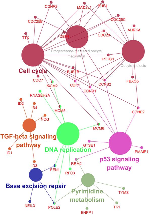
Vitamin C regulates cell cyclle and autophagy through Cyclin D1                     3

Western blotting analysis
Following vitamin C treatment with indicated concentration
for 24 h, cells were lysed in RIPA lysis buffer supplemented
with a proteasome and phosphatase inhibitor (#R0010, So-
larbio, Beijing, China). Comparable amounts of proteins were
separated by 10% SDS-PAGE and transferred to a PVDF mem-
brane (#SLVP07550, Millipore, Billerica, Massachusetts, USA). Af-
ter blocking with non-specific binding by 5% nonfat milk for 1
h at room temperature, the PVDF membranes were incubated
overnight at 4°C with primary antibodies for β-actin, Caspase 3,

                                                                                                                                                 Downloaded from https://academic.oup.com/bbb/advance-article/doi/10.1093/bbb/zbaa040/6104873 by guest on 13 February 2021
Caspase 9, Cyclin D1, Autophagy related 5 (ATG5), sequestosome
1/p62 protein (p62), Beclin 1 (Cell Signaling Technology, Boston,
USA), and Microtubule-associated proteins 1A/1B light chain 3
(LC3) (Abcam, Cambridge, UK). HRP conjugated secondary anti-
bodies (Solarbio, Beijing, China) were applied as secondary an-
tibody for 2 h at room temperature. After ECL development,
the immunoreactive bands were scanned by the chemilumines-
cence detection system (ImageQuant LAS 4000, GE Healthcare).
The relative band density was quantified using ImageJ software.

Real-time PCR
Total RNAs were extracted separately from the control and vita-
min C-treated cells using the Total RNA Extraction Kit according
to the manufacturer’s instructions. Following the reverse tran-
scription, real-time PCRs were further performed using the re-
sulting cDNAs as templates. Real-time reactions were run and
analyzed with ABI StepOne Plus machine. The amount of induc-
tion of mRNA was determined from threshold cycle values nor-
malized for β-actin expression and then normalized to the value
derived from cells of the control group. Primers used for qRT-PCR
were as follows:                                                    Figure 1. The KEGG functional annotation analysis of DEGs in GSE11919.

                                                                    cence intensity of staining was quantified by the ImageJ soft-
p62: ACATGGGGCTTGAGAAAGGG, CGGAGGTGGAAGTACACACG
                                                                    ware.
ATG5: TGTGCTTCGAGATGTGTGGT, AGCAAATAGTATGGTTCTGT
LC3: CCGACCGCTGTAAGGAGGTA, AGGACGGGCAGCTGCTT
Beclin1: AGCTGGATGATGAGCTGAAGAG, GATTGTGCCAAACT-                    Statistical analysis
   GTCCACTG
β-actin:   GATTGCCTCAGGACATTTCTG,  GATTGCTCAGGA-                    All data were presented as means (±standard deviation) of at
   CATTTCTG                                                         least 3 independent experiments. The 1-way ANOVA was ap-
                                                                    plied for comparison among 3 groups following Tukey’s test.
                                                                    For all the analyses, P < .05 was considered significant differ-
Immunofluorescence microscopy                                       ence. Statistical analyses were performed using the GraphPad
                                                                    Prism 7.0.
The cells were grown on glass cover slips and then incubated
with vitamin C for 24 h. After washing with PBS, the cells were
fixed with 4.0% paraformaldehyde in 1× PBS buffer for 30 min
                                                                    Results
at room temperature. The fixed cells were further washed and        Vitamin C has a close relationship
permeabilized by 0.3% Tween-20 in 1× PBS buffer for 10 min. The     with cell cycle pathway
cells were incubated with 1% bovine serum albumin and 1% nor-
mal goat serum in 1× PBS buffer for 3 h and subsequently incu-      In order to verify our hypothesis that vitamin C affects the cell
bated with anti-LC3 antibody overnight at 4 °C, followed by Alexa   signaling pathway, we first analyzed the GSE11919 dataset in the
Fluor 488 goat anti-rabbit IgG as a secondary antibody. The sam-    GEO database of NCBI, which could be used to test our hypoth-
ples were imaged by microscopy using the Cytation 5 Imaging         esis since it is related to differential gene expression after the
Reader. The fluorescence intensity of LC3 was quantified using      skin fibroblasts treated by vitamin C. This gene chip contains
the ImageJ software.                                                3 samples treated with vitamin C and 3 corresponding control
                                                                    samples. A total of 221 genes with |log FC|> 2 and P < .05 were
                                                                    identified by GEO2R tool analysis..
                                                                        As shown in Figure 1, the most significantly enriched
Monodansylcadaverine staining
                                                                    pathways of the DEGs were analyzed by ClueGO in Cy-
Cells were stained with Monodansylcadaverine (MDC) (0.05            toscape. The DEGs were enriched in cell cycle, DNA replica-
mmol/L) for 10 min at 37 °C. After washing with 1× PBS buffer,      tion, p53 signaling pathway, TGF-beta signaling pathway, pyrim-
the cells were observed under fluorescence microscopy. Fluores-     idine metabolism, malaria, base excision repair, homologous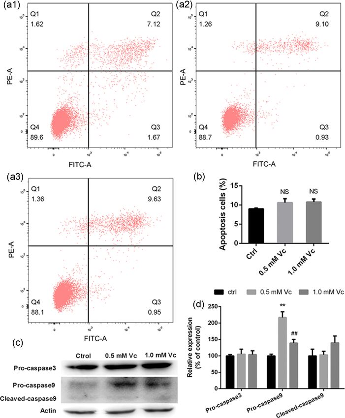
4      Bioscience, Biotechnology, and Biochemistry, 2021, Vol. 0, No. 0

                                                                                                                                                           Downloaded from https://academic.oup.com/bbb/advance-article/doi/10.1093/bbb/zbaa040/6104873 by guest on 13 February 2021
Figure 2. GO enrichment analysis of DEGs in GSE11919. (a) Biological processes. (b) Cellular components. (c): Molecular functions.

recombination, fanconi anemia pathway, oocyte meiosis, and                            experiments in MKN45 cells to uncover the relationship between
progesterone-mediated oocyte maturation.                                              cell cycle and anticancer activity of vitamin C.
    The GO describes function on biological process (BP), cellular
component (CC) and molecular function (MF). Using funrich tool
execute GO function enrichment, we found that DEGs’ GO en-
                                                                                      Vitamin C inhibits the viability of MKN45 cells without
richment analysis of BP was chromosome segregation, positive
                                                                                      affecting apoptosis in certain concentration
regulation of cytokinesis, microtubule-based movement, mitotic
sister chromatid segregation, mitotic cytokinesis, mitotic spin-                      In vitro antitumor capabilities of vitamin C were evaluated us-
dle organization, cell division, and regulation of attachment of                      ing the CCK8 assays. Our findings indicated that the cell viabil-
spindle microtubules to kinetochore (Figure 2a); The CCs of them                      ity decreased in a dose-dependent manner when treated with
were midbody, spindle microtubule, cytoplasm, spindle mid-                            vitamin C. The IC50 of vitamin C in MKN45 cells was 1.361 mM
zone, kinesin complex, kinetochore, nucleus, chromosome pas-                          (Figure 3a). We selected the concentrations of 0.5 mM and 1.0 mM
senger complex, nucleoplasm, chromosome, and centromeric                              for follow-up tests. The typical morphological change character-
region (Figure 2b). The significantly enriched MFs of them were                       istic of apoptosis was not observed after Hoechst 33 258 staining.
ATP binding, microtubule motor activity, ATP-dependent micro-                         All groups showed homogeneous staining (Figure 3b). Vitamin C
tubule motor activity, plus-end-directed, ATPase activity, tubulin                    did not increase the percentage of MKN45 cells with chromatin
binding, chromatin binding, and double-stranded DNA binding                           condensation (Figure 3c).
(Figure 2c). We found that most of the functional enrichment re-                          As shown in Figure 4a and b, flow cytometry assay showed
sults were associated with cell division and there were several                       that compared to the apoptosis proportion of MKN45 cells in
pathways about cancers.                                                               the control group (8.79 ± 0.20%), the apoptosis proportion in the
    In order to further verify our hypothesis that vitamin C may                      0.5 and 1.0 mM vitamin C treated groups were10.03 ± 0.76% and
directly affect cell cycle pathway in cells, we verified our hy-                      10.582 ± 1.01%, respectively. The apoptosis related protein ex-
pothesis in gastric cancer cells. Thus, we did the subsequent                         pression levels of pro-caspase 9 were increased, while the levels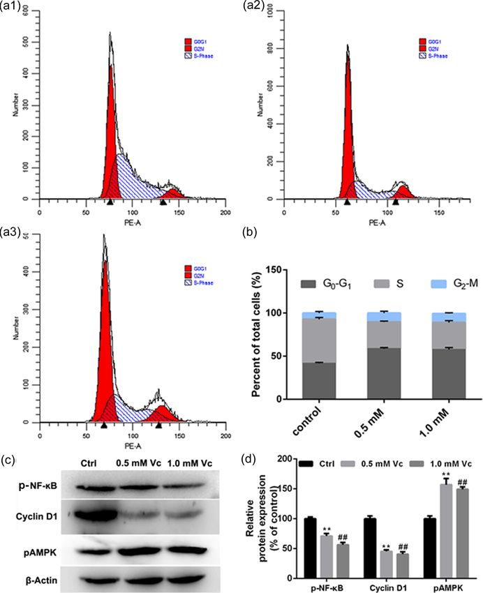
Vitamin C regulates cell cyclle and autophagy through Cyclin D1                            5

                                                                                                                                                                           Downloaded from https://academic.oup.com/bbb/advance-article/doi/10.1093/bbb/zbaa040/6104873 by guest on 13 February 2021
Figure 3. Effects of vitamin C on cell viability in MKN45 cells. (a)The effect of vitamin C on MKN45 cells by CCK-8. (b, c) MKN45 cells were stained with Hoechst 33 258
(Bar = 50 μm).

of pro-caspase 3, activated caspase 3, and activated caspase 9 in                      cycle, and the burgeoning autophagy, thus we designed exper-
cells were decreased (Figure 4c and d).                                                iments to analyze autophagy of gastric cancer cells treated by
                                                                                       vitamin C in addition to the cell cycle pathway.
                                                                                           LC3 is the first identified mammalian protein that local-
Vitamin C causes cell cycle arrest of MKN45 cells                                      izes at the autophagosome membrane and is essential for
                                                                                       autophagy (Li et al. 2020). We imaged the distribution of LC3 by
To explore whether vitamin C mediated the inhibition of MKN45
                                                                                       immunofluorescence, which was considered as the most spe-
cell growth via cell cycle signaling, we analyzed the cell cycle dis-
                                                                                       cific autophagosomal marker. As shown in Figure 6a and c, we
tribution subsequent to the cells treated with indicated concen-
                                                                                       found that the intensities of LC3 were significantly increased in
trations of vitamin C for 24 h with flow cytometry (Figure 5a). As
                                                                                       vitamin C-treated cells. We also checked the autolysosome for-
shown in Figure 5b, the proportion of MKN45 cells in the G0/G1
                                                                                       mation by staining cells with MDC (Mizushima, 2004), and found
phase was respectively increased to 58.71 ± 1.18% (0.5 mM) and
                                                                                       that the MDC fluorescent intensities were significantly increased
57.63 ± 2.33% (1.0 mM), compared with 41.63 ± 1.21% in the con-
                                                                                       in vitamin C-treated cells, as shown in Figure 6b and d. There
trol group. Vitamin C exposure also caused a decrease in S phase
                                                                                       are 2 cellular forms of LC3, LC3-I, which is the precursor, and
cell population from 51.03 ± 2.11% to 30.81 ± 0.21% (0.5 mM) and
                                                                                       LC3-II, which is bound to PE in the autophagosome membrane.
to 31.33 ± 2.27% (1.0 mM) in MKN45 cells, which indicated that
                                                                                       Thus, the LC3 is detected as 2 bands by Western blotting analy-
vitamin C inhibited the cell growth by inducing cell cycle arrest.
                                                                                       sis. We used Western blotting to detect the expression of ATG5,
To support our observations from the cell cycle analyses, we fur-
                                                                                       p62, Beclin1, LC3-I, and LC3-II in MKN45 cells treated with vita-
ther found that there was a significant decrease in the expres-
                                                                                       min C (Figure 6e and f). These results showed that the expres-
sion of Cyclin D1 (Figure 5c), which is a check-point protein of G1
                                                                                       sion levels of ATG5, LC3-I, and LC3-II increased, while the ex-
to S phase. Moreover, the transcription factor NF-ĸB plays an im-
                                                                                       pressions of p62 and Becin1 were decreased. At the transcription
portant role in the regulation of cyclin D1, which was decreased
                                                                                       level, the autophagy-related genes other than the LC3 did not
in vitamin C group (Figure 5d). Thus, insufficient expression of
                                                                                       show dose-dependent responses to vitamin C, indicating that vi-
cyclin D1 caused the cell cycle arrest at G0/G1 phase, which ad-
                                                                                       tamin C affected autophagy mainly by protein level and partially
dressed our hypothesis without bias. As shown in Supplemen-
                                                                                       by the transcription level (Figure 6g). All the results collectively
tary Table 1, the effects of vitamin C were common in other gas-
                                                                                       showed that vitamin C treatment decreased autophagy in the
tric cancer cell lines.
                                                                                       MKN45 cells. Cyclin D1 can restrain autophagy by inhibiting the
                                                                                       activation of pAMPK, which is an activator of autophagy (Brown
Vitamin C increases the autophagy level in MKN45 cells                                 et al. 2012). As shown in Figure 5c and d, the level of pAMPK in-
                                                                                       creased with the decrease of Cyclin D1. Thus, vitamin C affects
It is generally used to discover the targeting effect of the anti-                     the autophagy by Cyclin D1 in gastric cancer cells that may have
tumor medicine to the tumor cells by detecting apoptosis, cell                         a protective effect on tumor cells.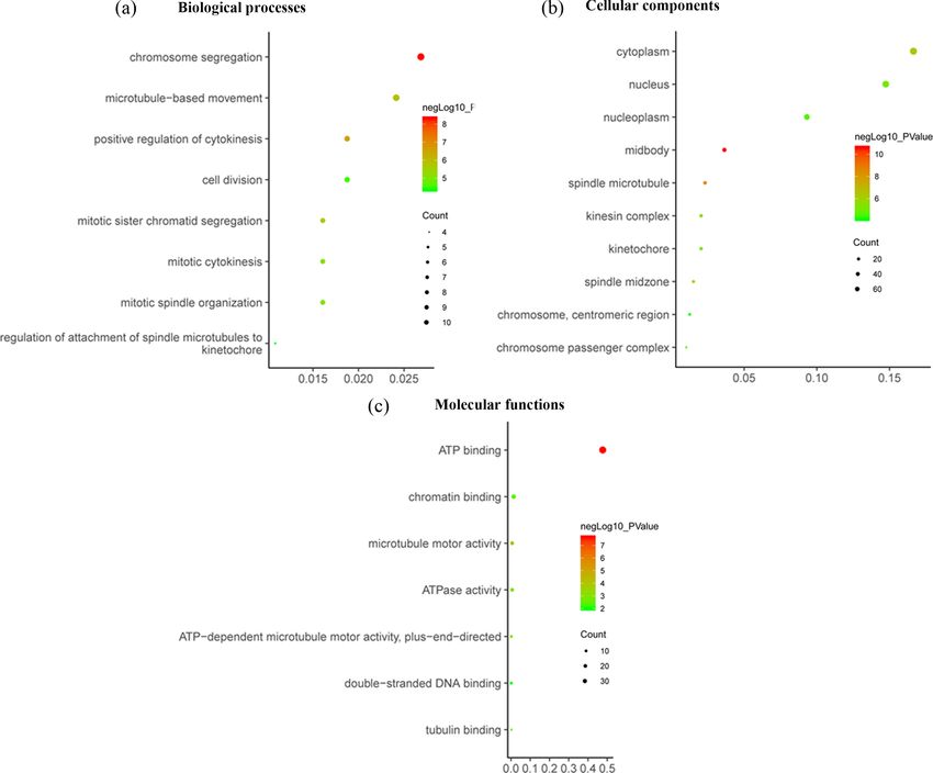
6      Bioscience, Biotechnology, and Biochemistry, 2021, Vol. 0, No. 0

                                                                                                                                                               Downloaded from https://academic.oup.com/bbb/advance-article/doi/10.1093/bbb/zbaa040/6104873 by guest on 13 February 2021
Figure 4. Effects of vitamin C on apoptosis in MKN45 cells. (a, b) apoptosis was determined by flow cytometry (a1: Control, a2: 0.5 mM vitamin C, a3: 1.0 mM
vitamin C). (c, d) Expression of caspase proteins. (** P < .01, 0.5 mM versus control; ## P < .01, 1.0 mM versus control).

Discussion                                                                      cells. Our results indicated that treatment of MKN45 cells with
                                                                                0.25–10 mM vitamin C for 24 h led to an obvious dose-dependent
The mechanism of vitamin C promoting a wide range of benefi-                    decrease of cell viability. Although cases of apparent effects of
cial effects remains exclusive. The DEGs between drug-treated                   malignancies to intravenous high dose vitamin C therapy
group and control group based bioinformatics analysis has                       without serious side effects in patients have been reported
played a critical role in elucidating drug mode of action. In the               (Padayatty et al. 2006), there was no sufficient mechanism
present study, we first analyzed a gene expression profile chip                  study.
on vitamin C-treated human cells, and further found that the                        Cell cycle therapy targeting cyclins has broad prospects (Otto
DEGs were related to the function of the nucleus and mitotic cy-                and Sicinski, 2017, O’Leary et al. 2016). The experimental re-
tokinesis through the enrichment of GO function. We also found                  sults revealed vitamin C attest the cell cycle in the G0/G1 phase
that the genes related to cell cycle signal pathway were dramat-                through reducing the expression of Cyclin D1 in gastric cancer
ically altered after vitamin C treatment, which provided a basis                cells. The cyclin D1 forms active complexes with either CDK4 or
for further investigate.                                                        CDK6, which in turn phosphorylates the retinoblastoma protein
    Generally, the physiological concentration of vitamin C is un-              and drives G1 to S phase progression (Otto and Sicinski, 2017).
der 0.1 mM, while the concentration of vitamin C to kill can-                   Sahar et al. reported that vitamin C alone cannot affect the ac-
cer cells in vitro is about 1–10 mM depending on different cell                 tivity of Cyclin B unless combined with 5-azacytidin (Sajadian et
lines (Park, 2013). Intravenous administration has been widely                  al. 2016). We found that vitamin C affects Cyclin D1. The cell cy-
used clinically to achieve a high plasma concentration of vita-                 cle regulator cyclin D1 is an NF-κB target gene (Lemieszek et al.
min C (up to approximately 14 mM) to cause toxicity to cancer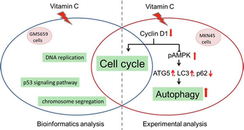
Vitamin C regulates cell cyclle and autophagy through Cyclin D1                  7

                                                                                                                                                          Downloaded from https://academic.oup.com/bbb/advance-article/doi/10.1093/bbb/zbaa040/6104873 by guest on 13 February 2021
Figure 5. Effects of vitamin C on the cell cycle of MKN45 cells. a, b: Cell cycle was determined by flow cytometry (a1: Control, a2: 0.5 mM
vitamin C, a3: 1.0 mM vitamin C). c, d. NF-ĸB, cyclin D1 and pAMPK expression in MKN45 cells. (** P < .01, versus control; ## P < .01, versus control).

2019), and NF-κB also accounts for the inhibitory effects of vita-            markers of autophagy, and p62 is incorporated in the completed
min C on gastric cancer cells in this study.                                  autophagosomes and then degraded in autolysosomes (Levy
    It was showed that the Cyclin D1 phosphorylated LKB1 and                  et al. 2017; Ichimura and Komatsu 2010). The present study
consequently reduced AMPK that activates autophagy in cancer                  demonstrated that the altered Cyclin D1 and pAMPK by vita-
cells (Casimiro et al. 2017). Another study reported that Cyclin              min C activated autophagy via the upregulation of ATG5 and LC3,
D1 was degraded by autophagy in HCC, which caused low cell                    increase of the conversion of LC3-I and LC3-II, and decrease of
proliferation and rate of cell cycle arrest (Wu and Lan 2018). Our            p62. On one hand, reduced cyclinD1 blocks the cell cycle process;
study was in accordance with the former process that the reduc-               on the other hand, reduced cyclinD1 paradoxically enhances au-
tion of Cyclin D1 showed dose dependent responses to vitamin                  tophagy.
C. Meanwhile, the expression of pAMPK elevated when Cyclin                       In addition, Beclin1, as a homologous gene of yeast gene
D1 decreased, indicating that vitamin C restrained autophagy by               Atg6/Vps30, is an important participant in autophagosome for-
cyclin D1 to modulate the activation of AMPK.                                 mation (Fogel et al. 2013). Beclin-1 can also regulate occurrence
    Autophagy was a double-edged sword, which either inhib-                   and progress of tumor through non-autophagy-dependent path-
ited or promoted cancer cell proliferation or tumorigenesis in                ways, such as the growth factor receptor signaling pathway, cell
model systems (Levy et al. 2017). It depended on different stage              mitosis, DNA damage repair and cell apoptosis (Boutouja et al.
of certain cancer or different cancer types. LC3 and ATG5 pro-                2017). It is noteworthy that Beclin1 protein expression level was
teins, especially LC3-II, are recognized as the most common                   decreased in this study. Beclin1 may serve as a key factor for
8 Bioscience, Biotechnology, and Biochemistry, 2021, Vol. 0, No. 0
Downloaded from https://academic.oup.com/bbb/advance-article/doi/10.1093/bbb/zbaa040/6104873 by guest on 13 February 2021
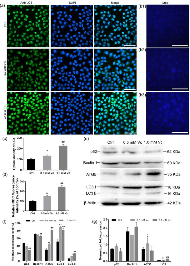
Figure 6. Effects of vitamin C on the autophagy in MKN45 cells. Autophagy was determined with immunofluorescence staining for LC3 puncta (a, c) and MDC staining
quantify (b, d) (b1: control, b2: 0.5 mM vitamin C, b3: 1.0 mM vitamin C). (e, f) Expression of ATG5, p62, Beclin1, LC3I and LCII, (g) real-time PCR (Bar = 50 μm; * P < .05,
versus control; ** P < .01, vs control; ## P < .01, versus control).Vitamin C regulates cell cyclle and autophagy through Cyclin D1 9
vitamin C to regulate cell cycle and autophagy, which needs to Du JA, Martin SM, Levine M et al. Mechanisms of ascorbate-
be further investigated. induced cytotoxicity in pancreatic cancer. . Clin Cancer Res
To sum up, anticancer mechanism of vitamin C to arrest cell 2010;16(2):509-20.
cycle and activate autophagy is related to Cyclin D1 and pAMPK. Duarte TL, Cooke MS, Jones GDD. Gene expression profiling re-
This research provides a theoretical basis for the relationship veals new protective roles for vitamin C in human skin cells.
between gastric cancer and vitamin C treatment. The limitation Free Radic Biol Med 2009;46(1):78-87.
of this study is that the direct target genes and signaling path- Fogel AI, Dlouhy BJ, Wang CX et al. role of membrane association
ways are not excavated. Importantly, further study with vitamin and atg14-dependent phosphorylation in Beclin-1-mediated
C treatment will give more insight into the anticancer effects autophagy. Mol Cell Biol 2013;33(18):3675-88.
both in vivo and in vitro. Goodman KJ, Correa P, Aux HJT et al. Nutritional factors and Heli-
Downloaded from https://academic.oup.com/bbb/advance-article/doi/10.1093/bbb/zbaa040/6104873 by guest on 13 February 2021
cobacter pylori infection in Colombian children. J Pediatr Gastr
Nutr 1997;25(5):507-15.
Supplementary material
Hoang BV, Lee J, Choi IJ et al. Effect of dietary vitamin C on gas-
Supplementary material is available at Bioscience, Biotechnology, tric cancer risk in the Korean population. World J Gastroenterol
and Biochemistry online. 2016;22(27):6257-67.
Ichimura Y, Komatsu M. Selective degradation of p62 by au-
tophagy. Semin Immunopathol 2010;32(4):431-6.
Author contribution
Kim J, Lee SD, Chang B et al. Enhanced antitumor activity
R.C.-X., W.C.-L., and Y.C.-Q. designed the study, analyzed data, of vitamin C via p53 in Cancer cells. Free Radic Biol Med
and prepared the manuscript. R.C.-X. and W.C.-L. performed the 2012;53(8):1607-15.
experiments. All authors approved the final manuscript. Lavecchia C, Ferraroni M, Davanzo B et al. Selected mcronutri-
ent intake and the risk of gastric-cancer. Cancer Epidem Biomar
1994;3(5):393-8.
Funding Lemieszek MK, Nunes FM, Marques G et al. Cantharellus cibar-
This work was supported by the Education Department of ius branched mannans inhibits colon cancer cells growth by
Shanxi Province under Grant Shanxi “1331 Project” Key Innova- interfering with signals transduction in NF -kB pathway. Int J
tive Research Team and Shanxi Province Science Foundation for Biol Macromol 2019;134:770-780.
Youths under Grant 201801D221284. Levy JMM, Towers CG, Thorburn A. Targeting autophagy in can-
cer. Nat Rev Cancer 2017;17(9):528-42.
Li X, He S, Ma B. Autophagy and autophagy-related proteins in
Disclosure statement cancer. Mol Cancer 2020;19(1):12.
No potential conflict of interest was reported by the authors. Mizushima N. Methods for monitoring autophagy. Int J Biochem
Cell B 2004;36(12):2491-502.
O’Leary B, Finn RS, Turner NC. Treating cancer with selective
References CDK4/6 inhibitors. Nat Rev Clin Oncol 2016;13(7):417-30.
Botterweck AAM, van den Brandt PA, Goldbohm RA. Vitamins, Otto T, Sicinski P. Cell cycle proteins as promising targets in can-
carotenoids, dietary fiber, and the risk of gastric carcinoma - cer therapy. Nat Rev Cancer 2017;17(2):93-115.
Results from a prospective study after 6.3 years of follow-up. Padayatty SJ, Riordan HD, Hewitt SM et al. Intravenously adminis-
Cancer-Am Cancer Soc 2000;88(4):737-48. tered vitamin C as cancer therapy: three cases. Can Med Assoc
Boutouja F, Brinkmeier R, Mastalski T et al. Regulation of the J 2006;174(7):937-42.
tumor-suppressor BECLIN 1 by distinct ubiquitination cas- Park S. The effects of high concentrations of vitamin c on cancer
cades. Int J Mol Sci 2017;18(12):2541. cells. Nutrients 2013;5(9):3496-505.
Bray F, Ferlay J, Soerjomataram I et al. Global cancer statis- Sajadian SO, Tripura C, Samani FS et al. Vitamin C enhances epi-
tics 2018: GLOBOCAN estimates of incidence and mortality genetic modifications induced by 5-azacytidine and cell cy-
worldwide for 36 cancers in 185 countries. CA Cancer J Clin cle arrest in the hepatocellular carcinoma cell lines HLE and
2018;68(6):394-424. Huh7. Clin Epigenet 2016;8.
Brown NE, Jeselsohn R, Bihani T et al. Cyclin D1 activity regulates Seo MS, Kim JK, Shim JY. High-Dose vitamin C promotes regres-
autophagy and senescence in the mammary epithelium. Can- sion of multiple pulmonary metastases originating from hep-
cer Res 2012;72(24):6477-89. atocellular carcinoma. Yonsei Med J 2015;56(5):1449-52.
Cameron E, Pauling L. Supplemental ascorbate in the support- Shenoy N, Creagan E, Witzig T et al. Ascorbic acid in cancer treat-
ive treatment of cancer: Prolongation of survival times in ment: Let the phoenix fly. Cancer Cell 2018;34(5):700-6.
terminal human cancer. Proc Natl Acad Sci USA 1976;73(10): Ullah MF, Khan HY, Zubair H et al. The antioxidant ascorbic
3685-9. acid mobilizes nuclear copper leading to a prooxidant break-
Casimiro MC, Di Sante G, Di Rocco A et al. Cyclin D1 Restrains age of cellular DNA: implications for chemotherapeutic ac-
oncogene-induced autophagy by regulating the AMPK-LKB1 tion against cancer. Cancer Chemother Pharmacol 2011;67(1):
signaling axis. Cancer Res 2017;77(13):3391-405. 103-10.
Chen Q, Espey MG, Krishna MC et al. Pharmacologic ascorbic acid Van Cutsem E, Sagaert X, Topal B et al. Gastric cancer. Lancet North
concentrations selectively kill cancer cells: Action as a pro- Am Ed 2016;388(10060):2654-64.
drug to deliver hydrogen peroxide to tissues. Proc Natl Acad Wiman KG, Zhivotovsky B. Understanding cell cycle and cell
Sci USA 2005;102(38):13604-9. death regulation provides novel weapons against human dis-
Deubzer B, Mayer F, Kuci Z et al. H2O2-mediated cytotoxicity of eases. J Intern Med 2017;281(5):483-95.
pharmacologic ascorbate concentrations to neuroblastoma Wu SY, Lan SH et al. Hepatocellular carcinoma-related cyclin
cells: potential role of lactate and ferritin. Cell Physiol Biochem D1 is selectively regulated by autophagy degradation system.
2010;25(6):767-74. Hepatology 2018;68(1):141-54.You can also read