Vitamin D and prostate cancer prevention and treatment
←
→
Page content transcription
If your browser does not render page correctly, please read the page content below
ARTICLE IN PRESS                                                                                       TEM 98

                         Review                            TRENDS in Endocrinology and Metabolism               Vol.not known No.not known Month 0000                           1

Vitamin D and prostate cancer
prevention and treatment
Tai C. Chen and Michael F. Holick
Vitamin D, Skin and Bone Research Laboratory, Section of Endocrinology, Diabetes and Nutrition, Department of Medicine, Boston
University School of Medicine, Boston, MA 02118, USA

Human prostate cells contain receptors for 1a,25-dihy-                                         and prodifferentiation activities of 1a,25(OH)2D and its
droxyvitamin D, the active form of vitamin D. Prostate                                         analogs in prostate cells in vitro and in vivo [6 –8].
cancer cells respond to vitamin D3 with increases in                                              Here we summarize recent findings of: (1) the associ-
differentiation and apoptosis, and decreases in prolifer-                                      ation between vitamin D deficiency, UVR exposure and the
ation, invasiveness and metastasis. These findings                                             risk of prostate cancer; (2) the mechanism of 1a,25(OH)2D
strongly support the use of vitamin D-based therapies                                          action; (3) the identification of 1a-OHase in the prostate
for prostate cancer and/or as a second-line therapy if                                         and its implications; (4) the evaluation of antiproliferative
androgen deprivation fails. The association between                                            activity of 1a,25(OH)2D3 and its analogs in prostate cells in
either decreased sun exposure or vitamin D deficiency                                          culture, in animal models and in clinical trials; and (5) the
and the increased risk of prostate cancer at an earlier                                        controversy that surrounds the association between VDR
age, and with a more aggressive progression, indicates                                         polymorphism and the risk of prostate cancer.
that adequate vitamin D nutrition should be a priority
for men of all ages. Here we summarize recent advances                                         Vitamin D deficiency, UV exposure and the risk of
in epidemiological and biochemical studies of the endo-                                        prostate cancer
crine and autocrine systems associated with vitamin D                                          An association between vitamin D deficiency and prostate
and their implications for prostate cancer and in the                                          cancer was reported by Ahonen et al. in a 13-year follow-up
evaluation of vitamin D3 and its analogs in preventing                                         of 19 000 middle-aged men in the Helsinki Heart Study [9].
and/or treating prostate cancer.                                                               In this study, 149 cases of prostate cancer were identified
                                                                                               and matched to 566 sample controls. The study showed
Vitamin D2 (ergocalciferol) is derived from fungi and                                          that low circulating levels of 25(OH)D (, 40 nmol l21 or
plants, whereas vitamin D3 (cholecalciferol) is produced in                                    16 ng ml21) were associated with an increased risk of
the skin. Both forms (referred to here as vitamin D) are                                       subsequent earlier onset and more aggressive progression
hydroxylated to create the active hormone. The first                                           of prostate cancer, especially before the age of 52.
hydroxylation step, which forms 25(OH)D (see Glossary),                                           UVR exposure has a significant protective effect in
occurs in the liver. 25(OH)D is then further hydroxylated                                      prostate cancer [10,11]. Luscombe et al. [10] showed that
at the 1a-position by 25(OH)D-1a-hydroxylase (1a-OHase,                                        cancer patients with the lowest quartile of sun exposure
also known as CYP27B1) in the kidney to form 1a,25(OH) 2 D ,                                   developed cancer at a median age of 67.7 years compared
the active form of vitamin D (Fig. 1) [1]. The cDNAs that                                      with 72.1 years in patients in other quartiles. Although the
encode 1a-OHase in mice, rats and humans have been                                             mechanism of this association is unclear, it is likely that
cloned [2,3], and 1a,25(OH)2D is now known to play                                             increased cutaneous synthesis of vitamin D3 increases the
important roles in the regulation of . 60 genes, including                                     circulating levels of 25(OH)D3 and the subsequent
those associated with calcium homeostasis, immune                                              formation of 1a,25(OH)2D3 in the prostate by prostatic
responses, blood pressure control, cell proliferation,                                         1a-OHase [12]. 1a,25(OH)2D3 then interacts with VDR in
differentiation and apoptosis. [1,3,4].
   Prostate cancer is the most commonly diagnosed and
                                                                                                  Glossary
the second most fatal cancer in American men. The inverse
                                                                                                  1a,25(OH)2D3: 1a,25-dihydroxyvitamin D3
correlation ðP , 0:0001Þ between the mortality rate of                                            25(OH)D3: 25-hydroxyvitamin D3
prostate cancer and exposure to ultraviolet radiation                                             EB1089: Seocalcitol, 1a,25-dihydroxy-22,24-diene-24,26,27-trihomovitamin D3
(UVR) in the US population, as well as the greater risk                                           CDK: cyclin-dependent kinase
                                                                                                  CKI: cyclin-dependent kinase inhibitor
of prostate cancer in Afro-Caribbean men indicate that one                                        E2F: early gene 2 factor
precipitating factor for prostate cancer might be vitamin D                                       IGFBP: insulin-like growth factor binding protein
insufficiency [5]. The biochemical evidence to support a                                          p21waf1: cyclin-dependent kinase inhibitor p21Cip1/Waf1
                                                                                                  p27: cyclin-dependent kinase inhibitor p27Kip1
role for vitamin D in prostate cancer includes the                                                p53: p53 tumor suppressor
demonstration of VDR and the antiproliferative, apoptotic                                         RFLP: restriction fragment length polymorphism
                                                                                                  VDR: vitamin D receptor
                                                                                                  VDRE: vitamin D response element
   Corresponding authors: T.C. Chen (taichen@bu.edu),
M.F. Holick (mfholick@bu.edu).

http://tem.trends.com 1043-2760/$ - see front matter q 2003 Elsevier Ltd. All rights reserved. doi:10.1016/j.tem.2003.09.004
ARTICLE IN PRESS TEM 98
2 Review TRENDS in Endocrinology and Metabolism Vol.not known No.not known Month 0000
SOLAR SOLAR
UVB UVB
7-DHC PreD3 Lumisterol
Skin Tachysterol
Heat
Chylomicrons 5,6-transvitamin D
Diet Vitamin D3
Suprasterol 1 & 2
– Maintains normal
25-OHase
cell proliferation
Liver
1αOHase VDR
25(OH)D3 1,25(OH)2D3
Pi and other
factors
–
1-OHase
+/–
Kidney
1,25(OH)2D3 1,25(OH)2D3
24-OHase
PTH Calcitroic
acid
–
Bone PTH
Intestine
Parathyroid
glands
Ca2+ HPO42– Ca2+ HPO42–
Blood
Calcium Absorption
Calcification Phosphorus
TRENDS in Endocrinology & Metabolism
Fig. 1. Synthesis and metabolism of vitamin D3. Vitamin D3 is either ingested in the diet or produced in the skin after exposure to the ultraviolet B portion (UVB) of the solar
spectrum (290– 315 nm), which converts 7-dehydrocholesterol (7-DHC) to previtamin D3 (PreD3). The cutaneous synthesis of vitamin D3 is inversely related to latitude, skin
pigmentation and age. To be biologically active, vitamin D3 must be hydroxylated, first in the liver by vitamin D-25-hydroxylase (25-OHase, also known as CYP27A1) to
form 25-hydroxyvitamin D3 [25(OH)D3], the major circulating metabolite of vitamin D3, and then in the kidney at the 1a-position, which is catalyzed by 25(OH)D-1a-hydroxyl-
ase (1a-OHase, also known as CYP27B1) to form 1a,25-dihydroxyvitamin D3 [1a,25(OH)2D3], the active form of vitamin D3. Both 25-OHase and 1a-OHase are mitochondrial
cytochrome P-450 enzymes. Parathyroid hormone (PTH) and low serum concentrations of phosphorus (Pi) both enhance the production of 1a,25(OH)2D3. Once formed,
1a,25(OH)2D3 regulates serum concentrations of Ca2þ and phosphorus by increasing the efficiency of Ca2þ and phosphorus absorption from the intestine and by mobilizing
Ca2þ stores from bone. 1a,25(OH)2D3 also downregulates the expression of PTH and 1a-OHase in a feedback mechanism that regulates the synthesis of 1a,25(OH)2D3. Ulti-
mately, vitamin D maintains Ca2þ and phosphorus levels within the normal serum range, to sustain a variety of metabolic functions, physiologic functions and bone health.
Reproduced with permission from the American Journal of Clinical Nutrition q Am J Clin Nutr. American Society for Clinical Nutrition [67].
the prostate and induces cell-cycle arrest and apoptosis called a vitamin D-response element (VDRE) in the
[6 – 8]. Alternatively, either vitamin D photoisomers that promoter region of vitamin D-regulated genes and
are produced in the skin [1] or humoral factors that they ultimately regulate the rate of RNA polymerase
are unrelated to vitamin D could be responsible for the II-mediated transcription of these genes (Fig. 2) [13].
UVR effect. Evidence that VDR is required for the antiproliferative
effects of 1a,25(OH)2D3 in prostate cancer-cell lines has
Mechanism of vitamin D action in prostate cancer cells been obtained using stable transfection of cDNA that
The antiproliferative effect of vitamin D in prostate cells is encodes the VDR into JCA-1 cells, a human prostatic
mediated through VDR, which is a member of the carcinoma cell line [6]. This causes proportional increases
steroid/nuclear receptor superfamily. In target cells, in antiproliferative effects and activity of 25(OH)D-24-
VDR binds 1a,25(OH)2D with high affinity and specificity, hydroxylase (24-OHase, also known as CYP24), a mito-
and then interacts with the retinoid X receptor (RXR). This chondrial cytochrome P-450 enzyme, by 1a,25(OH)2D3.
heterodimeric complex contains two characteristic zinc- Conversely, stable transfection of antisense VDR cDNA
finger motifs that bind to a specific DNA-sequence motif, into ALVA-31 cells derived from human prostate cancer
http://tem.trends.comARTICLE IN PRESS TEM 98
Review TRENDS in Endocrinology and Metabolism Vol.not known No.not known Month 0000 3
the growth of prostate-cancer cells [7], 1a,25(OH)2D3 does
Prostate cell inhibit prostate-cell growth in a Gg/T-15 transgenic mouse
1α,25(OH)2D3 25(OH)D3
line that contains the human fetal globin promoter linked
to the SV40 large-T antigen [16]. Moreover, the growth of
Mitochondria DU145 cells, which lack functional Rb and have high levels
1α-OHase
of 24-OHase, is inhibited by 1a,25(OH)2D3 in the presence
25(OH)D3 1α,25(OH)2D3
of Liarozole [an inhibitor of 24-OHase that prevents the
24-hydroxylation of 1a,25(OH)2D3 and so prolongs the
half-life of 1a,25(OH)2D3 [7]. These findings indicate that
Nucleus 1a,25(OH)2D3-mediated growth inhibition in DU145 cells
1α,25(OH)D3 and in cell lines generated using the SV40 large-T antigen
VDR RXR
might be mediated by an alternative mechanism.
Apoptotic actions
Gene transcription
Under some experimental conditions 1a,25(OH)2D also
induces apoptosis in LNCaP cells [8,15,17,18]. Using the
Apoptosis Differentiation Cell-cycle arrest
Bcl-2, Bcl-XL, Mcl-1 PSA, AR CDK2, p21, p27, p53,
terminal deoxynucleotide transferase-mediated dUTP
BAG1L, XIAP, cIAP1 Ki67, E-Cadherin nick end labeling (TUNEL) assay followed by flow
cIAP2 cytometric analysis to quantify DNA fragmentation,
Blutt et al. [18] have observed apoptosis after treating
TRENDS in Endocrinology & Metabolism LNCaP cells with 1a,25(OH)2D3. This is accompanied by
downregulation of two antiapoptotic proteins, Bcl2 and
Fig. 2. Mechanism of vitamin D3 activity. Both 25-hydroxyvitamin D3 [25(OH)D3] BclXL, and is prevented by overexpression of the gene that
and 1a,25-dihydroxyvitamin D3 [1a,25(OH)2D3] are transported into prostate cells.
25(OH)D3 is then converted to 1a,25(OH)2D3 by 25(OH)D-1a-hydroxylase
encodes Bcl2. Other antiapoptotic proteins (Mcl-1, BAG1L,
(1a-OHase) in mitochondria. Binding of 1a,25(OH)2D3 to the vitamin D receptor XIAP, cIAP1 and cIAP2) are also downregulated by
(VDR) causes the VDR to heterodimerize with the retinoid X receptor (RXR). The 1a,25(OH)2D3 in LNCaP cells but proapoptotic Bax and
VDR –RXR heterodimer binds to specific vitamin D-response elements in the pro-
moter region of vitamin D3-responsive genes and induces gene transcription. The
Bak are unaltered [17]. This downregulation leads to the
gene products include proteins involved in apoptosis (Bcl-2, Bcl-XL, Mcl-1, BAG1L, activation of caspase-3 and caspase-9, the apical proteases
XIAP, clAP1 and clAP2), differentiation [prostate specific antigen (PSA) and the in the mitochondrial pathway for apoptosis [17]. Neither
androgen receptor (AR)], and cell-cycle regulation, including cyclin-dependent
kinase (CDK), CDK inhibitors (p21 and p27), tumor suppression (p53 and E-Cad- apoptosis nor changes in synthesis of pro-apoptotic protein
herin), and cell proliferation-associated nuclear antigen (Ki 67). have been observed in DU145 cells treated with
1a,25(OH)2D3. Thus, both growth arrest and apoptosis
attenuates the ability of 1a,25(OH)2D3 to inhibit cell are involved in growth regulation of LNCaP cells in
growth and induce 24-OHase [6]. response to 1a,25(OH)2D3.
Antiproliferative actions Interaction between vitamin D and other hormones
The precise pathways through which 1a,25(OH)2D trans- 1a,25(OH)2D does not act alone in regulating prostate cell
duces signals in prostate cells are less well understood. proliferation. RARs and ARs are involved in regulating the
Recent studies indicate that 1a,25(OH)2D might act growth of some cancer cell lines [7]. Weigel and associates
through different pathways to inhibit cell proliferation in [8] have demonstrated that 1a,25(OH)2D3 and 9-cis RA, a
different cell types. For example, LNCaP cells, which are ligand of RXR, act synergistically to inhibit the growth of
androgen-sensitive prostate-cancer cells, accumulate in LNCaP cells and cause cells to accumulate in G0. This
the G0– G1 phase of the cell cycle after treatment with appears to be dependent on functional p53 [15]. Zhao et al.
1a,25(OH)2D3 [8,14] but no such accumulation is observed showed that 1a,25(OH)2D3 and 9-cis RA increase the
in ALVA-31 and PC-3 cells, even though the growth of both expression of mRNA that encodes the androgen receptor
is inhibited by 1a,25(OH)2D3 [14]. 1a,25(OH)2D3-induced (AR) and act synergistically to inhibit LNCaP cell growth
cell-cycle arrest of LNCaP cells in the G0– G1 phase [19]. Because both actions are prevented by the pure AR
involves decreased phosphorylation of the retinoblastoma antagonist, Casodex, they concluded that growth inhi-
gene-encoded protein (Rb). This is followed by a reduction bition of LNCaP cells by 1a,25(OH)2D3 and/or 9-cis RA is
in the activity of the E2F transcription factor, which leads mediated by an AR-dependent mechanism and preceded
to increased activity of p21waf1, the CDK inhibitor, and by the induction of AR gene expression. To re-examine the
decreased CDK2 activity. 1a,25(OH)2D3-induced arrest of role of androgens in the antiproliferative effects of
LNCaP cells in G0 might also require functional p53 [15]. 1a,25(OH)2D3 in prostate cancer cells, Yang et al. [20]
In p53-negative PC-3 cells and a line of LNCaP cells (called have utilized two androgen-independent cell models of
LN-56) in which p53 function is impaired by stable prostate cancer, ALVA-AR and LNCaP-104R1, that con-
transfection with the genetic suppressor element 56, tain functional ARs and VDRs. They found that neither
1a,25(OH)2D3 does not cause G0 arrest. This allows growth of ALVA-AR nor of control ALVA-NEO cells is
these cells to quickly regain normal growth capabilities inhibited substantially by 1a,25(OH)2D3 either in the
when 1a,25(OH)2D3 is withdrawn from the media [15]. presence or absence of androgen, which indicates that the
Although abrogating Rb function with the SV40 large-T resistance of ALVA-AR to 1a,25(OH)2D3-mediated growth
antigen compromises the ability of 1a,25(OH)2D3 to inhibit inhibition is not caused by lack of AR. They also found that
http://tem.trends.comARTICLE IN PRESS TEM 98
4 Review TRENDS in Endocrinology and Metabolism Vol.not known No.not known Month 0000
1a,25(OH)2D3 inhibits the growth of LNCaP-104R1 cells
100
by increasing the concentration of P27 and its subsequent
association with CDK2, which leads to an increase in
the proportion of cells in the G0 – G1 phase of the cell 80 ∗ ∗
cycle in the absence of androgen. This effect is not
blocked by Casodex, which indicates that AR is not
(% of control)
Cell number
60
required for the effects of 1a,25(OH)2D3 in LNCaP-104R1
cells. Thus, 1a,25(OH)2D3 can inhibit the growth of ∗∗ ∗∗
prostate-cancer cells by both androgen-dependent and 40
androgen-independent mechanisms [7].
20
Prodifferentiation and other actions
In addition to inhibiting cell growth and causing apoptosis,
1a,25(OH)2D3 stimulates the secretion of prostate specific- 0
antigen (PSA) in LNCaP cells [6,7] and the expression of 10–8 10–7 10–6
E-cadherin [21], a tumor-suppressor gene. It also inhibits Concentration (M)
angiogenesis [22] and reduces the invasiveness of DU-145
prostate cancer cells in an in vitro cell-invasion model [23]. Fig. 3. Effect of 1a,25(OH)2D3 (dark green) and 25(OH)D3 (light green) on cell pro-
liferation in primary cultures of prostate cells. Data are mean ^ SD of nine determi-
nations. *P , 0:05; ** P , 0:001 versus controls. There is no significant difference
Autocrine function of 1a-OHase between 1a,25(OH)2D3 and 25(OH)D3 at the doses studied. Reproduced with per-
The inverse relationship between lower serum levels of mission from Clinical Cancer Research [28].
1a,25(OH)2D and higher prostate cancer risk documented
activity might be associated with the initiation and
in initial reports [24] have not been observed by other
progression of prostate cancer.
investigators [7,8]. This discrepancy highlights the poss-
The importance of 1a-OHase in regulating the growth of
ible importance of intraprostate concentrations of
prostate cells is substantiated by the findings that both
1a,25(OH)2D rather than serum levels as the risk factor
1a,25(OH)2D3 and 25(OH)D3 cause a dose-dependent
for prostate cancer. Under physiological conditions,
growth inhibition of the primary cultured cells derived
1a,25(OH)2D in the serum is produced mainly by the
from human prostate tissue (Fig. 3) [27 –29]. Because
renal 1a-OHase, which is tightly regulated [1,3]. The
25(OH)D3 has relatively low binding affinity for the
levels of 1a,25(OH)2D do not fluctuate significantly with
VDR [1/500 that of 1a,25(OH)2D3], it has little or no
changing levels of serum 25(OH)D, except during vitamin
D insufficiency [1]. Therefore, it is difficult to understand antiproliferative activity in cells with little or no 1a-
why vitamin D deficiency with low circulating levels of OHase activity, such as LNCaP cells [30]. The most
25(OH)D and normal 1a,25(OH)2D levels is associated likely explanation for the 25(OH)D3 response in the
with the rate of prostate cancer mortality. One explanation primary cell cultures is that 25(OH)D3 is converted to
is that prostate cells contain 1a-OHase that converts 1a,25(OH)2D3 by an 1a-OHase that is present in
25(OH)D to 1a,25(OH)2D locally. Thus, the concentration prostate cells.
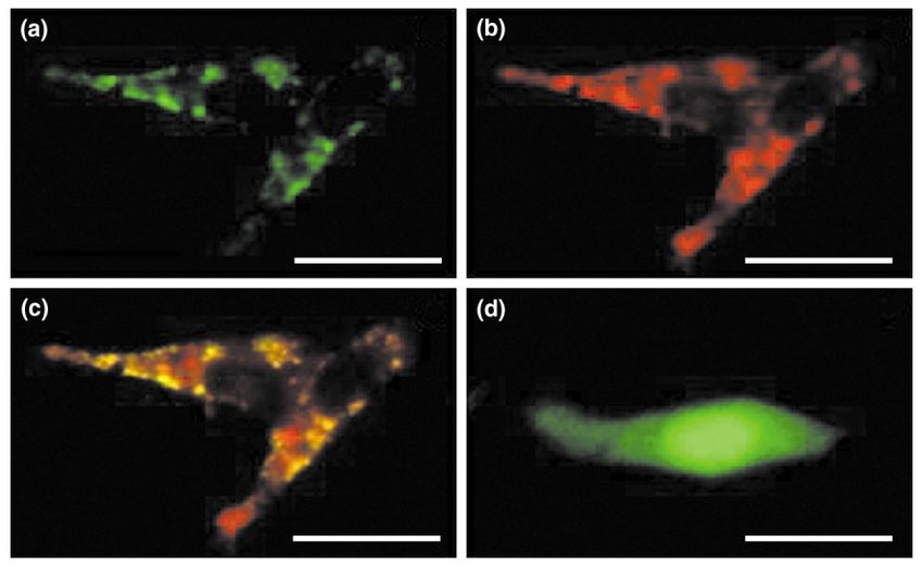
of 1a,25(OH)2D in the prostate might be influenced by the To further investigate the association between the loss
serum level of 25(OH)D. of 1a-OHase activity and prostate cancer, we transfected
Extrarenal synthesis of 1a,25(OH)2D from 25(OH)D in, LNCaP cells with a human 1a-OHase –green fluorescent
for example, skin and activated macrophages is well protein (GFP) fusion construct to confirm that the protein
known [1,3], and it is now recognized that two human is expressed and appears in the mitochondria (Fig. 4).
prostate cancer cell lines, DU145 and PC-3, as well as cells Alternatively, cells were transfected with cDNA encoding
derived from a normal prostate and a prostate with BPH human 1a-OHase to study their responses to 25(OH)D
also have 1a-OHase activity and synthesize 1a,25(OH)2D3 (Fig. 5). Transient and stable transfection markedly
from 25(OH)D3. However, 1a-OHase activity has not been increases the activity of 1a-OHase and so confers
detected in LNCaP cells [12]. inhibition of cell growth by 25(OH)D3 (Fig. 5) [26].
Comparing the activity of 1a-OHase in primary
cultures of prostate epithelial cells derived from four Are vitamin D analogs useful for treating prostate
patients with prostate cancer (CaP), two BPH patients and cancer?
three normal donors demonstrates that the normal Numerous reports demonstrate that 1a,25(OH)2D3
cultures had an average activity of 3.0 ^ stimulates differentiation and inhibits the prolifer-
0.36 pmol mg protein21 h21 (mean ^ SD ), whereas BPH ation, invasiveness and metastasis of prostate cancer
and prostate cancer cultures had an average activity of cells [6 –8,21– 23]. In addition, 1a,25(OH)2D3 and its
1.2 ^ 0.28 and 0.46 ^ 0.15 pmol mg protein21 h21, synthetic analogs prolong survival time in murine models
respectively. Therefore, compared with primary cultures of leukemia, and have been used successfully for treating
of normal prostate cells, enzyme activity is 60% and 85% psoriasis [1]. These findings strongly support the use of
lower in the primary cultured BPH and prostate cancer 1a,25(OH)2D3 and its analogs to treat prostate cancer and/
cells, respectively [25,26]. Similar results have been or 1a,25(OH)2D3 as a second line of therapy when
reported by Hsu et al. [27]. These findings have important androgen deprivation fails. However, the results of several
implications and indicate that the loss of 1a-OHase clinical trials indicate that the dose of 1a,25(OH)2D3
http://tem.trends.comARTICLE IN PRESS TEM 98
Review TRENDS in Endocrinology and Metabolism Vol.not known No.not known Month 0000 5
Fig. 4. Location of a fusion protein between 25-hydroxyvitamin D3-1a-hydroxylase (1a-OHase) and green fluorescent protein (GFP) (1a-OHase–GFP) in LNCaP cells. (a-c)
LNCaP cells were transfected with either the 1a-OHase–GFP plasmid or (d) GFP plasmid for 24 hours. Cells transfected with 1a-OHase– GFP were treated with MitoTracker
Orange (400 nM ) for 15 min and observed live with scanning laser confocal microscopy (600 £ ). (a) The green fluorescence (530-nm filter) is perinuclear and punctuate, con-
sistent with localization in the mitochondria. (b) The cell in (a), stained with the mitochondria-specific red fluorescent indicator MitoTracker (580-nm filter). (c) images (a)
and (b) superimposed. Colocalization of the 1a-OHase –GFP green fluorescence with the mitochondrial red fluorescence, which appears yellow-green, confirms that 1a-
OHase –GFP is synthesized in the mitochondria. (d) A live LNCaP cell transfected with the control GFP plasmid, viewed using a fluorescein filter. There is uniform green flu-
orescence throughout the cytoplasm, consistent with synthesis of GFP in the cytoplasm. Scale bar, 50 mm. Reproduced with permission from [26].
cannot be increased to . 0.5 mg twice a day because of their antiproliferative and prodifferentiating activities,
hypercalcemia and hypercalciuria [7,8,31,32]. However, and reducing or eliminating their calcemic effects
the time taken for PSA to double is at least doubled after [7,21,28 – 35]. Several studies have been published that
treatment with 1a,25(OH)2D3 [32]. Therefore, analogs of investigate the in vivo response of vitamin D analogs in
1a,25(OH)2D3 that have less calcemic activity and more prostate cancer [35 –38]. In general, the analogs are either
potent antiproliferative and prodifferentiatory activity are slightly more potent than or equipotent to 1a,25(OH)2D3,
attractive therapeutic agents. but slightly less calcemic than 1a,25(OH)2D3. A phase I
During the past two decades, . 2000 analogs of trial of 1a-hydroxyvitamin D2 in patients with advanced,
1a,25(OH)2D have been synthesized chemically [31,33] hormone-refractory prostate cancer has been conducted,
and their biological properties evaluated systematically which shows that five out of 25 patients achieved
in a variety of assay systems, with the goal of enhancing disease stabilization for $ 6 months, with main
toxicities being hypercalcemia and renal insufficiency
[39]. So far, no analogs of 1a,25(OH)2D3 effectively
(a) (b) prevent or inhibit prostate-cancer growth without signifi-
120
cant calcemic side-effects.
incorporation
100 Another approach to decreasing the side-effects of
1a,25(OH)2D3 and increasing its antiproliferative potency
(% of control)
80
∗ is to use 1a,25(OH)2D3 in combination with other agents,
60 such as retinoids [8], platinum compounds [40], inhibitors
∗
3H-thymidine
of histone deacetylase (sodium butyrate and trichostatin A)
40 [41] and docetaxel [42]. It has been shown that
20 1a,25(OH)2D3 and cisplatin, the most widely used plati-
num-based chemotherapeutic agent, act synergistically to
0 inhibit the growth of PC3 and DU-145 cancer cells [40],
l
1
1
S)
)
e
tro
(S
and that cisplatin enhances 1a,25(OH)2D3-induced apop-
3.
3.
as
(A
on
R
R
e
H
PC
PC
as
e
-O
C
totic signaling through mitogen-activated protein kinase
as
H
1α
le
H
-O
ab
kinase kinase (MEKK-1) [43]. Synergistic growth inhi-
-O
1-
1α
3.
St
1α
1-
R
bition of LNCaP, PC-3 and DU-145 prostate cancer cells by
1-
PC
3.
3.
R
1a,25(OH)2D3 and its 19-nor-hexafluoride analogs in
R
PC
le
PC
ab
St
combination with either sodium butyrate or trichostatin
A has also been observed [41]. The mechanism, which
Fig. 5. Effect of 25-hydroxyvitamin D3 [25(OH)D3] (1028 M ) on the incorporation of
3
H-thymidine into DNA of LNCaP cells transfected with cDNA encoding 25(OH)D-
involves histone deacetylation, appears to induce apopto-
1a-hydroxylase (1a-OHase). (a) LNCaP cells were transfected transiently with PCR sis by restoring the normal 1a,25(OH)2D3-mediated pro-
3.1 vector, antisense (AS) or sense (S) 1a-OHase cDNA. (b) LNCaP cells were stably apoptotic signals that are lost during prostate cancer
transfected with either PCR 3.1 vector or with sense 1a-OHase cDNA. Data are pre-
sented as % of mock transfected control in the absence of 25(OH)D3. Data are development. The combination of a weekly, oral high-dose
mean ^ SD , n ¼ 8, *P , 0:05: Reproduced with permission from [26]. (0.5 mg kg21) of calcitriol and weekly docetaxel
http://tem.trends.comARTICLE IN PRESS TEM 98
6 Review TRENDS in Endocrinology and Metabolism Vol.not known No.not known Month 0000
(36 mg m22) is well tolerated and effective in achieving a population-based case-control study in Shanghai, China
PSA response in 30 out of 37 metastatic, androgen- [59]. No significant association was observed between
independent prostate-cancer patients [42]. either BsmI or FokI polymorphisms in the VDR gene and
prostate cancer risk. However, there was a decreased risk
VDR polymorphism and prostate cancer risk of prostate cancer in men with the highest tertile of plasma
Following the initial observation that indicated an IGFBP-1 or -3 who have the ff FokI genotype but not FF
association between polymorphisms in the VDR gene and Ff genotypes. These results indicate that the IGF
and the risk of osteoporosis [44], many studies have and vitamin D systems might interact to affect prostate
examined whether the same polymorphisms are related to cancer risk [62].
the risk of prostate cancer [45]. Polymorphisms have been
identified in exons 2, 8, and 9 of the VDR gene, and involve Conclusion and perspectives
FokI, BsmI, and TaqI RFLP s, respectively. The FokI RFLP It has been known for more than two decades that
generates a VDR with three additional amino acids at the 1a,25(OH)2D3 is one of the most effective compounds for
N terminus, whereas the BsmI, and TaqI RFLPs do not inhibiting proliferation and inducing terminal differen-
affect the coding sequence. A microsatellite polymorphism tiation of normal and cancer cells that contain VDRs,
in the 30 -untranslated region that does not alter the VDR including prostate cells. There has been progress in
coding sequence has also been identified. Following the understanding how 1a,25(OH)2D3 inhibits cell growth
first report that indicated a positive association between and causes apoptosis in LNCaP cells but not in cells from
TaqI RFLP and prostate-cancer risk in a study from North other cancer cell lines, primary cultures and in vivo. More
Carolina [46], there have been at least seven other studies studies are required to examine the in situ interaction
that show a positive association between prostate cancer between vitamin D and other hormones and/or growth
and TaqI [47 – 49], BsmI [50 – 51], FokI, [52] and poly-A factors in the prostate.
microsatellite [51,53] polymorphisms (Table 1). However, If a positive association between polymorphisms in the
the associations between prostate-cancer risk and poly- VDR gene and prostate-cancer risk is established, the VDR
morphisms are called into question in a similar number of genotype could potentially be used to identify men who are
studies [54 – 60]. There are several explanations for these more likely to develop clinically significant prostate cancer
conflicting findings. For example, they could be caused by: and to intervene in these men to reduce the morbidity and
(1) differences in the selection of the patient and control mortality that result [63].
groups; (2) limitations in the sample size; (3) inadequate It is also recognized that an increase in the incidence
control of confounding factors; and (4) variation in the and mortality of many common solid tumors, including
prevalence of environmental risk factors and etiological prostate cancer is associated with both limited exposure to
factors across populations. In addition, the skin types of sunlight and vitamin D deficiency [1,9– 11,64]. However,
patients and controls that determine the pigmentation the exact association between latitude, sun exposure and
response and vitamin D3 synthesis in response to solar increased concentrations of 25(OH)D was not well under-
UVB irradiation has not been identified, and this might be stood until the relatively recent observation that prostate
crucial for determining the outcome of polymorphism cells contain the enzyme that converts 25(OH)D to
studies [61]. 1a,25(OH)2D [12]. Synthesis of 1a,25(OH)2D in the
Regarding gene– gene interaction, the combined effects prostate indicates that increasing circulating levels of
of the insulin-like growth factor (IGF) system and vitamin 25(OH)D, either by adequate exposure to sunlight or oral
D on prostate cancer risk have been investigated in a supplementation, might provide a simple way to increase
Table 1. Polymorphisms in the vitamin D receptor gene and prostate cancer risk
Location No. cases/controls Polymorphism Odds ratio 95% CI Refs
Positive association
North Carolina 108/170 TaqI 0.34 0.16 –0.76 ðP , 0:01Þ [46]
France/Germany 105/132 TaqI 0.5 0.27 –0.92 ðP , 0:026Þ [47]
Japan 115/133 TaqI 2.52 1.21 –5.27 ðP , 0:009Þ [48]
Portugal 163/211 TaqI 2.11 1.15 –3.88 ðP , 0:015Þ [49]
Los Angles 151/174 BsmI/Poly-A 0.7 0.3 –1.6 [50]
Japan 222/326 BsmI 3.31 2.05 –5.32 ðP , 0:0001Þ [51]
Poly –A 0.44 0.198 –0.966 ðP , 0:041Þ [51]
Los Angles 57/169 Poly –A 4.61 1.34 –15.82 [53]
California 191/191 FokI 0.43 0.428 –0.438 ðP ¼ 0:015Þ [52]
Negative association
US 372/591 BsmI, Taq I 0.86–0.92 0.57 –1.29 [54]
Maryland 41/41 Poly-A 1.3 0.4 –4.3 [55]
Taq I 0.7 0.2 –2.6 [55]
Japan 60/60 TaqI 1.3 0.6 –2.8 [56]
Japan 100/202 TaqI 0.9 0.67 –1.01 [57]
Poly-A 0.9 0.67 –1.01 [57]
North Carolina 77/183 Taq I 1.4 0.7 –2.8 [58]
Poly-A 1.2 0.6 –2.5 [58]
China 191/304 BsmI, FokI 1.01–1.13 0.3 –3.67 [59]
Austria 190/190 Taq I 1.76 0.9 –3.45 ðP ¼ 0:07Þ [60]
http://tem.trends.comARTICLE IN PRESS TEM 98
Review TRENDS in Endocrinology and Metabolism Vol.not known No.not known Month 0000 7
synthesis of 1a,25(OH)2D in the prostate and, therefore, D3-induced G0 arrest but is not required for G1 accumulation or
decrease the risk of prostate cancer. Chronic vitamin D apoptosis of LNCaP prostate cancer cells. Endocrinology 144, 50– 60
16 Perez-Stable, C.M. et al. (2002) The Gg/T-15 transgenic mouse model of
insufficiency in young and middle-aged men [65] might androgen-independent prostate cancer: Target cells of carcinogenesis
increase their risk of prostate cancer. Similar to the and the effect of the vitamin D analogue EB 1089. Cancer Epidemiol.
recommendation that men .50 years of age should be Biomarkers Prev. 11, 555 – 563
screened for PSA, surveillance of serum 25(OH)D should 17 Guzey, M. et al. (2002) Apoptosis induction by 1a,25-dihydroxyvitamin
be performed annually in men . 30 years, especially those D3 in prostate cancer. Mol. Cancer Ther. 1, 667 – 677
18 Blutt, S.E. et al. (2000) Calcitriol-induced apoptosis in LNCaP cells is
who are at higher risk of chronic vitamin D deficiency, such blocked by overexpression of bcl-2. Endocrinology 141, 10 – 17
as African Americans and indoor workers. Thus, adequate 19 Zhao, X.Y. et al. (1997) 1 alpha,25-dihydroxyvitamin D3 actions in
vitamin D nutrition should be maintained, not only for LNCaP human prostate cancer cells are androgen-dependent. Endo-
bone health in men and women, but because it might crinology 138, 3290– 3298
20 Yang, E.S. et al. (2002) Vitamin D-mediated growth inhibition of an
decrease the risk of prostate cancer in men and mitigate
androgen-ablated LNCaP cell line model of human prostate cancer.
metastatic activity should it develop. Mol. Cell. Endocrinol. 186, 69 – 79
The knowledge that the prostate synthesizes 21 Campbell, M.J. et al. (1997) Inhibition of proliferation of prostate
1a,25(OH)2D and that prostate-cancer cells respond to cancer cells by a 19-nor-hexafluoride vitamin D3 analogue involves the
1a,25(OH)2D offers new strategies to help reduce the induction of p21wafl, p27kipl and E-cadherin. J. Mol. Endocrinol. 19,
15 – 27
incidence of this devastating disease. The promising
22 Majeski, S. et al. (1996) Vitamin D is a potent inhibitor of tumor cell-
results of EB1089 in treating liver cancer offers hope induced angiogenesis. J. Investig. Dermatol. Symp. Proc. 1, 97 – 101
that noncalcemic analogs of 1a,25(OH)2D3 can be 23 Schwartz, G.G. et al. (1997) 1a,25-Dihydroxyvitamin D3 (calcitriol)
developed that might be combined with other chemo- inhibits the invasiveness of human prostate cancer cells. Cancer
preventing agents to treat prostate cancer without Epidemiol. Biomarkers Prev. 6, 727 – 732
24 Corder, E.H. et al. (1993) Vitamin D and prostate cancer: a
serious side-effects [66].
prediagnostic study with stored sera. Cancer Epidemiol. Biomarkers
Prev. 2, 467– 472
Acknowledgements 25 Chen, T.C. et al. (2000) Enhancement of 25-hydroxyvitamin D-1-alpha-
This work was supported in part by grants 4118PP1017 and 41211159016 hydroxylase activity in prostate cells by gene transfection: a novel
from The Commonwealth of Massachusetts, US Army DAMD17-01-1- approach for the treatment of prostate cancer. In Vitamin D Endocrine
0025, and MO1RR00533 from NIH. System: Structural, Biological, Genetic and Clinical Aspects (Norman,
A.W. et al., eds), pp. 525– 528, University of California, Riverside
References 26 Whitlatch, L.W. et al. (2002) 25-Hydroxyvitamin D-1a-hydroxylase
1 Holick, M.F. (2002) Vitamin D: the underappreciated D-lightful activity is diminished in human prostate cancer cells and is enhanced
hormone that is important for skeletal and cellular health. Curr. by gene transfer. J. Steroid Biochem. Mol. Biol. 81, 135 – 140
Opin. Endocrinol. Diabetes 9, 87 – 98 27 Hsu, J.Y. et al. (2001) Reduced 1a-hydroxylase activity in human
2 Miller, W.L. and Portale, A.A. (2000) Vitamin D 1a-hydroxylase. prostate cancer cells correlates with decreased susceptibility to 25-
Trends Endocrinol. Metab. 11, 315 – 319 hydroxyvitamin D3-induced growth inhibition. Cancer Res. 61,
3 Omdahl, J.L. et al. (2002) Hydroxylase enzyme of the vitamin D 2852– 2856
pathway: expression, function, and regulation. Annu. Rev. Nutr. 22, 28 Chen, T.C. et al. (2000) The in vitro evaluation of 25-hydroxyvitamin
139 – 166 D3 and 19-nor-1a,25-dihydroxyvitamin D2 for prostate cancer therapy.
4 Li, Y.C. et al. (2002) 1,25-Dihydroxyvitamin D3 is a negative endocrine Clin. Cancer Res. 6, 901 – 908
regulator of the renin-angiotensin system. J. Clin. Invest. 110, 29 Barreto, A.M. et al. (2000) 25-Hydroxyvitamin D3, the prohormone of
229 – 238 1,25-dihydroxyvitamin D3, inhibits the proliferation of primary
5 Schwartz, G.G. and Hulka, B.S. (1990) Is vitamin D deficiency a risk prostatic epithelial cells. Cancer Epidemiol. Biomarkers Prev. 9,
factor for prostate cancer? (Hypothesis). Anticancer Res. 10, 265– 270
1307 – 1311 30 Skowronski, R.J. et al. (1995) Actions of vitamin D3 analogs on human
6 Miller, G.J. (1999) Vitamin D and prostate cancer: biologic interactions prostate cancer cell lines: Comparison with 1,25-dihydroxyvitamin D3.
and clinical potentials. Cancer Metastasis Rev. 17, 353– 360 Endocrinology 136, 20 – 26
7 Zhao, X.Y. and Feldman, D. (2001) The role of vitamin D in prostate 31 Bouillon, R. et al. (1995) Function relationships in the vitamin D
cancer. Steroids 66, 293 – 300 endocrine system. Endocr. Rev. 16, 200 – 257
8 Polek, T.C. and Weigel, N.L. (2002) Vitamin D and prostate cancer. 32 Gross, C. et al. (1998) Treatment of early recurrent prostate cancer
J. Androl. 23, 9 – 17 with 1,25-dihydroxyvitamin D3 (calcitriol). J. Urol. 159, 2035 – 2039
9 Ahonen, M.H. et al. (2000) Prostate cancer risk and prediagnostic 33 Guyton, K.Z. et al. (2001) Cancer chemoprevention using natural
serum 25-hydroxyvitamin D levels (Finland). Cancer Causes Control vitamin D and synthetic analogs. Annu. Rev. Pharmacol. Toxicol. 41,
11, 847– 852 421– 442
10 Luscombe, C.J. et al. (2001) Exposure to ultraviolet radiation: 34 Schwartz, G.G. et al. (1994) Human prostate cancer cells: inhibition of
association with susceptibility and age at presentation with prostate proliferation by vitamin D analogs. Anticancer Res. 14, 1077– 1082
cancer. Lancet 358, 641 – 642 35 Schwartz, G.G. et al. (1995) 1,25-Dihydroxy-16-ene-23-yne-vitamin D3
11 Grant, W.B. (2002) An estimate of premature cancer mortality in the and prostate cancer cell proliferation in vivo. Urology 46, 365 – 369
U.S. due to inadequate doses of solar ultraviolet-B radiation. Cancer 36 Lucia, M.S. et al. (1995) Chemopreventive activity of tamoxifen, N-(4-
94, 1867 – 1875 hydroxyphenyl) retinamide and the vitamin D analogue Ro24-5531 for
12 Schwartz, G.G. et al. (1998) Human prostate cells synthesize 1,25- androgen-promoted carcinomas of the rat seminal vesicle and prostate.
dihydroxyvitamin D3 from 25-hydroxy vitamin D3. Cancer Epidemiol. Cancer Res. 55, 5621– 5627
Biomarkers Prev. 7, 391– 395 37 Lokeshwar, B.L. et al. (1999) Inhibition of prostate cancer metastasis
13 Freedman, L.P. (1999) Multimeric coactivator complexes for steroid/ in vivo: a comparison of 1a,25-Dihydroxyvitamin D3 (calcitriol) and
nuclear receptors. Trends Endocrinol. Metab. 10, 403 – 407 EB1089. Cancer Epidemiol. Biomarkers Prev. 8, 241 – 248
14 Zhuang, S.H. and Burnstein, K.L. (1998) Antiproliferative effect of 1a, 38 Blutt, S.E. et al. (2000) A calcitriol analogue, EB1089, inhibits the
25-dihydroxyvitamin D3 in human prostate cancer cell line LNCaP growth of LNCaP tumors in nude mice. Cancer Res. 60, 779 – 782
involves reduction of cyclin-dependent kinase 2 activity and persistent 39 Liu, G. et al. (2002) Phase I trial of 1a-hydroxyvitamin D2 in patients
G1 accumulation. Endocrinology 139, 1197 – 1207 with hormone refractory prostate cancer. Clin. Cancer Res. 8,
15 Polek, T.C. et al. (2003) p53 is required for 1,25-dihydroxyvitamin 2820– 2827
http://tem.trends.comARTICLE IN PRESS TEM 98
8 Review TRENDS in Endocrinology and Metabolism Vol.not known No.not known Month 0000
40 Moffatt, K.A. et al. (1999) 1a,25-dihydroxyvitamin D3 and platinum genetic polymorphisms in vitamin D receptor and androgen receptor.
drugs act synergistically to inhibit the growth of prostate cancer cell J. Natl. Cancer Inst. 89, 166 – 170
lines. Clin. Cancer Res. 5, 695 – 703 54 Ma, J. et al. (1998) Vitamin D receptor polymorphisms, circulating
41 Rashid, S.F. et al. (2001) Synergistic growth inhibition of prostate vitamin D metabolites, and risk of prostate cancer in United States
cancer cells by 1a,25-dihydroxyvitamin D3 and its 19-nor-hexafluoride physicians. Cancer Epidemiol. Biomarkers Prev. 7, 385 – 390
analogs in combination with either sodium butyrate or trichostatin A. 55 Kibel, A.S. et al. (1998) Vitamin D receptor polymorphisms and lethal
Oncogene 20, 1860– 1892 prostate cancer. J. Urol. 160, 1405– 1409
42 Beer, T.M. et al. (2003) Weekly high-dose calcitriol and docetaxel in 56 Furuya, Y. et al. (1999) Vitamin D receptor gene polymorphism in
metastatic androgen-independent prostate cancer. J. Clin. Oncol. 21, Japanese patients with prostate cancer. Endocr. J. 46, 467– 470
123 – 128 57 Watanabe, M. et al. (1999) Significance of vitamin D receptor gene
43 Hershberger, P.A. et al. (2002) Cisplatin potentiates 1,25-dihydrox- polymorphism for prostate cancer risk in Japanese. Anticancer Res. 19,
yvitamin D3-induced apoptosis in association with increased mitogen- 4511 – 4514
activated protein kinase kinase kinase 1 (MEKK-1) expression. Mol. 58 Blazer, D.G. et al. (2000) Vitamin D receptor polymorphisms and
Cancer Ther. 1, 821 – 829 prostate cancer. Mol. Carcinog. 27, 18 – 23
44 Morrison, N.A. et al. (1994) Prediction of bone density from vitamin D 59 Chokklingam, A.P. et al. (2001) Vitamin D Receptor gene polymorph-
isms, insulin-like growth factors, and prostate cancer risk: a
receptor alleles. Nature 367, 284 – 287
population-based case-control study in China. Cancer Res. 61,
45 Coughlin, S.S. and Hall, I.J. (2002) A review of genetic polymorphisms
4333– 4336
and prostate cancer risk. Ann. Epidemiol. 12, 182 – 196
60 Gsur, A. et al. (2002) Vitamin D receptor gene polymorphism and
46 Taylor, J.A. et al. (1996) Association of prostate cancer with vitamin D
prostate cancer risk. Prostate 51, 30 – 34
receptor gene polymorphism. Cancer Res. 56, 4108 – 4110
61 Luscombe, C.J. et al. (2001) Outcome in prostate cancer associations
47 Correa-Cerro, L. et al. (1999) Vitamin D receptor polymorphisms as
with skin type and polymorphism in pigmentation-related genes.
markers in prostate cancer. Hum. Genet. 105, 281– 287
Carcinogenesis 22, 1343 – 1347
48 Hamasaki, T. et al. (2001) Clinical and pathological significance of
62 Boyle, B.J. et al. (2001) Insulin-like growth factor binding protein-3
vitamin D receptor gene polymorphism for prostate cancer which is
mediates 1 alpha,25-dihydroxyvitamin D3 growth inhibition in the
associated with a higher mortality in Japanese. Endocr. J. 48, 543– 549 LNCaP prostate cancer cell line through p21/waf1. J. Urol. 165,
49 Medeiros, R. et al. (2002) The role of vitamin D receptor gene 1319– 1324
polymorphisms in the susceptibility to prostate cancer of a southern 63 Rebbeck, T.R. (2002) Inherited genotype and prostate cancer outcomes.
European population. J. Hum. Genet. 47, 413 – 418 Cancer Epidemiol. Biomarkers Prev. 11, 945– 952
50 Ingles, S.A. et al. (1998) Association of prostate cancer with vitamin D 64 Apperly, F.L. (1941) The relation of solar radiation to cancer mortality
receptor haplotypes in African-Americans. Cancer Res. 58, 1620– 1623 in North America. Cancer Res. 1, 191 – 195
51 Habuchi, T. et al. (2000) Association of vitamin D receptor gene 65 Tangpricha, V. et al. (2002) Vitamin D in-sufficiency among free-living
polymorphism with prostate cancer and benign prostatic hyperplasia healthy young adults. Am. J. Med. 112, 659– 662
in a Japanese population. Cancer Res. 60, 305 – 308 66 Hansen, C.M. et al. (2000) Seocalcitol (EB1089): a vitamin D analogue
52 Xu, Y. et al. (2003) Vitamin D receptor start codon polymorphism (FokI) of anti-cancer potential. Background, design, synthesis, pre-clinical
and prostate cancer progression. Cancer Epidemiol. Biomarkers Prev. and clinical evaluation. Curr. Pharm. Des. 6, 803 – 828
12, 23 – 27 67 Holick, M.F. (1994) Vitamin D – new horizons for the 21st century. Am.
53 Ingles, S.A. et al. (1997) Association of prostate cancer risk with J. Clin. Nutr. 60, 619 – 630
http://tem.trends.comYou can also read