IMAGING OF SARS-COV-2 INFECTED VERO E6 CELLS BY HELIUM ION MICROSCOPY
←
→
Page content transcription
If your browser does not render page correctly, please read the page content below
Imaging of SARS-CoV-2 infected Vero E6 cells by
helium ion microscopy
Natalie Frese1, Patrick Schmerer2, Martin Wortmann3, Matthias Schürmann4,
Matthias König2, Michael Westphal1, Friedemann Weber2, Holger Sudhoff4
and Armin Gölzhäuser*1

  Full Research Paper                                                                                                              Open Access

  Address:                                                             Beilstein J. Nanotechnol. 2021, 12, 172–179.
  1Physics of Supramolecular Systems and Surfaces, Faculty of          https://doi.org/10.3762/bjnano.12.13
  Physics, Bielefeld University, Bielefeld, Germany, 2Institute of
  Virology, Faculty of Veterinary Medicine, Justus-Liebig-University   Received: 02 December 2020
  Giessen, Germany, 3Faculty of Engineering and Mathematics,           Accepted: 28 January 2021
  Bielefeld University of Applied Sciences, Bielefeld, Germany and     Published: 02 February 2021
  4University Clinic for Otolaryngology, Head and Neck Surgery,

  Medical Faculty OWL at Bielefeld University, Germany                 This article is part of the thematic issue "Ten years of the helium ion
                                                                       microscope".
  Email:
  Armin Gölzhäuser* - ag@uni-bielefeld.de                              Guest Editors: G. Hlawacek and A. Wolff

  * Corresponding author                                               © 2021 Frese et al.; licensee Beilstein-Institut.
                                                                       License and terms: see end of document.
  Keywords:
  bioimaging; cell membrane; charge compensation; helium ion
  microscopy; SARS-CoV-2; Vero E6 cells

Abstract
Helium ion microscopy (HIM) offers the opportunity to obtain direct views of biological samples such as cellular structures, virus
particles, and microbial interactions. Imaging with the HIM combines sub-nanometer resolution, large depth of field, and high sur-
face sensitivity. Due to its charge compensation capability, the HIM can image insulating biological samples without additional
conductive coatings. Here, we present an exploratory HIM study of SARS-CoV-2 infected Vero E6 cells, in which several areas of
interaction between cells and virus particles, as well as among virus particles, were imaged. The HIM pictures show the three-
dimensional appearance of SARS-CoV-2 and the surface of Vero E6 cells at a multiplicity of infection of approximately 1 with
great morphological detail. The absence of a conductive coating allows for a distinction between virus particles bound to the cell
membrane and virus particles lying on top of the membrane. After prolonged imaging, it was found that ion-induced deposition of
hydrocarbons from the vacuum renders the sample sufficiently conductive to allow for imaging even without charge compensation.
The presented images demonstrate the potential of the HIM in bioimaging, especially for the imaging of interactions between
viruses and their host organisms.

Introduction
The last decade of helium ion microscopy (HIM) was character-            engineering [1]. Although HIM soon proved to be a promising
ized by a rapid exploration of its sub-nanometer imaging and             tool in the life sciences, the examination of biological samples
ion-beam nanofabrication capabilities in materials science and           by HIM proceeded at a much slower pace. In recent years, it has

                                                                                                                                                 172
Beilstein J. Nanotechnol. 2021, 12, 172–179.
been used in the field of cell biology for imaging various human trons. Such coatings, albeit only a few nanometers thick, can
and animal cells. These include cartilage [2], cancer [3], liver significantly alter and conceal fine details of biological nano-
[4], kidney [5] and stem cells [6], as well as fibrin fibers [7]. To structures [2], which is noticeable in SEM images of virus parti-
visualize viruses and their host organisms, HIM has so far been cles [19,24]. Since in the HIM positive charge accumulates on
applied to image T4 phage-infected E. coli bacteria [8], various insulating samples, a low-energy electron flood gun can be used
phases of the life cycle of the bacterial predator Bdellovibrio for charge compensation, which irradiates the sample with a
bacteriovorus [9] and the vesicular structure of ethane- diffuse beam of electrons. This eliminates the need for a
oxidizing archaea [10]. A comprehensive review on the subject conductive coating, and allows for a direct view on nanoscale
of bioimaging with HIM has recently been published by structures [6,25]. Here, we demonstrate the benefits of high-
Schmidt and co-workers [11]. resolution HIM by imaging SARS-CoV-2 interacting with Vero
E6 cells without any conductive coating. The presented images
In this work, we use HIM to investigate Vero E6 cells infected allow for the identification of SARS-CoV-2 virus particles,
with the novel severe acute respiratory syndrome coronavirus 2 their interaction with the cell membrane and a distinction be-
(SARS-CoV-2). Several members of the family Coronaviridae tween virus particles bound to the cell surface from those lying
have been described in the human population and usually cause on it.
mild respiratory disease. SARS-CoV-2 demonstrated a world-
wide spread causing a significant global public health emer- Experimental
gency [12,13]. As of January 18th, 2021, more than 95 million Vero E6 cells were cultivated in Dulbecco's modified Eagle's
cases worldwide have been confirmed with the infection and medium (Thermo Fisher Scientific) supplemented with 10%
over two million infected patients have died [14]. African green fetal bovine serum (Capricorn Scientific) in a 5% CO2 atmo-
monkey kidney Vero E6 cells have been reported to support sphere at 37 °C. SARS-CoV-2 (strain SARS-CoV-2 /München-
SARS-CoV-2 replication in culture, while many more cell lines 1.2/2020/984, p.2) [26] was grown on Vero E6 cells and titrated
have been reported to be refractory to SARS-CoV-2 infection as described [27]. Infection experiments were done under
[15]. Both scanning electron microscopy (SEM) and transmis- biosafety level 3 conditions with enhanced respiratory personal
sion electron microscopy (TEM) have been used to image protection equipment.
SARS-CoV-2 [16-20]. While TEM achieves unsurpassed reso-
lution and can visualize macromolecular structures such as For HIM, cells were seeded onto coverslips placed in 24-well
spike glycoproteins or transmembrane proteins [21], SEM plates. The coverslips were previously sputter coated with
provides topographic images of infected cells and virus parti- 30 nm of gold to improve charge neutralization during HIM
cles distributed on their surface, albeit only after the samples imaging. After 24 h, nearly confluent monolayers were infected
have been coated with a conductive layer. In contrast, the HIM with SARS-CoV-2 at a multiplicity of infection (MOI) of
delivers a topographic image of the uncoated surface morpholo- approximately 1 or mock-infected using cell culture medium.
gy of cells and virus particles, allowing one to identify and in- Following an incubation period of 18 h in a cell culture
vestigate sites at which a cell interacts with the virus. While its incubator (37 °C), cells were washed with 0.1 M sodium
principle of operation is very similar to SEM, HIM utilizes a cacodylate (NaCac, pH 7.4) and fixed in 2% (v/v) glutaralde-
beam of positively charged helium ions (He+) instead of nega- hyde, 2% (w/v) paraformaldehyde in NaCac buffer at room
tively charged electrons to excite and detect secondary elec- temperature for 30 min. After fixation at room temperature, the
trons from the sample surface. Due to the high brightness and samples were transferred to the normal laboratory area and then
low energy spread of its atomically sharp gas field ion source, fixed at 4 °C with fresh fixatives. The coverslips were subse-
the smallest attainable focused spot size is about 0.3 nm [22]. quently washed and dehydrated in a graded series of ethanol
With its significantly smaller convergence angle compared to (50%, 70%, 95%, 99.5% (2×)), transferred to water-free ace-
SEM, HIM achieves a much larger depth of field, which is par- tone and critical point dried in carbon dioxide.
ticularly useful for imaging three-dimensional structures [22].
Due to their higher mass, He+ ions penetrate deeper into the HIM was performed with an Orion Plus microscope (Carl
sample and do not spread as wide as electrons, resulting in a Zeiss) at an acceleration voltage of about 36 kV and a working
smaller escape volume of the secondary electrons and a higher distance of 20 mm. The spot control was set to 6 to obtain a
surface resolution of the HIM, compared to the SEM [23]. A beam current of 0.2 to 0.4 pA. To avoid charging effects during
further benefit of HIM is its charge compensation capability secondary electron detection, an electron flood gun was used
during secondary electron detection. SEM imaging of biologi- after each line scan, if not stated otherwise, with a flood energy
cal specimen usually necessitates a thin conductive coating to of 540 eV, flood time of 10 µs and a focus of 107 V. It should
prevent negative charge accumulation from the impinging elec- be mentioned that the flood gun parameters have to be opti-
173Beilstein J. Nanotechnol. 2021, 12, 172–179.
mized for each magnification level. All HIM images were re- cles. This is in accordance with a study of Bojkova et al. [28],
corded with 1024 × 1024 pixels. Before imaging, each sample which demonstrated the presence of newly synthesized viral
was stored in the vacuum chamber of the microscope at particles of SARS-CoV-2 even 10 h after initial infection. The
3.3 × 10 −7 mbar for at least 24 h to remove most volatile cell membrane of the infected cell is covered with the virus par-
organic contaminants. ticles, which are predominantly spherically shaped. Holes in the
cell membrane, illustrated in Figure 1b4 and Figure S1 of Sup-
Results and Discussion porting Information File 1, have previously been observed in
A comparison between a native and an infected Vero E6 cell at uncoated mammalian cells and indicate lipid nanodomains or
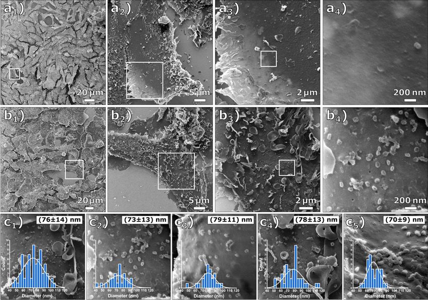
multiple magnification levels is shown in Figure 1. Figure 1a caveolea [6]. Figure 1c shows an evaluation of the virus parti-
shows a sequence of four HIM images of native Vero E6 cells cle size in five arbitrarily chosen regions on the MOI 1 sample
(mock-infected). Figure 1b displays a sequence of HIM images resulting in an average diameter of the virus particles of
of Vero E6 cells after they have been exposed to SARS-CoV-2 75 ± 13 nm, noting that this value has been obtained from
at a multiplicity of infection of approximately 1 (MOI 1) and an viruses after fixation and critical point drying.
incubation time of 18 h. The surface of the infected cells is
covered by a number of micrometer-sized vesicles and seg- As He+ ions can penetrate several hundred nanometers into the
ments of cell membranes, which is a first indication that apopto- sample [29], the outer rim of the cells appears brighter because
sis occurred during viral replication. Regularly shaped particles the ions pass through the cells and generate additional second-
below 100 nm diameter on the cell membrane shown in ary electrons at the back of the cells and in the gold-coated
Figure 1b4 were only abundant on the cells of the MOI 1 sam- specimen slide [30]. The edges appear brightest where the cells
ple and were therefore identified as SARS-CoV-2 virus parti- bend upwards from the substrate. The edge resolution in two
Figure 1: Comparative HIM images of Vero E6 cells that were mock-infected and infected at MOI 1. (a1–4) Mock-infected cells at different magnifica-
tions (FOV 200 µm, 45 µm, 15 µm, and 1.7 µm) and (b1–4) cells infected at MOI 1 at different magnifications (FOV 250 µm, 45 µm, 15 µm, and
1.7 µm). The cell membrane is covered with the virus particles. (c1–5) Determined virus particle diameter distributions. The inserted histograms show
the respective image evaluation with normal distribution, mean value, and standard deviation. The average diameter of all evaluated images is
75 ± 13 nm.
174Beilstein J. Nanotechnol. 2021, 12, 172–179.
highly magnified images, shown in Figure S2 of Supporting shown in Figure 2a1–3, where a location on a MOI 1-infected
Information File 1, has been determined by plotting the corre- Vero E6 sample was first imaged at a field of view (FOV) of
sponding gray-scale values over the edges of two holes, result- 23 µm (Figure 2a 1 ), followed by two higher magnification
ing in values of 1.3 and 2.1 nm. The edge resolution of the images with a FOV of 4.5 µm and a FOV of 1 µm (Figure 2a2).
images is determined by an interplay between the size of the Figure 2a3 shows the same region as Figure 2a1, but the parts
focused He+ beam and the widening of the beam within the that were previously imaged at high magnification (FOV of
sample material. The obtained values are typical for biological 4.5 μm) with a dose of 1.4 × 10¹⁶ ions/cm2 appear noticeably
materials [6-8,11]. brighter. This is caused by He+ beam-induced carbonaceous
deposits resulting in a thin conductive coating. In addition to the
An effect frequently occurring during HIM imaging with charge improved conductivity of the specimen, the deposited layer may
compensation can be observed in the sequence of HIM images contribute to the electron density of the surface, thus increasing
Figure 2: Effect of carbon deposition during HIM imaging. (a1) HIM image (FOV 20 µm) of a cell infected at MOI 1 with charge compensation.
(a2) HIM images at high magnification (FOV 4.5 µm and 1 µm) with charge compensation. (a3) The same image section as (a1) after imaging the
regions in (a2). Due to increased conductivity, this region appears significantly brighter than the rest of the image. (b1–3) HIM images of a cell infected
at MOI 1 at different magnifications (FOV 20 µm, 5 µm, and 450 nm) with charge compensation. (c1–3) HIM images of the same cell (FOV 20 µm,
2 µm, and 450 nm) after imaging the magnified sections in (b). (c1) and (c2) were imaged with and (c3) was imaged without charge compensation.
175Beilstein J. Nanotechnol. 2021, 12, 172–179.
secondary electron yield. This effect, commonly referred to as appear again brighter. After imaging Figure 2c2 with a dose of
electron- and/or ion beam-induced deposition, is commonly ob- 1.9 × 10 17 ions/cm 2 , the flood gun was turned off, which
served in charged-particle microscopes. In electron micro- allowed imaging of Figure 2c3 without any external charge
scopes, deposition rates of up to 3 Å/s at high current densities compensation. From the quality of this image, it can be inferred
have been reported. As the deposition rate quickly reaches an that the deposited carbon layer rendered the sample sufficiently
equilibrium with rising current density, it can be assumed that conductive. However, small structures are still visible on the
the limiting factor is the density of residual hydrocarbons in the membrane surface, which may originate from surface topogra-
vacuum [31]. In the HIM, residual gas as well as the specimen phy or material contrast. The deposited carbon film is presum-
itself are considered the main contributors of hydrocarbons ably thinner than typical conductive metal or carbon coatings
[32,33]. Due to the much larger mass of He+ ions compared to for SEM imaging, and it does not show any surface masking
electrons, their sputter rate is typically much higher. Since and clustering as seen on the gold substrate in the upper left of
organic compounds are ablated from the sample surface, hydro- Figure 2b2. The energy of the incident hydrocarbons is much
carbon deposition is likely to be more pronounced when lower compared to the energy of sputter-deposited metals.
imaging biological samples in HIM. A schematic illustration of However, it is possible that this unintended, but sometimes use-
this effect can be seen in Figure S3 of Supporting Information ful, carbon deposition can be reduced by HIM imaging in ultra-
File 1. high vacuum [34-36].
Figure 2b1–3 shows an infected Vero E6 cell at different magni- The cell structures shown in the HIM images of Figure 3a are
fication levels. Figure 2b3 depicts the highest magnification sharply resolved over tens of micrometers, which demonstrates
(FOV 450 nm) of the cell seen in Figure 2b1, showing the virus the high depth of field of HIM compared to SEM [37]. In image
particles on top of the cell membrane in a side view. Note that 3a3, at the surface of the cell, a cluster of virus particles seems
after the zoom-out in Figure 2c1, the previously imaged regions to be bound to the cell membrane (arrow). We suggest that this
Figure 3: HIM images of cells infected at MOI 1 imaged with charge compensation. (a1–3) Different magnifications of an infected cell (FOV 17 µm,
3.5 µm, and 1.3 µm). At the high magnification in (a3), clusters of virus particles (arrow) and junctions (arrowheads) between the virus particle and the
cell membrane become visible. (b1–3) Different magnifications of an infected cell (FOV 18 µm, 2 µm, and 850 nm). While some of the virus particles
appear to be bound to the cell membrane (arrowheads), others seem to just lie on top of it (arrow).
176Beilstein J. Nanotechnol. 2021, 12, 172–179.
resembles the particle clustering by host defense protein BST-2 particles lying on top of the cell membrane. The images unveil
as it was observed for human coronavirus229E and quantified the three-dimensional appearance of SARS-COV-2 and the sur-
in HeLa cells by Wang and co-workers [38]. However, the face of Vero E6 cells at MOI 1 with an edge resolution of up to
metal coating applied by Wang et al. is clearly visible at high 1.3 nm. Additionally, it is shown that ion-induced deposition
resolution in the SEM images as a rough layer on the cell mem- renders the sample surface sufficiently conductive to be imaged
brane that hides the true topography [25,39]. In contrast, the without charge compensation. The presented images demon-
HIM images presented here not only allow for the quantifica- strate the potential of the HIM in bioimaging, especially for the
tion of particles and clusters, but also enable an unveiled view imaging of interactions between viruses and their host organ-
on the interaction of virus particles with the cell membrane. The isms. HIM thus represents a versatile complement to conven-
presented particle cluster seems to have a coalesced appearance, tional methods in the life sciences.
which might be caused by the virus–virus and virus–membrane
interactions mediated through agglutinating BST-2 [40,41].
Some viral particles appear to be connected to the cell mem- Supporting Information
brane by a continuous junction (arrowheads). Figure 3b shows
another cell on the MOI 1 sample at different magnification Supporting Information File 1
levels. At the highest magnification shown in Figure 3b3 (FOV Additional experimental data.
850 nm), these junctions can also be observed (arrowheads). [https://www.beilstein-journals.org/bjnano/content/
We assume that this resembles the tubulating cell membrane, supplementary/2190-4286-12-13-S1.pdf]
which is stabilized by BST-2 to prevent viral scission. This al-
ternative BST-2 interaction was already described for HIV-
infected cells via immuno-TEM [42] but has not yet been ob- Acknowledgements
served for SARS-CoV-2. Aside from this observation, the HIM The authors thank André Beyer and Daniel Emmrich for valu-
images allow for the distinction between viruses bound to the able discussions.
membrane and virus particles lying on top of the membrane
(Figure 3b, arrows). Compared to a SEM study in which all Funding
visible virus particles on a cell membrane were quantified [38], F.W. is funded by the LOEWE Centre for Novel Drug Targets
HIM images could provide additional information about bound against Poverty-Related and Neglected Tropical Infectious
and unbound particles, resulting in more accurate data by Diseases (DRUID), which is part of the excellence initiative of
counting only the bound particles. The presented images the Hessen State Ministry of Higher Education, Research and
demonstrate that the HIM is well suited for the imaging of the Arts (HMWK), the RAPID consortium of the Federal
virus–membrane and virus–virus interactions, for example, Ministry of Education and Research (BMBF, grant number
when the virus particles are bound to the cell membrane or/and 01KI1723E), and the European Union’s Horizon 2020 research
have a coalesced appearance. and innovation program under grant agreement No 101003666
(OPENCORONA). This work was further conducted within the
It is known that the spike glycoproteins can be visualized by framework of the COST Action CA19140 (FIT4NANO).
TEM. As the HIM images depicted the virus particles without
conductive coating, it is an interesting question, whether or not ORCID® iDs
the spike glycoproteins could, in principle, be resolved in HIM Martin Wortmann - https://orcid.org/0000-0002-5101-4643
images. Inspecting the highest magnification images, Figure 2b3 Matthias König - https://orcid.org/0000-0001-5877-6643
Friedemann Weber - https://orcid.org/0000-0001-9737-337X
and Figure 2c3, we do not see unequivocal evidence of struc-
Holger Sudhoff - https://orcid.org/0000-0002-9274-5645
tures indicating the spike glycoproteins. However, it is conceiv-
Armin Gölzhäuser - https://orcid.org/0000-0002-0838-9028
able that a dedicated sample preparation could preserve their
structure for imaging in HIM.
Preprint
Conclusion A non-peer-reviewed version of this article has been previously published
In this study, HIM images of Vero E6 cells without infection as a preprint: https://doi.org/10.3762/bxiv.2020.136.v1
and infected with SARS-CoV-2 are presented. On infected
cells, the ultrastructure of the cell–virus interaction, as well as References
interaction among virus particles, is shown. The absence of a 1. Hlawacek, G.; Gölzhäuser, A., Eds. Helium Ion Microscopy;
previously applied conductive coating allows for the distinction NanoScience and Technology; Springer International Publishing:
Cham, Switzerland, 2016. doi:10.1007/978-3-319-41990-9
between virus particles bound to the cell membrane and virus
177Beilstein J. Nanotechnol. 2021, 12, 172–179.
2. Vanden Berg-Foels, W. S.; Scipioni, L.; Huynh, C.; Wen, X. 19. Bouhaddou, M.; Memon, D.; Meyer, B.; White, K. M.; Rezelj, V. V.;
J. Microsc. (Oxford, U. K.) 2012, 246, 168–176. Correa Marrero, M.; Polacco, B. J.; Melnyk, J. E.; Ulferts, S.;
doi:10.1111/j.1365-2818.2012.03606.x Kaake, R. M.; Batra, J.; Richards, A. L.; Stevenson, E.; Gordon, D. E.;
3. BAZOU, D.; BEHAN, G.; REID, C.; BOLAND, J. J.; ZHANG, H. Z. Rojc, A.; Obernier, K.; Fabius, J. M.; Soucheray, M.; Miorin, L.;
J. Microsc. (Oxford, U. K.) 2011, 242, 290–294. Moreno, E.; Koh, C.; Tran, Q. D.; Hardy, A.; Robinot, R.; Vallet, T.;
doi:10.1111/j.1365-2818.2010.03467.x Nilsson-Payant, B. E.; Hernandez-Armenta, C.; Dunham, A.;
4. Chen, X.; Udalagama, C. N. B.; Chen, C.-B.; Bettiol, A. A.; Weigang, S.; Knerr, J.; Modak, M.; Quintero, D.; Zhou, Y.; Dugourd, A.;
Pickard, D. S.; Venkatesan, T.; Watt, F. Biophys. J. 2011, 101, Valdeolivas, A.; Patil, T.; Li, Q.; Hüttenhain, R.; Cakir, M.;
1788–1793. doi:10.1016/j.bpj.2011.08.028 Muralidharan, M.; Kim, M.; Jang, G.; Tutuncuoglu, B.; Hiatt, J.;
5. Rice, W. L.; Van Hoek, A. N.; Păunescu, T. G.; Huynh, C.; Goetze, B.; Guo, J. Z.; Xu, J.; Bouhaddou, S.; Mathy, C. J. P.; Gaulton, A.;
Singh, B.; Scipioni, L.; Stern, L. A.; Brown, D. PLoS One 2013, 8, Manners, E. J.; Félix, E.; Shi, Y.; Goff, M.; Lim, J. K.; McBride, T.;
e57051. doi:10.1371/journal.pone.0057051 O’Neal, M. C.; Cai, Y.; Chang, J. C. J.; Broadhurst, D. J.; Klippsten, S.;
6. Schürmann, M.; Frese, N.; Beyer, A.; Heimann, P.; Widera, D.; De wit, E.; Leach, A. R.; Kortemme, T.; Shoichet, B.; Ott, M.;
Mönkemöller, V.; Huser, T.; Kaltschmidt, B.; Kaltschmidt, C.; Saez-Rodriguez, J.; tenOever, B. R.; Mullins, R. D.; Fischer, E. R.;
Gölzhäuser, A. Small 2015, 11, 5781–5789. Kochs, G.; Grosse, R.; García-Sastre, A.; Vignuzzi, M.; Johnson, J. R.;
doi:10.1002/smll.201501540 Shokat, K. M.; Swaney, D. L.; Beltrao, P.; Krogan, N. J. Cell 2020, 182,
7. Greiner, J. F. W.; Hauser, S.; Widera, D.; Müller, J.; Qunneis, F.; 685–712.e19. doi:10.1016/j.cell.2020.06.034
Zander, C.; Martin, I.; Mallah, J.; Schuetzmann, D.; Prante, C.; 20. Prasad, S.; Potdar, V.; Cherian, S.; Abraham, P.; Basu, A.;
Schwarze, H.; Prohaska, W.; Beyer, A.; Rott, K.; Hütten, A.; ICMR-NIV NIC Team. Indian J. Med. Res. 2020, 151, 241–243.
Gölzhäuser, A.; Sudhoff, H.; Kaltschmidt, C.; Kaltschmidt, B. doi:10.4103/ijmr.ijmr_577_20
Eur. Cells Mater. 2011, 22, 403–419. doi:10.22203/ecm.v022a30 21. Wolff, G.; Limpens, R. W. A. L.; Zevenhoven-Dobbe, J. C.; Laugks, U.;
8. Leppänen, M.; Sundberg, L.-R.; Laanto, E.; de Freitas Almeida, G. M.; Zheng, S.; de Jong, A. W. M.; Koning, R. I.; Agard, D. A.;
Papponen, P.; Maasilta, I. J. Adv. Biosyst. 2017, 1, 1700070. Grünewald, K.; Koster, A. J.; Snijder, E. J.; Bárcena, M. Science 2020,
doi:10.1002/adbi.201700070 369, 1395–1398. doi:10.1126/science.abd3629
9. Said, N.; Chatzinotas, A.; Schmidt, M. Adv. Biosyst. 2019, 3, 1800250. 22. Ward, B. W.; Notte, J. A.; Economou, N. P.
doi:10.1002/adbi.201800250 J. Vac. Sci. Technol., B: Microelectron. Nanometer Struct.–Process., M
10. Chen, S.-C.; Musat, N.; Lechtenfeld, O. J.; Paschke, H.; Schmidt, M.; eas., Phenom. 2006, 24, 2871. doi:10.1116/1.2357967
Said, N.; Popp, D.; Calabrese, F.; Stryhanyuk, H.; Jaekel, U.; 23. Hlawacek, G.; Veligura, V.; van Gastel, R.; Poelsema, B.
Zhu, Y.-G.; Joye, S. B.; Richnow, H.-H.; Widdel, F.; Musat, F. Nature J. Vac. Sci. Technol., B: Nanotechnol. Microelectron.: Mater., Process.,
2019, 568, 108–111. doi:10.1038/s41586-019-1063-0 Meas., Phenom. 2014, 32, 020801. doi:10.1116/1.4863676
11. Schmidt, M.; Byrne, J. M.; Maasilta, I. J. Beilstein J. Nanotechnol. 24. “New Images of Novel Coronavirus SARS-CoV-2 Now Available”,
2021, 12, 1–23. doi:10.3762/bjnano.12.1 NIAID Media Team, Feb 17, 2020, USA.
12. Corman, V. M.; Lienau, J.; Witzenrath, M. Internist 2019, 60, https://www.niaid.nih.gov/news-events/novel-coronavirus-sarscov2-ima
1136–1145. doi:10.1007/s00108-019-00671-5 ges (accessed Dec 1, 2020).
13. Wu, F.; Zhao, S.; Yu, B.; Chen, Y.-M.; Wang, W.; Song, Z.-G.; Hu, Y.; 25. Joens, M. S.; Huynh, C.; Kasuboski, J. M.; Ferranti, D.; Sigal, Y. J.;
Tao, Z.-W.; Tian, J.-H.; Pei, Y.-Y.; Yuan, M.-L.; Zhang, Y.-L.; Dai, F.-H.; Zeitvogel, F.; Obst, M.; Burkhardt, C. J.; Curran, K. P.;
Liu, Y.; Wang, Q.-M.; Zheng, J.-J.; Xu, L.; Holmes, E. C.; Zhang, Y.-Z. Chalasani, S. H.; Stern, L. A.; Goetze, B.; Fitzpatrick, J. A. J. Sci. Rep.
Nature 2020, 579, 265–269. doi:10.1038/s41586-020-2008-3 2013, 3, 3514. doi:10.1038/srep03514
14. Coronavirus COVID-19 Dashboard, Johns Hopkins University. 26. Rothe, C.; Schunk, M.; Sothmann, P.; Bretzel, G.; Froeschl, G.;
https://gisanddata.maps.arcgis.com/apps/opsdashboard/index.html#/bd Wallrauch, C.; Zimmer, T.; Thiel, V.; Janke, C.; Guggemos, W.;
a7594740fd40299423467b48e9ecf6 (accessed Jan 18, 2021). Seilmaier, M.; Drosten, C.; Vollmar, P.; Zwirglmaier, K.; Zange, S.;
15. Takayama, K. Trends Pharmacol. Sci. 2020, 41, 513–517. Wölfel, R.; Hoelscher, M. N. Engl. J. Med. 2020, 382, 970–971.
doi:10.1016/j.tips.2020.05.005 doi:10.1056/nejmc2001468
16. Zhu, N.; Zhang, D.; Wang, W.; Li, X.; Yang, B.; Song, J.; Zhao, X.; 27. Felgenhauer, U.; Schoen, A.; Gad, H. H.; Hartmann, R.;
Huang, B.; Shi, W.; Lu, R.; Niu, P.; Zhan, F.; Ma, X.; Wang, D.; Xu, W.; Schaubmar, A. R.; Failing, K.; Drosten, C.; Weber, F. J. Biol. Chem.
Wu, G.; Gao, G. F.; Tan, W. N. Engl. J. Med. 2020, 382, 727–733. 2020, 295, 13958–13964. doi:10.1074/jbc.ac120.013788
doi:10.1056/nejmoa2001017 28. Bojkova, D.; Klann, K.; Koch, B.; Widera, M.; Krause, D.; Ciesek, S.;
17. Algarroba, G. N.; Rekawek, P.; Vahanian, S. A.; Khullar, P.; Palaia, T.; Cinatl, J.; Münch, C. Nature 2020, 583, 469–472.
Peltier, M. R.; Chavez, M. R.; Vintzileos, A. M. Am. J. Obstet. Gynecol. doi:10.1038/s41586-020-2332-7
2020, 223, 275–278. doi:10.1016/j.ajog.2020.05.023 29. Cohen-Tanugi, D.; Yao, N. J. Appl. Phys. 2008, 104, 063504.
18. Harcourt, J.; Tamin, A.; Lu, X.; Kamili, S.; Sakthivel, S. K.; Murray, J.; doi:10.1063/1.2976299
Queen, K.; Tao, Y.; Paden, C. R.; Zhang, J.; Li, Y.; Uehara, A.; 30. Bell, D. C. Microsc. Microanal. 2009, 15, 147–153.
Wang, H.; Goldsmith, C.; Bullock, H. A.; Wang, L.; Whitaker, B.; doi:10.1017/s1431927609090138
Lynch, B.; Gautam, R.; Schindewolf, C.; Lokugamage, K. G.; 31. Ennos, A. E. Br. J. Appl. Phys. 1953, 4, 101–106.
Scharton, D.; Plante, J. A.; Mirchandani, D.; Widen, S. G.; doi:10.1088/0508-3443/4/4/302
Narayanan, K.; Makino, S.; Ksiazek, T. G.; Plante, K. S.; 32. Isaacson, M. Ultramicroscopy 1979, 4, 193–199.
Weaver, S. C.; Lindstrom, S.; Tong, S.; Menachery, V. D.; doi:10.1016/s0304-3991(79)90193-1
Thornburg, N. J. Emerging Infect. Dis. 2020, 26, 1266–1273. 33. Hren, J. J. Ultramicroscopy 1978, 3, 375–380.
doi:10.3201/eid2606.200516 doi:10.1016/s0304-3991(78)80057-6
178Beilstein J. Nanotechnol. 2021, 12, 172–179.
34. van Gastel, R.; Barriss, L.; Sanford, C.; Hlawacek, G.; Scipioni, L.;
Merkle, A. P.; Voci, D.; Fenner, C.; Zandvliet, H. J. W.; Poelsema, B.
Microsc. Microanal. 2011, 17 (Suppl. 2), 928–929.
doi:10.1017/s1431927611005514
35. Hlawacek, G.; Veligura, V.; Lorbek, S.; Mocking, T. F.; George, A.;
van Gastel, R.; Zandvliet, H. J. W.; Poelsema, B.
Beilstein J. Nanotechnol. 2012, 3, 507–512. doi:10.3762/bjnano.3.58
36. Veligura, V.; Hlawacek, G.; van Gastel, R.; Zandvliet, H. J. W.;
Poelsema, B. Beilstein J. Nanotechnol. 2012, 3, 501–506.
doi:10.3762/bjnano.3.57
37. Wirtz, T.; De Castro, O.; Audinot, J.-N.; Philipp, P.
Annu. Rev. Anal. Chem. 2019, 12, 523–543.
doi:10.1146/annurev-anchem-061318-115457
38. Wang, S.-M.; Huang, K.-J.; Wang, C.-T. Virology 2014, 449, 287–296.
doi:10.1016/j.virol.2013.11.030
39. Caldas, L. A.; Carneiro, F. A.; Higa, L. M.; Monteiro, F. L.;
da Silva, G. P.; da Costa, L. J.; Durigon, E. L.; Tanuri, A.; de Souza, W.
Sci. Rep. 2020, 10, 16099. doi:10.1038/s41598-020-73162-5
40. Mahauad‐Fernandez, W. D.; Okeoma, C. M. Immun., Inflammation Dis.
2016, 4, 4–23. doi:10.1002/iid3.92
41. Berry, K. N.; Kober, D. L.; Su, A.; Brett, T. J. BioEssays 2018, 40,
1800086. doi:10.1002/bies.201800086
42. Hammonds, J.; Wang, J.-J.; Yi, H.; Spearman, P. PLoS Pathog. 2010,
6, e1000749. doi:10.1371/journal.ppat.1000749
License and Terms
This is an Open Access article under the terms of the
Creative Commons Attribution License
(https://creativecommons.org/licenses/by/4.0). Please note
that the reuse, redistribution and reproduction in particular
requires that the author(s) and source are credited and that
individual graphics may be subject to special legal
provisions.
The license is subject to the Beilstein Journal of
Nanotechnology terms and conditions:
(https://www.beilstein-journals.org/bjnano/terms)
The definitive version of this article is the electronic one
which can be found at:
https://doi.org/10.3762/bjnano.12.13
179You can also read