High-Frequency Ultrasonography-Possibilities and Perspectives of the Use of 20 MHz in Teledermatology - Frontiers
←
→
Page content transcription
If your browser does not render page correctly, please read the page content below
REVIEW
                                                                                                                                          published: 22 February 2021
                                                                                                                                      doi: 10.3389/fmed.2021.619965

                                              High-Frequency
                                              Ultrasonography—Possibilities and
                                              Perspectives of the Use of 20 MHz in
                                              Teledermatology
                                              Adriana Polańska 1*, Dorota Jenerowicz 2 , Elżbieta Paszyńska 3 , Ryszard Żaba 1 ,
                                              Zygmunt Adamski 2 and Aleksandra Dańczak-Pazdrowska 2
                                              1
                                               Department of Dermatology and Venereology, Poznań University of Medical Sciences, Poznań, Poland, 2 Department of
                                              Dermatology, Poznań University of Medical Sciences, Poznań, Poland, 3 Department of Integrated Dentistry, Poznań
                                              University of Medical Sciences, Poznań, Poland

                                              High-frequency ultrasonography (HF-USG) is a non-invasive and in vivo method of
                                              visualization of the skin and upper part of subcutaneous tissue based on ultrasounds
                                              above 20 MHz. Although initially HF-USG was introduced to measure skin thickness, it
                                              currently gained widespread acceptance in dermato-oncology, primarily when used to
                                              determine skin tumor margins. Moreover, its application in different dermatology fields is
                          Edited by:
                      H. Peter Soyer,         known, particularly as a rapidly evolving method in the objective evaluation of the severity
                     The University of        of various chronic skin diseases. Among different specialties, teledermatology belongs to
                 Queensland, Australia
                                              leading and continually developing areas of successful telemedicine applications. Various
                       Reviewed by:
                      Katie June Lee,         skin conditions are visible to the human eye, which makes them particularly suitable for
   University of Queensland, Australia        telemedicine. However, HF-USG enables specialists to look into deeper skin layers, thus
                       Marika Quadri,
                                              extending diagnostic options. On the other hand, teledermatology creates the possibility
    University of Modena and Reggio
                           Emilia, Italy      of sending images for consultation and facilitates the therapeutic decision as HF-USG
                   *Correspondence:           can be used in an asynchronous store and forward manner. It seems that HF-USG
                     Adriana Polańska        and teledermatology may be regarded as a truly matched pair. The aim of this work
              adriana-polanska@wp.pl
                                              is to present current applications of 20-MHz ultrasonography in dermatology, including
                    Specialty section:        skin neoplasms and chronic skin diseases. Moreover, the authors aimed to analyze the
          This article was submitted to       possibilities of HF-USG use as a valuable tool in teledermatology, especially in diagnosing
                           Dermatology,
                a section of the journal
                                              and monitoring patients suffering from long-lasting skin conditions.
                   Frontiers in Medicine
                                              Keywords: teledermatology, melanoma, atopic dermatitis, mycosis fungoides, high-frequency ultrasonography
         Received: 21 October 2020
         Accepted: 11 January 2021
        Published: 22 February 2021           INTRODUCTION
                             Citation:
            Polańska A, Jenerowicz D,        According to the World Health Organization (WHO), telemedicine utilizes communication
 Paszyńska E, Żaba R, Adamski Z and         technologies in healthcare to exchange medical information for the diagnosis, treatment,
        Dańczak-Pazdrowska A (2021)          prevention, research, evaluation, and education over a distance (1). Therefore, it can be considered
                       High-Frequency
                                              a merge of expertise and communication technology, giving patients the possibility of being
   Ultrasonography—Possibilities and
 Perspectives of the Use of 20 MHz in
                                              examined, monitored, and managed by a medical expert in a distant location. Electronic transfer
                      Teledermatology.        of information may be accomplished in various ways. Still, two basic types currently in practice
                Front. Med. 8:619965.         are “real-time” or “live interactive” (LI) (enables direct communication sender–recipient with an
     doi: 10.3389/fmed.2021.619965            immediate result) and “store-and-forward” (SAF) telemedicine (digital images and patient data are

Frontiers in Medicine | www.frontiersin.org                                         1                                        February 2021 | Volume 8 | Article 619965
Polańska et al.                                                                                                        HF-USG Use in Teledermatology

captured, transferred, and stored) (2–4). Teledermatology
belongs to the earliest and leading areas of the successful use of
telemedicine solutions. Nowadays, it is applied among all kinds
of medical facilities, including hospitals, primary care, or nursing
homes (5, 6).
    The principle aim of teledermatology seems to be to consult
and to educate. Digital communication brings possibilities for              FIGURE 1 | The HF-USG image of healthy skin of the forearm. Red
the exchange of medical information between the patient and                 arrow—the echo from the surface of the epidermis; black arrow—dermis;
the specialist. What is more, a recent modality—“patient-assisted           yellow arrow—subcutaneous tissue.

teledermatology,” or “home-based teledermatology”—has been
vigorously developing (7). After the first visit and face-to-face
diagnosis by the dermatologist, a patient is then supposed
to send pictures documenting the skin’s condition. This form               the minimum distance that can be differentiated between two
combines SAF teledermatology and mobile technology tools. It               reflectors located parallel to the ultrasound beam’s direction. In
is particularly useful for patients with chronic diseases, such as         comparison, the lateral one is the minimum distance that can
psoriasis, atopic dermatitis, vitiligo, or leg ulcers.                     be distinguished between two reflectors situated perpendicular
    One of the emerging technologies that may find application             to the ultrasound beam’s path. Fifty MHz can achieve 20 and
in teledermatology and implement WHO assumptions seems                     100 µm resolution, respectively, while 20 MHz can achieve 80
to be high-frequency ultrasonography (HF-USG). Although                    and 200 µm (17). The emission of an ultrasound beam by a
initially HF-USG was introduced to measure skin thickness,                 piezoelectric transducer and its reception enables the analysis
currently it has gained widespread acceptance in dermato-                  of echoes in various presentations. The oldest presentation is
oncology, particularly to determine margins of skin tumors (8–             of the A-type—it analyzes the echo amplitude as a function of
10). Moreover, its application in different dermatology fields is          time and allows measurements to be made. However, most often,
known, particularly as a rapidly evolving method in the objective          the B-type presentation is used to analyze the ultrasound image,
evaluation of the severity of the various chronic skin diseases            which is an overlay of many A-type presentations in real time,
(11–13). The possibility of sending images for consultation may            in which echoes have been converted into glowing spots and
facilitate therapeutic decisions as HF-USG can be used in a                mapped on the screen using a grayscale (256-degree grayscale)
SAF manner.                                                                (18, 19). Thanks to the B-type presentation, it is possible to
    We present the actual applications of 20-MHz                           analyze the echogenicity. The echogenic structures appear bright
ultrasonography in dermatology and analyze the possibility                 on ultrasound images (the higher the amplitude of the reflected
of its use as a valuable tool in teledermatology, especially in            wave, the brighter the pixel) (14, 16, 18).
managing patients suffering from chronic skin diseases.                        The ultrasound image of healthy skin corresponds to the skin
                                                                           layers visible on histological examination. We can distinguish
                                                                           three layers that differ in echogenicity (14, 18, 19) (Figure 1).
                                                                           The first layer, usually called entrance echo, is hyperechoic and
BASIC PRINCIPLES OF HF-USG
                                                                           reflects the upper (mainly dead) parts of the epidermis. Below,
The pioneering use of the ultrasound beam in dermatology was               there is a layer of less echogenicity—which corresponds to the
carried out in the 1980s by Alexander and Miller with a 15-MHz             lower layers of the epidermis, the dermoepidermal border, and,
probe (8). As is known, routine imaging of the abdominal organs            above all, the dermis, within which echoes of varying intensity are
is possible with the use of ultrasound waves in the 3–5-MHz                visible. The lowest layer can be easily distinguishable because it is
range, while for the assessment of more superficial structures             hypoechogenic in nature and corresponds to the subcutaneous
(such as lymph nodes, testes, or the thyroid gland), frequencies           tissue, which (depending on the anatomical location and sex) can
in the range 7.5–15 MHz are needed. The imaging of the skin                be visualized to various depths (14, 18, 19).
became possible with the invention of transducers emitting
waves with higher vibration ranges, and now ultrasound scanners
of 20 MHz (High Frequency Ultrasonography, HF-USG) and                     DIAGNOSTIC PROPERTIES OF HF-USG IN
higher (Ultra High Frequency Ultrasonography, UHF-USG) are                 TELEDERMATOLOGY
used for skin examination (14). According to the Guidelines
for Performing Dermatologic Ultrasound Examinations, the                   According to an interesting report by Warshaw et al. (20),
minimum frequency for dermatological analysis should be 15                 performed for The American Veterans Health Services Research,
MHz (15). However, considering the multifrequency property of              in-person dermatology diagnostic accuracy is of a higher quality
available probes, the use of the range from 15 to 22 MHz for better        than teledermatology. While overall management accuracy rates
visualization of deeper lesions is suggested (15).                         seem to be equivalent, in the case of malignant and premalignant
   The use of 20-MHz heads allows for images of structures                 lesions, rates for teledermatology and teledermatoscopy are
at a depth of 8–10 mm and, according to some manufacturers,                inferior to the usual care. It is then recommended to be cautious
even up to 15 mm (14, 16). The higher the frequency, the                   when using teledermatology in such cases. These observations
better the picture resolution. The axial resolution is defined as          have to be considered in regard to implementing HF-USG in

Frontiers in Medicine | www.frontiersin.org                            2                                       February 2021 | Volume 8 | Article 619965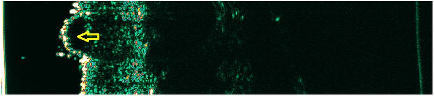
Polańska et al.                                                                                                         HF-USG Use in Teledermatology

 FIGURE 2 | Cellular nevus. Yellow arrow—hypoechogenic mass                   FIGURE 4 | Basal cell carcinoma. Yellow arrow—hypoechogenic mass
 corresponding to the nevus.                                                  corresponding to the basal cell carcinoma.

                                                                                 It is worth noting that the usefulness of HF-USG for assessing
                                                                             the thickness of BCC and melanoma infiltration has been well-
                                                                             documented and is now considered a recognized indication
                                                                             for HF-USG (14, 28–32). The preoperative determination of
                                                                             the surgical margins determines the future management and
                                                                             patients’ prognosis (particularly in the case of melanoma).
 FIGURE 3 | Melanoma. Yellow arrow—hypoechogenic mass corresponding to
                                                                             Previous studies confirm the agreement between a histological
 the melanoma.
                                                                             and ultrasonographic measurement with a high compliance
                                                                             rate (28–33). Therefore, the HF-USG assessment of tumor size
                                                                             may be then transferred to a dermato-oncologist or surgeon
                                                                             during teleconsultation to plan the future procedure. This way,
teledermatology modalities, particularly concerning pigmented
                                                                             teledermatology may globally contribute to better outcomes and
lesions and tumors.
                                                                             radical treatment. However, it should be considered that the
    Various skin pathologies change the echogenicity of its
                                                                             thickness of the tumor determined by ultrasound is slightly
individual layers, which can be used to analyze the ultrasound
                                                                             greater than the thickness determined histologically, due to
image. However, the lack of sufficient resolution does not allow
                                                                             shrinkage of the material during histological preparation and
a full interpretation of the image with microscopic examination
                                                                             accumulation of inflammatory cells underneath the tumor (10,
accuracy. Hypoechoic structures can be both of neoplastic
                                                                             24, 28). Another limitation of the proper margin assessment is
and inflammatory origin (14, 21, 22). Moreover, an identical
                                                                             the tumor size, mainly when it crosses the dermis border and
ultrasound image is observed in melanoma and a benign
                                                                             penetrates the subcutaneous tissue, which is also hypoechogenic.
melanocytic nevus. Due to the insufficient resolution of HF-USG,
                                                                             However, the use of a multifrequency probe may reduce this
the process cannot be distinguished at the cell level (Figures 2,
                                                                             limitation. The preoperative evaluation of the tumor margins
3). Although literature has tried to identify more specific features
                                                                             may also be difficult in patients with evident photoaging,
for various skin neoplasms, the final diagnosis should still be
                                                                             where elastosis decreases the skin echogenicity, similarly to the
supported by histological examination. In melanoma, echolucent
                                                                             neoplasmatic cells (14).
areas can be observed, with a shape depending on the subtype.
                                                                                 Due to the low diagnostic properties of HF-USG in
The nodular form may have a spherical arrangement, while
                                                                             oncodermatology, the combination of this method with a
superficial spreading melanoma presents as an echolucent area
                                                                             preliminary dermoscopic evaluation may be extremely useful.
parallel to the entry echo. However, there are no ultrasonographic
                                                                             Therefore, an initial diagnosis of probable pathology should
features pathognomonic for melanoma (14).
                                                                             be performed using a dermoscope, followed by an evaluation
    What is more, the accuracy of ultrasound analysis may be
                                                                             of disease extent using HF-USG. Acquired data, provided in
increased with a Color Doppler ultrasound. This modality is
                                                                             the form of telecommunication, may significantly improve the
recommended, especially for discriminating between vascular or
                                                                             diagnostic and therapeutic process. These observations remain
non-vascular lesions and recognizing the benign or malignant
                                                                             consistent with teledermatology’s role in the process of triaging
nature of skin tumors (15, 23, 24). In previous studies, vascular
                                                                             (particularly the SAF method), which influences the cost-
signal use in assessing melanoma correlated with the Breslow
                                                                             effectiveness of therapies and may limit waiting time in the case
index and metastatic potential (23). The addition of Color-
                                                                             of urgent patients (34). Moreover, the inclusion of dermatoscopic
Doppler in pigmented lesion analysis may have a significant
                                                                             images improves triaging decisions, including a need for an
prognostic value (25, 26).
                                                                             excision for both melanomas and other skin cancers (35).
    In basal cell carcinoma (BCC), the HF-USG can detect the
presence of hyperechoic spots, the so-called cotton flowers;
however, in the authors’ opinion, this is not a phenomenon that              THE MONITORING PROPERTIES OF
occurs in all tumors and therefore cannot be defining for BCC                HF-USG IN TELEDERMATOLOGY
(14, 21, 27) (Figure 4). In squamous cell carcinoma (SCC) and
actinic keratosis, scaling may disturb the image’s interpretation.           Due to the limited diagnostic value of ultrasonography, the
This seems to be a crucial limitation of HF-USG in evaluating                monitoring properties of HF-USG are noteworthy. They allow
hyperkeratotic tumors (10, 18, 19).                                          for observing changes dynamically—occurring in the skin over

Frontiers in Medicine | www.frontiersin.org                              3                                      February 2021 | Volume 8 | Article 619965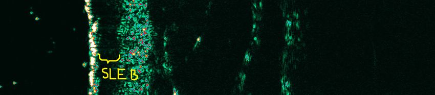
Polańska et al. HF-USG Use in Teledermatology
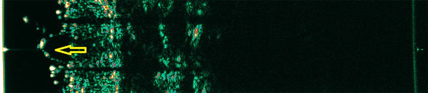
of evidence-based medicine (41). We presented that SLEB was
detected within lesional skin and in non-affected skin regions,
revealing the presence of subclinical inflammation (42, 43). Such
patients may significantly benefit most from proactive treatment
(40, 42). Information on the decrease of SLEB or lack of its change
could be provided in the form of a teleconference to the attending
FIGURE 5 | Subepidermal low echogenic band (SLEB) within the plaque of physician with information about the need to intensify the
mycosis fungoides before treatment (the hypoechogenic band corresponding treatment process. It could also be combined with an emerging
to the SLEB was marked). teledermatology branch—patient-assisted teledermatology (also
called home-based teledermatology)—which involves the SAF
method and mobile technology tools. It is regarded as especially
useful for patients with chronic dermatoses (psoriasis, atopic
dermatitis, vitiligo, leg ulcers) and allows the specialist to monitor
the phase and the degree of skin lesions. The above method is
relatively new (started in 2009) and is used for both diagnosis and
follow-up (20, 35).
SLEB thickness objective measurement as a useful, accurate
FIGURE 6 | The thinning of the SLEB after treatment. Lack of complete
disappearance of SLEB still shows the presence of neoplastic T cell infiltrations
parameter for skin assessment was also performed in psoriasis
(the hypoechogenic band corresponding to the SLEB was marked). (14, 36). Queille-Roussel et al. (36) evaluated an innovative
aerosol foam formulation’s anti-psoriatic effect and analyzed
this parameter’s thickness. They found a reduction of SLEB in
week 4. We also assessed monitoring properties of HF-USG in
time, mainly due to the introduced treatment (Figures 5, 6). a prospective, observational study, which included 58 patients
Inflammatory and neoplastic cells cause a reduction of skin diagnosed with recurrent chronic small plaque psoriasis treated
echogenicity and may result in the formation of a linear with a two-compound ointment containing calcipotriol and
band underneath the entry echo, known as the subepidermal betamethasone dipropionate or NB-UVB (311 nm) (11). The
low echogenic band (SLEB) (11, 12, 14). Originally, SLEB SLEB was significantly decreased in both treated groups. The
was observed in photodamaged skin due to glycosaminoglycan lack of its decrease, similarly to atopic dermatitis, indicated
accumulation, which possesses an increased water-binding no improvement in the dermatological condition or the need
capacity (14, 18). The SLEB formation was well-documented in to change/intensify therapy (11, 14). Such information could
inflammatory diseases, including psoriasis and atopic dermatitis be consulted with a dermatologist without the need to travel
(11–13, 36, 37). It is not a parameter specific for any skin and access the specialist advice more easily, especially in rural
disease, but its changes over time are of prognostic significance, areas. What is essential, a high correlation between SLEB and
especially in patients with chronic skin diseases. The areas of PASI index indicates that HF-USG quickly complements the
reduced echogenicity are mainly related to swelling of the skin measurement of the severity of skin lesions in psoriasis in an
and inflammatory cell infiltration. The SLEB thickness in atopic objective way (11).
dermatitis correlated with the degree of several histopathologic Monitoring properties of HF-USG seem to be particularly
findings like epidermal hyperplasia, epidermal hyperkeratosis, useful in assessing the therapeutical outcomes in patients with
the degree of parakeratosis, and the degree of spongiosis as well mycosis fungoides. They can be used in teleconsultations also
as the intensity of inflammatory infiltrates (38). However, in with a non-dermatologist (i.e., hematologist). Neoplastic cells
psoriatic plaque, SLEB’s uniformity can be disturbed by streaky are responsible for decreasing echogenicity, which in mycosis
shadows perpendicular to the entry echo (most likely caused by fungoides also manifests in the form of SLEB, similarly to
air bubbles trapped between the scales). It is observed that SLEB inflammatory diseases (12, 14, 44–46). We have found strong
thickness corresponds to the severity of skin lesions and can be correlations between SLEB thickness and the thickness of
used to monitor patients, also potentially as a teledermatology subepidermal infiltration in histopathological examination (47).
consultation (14, 39). As it is known, in mycosis fungoides, the proper staging is
In one of our first studies, monitoring properties of HF- essential in planning the therapy. It should include a detailed
USG were assessed for atopic dermatitis and so-called proactive evaluation of the skin lesion types and an assessment of the
therapy, using tacrolimus ointment (40). Six-month ultrasound pathological process’ extent (45, 47). For 5 years, we followed
monitoring was performed in a group of 39 patients with up patients based on HF-USG, where SLEB was the assessed
atopic dermatitis (mean age 26 years). It revealed the significant parameter (48). We evaluated well-known forms of therapies
decrease of SLEB value during the undergone therapy, which in mycosis fungoides (the effectiveness of UVA1 and PUVA).
correlated with the disease severity assessed by measuring scales We monitored these patients’ clinical responses compared to
(40). We found that HF-USG allowed clinicians to visualize the modified Severity Weighted Assessment Tool (mSWAT)
pathologic changes of all skin in vivo. As a non-invasive and (12). Complete response in examined patients correlated with
independent of subjective judgment method, HF-USG should the entire disappearance of SLEB (12, 48). SLEB thickness was
be added to the overall patient evaluation, especially in the era associated with disease severity and was wider in stage IIA
Frontiers in Medicine | www.frontiersin.org 4 February 2021 | Volume 8 | Article 619965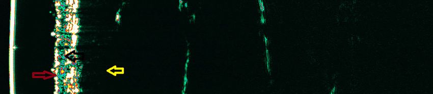
Polańska et al.                                                                                                                      HF-USG Use in Teledermatology

patients than in stage IA and IB patients (48). For the first time,                    of multiple scans without the need to prepare the patient in
we revealed that HF-USG is a tool for evaluating the patient’s                         advance. It seems that in the era of scrupulously determined
response and can be applied in routine clinical practice.                              scales determining the extent and severity of skin lesions during
   What is more, using HF-USG, there is a possibility                                  various treatments of skin diseases, especially to qualify patients
to distinguish a lesion of the type of post-inflammatory                               for biological therapies and research programs, HF-USG can find
hyper/hypopigmentation from a non-specific infiltrative lesion                         an application. In the case of teledermatology, the possibility
(12, 47, 48). Performing ultrasound measurements enables                               of transmitting images in between clinicians and of archiving
the patient to be consulted with a dermatologist/hematologist                          images and analyzing them over time contributes an extremely
without a face-to-face visit and can significantly improve the                         useful visual in vivo method. Teleconsultation with the use
therapeutic process. In the case of irradiated patients, a routine                     of HF-USG may improve the treatment process of chronic
practice in our clinic is the ultrasound supervision of patients                       dermatological diseases requiring long-term care. The possibility
and making decisions about additional radiation if a visible SLEB                      of simultaneous dermoscopy and ultrasound of the skin may have
is still observed. Again, collected data can be then sent to a                         a prognostic significance in skin tumors.
photodermatology center.                                                                   Among the main limitations of HF-USG, its high price
   The practical application of HF-USG is its use in hidradenitis                      should be mentioned, as well as restricted availability of the
suppurativa (49). Recent studies proposed this method to                               apparatus in a doctor’s office. As in the case of dermoscopy,
optimize staging, treatment planning, and monitoring patients                          acquiring the ability to interpret an ultrasonographic image
suffering from this chronic disease (49). Wortsman et al.                              requires appropriate training. Lately, also other techniques of
(50) proposed a three-point sonographic scoring system for                             non-invasive skin visualization are gaining interest in regard
hidradenitis suppurativa, based on the number and distribution                         to associations with teledermatology—for example, reflectance
of fluid collections, fistulous tracts, and pseudocystic nodules,                      confocal microscopy (RCM). RCM allows an in vivo evaluation
widening of the hair follicles, and alterations in the dermal                          of various skin lesions on the cellular level—nearly to histologic
thickness/echogenicity. According to the authors, the proper                           resolution. In comparison to this technique, it has to be
staging of these patients resulted in a management modification                        emphasized that HF-USG does not possess sufficient resolution
in 82% of cases (51). It seems that HF-USG in relation                                 and its diagnostic properties are much lower (52).
to hidradenitis suppurativa may also be conducted in the
form of teleconsultation with other specialists (diagnostic and                        AUTHOR CONTRIBUTIONS
radiological center, dermatological surgeons). The possibility of
an ultrasound consultation can improve diagnosis and help                              All authors listed have made a substantial, direct and intellectual
choose an appropriate therapeutic option (depending on the                             contribution to the work, and approved it for publication.
severity of skin lesions).
                                                                                       ACKNOWLEDGMENTS
THE ADVANTAGES AND LIMITATIONS OF
HF-USG                                                                                 The authors are entirely grateful to Dr. Tomasz Maksymiuk
                                                                                       (MD, Ph.D.), who provided English grammar editing and
The most significant advantages of HF-USG in a monitoring                              really improved the quality and understanding of the
examination are non-invasiveness, safety, and the possibility                          whole manuscript.

REFERENCES                                                                              8. Alexander H, Miller DL. Determining skin thickness with pulsed
                                                                                           ultrasound. J Invest Dermatol. (1979) 72:17–9. doi: 10.1111/1523-1747.ep125
 1. Telemedicine: Opportunities and Developments in Member States: Report on               30104
    the Second Global Survey on eHealth 2009. World Health Organization (2010).         9. Pellacani G, Seidenari S. Preoperative melanoma thickness determination
 2. Waller M, Stotler C. Telemedicine: a primer. Curr Allergy Asthma Rep. (2018)           by 20-MHz sonography and digital videomicroscopy in combination. Arch
    25:54. doi: 10.1007/s11882-018-0808-4                                                  Dermatol. (2003) 139:293–8. doi: 10.1001/archderm.139.3.293
 3. Bischoff A. Benefits and risks from telemedicine. MMW Fortschr Med. (2016)         10. Hoffmann K, el-Gammal S, Winkler K, Jung J, Pistorius K, Altmayer P.
    158:18–9. doi: 10.1007/s15006-016-8250-9                                               Skin tumours in high-frequency ultrasound. In: Altmeyer P, el-Gammal S,
 4. Zaba R. Telemedycyna w dermatologii. Medicus Mundi Polonia. (2009) 28:7–               Hoffmann K, editors. Ultrasound in Dermatology. Berlin; New York, NY:
    8.                                                                                     Springer- Verlag (1992). p. 181–202.
 5. Coates SJ, Kvedar J, Granstein RD. Teledermatology: from historical                11. Polańska A, Gaura T, Bowszyc-Dmochowska M, Osmola-Mańkowska
    perspective to emerging techniques of the modern era: part I: history,                 A, Olek-Hrab K, Adamski Z, et al. Calcipotriol/betamethasone
    rationale, and current practice. J Am Acad Dermatol. (2015) 72:563–                    ointment compared to narrow-band UVB in plaque psoriasis:
    74. doi: 10.1016/j.jaad.2014.07.061                                                    first clinical and ultrasonographic study. Int J Dermatol. (2019)
 6. Jankowski M, Klimczak-Wieczorek A, Kloc M, Matuszewski M, Rozum J.                     58:108–13. doi: 10.1111/ijd.14150
    Telemedycyna w Polsce – mozliwości i szanse rozwoju. Fundacja im. Lesława         12. Polańska A, Osmola-Mańkowska A, Olek-Hrab K, Molińska-Glura M,
    A. Pagi, Warszawa (2016).                                                              Adamski Z, Zaba R, et al. High-frequency ultrasonography in objective
 7. Kanthraj GR. Patient-assisted teledermatology practice: what is it? When,              evaluation of the efficacy of PUVA and UVA 1 phototherapy in mycosis
    where, and how it is applied? Indian J Dermatol Venereol Leprol. (2015)                fungoides. Arch Dermatol Res. (2017) 309:645–51. doi: 10.1007/s00403-017-1
    17:136–43. doi: 10.4103/0378-6323.152172                                               767-7

Frontiers in Medicine | www.frontiersin.org                                        5                                         February 2021 | Volume 8 | Article 619965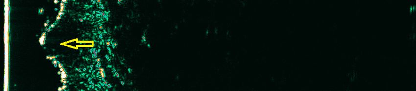
Polańska et al.                                                                                                                           HF-USG Use in Teledermatology

13. Wortsman X, Moreno C, Soto R, Arellano J, Pezo C, Wortsmanet J.                         33. Seidenari S. High-frequency sonography combined with image analysis:
    Ultrasound in-depth characterization and staging of hidradenitis suppurativa.               a non-invasive objective method for skin evaluation and desription. Clin
    Dermatol Surg. (2013) 39:1835–42. doi: 10.1111/dsu.12329                                    Dermatol. (1995) 13:349–59. doi: 10.1016/0738-081X(95)00074-P
14. Polańska A, Dańczak-Pazdrowska A, Jałowska M, Adamski Z, Zaba R.                      34. Congalton AT, Oakley AM, Rademaker M, Bramley D, Martin R. Successful
    Current applications of high-frequency ultrasonography in dermatology. Adv                  melanoma triage by a virtual lesion clinic (teledermoscopy). J Eur Acad
    Dermatol Allergol. (2017) 6:535–42. doi: 10.5114/ada.2017.72457                             Dermatol Venereol. (2015) 29:2423–8. doi: 10.1111/jdv.13309
15. Wortsman X, Alfageme F, Roustan G, Arias-Santiago S, Martorell A,                       35. Kanthraj GR. Classification and design of teledermatology practice: what
    Catalano O, et al. Guidelines for performing dermatologic ultrasound                        dermatoses? Which technology to apply? J Eur Acad Dermatol Venereol.
    examinations by the DERMUS group. J Ultrasound Med. (2016) 35:577–                          (2009) 23:865–75. doi: 10.1111/j.1468-3083.2009.03136.x
    80. doi: 10.7863/ultra.15.06046                                                         36. Queille-Roussel C, Olesen M, Villumsen J, Lacour J. Efficacy of an
16. Dill-Müller D, Maschke J. Ultrasonography in dermatology. J Dtsch Dermatol                  innovative aerosol foam formulation of fixed combination calcipotriol plus
    Ges. (2007) 5:689–707. doi: 10.1111/j.1610-0387.2007.06453.x                                betamethasone dipropionate in patients with psoriasis vulgaris. Clin Drug
17. Ng A, Swanevelder J. Resolution in ultrasound imaging. Continuing Educ.                     Investig. (2015) 35:239–45. doi: 10.1007/s40261-015-0269-7
    Anaesthesia Crit Care Pain. (2011) 11:186–92. doi: 10.1093/bjaceaccp/mkr030             37. Yazdanparast T, Yazdani K, Humbert P, Khatami A, Nasrollahi SA,
18. Jasaitiene D, Valiukeviciene S, Linkeviciute G, Raisutis R, Jasiuniene E,                   Firouzabadi LI, et al. Biophysical measurements and ultrasonographic
    Kazyset R. Principles of high-frequency ultrasonography for investigation                   findings in chronic dermatitis in comparison with uninvolved skin. Indian J
    of skin pathology. J Eur Acad Dermatol Venereol. (2011) 25:375–                             Dermatol. (2019) 64:90–6. doi: 10.4103/ijd.IJD_464_17
    82. doi: 10.1111/j.1468-3083.2010.03837.x                                               38. Polańska A, Dańczak-Pazdrowska A, Silny W, Wozniak A, Maksin
19. Jemec GB, Gniadecka M, Ulrich J. Ultrasound in dermatology. Part I: high                    K, Jenerowicz D. Comparison between high-frequency ultrasonography
    frequency ultrasound. Eur J Dermatol. (2000) 10:492–7.                                      (Dermascan C, version 3) and histopathology in atopic dermatitis. Skin Res
20. Warshaw E, Greer N, Hillman Y, Hagel E, MacDonald R, Rutks I, et                            Technol. (2013) 19:432–7. doi: 10.1111/srt.12064
    al. Teledermatology for Diagnosis and Management of Skin Conditions: A                  39. Gupta AK, Turnbull DH, Harasiewicz KA, Shum DT, Watteel GN, Foster
    Systematic Review of the Evidence. VA-ESP Project #09-009 (2009).                           FS, et al. The use of high-frequency ultrasound as a method of assessing
21. Wortsman X, Jemec GBE. High resolution ultrasound applications in                           the severity of a plaque of psoriasis. Arch Dermatol. (1996) 132:658–
    dermatology. Rev Chilena Dermatol. (2006) 22:37–45.                                         62. doi: 10.1001/archderm.1996.03890300076011
22. Harland CC, Kale SG, Jackson P, Mortimer PS, Bamberet JC.                               40. Polańska A, Silny W, Jenerowicz D, Knioła K, Molińska-Glura M, Dańczak-
    Differentiation of common benign pigmented skin lesions from                                Pazdrowska A. Monitoring of therapy in atopic dermatitis- observations
    melanoma by high-resolution ultrasound. Br J Dermatol. (2000)                               with the use of high-frequency ultrasonography. Skin Res Technol. (2015)
    143:281–9. doi: 10.1046/j.1365-2133.2000.03652.x                                            21:35–40. doi: 10.1111/srt.12153
23. Scotto di Santolo M, Sagnelli M, Mancini M, Scalvenzi M,                                41. Osmola-Mańkowska A, Polańska A, Silny W, Zaba R, Adamski A,
    Delfino M, Schonauer F, et al. High-resolution Color-Doppler                                Dańczak-Pazdrowska A. Topical tacrolimus vs medium-dose ultraviolet al
    ultrasound for the study of skin growths. Arch Dermatol Res. (2015)                         phototherapy in the treatment of atopic dermatitis—a preliminary study in
    307:559–66. doi: 10.1007/s00403-015-1538-2                                                  relation to parameters of the epidermal barrier function and high-frequency
24. Catalano O, Roldán FA, Varelli C, Bard R, Corvino A, Wortsman X. Skin                       ultrasonography. Eur Rev Med Pharmacol Sci. (2014) 18:3927–34.
    cancer: findings and role of high-resolution ultrasound. J Ultrasound. (2019)           42. Dańczak-Pazdrowska A, Polańska A, Silny W, Sadowska A, Osmola-
    22:423–31. doi: 10.1007/s40477-019-00379-0                                                  Mańkowska A, Czarnecka-Operacz M, et al. Seemingly healthy skin
25. Catalano O, Voit C, Sandomenico F, Mandato Y, Petrillo M, Franco R,                         in atopic dermatitis: observations with the use of high-frequency
    et al. Previously reported sonographic appearances of regional melanoma                     ultrasonography, preliminary study. Skin Res Technol. (2012)
    metastases are not likely due to necrosis. J Ultrasound Med. (2011) 30:1041–                18:162–7. doi: 10.1111/j.1600-0846.2011.00548.x
    9. doi: 10.7863/jum.2011.30.8.1041                                                      43. Polańska A, Dańczak-Pazdrowska A, Silny W, Jenerowicz D, Olek-Hrab K,
26. Lassau N, Lamuraglia M. Prognostic value of angiogenesis evaluated                          Osmaola-Mańkowska A. Nonlesional skin in atopic dermatitis is seemingly
    with high frequency and Color-Doppler sonography for preoperative                           healthy skin - observations using noninvasive methods. Wideochir Inne Tech
    assessment of primary cutaneous melanomas: correlation with                                 Maloinwazyjne. (2013) 8:192–9. doi: 10.5114/wiitm.2011.33633
    recurrence after a 5 year follow-up period. Cancer Imaging. (2006)                      44. Polańska A, Osmola-Mańkowska A, Olek-Hrab K. Do we need high-
    25:24–9. doi: 10.1102/1470-7330.2006.0009                                                   frequency ultrasonography in mycosis fungoides? Abstract Book P1065,
27. Wang SQ, Liu J, Zhu QL, Zhao CY, Qu T, Li F, et al. High-frequency ultrasound               EADV 2016 Vienna.
    features of basal cell carcinoma and its association with histological recurrence       45. Mandava A, Koppula V, Wortsman X, Catalano O, Alfageme F.
    risk. Chin Med J. (2019) 132:2021–6. doi: 10.1097/CM9.0000000000000369                      The clinical value of imaging in primary cutaneous lymphomas:
28. Tacke J, Haagen G, Hornstein OP, Tacke J, Haagen G, Hornstein                               role of high resolution ultrasound and PET-CT. Br J Radiol. (2019)
    P, et al. Clinical relevance of sonometry-derived tumour thickness in                       92:20180904. doi: 10.1259/bjr.20180904
    malignant melanoma: a statistical analysis. Br J Dermatol. (1995) 132:209–              46. Olek-Hrab K, Silny W, Dańczak-Pazdrowska A, Osmola-Mańkowska A,
    14. doi: 10.1111/j.1365-2133.1995.tb05015.x                                                 Sadowska A, Polańska A, et al. Ultraviolet A1 phototherapy for mycosis
29. Lassau N, Koscielny S, Avril MF, Margulis A, Duvillard P, De Baere T,                       fungoides. Clin Exp Dermatol. (2013) 38:126–30. doi: 10.1111/ced.12001
    et al. Prognostic value of angiogenesis evaluated with high-frequency and               47. Polańska A, Bowszyc-Dmochowska M, Olek-Hrab K, Adamski Z, Zaba R,
    color Doppler sonography for preoperative assessment of melanomas. Am J                     Dańczak-Pazdrowska A. High-frequency ultrasonography a new quantitative
    Roentgenol. (2002) 178:1547–51. doi: 10.2214/ajr.178.6.1781547                              method in evaluation of skin lymphomas-first comparative study in relation
30. Serrone L, Solivetti FM, Thorel MF, Eibenschutz L, Donati P,                                to histopathology. Skin Res Technol. (2019) 25:720–4. doi: 10.1111/srt.
    Catricalàet C. High frequency ultrasound in the preoperative staging                        12708
    of primary melanoma: a statistical analysis. Melanoma Res. (2002)                       48. Polańska A, Dańczak-Pazdrowska A, Olek-Hrab K, Osmola-
    12:287–90. doi: 10.1097/00008390-200206000-00013                                            Mańkowska A, Bowszyc-Dmochowska M, Zaba R, et al. High-frequency
31. Guitera P, Li LX, Crotty K, Fitzgerald P, Mellenbergh R,                                    ultrasonography-new non-invasive method in assessment of skin
    Pellacani G, et al. Melanoma histological Breslow thickness                                 lymphomas. Skin Res Technol. (2018) 24:517–21. doi: 10.1111/srt.
    predicted by 75-MHz ultrasonography. Br J Dermatol. (2008)                                  12450
    159:364–9. doi: 10.1111/j.1365-2133.2008.08681.x                                        49. Lacarrubba F, Dini V, Napolitano M, Venturini M, Caposiena Caro DR,
32. Bobadilla F, Wortsman X, Muñoz C, Segovia L, Espinoza M,                                    Molinelli E, et al. Ultrasonography in the pathway to an optimal standard
    Jemec G. Pre-surgical high resolution ultrasound of facial basal cell                       of care of hidradenitis suppurativa: the Italian Ultrasound Working Group
    carcinoma: correlation with histology. Cancer Imaging. (2008) 22:63–72.                     experience. J Eur Acad Dermatol Venereol. (2019) 33:10–4. doi: 10.1111/jdv.
    doi: 10.1102/1470-7330.2008.0026                                                            15847

Frontiers in Medicine | www.frontiersin.org                                             6                                         February 2021 | Volume 8 | Article 619965
Polańska et al. HF-USG Use in Teledermatology
50. Wortsman X. Imaging of hidradenitis suppurativa. Conflict of Interest: The authors declare that the research was conducted in the
Dermatol Clin. (2016) 34:59–68. doi: 10.1016/j.det.2015. absence of any commercial or financial relationships that could be construed as a
08.003 potential conflict of interest.
51. Nazzaro G, Passoni E, Guanziroli E, Casazza G, Muratori S, Barbareschiet
M, et al. Comparison of clinical and sonographic scores in a cohort Copyright © 2021 Polańska, Jenerowicz, Paszyńska, Żaba, Adamski and Dańczak-
of 140 patients with hidradenitis suppurativa from an Italian referral Pazdrowska. This is an open-access article distributed under the terms of the Creative
centre: a retrospective observational study. Eur J Dermatol. (2018) Commons Attribution License (CC BY). The use, distribution or reproduction in
28:845–7. other forums is permitted, provided the original author(s) and the copyright owner(s)
52. Witkowski A, Łudzik J, Soyer P. Telediagnosis with confocal microscopy: a are credited and that the original publication in this journal is cited, in accordance
reality or a Dream? Dermatol Clin. (2016) 4:505–12. doi: 10.1016/j.det.2016. with accepted academic practice. No use, distribution or reproduction is permitted
05.013 which does not comply with these terms.
Frontiers in Medicine | www.frontiersin.org 7 February 2021 | Volume 8 | Article 619965You can also read