Xanthohumol Attenuated Inflammation and ECM Degradation by Mediating HO-1/C/EBPβ Pathway in Osteoarthritis Chondrocytes - Frontiers
←
→
Page content transcription
If your browser does not render page correctly, please read the page content below
ORIGINAL RESEARCH
                                                                                                                                              published: 04 May 2021
                                                                                                                                      doi: 10.3389/fphar.2021.680585

                                           Xanthohumol Attenuated
                                           Inflammation and ECM Degradation
                                           by Mediating HO-1/C/EBPβ Pathway
                                           in Osteoarthritis Chondrocytes
                                           Ming Zhang 1, Rui Zhang 2, Tiansheng Zheng 3, Zhixi Chen 2, Guanglin Ji 3, Fang Peng 4 and
                                           Wei Wang 5*
                                           1
                                             Department of Orthopedics, Taizhou People’s Hospital, Taizhou, China, 2College of Pharmacy, Gannan Medical University,
                                           Ganzhou, China, 3Department of Orthopedics, The First Affiliated Hospital of Gannan Medical University, Ganzhou, China,
                                           4
                                             Department of Pathology, The Affiliated Ganzhou Hospital of Nanchang University, Ganzhou, China, 5Department of Hepatology,
                                           Taizhou People’s Hospital, Taizhou, China

                                           Osteoarthritis (OA) is the most frequent and disabling disease in developed countries. The
                                           progressive degeneration of articular cartilage characterized as thinner and erosive.
                                           Inflammation is well-known to be involved in OA development. However, there are no
                            Edited by:     effective therapeutic strategies to cure it. Xanthohumol (XH) is a natural prenylflavonoid
                     Gabriel A. Agbor,     isolated from hops and beer. The protective activity of XH against OA chondrocytes
    Institute of Medical Research and
Studies of Medicinal Plants, Cameroon
                                           inflammation and ECM degradation is unclear. In this article, we found that XH significantly
                         Reviewed by:
                                           inhibited inflammatory responses, attenuated catabolic enzymes expression, and
        Aristide Laurel Mokale Kognou,     ameliorated ECM degradation, as showed by decreased production of NO, PGE2,
  Institut de Recherches Médicales et
                                           TNFα, and IL-6, decreased expression of MMP-3/-13 and ADAMTS-4/-5, and
    d’Etudes des Plantes Médicinales,
                             Cameroon      increased expression of collagen-II and aggrecan. In addition, XH activated HO-1
                     David L Kooyman,      signaling and attenuated IL-1β-induced C/EBPβ. XH promoted the interaction between
              Brigham Young University,
                          United States
                                           HO-1 and C/EBPβ, inhibiting the nuclear translocation of C/EBPβ. HO-1 knockdown could
                  *Correspondence:
                                           abrogate the protective effects of XH in IL-1β-treated chondrocytes. Collectively, XH
                           Wei Wang        attenuated inflammatory responses and ECM degradation by mediating HO-1 and
                   aerie@njmu.edu.cn
                                           C/EBPβ signaling pathways in osteoarthritis chondrocytes.
                    Specialty section:     Keywords: xanthohumol, osteoarthritis, ECM degradation, C/EBP, HO-1, mmp-13
         This article was submitted to
                  Ethnopharmacology,
               a section of the journal    INTRODUCTION
            Frontiers in Pharmacology
          Received: 15 March 2021          Osteoarthritis (OA), one of the age-related debilitating and degenerative diseases, is often clinically
           Accepted: 23 April 2021         characterized by joint pain, limited movement, and transient morning stiffness (Shamdani et al.,
           Published: 04 May 2021          2020). Mechanistically, cartilage degradation, synovial inflammation, and subchondral bone
                             Citation:     remodeling are involved in the pathological development of OA (Dai et al., 2017). However, the
Zhang M, Zhang R, Zheng T, Chen Z,         underlying mechanisms are still unclear, and no effective treatments are available to cure OA
   Ji G, Peng F and Wang W (2021)          (Altman and Barthel, 2011).
Xanthohumol Attenuated Inflammation
                                               Inflammation and inflammatory responses have been considered as the main factors contributing
 and ECM Degradation by Mediating
            HO-1/C/EBPβ Pathway in
                                           to the pathological development of OA. Inflammatory cytokines, such as IL-1β and TNFα, are closely
          Osteoarthritis Chondrocytes.     involved in the aberrant metabolism and promote the catabolic activities of cartilage tissues in OA
       Front. Pharmacol. 12:680585.        (Nambi, 2020). Expectedly, IL-1β dramatically contributes to the cartilage destruction of OA, which
     doi: 10.3389/fphar.2021.680585        is associated with up regulation of C/EBPβ in chondrocytes (Nishimura et al., 2020). Increasing

Frontiers in Pharmacology | www.frontiersin.org                                   1                                            May 2021 | Volume 12 | Article 680585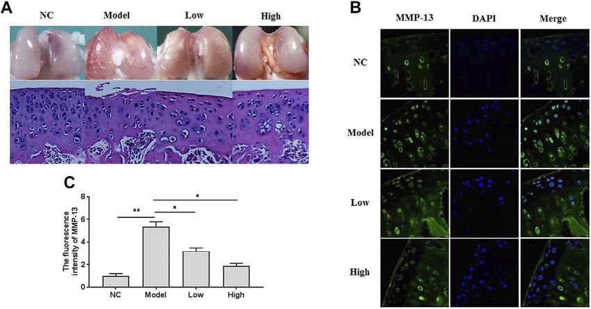
Zhang et al.                                                                                                    XH Attenuated ECM Degradation

evidence shows that IL-1β suppresses the production of the              no. 3082), HO-1 (Cat. no. 43966), MMP-3 (Cat. no. 14351),
component of extracellular matrix (ECM), synthesized by the             GAPDH (Cat. no. 5174S), and the horseradish peroxidase-
unique chondrocytes. The possible mechanisms might be                   labelled secondary antibody (Cat. no. 7074S) were purchased
associated with the inductive activity of IL-1β on the                  from Cell Signaling Technology. MMP-13 (Cat. no. 701287),
expression of matrix metalloproteinases (MMPs), thrombin                ADAMTS-4 (Cat. no. PA5-69140), ADAMTS-5 (Cat. no. PA5-
sensitive protein motifs (ADAMTS), cyclooxygenase-2 (COX-               32142), collagen-II (Cat. no. MA5-12789), and aggrecan (Cat. no.
2), and inducible nitric oxide synthase (iNOS), which are the           MA3-16888) were obtained from Invitrogen. Recombinant rat
main catabolic factors for the destruction of articular cartilage       IL-1β (Cat. no. ab9788) purchased from Abcam.
(Wu et al., 2019). Nuclear factor-erythroid 2-related factor
(Nrf2)/HO-1 is the critical factor mediating the expression of          Rat Knee OA Models
anti-oxidative enzymes and balancing the redox homeostasis              Rat knee OA models were duplicated using a classic
(Zhao et al., 2020). Up regulation of NRF2/HO-1 expression              osteoarthritic destabilization of the medial meniscus
may ameliorate IL-1β-induced inflammation and induction of               (DMM) model through surgery (Glasson et al., 2007).
ECM degradation (Gu et al., 2020). Promisingly, NRF2/HO-1 has           Simply, eight-week-old male Sprague-Dawley rats (200 g)
become the potential target for the therapeutic management              were anesthetized with 3% (w/v) pentobarbital (30 mg/kg)
of OA.                                                                  intraperitoneally. Then, surgery was conducted to cut the
   Non-steroidal anti-in flammatory drugs (NSAIDs) have been             medial meniscus tibial ligament through the medial patellar
widely used for treating OA, alleviating clinical symptoms, such        tendon of the capsule in the right knee joints. The sham group
as pain and swelling. However, they cannot inhibit the                  was performed without cutting off the medial meniscus tibial
progression of OA development but produce many serious                  ligament in the left knee joints. After eight weeks of post-
adverse effects (Bournia et al., 2017). It is emerging to explore       operation, rats were sacrificed, and joint cartilages were
the potential candidates for OA treatment. Recently, morroniside,       collected for gross observation, histochemical examination,
a bioactive compound isolated from Cornus officinalis, has been          and immunohistochemical evaluation. The low and high
showed to protect osteoarthritic cartilage by attenuation of            doses of XH for treating rats OA were 5.64 mg/kg and
inflammation in mice chondrocytes (Park et al., 2021).                   16.9 mg/kg, respectively, which were reported to be
Engeletin (dihydrokaempferol 3-rhamnoside) is reported to               corresponding to the values of 60 mg and 180 mg XH,
decrease TNFα-induced chondrocytes apoptosis and the                    respectively, for humans with a body weight of 66 kg
production of ROS in rat knee cartilage (Wang et al., 2021).            (Legette et al., 2012). Different doses of XH were dissolved
Acacetin can significantly ameliorate IL-1β-induced matrix               in 0.5% DMSO. The sham group received the same volumes of
metalloproteinases expression in chondrocytes by inhibiting             dosing vehicle as the treating groups.
NF-κB signaling pathways in mice (Chen et al., 2020a). The
therapeutic single compounds for osteoarthritis treatment have          Cell Culture
been reviewed recently (Lee et al., 2021). Xanthohumol (XH), a          Under sterile conditions, the articular cartilage of the articulatio
natural prenylflavonoid isolated from hops and beer, has been            genus, terminal femur, and upper tibia was collected, cut into
preventing hyaluronan overproduction and subsequent response            small pieces, and digested with 0.25% pancreatic enzyme and
in the early stage of OA (Stracke et al., 2011). Recently, XH has       0.2% collagenase II at 37°C for 4 h. Cells were cultured in
been reported to reduce the nephrotoxicity induced by cisplatin         Dulbecco’s modified Eagle’s minimum essential medium
by activating Nrf2/HO-1 and attenuating NF-κB signaling                 (DMEM) (low glucose) (Life Technologies, NY, United States)
pathways (Li et al., 2018). However, the biological activity of         supplemented with 10% fetal bovine serum (FBS), penicillin, and
XH in osteoarthritic chondrocytes is still unclear.                     streptomycin (Life Technologies) at 37°C with 5% CO2. 24 h later,
                                                                        the fresh complete culture medium was used for replacement.
                                                                        The cells employed for all the experiments were the second or the
MATERIALS AND METHODS                                                   third generation of the primary cartilage cells, ensuring consistent
                                                                        cell phenotypes. XH dissolved in DMSO was prepared, and the
General                                                                 final concentration of DMSO was 0.1% in the culture medium.
This study was approved by the Institutional Animal Care and
Use Committee of Taizhou People’s Hospital and performed in             Cell Viability Assays
strict accordance with the guideline of Animal Care and Use (No.        The CCK8 kit (Cat. no. C0038) (Beyotime, Shanghai, China) was
TZPH2006381962), according to the Declaration of Helsinki               used for testing the chondrocytes viability according to the
Principles.                                                             manufacturer’s instructions. Chondrocytes were grown in 96-
                                                                        well plates at a density of 5000 cells/well for 24 h. Different
Chemicals and Reagents                                                  concentrations of XH (0, 5, 10, 20, 40, and 80 μM) were used to
Xanthohumol (XH) (purity98%) (Cat. no. D108095) was                     pretreat the chondrocytes for 24 and 48 h, respectively. Then,
obtained from BIOBW (Beijing, China). Collagenase type II               CCK-8 solution (10 μl) was added into each well and incubated at
(Cat. no. C2-28) and dimethylsulfoxide (DMSO) (Cat. no.                 37°C for 4 h. After that, the absorbance was detected at the
D8418) were purchased from Sigma Aldrich (St Louis, MO,                 wavelength of 450 nm by using a microplate system (Leica
United States). The primary antibodies against C/EBPβ (Cat.             microsystems, Germany).

Frontiers in Pharmacology | www.frontiersin.org                     2                                     May 2021 | Volume 12 | Article 680585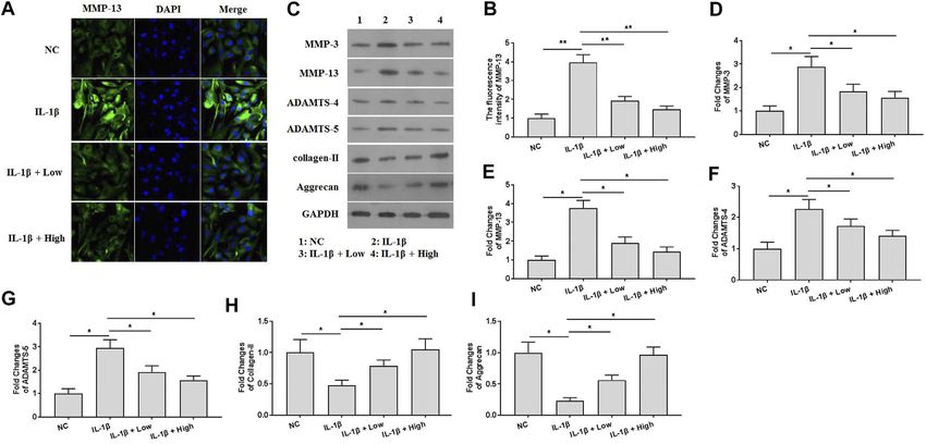
Zhang et al. XH Attenuated ECM Degradation
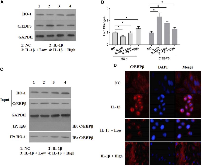
FIGURE 1 | The gross observation, histomorphological examination, and immunofluorescence assays in the knee articular cartilage. (A) The gross observation and
histomorphological examination (HE staining) of articular cartilage. (B) The immunofluorescence study of MMP-13 in cartilage. (C) The summary data of fluorescence
intensity of MMP-13 in situ. All experiments were performed in triplicate and data are presented as the mean ± standard deviation. *p < 0.05 and **p < 0.01. NC, negative
control; Model, the model group; Low, the group treated with XH (5.64 mg/kg); High, the group treated with XH (16.9 mg/kg).
Detection of NO, PGE2, TNFα, and IL-6 polyvinylidene fluoride membrane (Bio-Rad, CA,
Chondrocytes were grown in 6-well plates, and XH was added to United States). It was blocked by tris-buffered saline
treat for 24 h. Then, cells were treated with the recombinant IL-1β supplemented with 5% nonfat milk for 1 h at room
for another 24 h. The levels of NO metabolite nitrite were temperature. After washing three times with TBST (Tris-
detected as the production of NO by using sodium nitrite as buffered saline containing Tween 20), the membrane was
the standard (Au et al., 2007). Simply, 100 μl of the culture cut and incubated with the corresponding primary antibody
supernatant was permitted to react with Griess reagent in an at 4°C overnight. Then, it was washed with TBST for three
equal volume in 96-well microplate at room temperature for times and incubated with horseradish peroxidase-labelled
10 min in the dark. The absorbance was detected at the secondary antibody for 1 h at room temperature. Protein
wavelength of 540 nm by using a microplate system (Leica bands were detected using the enhanced chemiluminescence
microsystems, Germany). The productions of PGE2, TNFα, detection system (Bio-Rad Laboratories) and Quantity One
and IL-6 were determined by employment of ELISA kits software v4.6.2 (Bio-Rad Laboratories).
(Beyotime, Shanghai, China) according to the manufacturer’s
instructions. Co-immunoprecipitation Assay
Chondrocytes were harvested by the immunoprecipitation lysis
Cell Transfection buffer. Then, they were centrifugated at 12,000 rpm for 30 min
HO-1 siRNA and scramble siRNA (negative control), obtained 10% of chondrocytes lysates were kept and used as the input. The
from RiboBio (Guangzhou, China), were used for investigating remained proteins were immunoprecipitated by incubating with
the roles of HO-1 knockdown in the inflammatory responses in normal goat IgG or HO-1 in immunoprecipitation washing
chondrocytes. Cells were cultured in 6-well plates at a density of buffer at 4°C overnight. Next, they were incubated with the
1×10⁵ cells/well. Once they reached 60% confluence, cells were pre-cleared protein A-sepharose beads at 4°C for 2 h. The
used for the subsequent transfections. According to the eluted samples were then subjected to Western blot as
manufacturer’s instructions, transfections of HO-1 siRNA and mentioned above.
negative control were performed by using lipofectamine 3000
reagent (Thermo Fisher Scientific, Inc.) for 36 h at 50 nM. Immunofluorescence
Western blot assays were used for determining the transfection Chondrocytes were grown on glass coverslips for 24 h. After
efficiency. rinsing with PBS for three times, cells were fixed with 4%
paraformaldehyde for 15 min at room temperature and rinsed
Western Blotting with PBS again. 0.1% Triton X-100 was used to infiltrate cell and
RIPA lysis buffer (Beyotime Institute of Biotechnology) nuclear membranes for 5 min at room temperature. Then, cells
containing 1% PMSF was used to lyse cells. BCA protein were blocked by 5% protease-free bovine serum albumin (BSA)
assay kit (cat. no. 23250) (Pierce Biotechnology) was for 1 h at room temperature, rinsed with PBS, and incubated with
employed to quantify the total protein. 25 μg protein/lane the corresponding primary antibody at 4°C overnight. Cells were
was separated via gel electrophoresis and transferred to washed with PBS and incubated with fluorescein-conjugated goat
Frontiers in Pharmacology | www.frontiersin.org 3 May 2021 | Volume 12 | Article 680585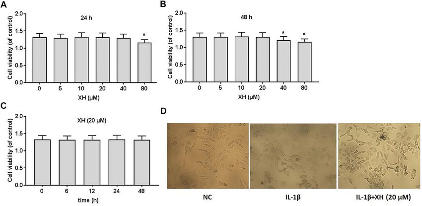
Zhang et al.                                                                                                                                   XH Attenuated ECM Degradation

 FIGURE 2 | Cell viability of XH on rat chondrocytes. CCK8 assays were conducted to determine the effects of XH (0, 5, 10, 20, 40, and 80 μM) in 24 h (A) and 48 h
 (B), respectively. (C) The effects of XH (20 μM) on cell viability in 48 h. (D) The cell morphology was observed after administration with IL-1β (10 ng/ml) with or without XH
 (20 μM) in 48 h. All experiments were performed in triplicate and data are presented as the mean ± standard deviation. *p < 0.05 and **p < 0.01.

anti-rabbit IgG antibody for 1 h at room temperature.                                       decreased thickness of articular cartilage were found in the model
Subsequently, cells were mounted in medium containing DAPI                                  group by hematoxylin-eosin (HE) staining. XH could protect
(Invitrogen) after washing with PBS. A confocal laser scanning                              articular cartilage from damage, as indicated by better cartilage
microscope (Leica Microsystems) was employed to observe the                                 surface, increased cell number, and thicker cartilage dose
slides, and the fluorescence intensity was detected by using                                 dependently. The immunofluorescence study of MMP-13 in situ
ImageJ software 2.1 (Bethesda, MD, United States).                                          (Figure 1B) showed that oral administration of XH might
                                                                                            significantly decrease the fluorescence intensity (Figure 1C) in a
Statistical Analysis                                                                        dose-dependent manner, compared with that in the model group.
All experiments were performed in triplicate and data are                                   These indicated that XH exhibited protective effects against de-
presented as the mean ± standard error of the mean. SPSS                                    structure in articular cartilage, which might be possibly associated
20.0 software (IBM Corp., Armonk, NY, United States) was                                    with down regulation of MMP-13 expression in chondrocytes.
used for statistical analysis. One-way ANOVA and followed by
Tukey’s post hoc test was used to make statistical comparisons                              Effects of XH on Cell Viability
between multiple groups. An unpaired Student’s t-test was used                              To detect the effects of XH on the viability of rat chondrocytes,
to make statistical comparisons between two groups. p < 0.05 was                            the different doses XH (0, 5, 10, 20, 40, and 80 μM) were
considered to indicate a statistically significant difference.                               administered to the cultured cells for 24 and 48 h, respectively,
                                                                                            before cell viability was detected using CCK8 assays. As indicated
                                                                                            in Figures 2A,B XH did not produce any cytotoxicity when the
RESULTS                                                                                     concentration of XH was less than 20 μM. After 48 h incubation,
                                                                                            XH at the dose of 40 μM slightly reduced the viability of
XH Exhibited Protective Activity Against OA                                                 chondrocytes, compared with that after 24 h incubation. At
Development in Rats                                                                         the dose of 20 μM, XH did not exhibit toxic effects on
To investigate the effects of XH on OA development, a surgical rat                          chondrocytes in 48 h (Figures 2C,D).
DMM model was duplicated and treated with XH (5.64 mg/kg and
16.9 mg/kg) for 8 weeks after operation by oral administration. As
showed in Figure 1A, gross observation and histomorphological
                                                                                            XH Decreased the Productions of
examination were studied. No obvious damages were seen in the                               Inflammatory Cytokines in IL-1β-Treated
negative control group. In contrast, the rough surface of cartilage                         Chondrocytes
with some erosion was observed in the model group. Consistently,                            To investigate the effects of XH on chondrocytes regarding
the decreased number and disorder distribution of chondrocytes and                          inflammation, cells were pretreated with IL-1β (10 ng/ml) and

Frontiers in Pharmacology | www.frontiersin.org                                         4                                               May 2021 | Volume 12 | Article 680585
Zhang et al. XH Attenuated ECM Degradation
FIGURE 3 | Effects of XH on the production of NO, PGE2, TNFα, and IL-6 in IL-1β-treated chondrocytes. (A) The production of NO was determined. (B) The
production of PGE2 was determined. (C) The production of TNFα was determined. (D) The production of IL-6 was determined. All experiments were performed in
triplicate and data are presented as the mean ± standard deviation. *p < 0.05 and **p < 0.01. NC, negative control; Low, XH (5 μM); High, XH (20 μM).
FIGURE 4 | XH attenuated ECM degradation in IL-1β-treated chondrocytes. (A) The immunofluorescence study of MMP-13 in IL-1β-treated chondrocytes. (B) The
summary data of fluorescence intensity of MMP-13. (C) The proteins expression of MMP-3, MMP-13, ADAMTS-4, ADAMTS-5, collagen-II, and aggrecan were detected
by western blotting. (D–I) were the quantified values of tested proteins. All experiments were performed in triplicate and data are presented as the mean ± standard
deviation. *p < 0.05 and **p < 0.01. NC, negative control; Low, XH (5 μM); High, XH (20 μM).
XH (5 and 20 μM) for 24 h. The productions of inflammatory could effectively attenuate the expression of MMP-13, which
cytokines, including NO, PGE2, TNFα, and IL-6 were was significantly induced by IL-1β. Consistently, IL-1β also
determined. As shown in Figure 3, IL-1β could significantly increased the expression of the proteins of MMP-3, MMP-13,
stimulate the expression of NO (Figure 3A), PGE2 (Figure 3B), ADAMTS-4, and ADAMTS-5 and decreased the expression of
TNFα (Figure 3C), and IL-6 (Figure 3D). In contrast, XH collagen-II and aggrecan in chondrocytes (Figures 4C,D).
exhibited protective activity and compromised the effects of Administration of XH could ameliorate IL-1β-induced up
IL-1β, decreasing the productions of these inflammatory regulation of MMP-3, MMP-13, ADAMTS-4, and ADAMTS-
cytokines in chondrocytes. 5 expression and down regulation of collagen-II and aggrecan
expression in chondrocytes.
XH Ameliorated IL-1β-Induced ECM
Degradation in Chondrocytes XH Inhibited the Activity of C/Ebpβ by
To explore the protective effects of XH against ECM Stimulating HO-1 Expression in
degradation in vitro, immunofluorescence and western IL-1β-Treated Chondrocytes
blotting assays were performed. The results of To further investigate the catabolic responses of ECM in IL-1β-
immunofluorescence assays (Figures 4A,B) showed that XH treated chondrocytes, the effects of XH on the activity of IL-1β-
Frontiers in Pharmacology | www.frontiersin.org 5 May 2021 | Volume 12 | Article 680585Zhang et al. XH Attenuated ECM Degradation
FIGURE 5 | XH increased the expression of HO-1 and attenuated the expression of C/EBPβ in IL-1β-treated chondrocytes. (A) The proteins expression of HO-1
and C/EBPβ were detected by western blotting. (B) The quantified values of tested proteins were indicated. (C) The co-immunoprecipitation of HO-1 and C/EBPβ was
studied. (D) The immunofluorescence study of C/EBPβ in IL-1β-treated chondrocytes. All experiments were performed in triplicate and data are presented as the mean ±
standard deviation. *p < 0.05 and **p < 0.01. NC, negative control; Low, XH (5 μM); High, XH (20 μM).
activated C/EBPβ were involved. Interestingly, the expression of suggested that XH inhibited the activity of C/EBPβ by
C/EBPβ was increased by IL-1β (Figures 5A,B). XH could stimulating HO-1 expression in IL-1β-treated chondrocytes.
effectively attenuate IL-1β-activated C/EBPβ expression in
chondrocytes. XH has been reported to be an activator for
NRF2/HO-1 signaling (Zimmermann et al., 2015). Whether
XH Ameliorated IL-1β-Induced ECM
HO-1 expression was involved in the protective activity of XH Degradation by Stimulating HO-1 Activity in
against IL-1β-induced C/EBPβ in chondrocytes, western blotting Chondrocytes
assays were conducted. The protein expression of HO-1 was To investigate the roles of XH-mediated HO-1 expression in
detected (Figures 5A,B). IL-1β decreased HO-1 expression. XH IL-1β-induced ECM degradation, HO-1 was knockdown by
reversed it effectively. Co-immunoprecipitation assays were siRNA. The decreased expression of HO-1 was observed,
conducted. XH promoted the interaction between HO-1 and indicating successful transfection (Figures 6A,B). In HO-1-
C/EBPβ (Figure 5C). The results of immunofluorescence assays knockdown chondrocytes, the productions of NO, PGE2,
(Figure 5D) demonstrated that IL-1β promoted translocation of TNFα, and IL-6 induced by IL-1β were not significantly
C/EBPβ into the nucleus, and XH could block C/EBPβ down regulated by XH (Figures 6C–F). Similarly, the
translocation induced by IL-1β in chondrocytes. These expression of the proteins of MMP-3, MMP-13, ADAMTS-
Frontiers in Pharmacology | www.frontiersin.org 6 May 2021 | Volume 12 | Article 680585Zhang et al. XH Attenuated ECM Degradation
FIGURE 6 | XH attenuated ECM degradation induced by IL-1β via up regulation of HO-1 in chondrocytes. (A) The expression of HO-1 was detected in HO-1
siRNA-transfected chondrocytes. (B) The quantified values of tested protein were indicated. The productions of inflammatory cytokines NO (C), PGE2 (D), TNFα (E), and
IL-6 (F) in HO-1 siRNA-transfected chondrocytes were determined by ELISA. (G) The proteins expression of MMP-3, MMP-13, ADAMTS-4, ADAMTS-5, collagen-II, and
aggrecan were detected in HO-1 siRNA-transfected chondrocytes. (H–M) were the quantified values of tested proteins. All experiments were performed in
triplicate and data are presented as the mean ± standard deviation. *p < 0.05 and **p < 0.01. NC, negative control; Con Si, control siRNA.
4, ADAMTS-5, collagen-II, and aggrecan in IL-1β-treated drugs, and steroid and biological response modifiers have been
chondrocytes showed no difference those in the negative developed for the main treatment strategy of OA (Saeki et al., 2012).
control group (Figures 6G–M). These suggested that the However, long-term administration of these agents may produce
protective mechanisms of XH against ECM degradation in serious side effects. Natural sources have been interested
cartilage might be associated with attenuation of inflammation scientifically for developing novel managing strategy (Jayakumar
and C/EBPβ expression induced by IL-1β. et al., 2020). In this study, we found that XH could effectively
decrease the productions of inflammatory cytokines NO, PGE2,
TNFα, and IL-6, stimulate HO-1 expression, attenuate the activity
DISCUSSION of C/EBPβ, promote the interaction of HO-1 and C/EBPβ, and
inhibit C/EBPβ nuclear translocation, resulting in amelioration of
Maintenance of homeostasis in articular cartilage is tightly mediated catabolic enzymes expression in IL-1β-treated chondrocytes.
by the anabolic and catabolic players in the metabolism of cartilage Chronic, low-grade, and unregulated inflammation has been
matrix. Recently, several inflammatory mediators have been involved in the pathophysiological development of several human
demonstrated to be the major players in joint articular matrix diseases, including osteoarthritis. Many studies showed that high
degradation by up regulating the expression of MMPs (Nguyen levels of inflammatory responses are associated with OA
et al., 2017), which are the important proteolytic enzymes degrading development in human or experimental animal models and
ECM and contributing the turnover of cartilage. Three main administration of anti-inflammatory agents is clinically implicated
therapeutic agents including NSAIDs, disease-modifying OA in OA treatment (Ansari et al., 2020). IL-1β and TNFα are often used
Frontiers in Pharmacology | www.frontiersin.org 7 May 2021 | Volume 12 | Article 680585Zhang et al. XH Attenuated ECM Degradation to be stimulators to primary chondrocytes or cartilage explants, inflammatory, anti-oxidative, anti-proliferative, and anti-angiogenic mimicking pathological conditions in vivo (Daheshia and Yao, activity (Harikumar et al., 2009; Krajka-Kuźniak et al., 2020). 2008). Often, they up regulate the expression of many catabolic Consistently, our study showed that XH could decrease the factors, such as COX-2, PGE2, IL-6, iNOS, MMPs, and ADAMTSs, productions of the inflammation cytokines. Interestingly, XH has and down regulate the expression of collagen-II and aggrecan also been an activator of NRF2/HO-1 signaling (Bai et al., 2019; (Ansari and Haqqi, 2016). In our study, we employed IL-1β as Krajka-Kuźniak et al., 2020). XH exhibits free radicals scavenging the stimulator to the primary isolated chondrocytes to duplicate cell activity by activating NRF2/HO-1 signaling, preventing neuro- models. As expected, IL-1β up regulated the expression of NO, degeneration (Wang et al., 2019). Our study found that XH PGE2, IL-6, MMP3, MMP-13, and ADAMTS-4/5 in chondrocytes increased the expression of HO-1 in chondrocytes. and down regulated the expression of collagen-II and aggrecan. It has been reported that the expression of NRF2/HO-1 is MMPs are the collagenases to degrade collagen in the showed to be negatively correlated with C/EBPβ in neonatal cartilage and bone. Aberrant expression of MMPs is linked satellite cell (Zecchini et al., 2019). In HO-1 knockout mice, to OA progression, and MMPs have become the potential the expression of C/EBPβ is significantly increased in targets for developing specific inhibitors implicated in clinic myelocytes (Bukowska-Strakova et al., 2017). These (Li et al., 2017). MMP-13 is the major enzyme to induce the indicate that C/EBPβ might be a downstream factor of degradation of collagen, particularly collagen (Knäuper et al., HO-1. Our study revealed that XH promoted the binding 1997). MMP-13 is the factor responsible for the early onset of of HO-1 to C/EBPβ and inhibited C/EBPβ nuclear OA, and its increased expression remained elevated translocation. To further investigate the roles of XH in throughout the 10 weeks study (Pickarski et al., 2011). protection against IL-1β-induced inflammatory responses Interestingly, MMP-13 acts as a biomarker to initiate the and ECM degradation, HO-1 siRNA was employed to degradation of various downstream matrix and collagen knockdown the expression of HO-1. Our study showed components, and it has been comprehensively reviewed (Li that HO-1 knockdown could abrogate the protective effects et al., 2017). ADAMTS-5 is the major aggrecanase, a critical of XH on IL-1β-treated chondrocytes. factor associating with OA pathogenesis, and it is much more However, there are some limitations in this study. The active than ADAMTS-4 catalytically (Fushimi et al., 2008). In selected doses were not obtained by our previous study but by our study, we found that the expression of MMP-13 was the reference from Legette et al. (2012), who calculated them significantly elevated in vivo and in vitro by according to the FDA-approved Guidance for Industry. It is immunofluorescence and western blotting assays. The necessary to re-confirm them under the different proteins expression of ADAMTS-4 and ADAMTS-5 were environment. Recently, it has been demonstrated that also increased in IL-1β-treated chondrocytes. DMSO exhibits anti-inflammatory and anti-oxidative Mechanistically, the regulation of the inflammatory activities (Kim et al., 2006; Cheleschi et al., 2018). In our cytokines, COX-2, iNOS, MMPs, and ADAMTSs is tightly study, DMSO was used as the vehicle to dissolve XH. The mediated by inflammatory signaling pathways (Sun et al., negative control group in rat OA models was applied to 2020). Treatment with IL-1β may trigger a cascade of events administration with the same volume vehicle. In vitro, the and induce the activation of C/EBPβ expression in effects of 0.1% DMSO on the productions of inflammatory chondrocyte (Tsushima et al., 2012). It has been reported cytokines were investigated, and they showed no significant that C/EBPβ in the nucleus can bind to the promoter region of changes (Supplementary Figure S1). This might be associated MMP-13 and induce its expression (Hayashida et al., 2009). with its low concentration. Then, the remaining experiments Thus, IL-1β up regulated MMP-13 expression by promoting were conducted without consideration of DMSO influence. the recruitment of C/EBPβ to the promoter of MMP-13. In However, DMSO still exhibited potential activity on our study, we consistently demonstrated that IL-1β activated inflammatory and oxidative stress. More efforts are still C/EBPβ expression and promoted C/EBPβ nuclear needed for further confirmation. translocation in rat chondrocytes. Natural products have gained increasing attention due to novel structures for drug development. CONCLUSION β-Hydroxyisoamylshikonin, a natural naphthoquinone, has been reported to exhibit anti-inflammatory and anti-oxidative Collectively, XH ameliorated IL-1β-induced inflammatory activity. β-Hydroxyisoamylshikonin can decrease the expression of responses and ECM degradation by activation of HO-1 NO, PEG2, IL-6, TNFα, iNOS, ADAMTS-5, and MMP13 in expression and inhibition of C/EBPβ activity in chondrocytes. chondrocytes by down regulation of NF-κB pathway and up regulation of NRF2/HO-1 pathway (Chen et al., 2020b). Hyperoside also decrease the productions of inflammatory DATA AVAILABILITY STATEMENT cytokines, down regulates the expression of MMPs and ADAMTS-5, and mediates the activity of NF-κB/MAPK and The original contributions presented in the study are included in NRF2/HO-1 signaling pathways (Sun et al., 2021). XH is a the article/Supplementary Material, further inquiries can be prenylated chalcone isolated from the hop plant and shows anti- directed to the corresponding author. Frontiers in Pharmacology | www.frontiersin.org 8 May 2021 | Volume 12 | Article 680585
Zhang et al.                                                                                                                                    XH Attenuated ECM Degradation

ETHICS STATEMENT                                                                              FUNDING
The animal study was reviewed and approved by the Institutional                               This study was financially supported by the National Natural
Animal Care and Use Committee of Taizhou People’s Hospital.                                   Science Foundation of China (81960883 and 81860261), the
                                                                                              National Science Foundation of Jiangxi Province
AUTHOR CONTRIBUTIONS                                                                          (20202BABL206122 and 20202BABL206134), Scientific
                                                                                              Research Fund of Jiangxi Provincial Education Department
WW and FP provided the idea of this paper. MZ, RZ, TZ, ZC, and                                (GJJ190789, GJJ190791, and GJJ190824), and Team
GJ conducted the experiments and revised and finalized the                                     Construction Projects from Gannan Medical University
paper. All authors approved the final paper.                                                   (TS202002).

                                                                                              Gu, M., Jin, J., Ren, C., Chen, X., Gao, W., Wang, X., et al. (2020). Akebia Saponin D
REFERENCES                                                                                        Suppresses Inflammation in Chondrocytes via the NRF2/HO-1/nf-Κb axis and
                                                                                                  Ameliorates Osteoarthritis in Mice. Food Funct. 11 (12), 10852–10863. doi:10.
Altman, R. D., and Barthel, H. R. (2011). Topical Therapies for Osteoarthritis.                   1039/d0fo01909g
   Drugs 71 (10), 1259–1279. doi:10.2165/11592550-000000000-00000                             Harikumar, K. B., Kunnumakkara, A. B., Ahn, K. S., Anand, P., Krishnan, S., Guha,
Ansari, M. Y., and Haqqi, T. M. (2016). Interleukin-1β Induced Stress Granules                    S., et al. (2009). Modification of the Cysteine Residues in IκBα Kinase and NF-
   Sequester COX-2 mRNA and Regulates its Stability and Translation in Human                      Κb (P65) by Xanthohumol Leads to Suppression of NF-Κb-Regulated Gene
   OA Chondrocytes. Sci. Rep. 6, 27611. doi:10.1038/srep27611                                     Products and Potentiation of Apoptosis in Leukemia Cells. Blood 113 (9),
Ansari, M. Y., Ahmad, N., and Haqqi, T. M. (2020). Oxidative Stress and                           2003–2013. doi:10.1182/blood-2008-04-151944
   Inflammation in Osteoarthritis Pathogenesis: Role of Polyphenols. Biomed.                   Hayashida, M., Okazaki, K., Fukushi, J., Sakamoto, A., and Iwamoto, Y. (2009).
   Pharmacother. 129, 110452. doi:10.1016/j.biopha.2020.110452                                    CCAAT/ENHANCER Binding Protein β Mediates Expression of Matrix
Au, R. Y., Al-Talib, T. K., Au, A. Y., Phan, P. V., and Frondoza, C. G. (2007).                   Metalloproteinase 13 in Human Articular Chondrocytes in Inflammatory
   Avocado Soybean Unsaponifiables (ASU) Suppress TNF-α, IL-1β, COX-2,                             Arthritis. Arthritis Rheum. 60 (3), 708–716. doi:10.1002/art.24332
   iNOS Gene Expression, and Prostaglandin E2 and Nitric Oxide Production                     Jayakumar, T., Saravana Bhavan, P., and Sheu, J-R. (2020). Molecular Targets of
   in Articular Chondrocytes and Monocyte/macrophages. Osteoarthritis and                         Natural Products for Chondroprotection in Destructive Joint Diseases. Ijms 21
   Cartilage 15 (11), 1249–1255. doi:10.1016/j.joca.2007.07.009                                   (14), 4931. doi:10.3390/ijms21144931
Bai, F., Zhang, B., Hou, Y., Yao, J., Xu, Q., Xu, J., et al. (2019). Xanthohumol              Kim, L. S., Axelrod, L. J., Howard, P., Buratovich, N., and Waters, R. F. (2006).
   Analogues as Potent Nrf2 Activators against Oxidative Stress Mediated                          Efficacy of Methylsulfonylmethane (MSM) in Osteoarthritis Pain of the Knee: a
   Damages of PC12 Cells. ACS Chem. Neurosci. 10 (6), 2956–2966. doi:10.                          Pilot Clinical Trial. Osteoarthritis and Cartilage 14 (3), 286–294. doi:10.1016/j.
   1021/acschemneuro.9b00171                                                                      joca.2005.10.003
Bournia, V-K., Kitas, G., Protogerou, A. D., and Sfikakis, P. P. (2017). Impact of             Knäuper, V., Cowell, S., Smith, B., López-Otin, C., O’Shea, M., Morris, H., et al.
   Non-steroidal Anti-inflammatory Drugs on Cardiovascular Risk: Is it the Same                    (1997). The Role of the C-Terminal Domain of Human Collagenase-3 (MMP-
   in Osteoarthritis and Rheumatoid Arthritis? Mod. Rheumatol. 27 (4), 559–569.                   13) in the Activation of Procollagenase-3, Substrate Specificity, and Tissue
   doi:10.1080/14397595.2016.1232332                                                              Inhibitor of Metalloproteinase Interaction. J. Biol. Chem. 272 (12), 7608–7616.
Bukowska-Strakova, K., Ciesla, M., Szade, K., Nowak, W. N., Straka, R., Szade, A.,                doi:10.1074/jbc.272.12.7608
   et al. (2017). Heme Oxygenase 1 Affects Granulopoiesis in Mice through                     Krajka-Kuźniak, V., Cykowiak, M., Szaefer, H., Kleszcz, R., and Baer-Dubowska,
   Control of Myelocyte Proliferation. Immunobiology 222 (3), 506–517. doi:10.                    W. (2020). Combination of Xanthohumol and Phenethyl Isothiocyanate
   1016/j.imbio.2016.10.018                                                                       Inhibits NF-Κb and Activates Nrf2 in Pancreatic Cancer Cells. Toxicol.
Cheleschi, S., Fioravanti, A., De Palma, A., Corallo, C., Franci, D., Volpi, N., et al.           Vitro 65, 104799. doi:10.1016/j.tiv.2020.104799
   (2018). Methylsulfonylmethane and Mobilee Prevent Negative Effect of IL-1β                 Lee, H., Zhao, X., Son, Y-O., and Yang, S. (2021). Therapeutic Single Compounds for
   in Human Chondrocyte Cultures via NF-Κb Signaling Pathway. Int.                                Osteoarthritis Treatment. Pharmaceuticals 14 (2), 131. doi:10.3390/ph14020131
   Immunopharmacology 65, 129–139. doi:10.1016/j.intimp.2018.10.004                           Legette, L., Ma, L., Reed, R. L., Miranda, C. L., Christensen, J. M., Rodriguez-
Chen, J., Wang, C., Huang, K., Chen, S., and Ma, Y. (2020a). Acacetin Suppresses                  Proteau, R., et al. (2012). Pharmacokinetics of Xanthohumol and Metabolites in
   IL-1β-Induced Expression of Matrix Metalloproteinases in Chondrocytes and                      Rats after Oral and Intravenous Administration. Mol. Nutr. Food Res. 56 (3),
   Protects against Osteoarthritis in a Mouse Model by Inhibiting NF-Κb                           466–474. doi:10.1002/mnfr.201100554
   Signaling Pathways. Biomed. Res. Int. 2020, 1–12. doi:10.1155/2020/2328401                 Li, H., Wang, D., Yuan, Y., and Min, J. (2017). New Insights on the MMP-13
Chen, X., Gu, M., Jin, J., Ren, C., Pan, Z., Wu, Y., et al. (2020b).                              Regulatory Network in the Pathogenesis of Early Osteoarthritis. Arthritis Res.
   β-Hydroxyisovalerylshikonin          Inhibits      IL-1β-induced      Chondrocyte              Ther. 19 (1), 248. doi:10.1186/s13075-017-1454-2
   Inflammation via Nrf2 and Retards Osteoarthritis in Mice. Food Funct. 11                    Li, F., Yao, Y., Huang, H., Hao, H., and Ying, M. (2018). Xanthohumol Attenuates
   (11), 10219–10230. doi:10.1039/d0fo02192j                                                      Cisplatin-Induced Nephrotoxicity through Inhibiting NF-Κb and Activating
Daheshia, M., and Yao, J. Q. (2008). The Interleukin 1β Pathway in the                            Nrf2 Signaling Pathways. Int. Immunopharmacology 61, 277–282. doi:10.1016/
   Pathogenesis of Osteoarthritis. J. Rheumatol. 35 (12), 2306–2312. doi:10.                      j.intimp.2018.05.017
   3899/jrheum.080346                                                                         Nambi, G. (2020). Does Low Level Laser Therapy Has Effects on Inflammatory
Dai, X., Song, R., and Xiong, Y. (2017). The Expression of ERK and JNK in Patients                Biomarkers IL-1β, IL-6, TNF-α, and MMP-13 in Osteoarthritis of Rat Models-
   with an Endemic Osteochondropathy, Kashin-Beck Disease. Exp. Cel Res. 359                      A Systemic Review and Meta-Analysis. Lasers Med. Sci. 36, 475–484. doi:10.
   (2), 337–341. doi:10.1016/j.yexcr.2017.08.015                                                  1007/s10103-020-03124-w
Fushimi, K., Troeberg, L., Nakamura, H., Lim, N. H., and Nagase, H. (2008).                   Nguyen, L., Sharma, A., Chakraborty, C., Saibaba, B., Ahn, M-E., and Lee, S-S.
   Functional Differences of the Catalytic and Non-catalytic Domains in Human                     (2017). Review of Prospects of Biological Fluid Biomarkers in Osteoarthritis.
   ADAMTS-4 and ADAMTS-5 in Aggrecanolytic Activity. J. Biol. Chem. 283                           Ijms 18 (3), 601. doi:10.3390/ijms18030601
   (11), 6706–6716. doi:10.1074/jbc.M708647200                                                Nishimura, R., Hata, K., Takahata, Y., Murakami, T., Nakamura, E., Ohkawa, M.,
Glasson, S. S., Blanchet, T. J., and Morris, E. A. (2007). The Surgical Destabilization           et al. (2020). Role of Signal Transduction Pathways and Transcription Factors
   of the Medial Meniscus (DMM) Model of Osteoarthritis in the 129/SvEv                           in Cartilage and Joint Diseases. Ijms 21 (4), 1340. doi:10.3390/ijms21041340
   Mouse. Osteoarthritis and Cartilage 15 (9), 1061–1069. doi:10.1016/j.joca.                 Park, E., Lee, C. G., Han, S. J., Yun, S. H., Hwang, S., Jeon, H., et al. (2021).
   2007.03.006                                                                                    Antiosteoarthritic Effect of Morroniside in Chondrocyte Inflammation and

Frontiers in Pharmacology | www.frontiersin.org                                           9                                               May 2021 | Volume 12 | Article 680585
Zhang et al. XH Attenuated ECM Degradation Destabilization of Medial Meniscus-Induced Mouse Model. Ijms 22 (6), 2987. Generation in Chondrocytes and Alleviates Osteoarthritis In Vivo. Jir 14, doi:10.3390/ijms22062987 745–760. doi:10.2147/jir.S297166 Pickarski, M., Hayami, T., Zhuo, Y., and Duong, L. T. (2011). Molecular Changes in Wu, Y., Lin, Z., Yan, Z., Wang, Z., Fu, X., and Yu, K. (2019). Sinomenine Articular Cartilage and Subchondral Bone in the Rat Anterior Cruciate Contributes to the Inhibition of the Inflammatory Response and the Ligament Transection and Meniscectomized Models of Osteoarthritis. BMC Improvement of Osteoarthritis in Mouse-Cartilage Cells by Acting on the Musculoskelet. Disord. 12, 197. doi:10.1186/1471-2474-12-197 Nrf2/HO-1 and NF-Κb Signaling Pathways. Int. Immunopharmacology 75, Saeki, Y., Matsui, T., Saisho, K., and Tohma, S. (2012). Current Treatments of 105715. doi:10.1016/j.intimp.2019.105715 Rheumatoid Arthritis: from the ’NinJa’ Registry. Expert Rev. Clin. Immunol. 8 Zecchini, S., Giovarelli, M., Perrotta, C., Morisi, F., Touvier, T., Di Renzo, I., et al. (5), 455–465. doi:10.1586/eci.12.35 (2019). Autophagy Controls Neonatal Myogenesis by Regulating the GH-IGF1 Shamdani, S., Chantepie, S., Flageollet, C., Henni-Chebra, N., Jouan, Y., Eymard, F., System through a NFE2L2- and DDIT3-Mediated Mechanism. Autophagy 15 et al. (2020). Heparan Sulfate Functions Are Altered in the Osteoarthritic (1), 58–77. doi:10.1080/15548627.2018.1507439 Cartilage. Arthritis Res. Ther. 22 (1), 283. doi:10.1186/s13075-020-02352-3 Zhao, Y-L., Zhao, W., Liu, M., Liu, L., and Wang, Y. (2020). TBHQ-overview of Stracke, D., Schulz, T., and Prehm, P. (2011). Inhibitors of Hyaluronan Export Multiple Mechanisms against Oxidative Stress for Attenuating from Hops Prevent Osteoarthritic Reactions. Mol. Nutr. Food Res. 55 (3), Methamphetamine-Induced Neurotoxicity. Oxidative Med. Cell Longevity 485–494. doi:10.1002/mnfr.201000210 2020, 1–10. doi:10.1155/2020/8874304 Sun, Y., Zuo, Z., and Kuang, Y. (2020). An Emerging Target in the Battle against Zimmermann, K., Baldinger, J., Mayerhofer, B., Atanasov, A. G., Dirsch, V. M., and Osteoarthritis: Macrophage Polarization. Ijms 21 (22), 8513. doi:10.3390/ Heiss, E. H. (2015). Activated AMPK Boosts the Nrf2/HO-1 Signaling Axis-A ijms21228513 Role for the Unfolded Protein Response. Free Radic. Biol. Med. 88 (Pt B), Sun, K., Luo, J., Jing, X., Xiang, W., Guo, J., Yao, X., et al. (2021). Hyperoside 417–426. doi:10.1016/j.freeradbiomed.2015.03.030 Ameliorates the Progression of Osteoarthritis: An In Vitro and In Vivo Study. Phytomedicine 80, 153387. doi:10.1016/j.phymed.2020.153387 Conflict of Interest: The authors declare that the research was conducted in the Tsushima, H., Okazaki, K., Hayashida, M., Ushijima, T., and Iwamoto, Y. (2012). absence of any commercial or financial relationships that could be construed as a CCAAT/enhancer Binding Protein β Regulates Expression of Matrix potential conflict of interest. Metalloproteinase-3 in Arthritis. Ann. Rheum. Dis. 71 (1), 99–107. doi:10. 1136/annrheumdis-2011-200061 Copyright © 2021 Zhang, Zhang, Zheng, Chen, Ji, Peng and Wang. This is an open- Wang, X., Ho, S-L., Poon, C-Y., Yan, T., Li, H-W., and Wong, M. S. (2019). access article distributed under the terms of the Creative Commons Attribution Amyloid-β Aggregation Inhibitory and Neuroprotective Effects of License (CC BY). The use, distribution or reproduction in other forums is permitted, Xanthohumol and its Derivatives for Alzheimer’s Diseases. Car 16 (9), provided the original author(s) and the copyright owner(s) are credited and that the 836–842. doi:10.2174/1567205016666190827123222 original publication in this journal is cited, in accordance with accepted academic Wang, H., Jiang, Z., Pang, Z., Qi, G., Hua, B., Yan, Z., et al. (2021). Engeletin practice. No use, distribution or reproduction is permitted which does not comply Protects against TNF-α-Induced Apoptosis and Reactive Oxygen Species with these terms. Frontiers in Pharmacology | www.frontiersin.org 10 May 2021 | Volume 12 | Article 680585
You can also read