Synthesis and Preclinical Evaluation of 68Ga-labeled Adnectin, 68Ga-BMS-986192 as a PET Agent for Imaging PD-L1 Expression - Journal of Nuclear ...
←
→
Page content transcription
If your browser does not render page correctly, please read the page content below
Journal of Nuclear Medicine, published on January 30, 2021 as doi:10.2967/jnumed.120.258384

       Synthesis and Preclinical Evaluation of 68Ga-labeled Adnectin,
      68
        Ga-BMS-986192 as a PET Agent for Imaging PD-L1 Expression

       Stephanie Robu*1, Antonia Richter†1, Dario Gosmann†2, Christof Seidl1, David Leung3, Wendy Hayes3,
      Daniel Cohen3, Paul Morin3, David J Donnelly3, Daša Lipovšek3, Samuel J Bonacorsi3, Adam Smith3, Katja
                  Steiger4,5, Christina Aulehner2, Angela M Krackhardt2,5, Wolfgang A Weber1,5,6

  1
            Technical University of Munich, Department of Nuclear Medicine, Klinikum rechts der Isar,
            Ismaningerstr. 22, 81675 München, Germany
  2
            Technical University of Munich, School of Medicine, Klinikum rechts der Isar, Clinic and Policlinic
            for Internal Medicine III
  3
            Bristol-Myers Squibb Research and Development, Route 206 & Province Line Rd., Princeton,
            08543 New Jersey, USA
  4
            Technical University of Munich, School of Medicine, Institute of Pathology
  5
            German Cancer Consortium (DKTK), partner-site Munich; and German Cancer Research Center
            (DKFZ) Heidelberg, Germany
  6
            TranslaTUM (Zentralinstitut für translationale Krebsforschung der Technischen Universität
            München), Ismaningerstr. 22, 81675 München, Germany

  †
      Contributed equally to this work.

  Word Count: 5083 words

  * to whom correspondence should be addressed

  Stephanie Robu
  Department of Nuclear Medicine
  Klinikum rechts der Isar
  Technical University Munich
  Ismaningerstr. 22
  81675 München
  Germany
  Phone: +49 89 4140 6315
  Fax: +49 89 4140 4989
  stephanie.robu@tum.de

                                   68
                                     Ga-BMS-986192 for PD-L1 PET Imaging                                 1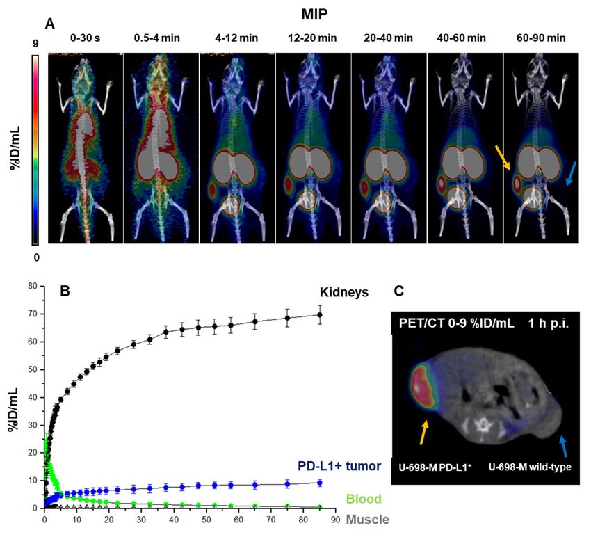
ABSTRACT

Blocking the interaction of the immune checkpoint molecules programmed cell death protein-1 (PD-1)

and its ligand, PD-L1, using specific antibodies has been a major breakthrough for immune oncology.

Whole-body PD-L1 expression positron emission tomography (PET) imaging may potentially allow for a

better prediction of response to PD-1 targeted therapies. Imaging of PD-L1 expression is feasible by PET

with the Adnectin protein 18F-BMS-986192. However, radiofluorination of proteins, such as BMS-986192

remains complex and labelling yields are low. The goal of this study was therefore the development and

preclinical evaluation of a 68Ga-labeled Adnectin protein (68Ga- BMS-986192) to facilitate clinical trials.

Methods

68
  Ga-labeling of DOTA-conjugated Adnectin (BXA-206362) was carried out in NaOAc-buffer at pH

5.5 (50°C, 15min). In vitro stability in human serum at 37°C was analyzed using Radio-thin layer

chromatography (Radio-TLC) and Radio-high performance liquid chromatography (Radio-HPLC). PD-L1

binding assays were performed using the transduced PD-L1 expressing lymphoma cell line U-698-M and

wild-type U-698-M cells as negative control. Immunohistochemical staining studies, biodistribution and

small animal PET studies of 68Ga-BMS-986192 were carried out using PD-L1-positive and negative U-698-

M-bearing NSG mice.

Results

68
  Ga-BMS-986192 was obtained with quantitative radiochemical yields (RCYs) >97% and with high

radiochemical purity (RCP). In vitro stability in human serum was ≥ 95% after 4h of incubation. High and

                          68
specific binding of            Ga-BMS-986192 to human PD-L1-expressing cancer cells was confirmed, which

closely correlates with the respective PD-L1 expression level determined by flow cytometry and IHC

                     68
staining. In vivo,    Ga-BMS-986192 uptake was high in PD-L1+ tumors (9.0±2.1%ID/g at 1hp.i.) and

kidneys (56.9±9.2% ID/g at 1hp.i.) with negligible uptake in other tissues. PD-L1 negative tumors

                                      68
                                       Ga-BMS-986192 for PD-L1 PET Imaging                               2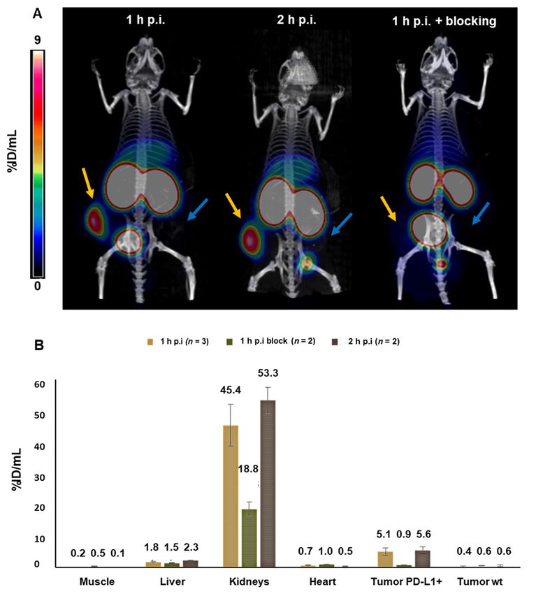
demonstrated only background uptake of radioactivity (0.6±0.1% ID/g). Co-injection of an excess of

unlabelled Adnectin reduced tumor uptake of PD-L1 by more than 80%.

Conclusion

68
 Ga-BMS-986192 enables easy radiosynthesis and shows excellent in vitro and in vivo PD-L1 targeting

characteristics. The high tumor uptake combined with low background accumulation at early imaging

time points demonstrate the feasibility of 68Ga-BMS-986192 for imaging of PD-L1 expression in tumors

and is encouraging for further clinical applications of PD-L1 ligands.

Key Words

PD-1/PD-L1 checkpoint inhibitors, PD-L1 PET Imaging, 68Ga-Adnectin, 68Ga-BMS-986192, 18F-BMS-986192

                               68
                                 Ga-BMS-986192 for PD-L1 PET Imaging                           3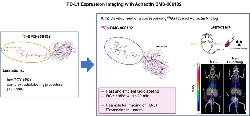
INTRODUCTION

The immune system is in principle capable to recognize and destroy cancer cells even in the presence of

larger tumor masses and multiple metastases (1). However, cells of the innate and adaptive immune

systems are frequently inhibited by molecular pathways that suppress their activation and effector

functions and allow tumor cells to escape immune recognition and attack (2). One major checkpoint that

is exploited by cancer cells to evade the immune system is the programmed death protein-1 (PD-1)

pathway. The negative costimulatory receptor PD-1 is expressed on the surface of activated T cells (3,4).

Its ligand the programmed death protein ligand 1 (PD-L1), a surface protein, is expressed on both

antigen-presenting cells and tumor cells of a variety of human cancers (5-7). PD-1/PD-L1 interaction

induces downregulation of T-cell activation and allows tumor cells to evade immune recognition and

elimination (8).

        Inhibition of the PD-1/PD-L1 interaction by antibodies has been a breakthrough for the

treatment of several common malignancies such as melanoma, non-small cell lung cancer, renal cell

carcinoma, and urothelial cancer (9-12). However, only a subgroup of the patients responds to this

immune checkpoint inhibitor treatment and the underlying reasons are not well understood (13-16).

Several studies demonstrated that response to PD-1/PD-L1-targeted immunotherapy correlates with PD-

L1 expression levels on tumor tissue of diverse malignancies as determined by immunohistochemistry

(17). However, the quantitative analysis of PD-L1 expression in the tumor tissue is challenging due to the

heterogeneous and dynamic expression of such immune checkpoint molecules (18-21). Prediction of

response to PD-1 targeted therapy by immunohistochemistry is therefore limited and in several tumor

types patients benefit from PD-1 targeted therapies in the absence of significant PD-L1 expression in the

tumor tissue (17,22-23).

                              68
                                Ga-BMS-986192 for PD-L1 PET Imaging                                 4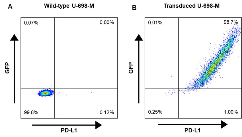
Imaging of PD-L1 expression can overcome several of the fundamental limitations of PD-L1

immunohistochemistry. First, imaging can provide a three-dimensional measure for overall PD-L1

expression of a tumor, whereas as only a very small fraction of the tumor tissues can be routinely

studied by immunohistochemistry. Second, imaging can assess PD-L1 expression at the whole-body level

and therefore allows for studies of the heterogeneity of PD-L1 expression across multiple metastases in

an individual patient. Third, imaging is non-invasive and can therefore not only provide information on

PD-L1 status before start of PD-L1 treatment but as well monitor changes of PD-L1 expression at multiple

times during treatment. Thus, PD-L1 imaging enables new approaches for studying PD1/PD-L1

pathophysiology in patients and may potentially allow for a better prediction of response to PD-1-

targeted therapies than immunohistochemistry (24).

        It is feasible to image and quantify PD-L1-expression within the tumor in patients with

radiolabeled antibodies (25,26). However, antibody-based imaging has several disadvantages such as the

long circulatory half-life of antibodies and their slow penetration in the tumor tissue which necessitates

imaging several days after injection. This requires the use of long-lived radioisotopes for radiolabelling

which cause the radiation exposure of antibody positron-emission tomography (PET) to be several-fold

higher than of PET with 18F-FDG.

        Adnectins are a family of engineered, target binding proteins with a size of ~ 10 kDa, derived

from the 10th type III domain of human fibronectin (27). Donnelly et al. synthesized and evaluated an 18F-

labeled anti-PD-L1 Adnectin (18F-BMS-986192) for PET imaging of the PD-L1 expression in vivo (28). 18F-

BMS-986192 showed high in vivo stability and excellent anti-PD-L1 targeting characteristics combined

with fast renal clearance of the tracer, resulting in high contrast imaging of PD-L1 positive lesions up to

hours p.i. (28). A first-in-human study with 18F-BMS-986192 in patients with advanced non-small cell lung

carcinoma demonstrated a correlation of 18F-Adnectin tumor uptake with PD-L1 expression confirmed by

                               68
                                Ga-BMS-986192 for PD-L1 PET Imaging                                  5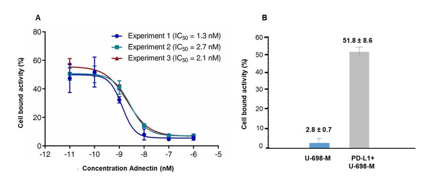
immunohistochemistry and response to nivolumab treatment on a lesional basis. The authors illustrated

       18
that    F-BMS-986192 SUVpeak was higher for responding lesions than non-responding, indicating a

correlation of 18F-BMS-986192 PD-L1 PET signal with PD-L1 expression and tumor-level response. (29).

            However, the multi-step synthesis of 18F-BMS-986192 with only moderate radiochemical yields is

challenging and time-consuming (28,30). To facilitate a broader clinical application of anti-PD-L1 PET

                                                   68
imaging the development of a corresponding          Ga-labeled analogue based on the Adnectin scaffold

                                                                                                 68
seemed the conclusive next step, due to the commercial availability of FDA approved               Ga/68Ga-

                                                                      68                              68
generators, the possibility of large-scale cyclotron production of     Ga and the fast and robust         Ga-

                                                                                      68
labeling technique. Herein, we report the synthesis and preclinical evaluation of         Ga-BMS-986192 in

terms of PD-L1-affinity, metabolic stability, micro-PET imaging and in vivo biodistribution in PD-L1-

positive and negative xenografts to investigate the feasibility of this tracer for in vivo imaging of PD-L1

expression in tumors.

MATERIALS AND METHODS
Materials

All reagents were obtained from Sigma Aldrich (Munich, Germany) unless otherwise stated. The DOTA-

conjugated anti-hPD-L1 Adnectin (BXA-206362), BXA-206362 was kindly provided by Bristol-Myers-

Squibb Pharmaceutical Research Institute (New Jersey, USA), formulated in PBS buffer (0.9 mg/mL, pH

                                                         68                          68
7.4) and tested to be endotoxin-free (1.5 EU/mg).          Ga was obtained from a      Ge/68Ga generator

(Galliapharm, Eckert & Ziegler AG, Berlin, Germany).

Synthesis of 68Ga-BMS-986192 and Quality Control

                                 68
                                   Ga-BMS-986192 for PD-L1 PET Imaging                                6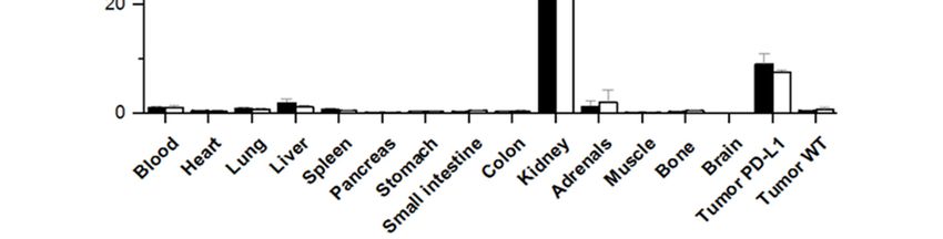
The generator was eluted in 1 mL fractions with 0.05 M aq. HCl (4 ml) containing 170 – 250 MBq of

68
 GaCl3. To this eluate 1 M NaOAc (100 µl, pH 5.5) and 200 µg DOTA-Adnectin (222 µL in PBS) were

added, resulting in a labeling solution at pH 5.5. The solution was mixed briefly and incubated for 15 min

           68
at 50°C.        Ga-BMS-986192 was purified by gel filtration on a PD-10 column (GE Healthcare,

Buckinghamshire, UK). Radiochemical yield and radiochemical purity was analyzed by radio-TLC and

radio-HPLC. Radio-TLC was performed using Varian silica impregnated ITLC chromatography paper

(Varian Inc., CA, USA) and 0.1 M aq. sodium citrate buffer (pH 5.5) as mobile phase. TLC strips were

analyzed on a B-FC-3600 TLC Scanner (Bioscan, Washington, USA). Analytical radio-size exclusion

chromatography (radio-SEC) of 68Ga-BMS-986192 was carried out using a bioZen SEC-2 (300 x 4.6 mm)

column (Phenomenex LTD, Aschaffenburg, Germany) on a Shimadzu HPLC system (0.1 M phosphate

buffer, pH 6.8, flow 0.35 ml/min) equipped with a NaI(TI) scintillation detector (2“ x 2“) and a SPD M20A

diode array UV/Vis detector.

In Vitro Stability of 68Ga-BMS-986192 in Human Serum

                                                     68
In vitro stability study was carried out by adding        Ga-BMS-986192 (app. 18 MBq to freshly prepared

human serum (1/8, v/v; SeronormTM Human, IGZ Instruments AG, Zürich), followed by incubation at 37

°C for up to 4 hours. To investigate the stability of 68Ga-BMS-986192 in human serum, radio-HPLC and

radio-TLC were performed at 0, 1, 2, 3, and 4 hours.

Cell Culture

The PD-L1 negative B-cell lymphoma cell line U-698-M was purchased from ATCC (Manassas, VA, USA).

Cultures were maintained in RPMI medium supplemented with 10% FBS and penicilline/streptomycine

(100IU/ml). Cells were grown at 37 °C in a humidified atmosphere of 5% CO2. To stably express PD-L1 the

                               68
                                Ga-BMS-986192 for PD-L1 PET Imaging                                  7
U-698-M cells were retrovirally transduced with a construct containing genes for human PD-L1 and GFP,

as previously described (31). Quantification of PD-L1 expression on transduced and wild-type U-698-M

cells was determined by fluorescence activated cell sorting (FACS) analysis using a anti-human CD274

antibody (Clone MIH1; BD Bioscience, Franklin Lakes, USA) as previously described (32).

Assessment of PD-L1 Binding Affinity and Specificity

The binding affinity of 68Ga-BMS-986192 towards human PD-L1 was determined in a competitive binding

experiment using stable transduced U-698-M PD-L1 cells with elevated PD-L1 expression. Briefly, a

                                     68
solution containing a mixture of          Ga-labeled (25 µL) and unlabeled DOTA-conjugated anti-hPD-L1

Adnectin (25 µL, competitor) with increasing concentrations (10-10 – 10-6 M) was added to the cells

(400.000 cells/vial). After 1 h incubation at 37 °C, cells were centrifuged at 600 x g (1,200 rpm, Biofuge

15) for 5 min, the supernatant of each vial was removed and cells were thoroughly washed 2 times with

250 µl PBS. The washing media was combined with the previously removed supernatant, representing

the amount of free radioligand. The amount of cell bound activity (cell pellet) as well as the amount of

free radioligand was measured in a 2470 Wizard2 ɤ-counter (PerkinElmer, MA, USA). Due to the high

structural similarity of 68Ga-labeled and unlabeled ligand a nearly identical affinity for PD-L1 is assumed,

resulting in homologous competitive binding. Specific binding was confirmed using non-transduced U-

689-M cells as a negative control.

U-698-M Tumor Model

All animal experiments were conducted according the current animal welfare regulations in Germany

(DeutschesTierschutzgesetz, approval (ROB-55.2-2532.Vet_02-15-216)). The transduced U-698-M PD-L1

positive and U-698-M wild-type cell line were detached from the surface of the culture flask using

                               68
                                Ga-BMS-986192 for PD-L1 PET Imaging                                   8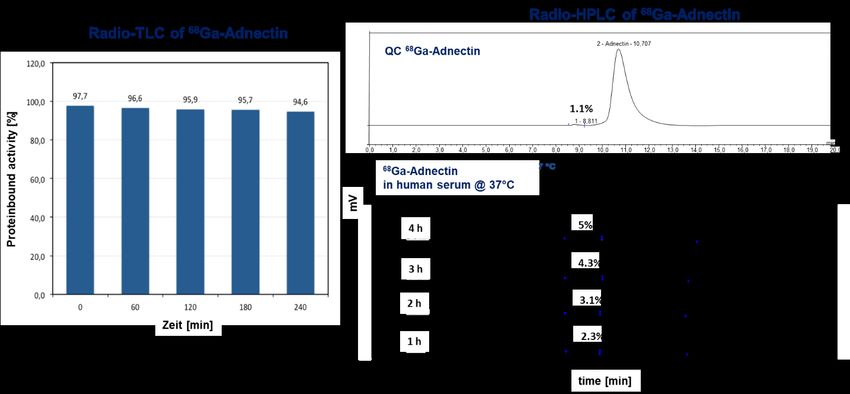
Trypsin/EDTA (0.05% and 0.02%) in PBS, centrifuged and resuspended in PBS. Approximately 1 x 10⁷

cells/200µL of the U-698-M PD-L1 positive cell line were inoculated subcutaneously on the right and U-

698-M wild-type cells on the left flank of 6 to 8 weeks old NSG mice (male, Charles River WIGA GmbH,

Sulzfeld, Germany). Tumors were grown for 2 to 3 weeks to reach 0.6- 1 cm in diameter.

Small-Animal PET-Imaging

Mice were anesthetized with isoflurane and intravenously injected via tail vein with app. 5-7 MBq (~ 4.5 -

             68
5.5 µg) of        Ga-BMS-986192. In vivo imaging studies were performed using a Siemens Inveon small-

animal PET/CT scanner. Static images were recorded at 1 h and 2 h p.i. with an acquisition time of 20

                                                                                       68
min. For blocking studies, unlabeled BXA-206362 (9 mg/kg) was coinjected with            Ga-BMS-986192.

Dynamic imaging was performed after on-bed injection for 1.5 h under isoflurane anesthesia.

Reconstruction of the images was carried out using 3-dimensional ordered-subsets expectation

maximum (OSEM3D) algorithm with scanner and attenuation correction. Data analysis was carried out

using Inveon Workplace software (Siemens). Regions of interest were drawn around areas with

increased uptake in transaxial slices for calculation of the average tracer concentration as percent

injected activity per milliliter (% IA/mL).

Ex Vivo Histology and Immunohistochemistry

Tumor tissues were fixed in 10% neutral-buffered formalin solution for at least 48 h, dehydrated under

standard conditions (Leica ASP300S, Wetzlar, Germany) and embedded in paraffin. Serial 2 µm-thin

sections prepared with a rotary microtome (HM355S, Thermo Fisher Scientific, Waltham, USA) were

collected and subjected to histological and immunohistochemical analysis. Hematoxylin-Eosin (HE)

staining was performed on deparaffinized sections with Eosin and Mayer’s Haemalaun according to a

standard protocol.

                                 68
                                   Ga-BMS-986192 for PD-L1 PET Imaging                              9
Immunohistochemistry was performed using a Bond RXm system (Leica, Wetzlar, Germany) with
primary antibodies against human PD-L1 (clone 28-8, ab205921, 1:500).
Ex-Vivo Biodistribution
About 5-7 MBq of 68Ga-BMS-986192 (4.5 - 5.5 µg) were injected into the tail vein of the U-698-M-PD-L1+
and U-698-M wild-type tumor bearing NSG mice under isoflurane anesthesia. Animals were sacrificed at
1h p.i. (n=4) and 2h p.i. (n=4), the organs of interest were dissected, and the activity in the weighed
tissues samples was quantified using a γ-counter.
RESULTS
68
Ga-Labeling and In Vitro Stability of 68Ga-BMS-986192 in Human Serum
High labeling efficiencies with quantitative radiochemical yields of > 97 % were obtained after 15 min.
After purification moderate specific activities (SA) of 11-16 GBq/µmol and radiochemical purity > 98%
was achieved.
68
Radio-chromatograms of Ga-BMS-986192 at various time points after mixing with human serum
revealed monomeric elution profiles with a minimal radioactive impurity of higher molecular weight
(8.97 min), which slightly increases up to 5% after 4 h. Radio-TLC analysis revealed moderate
transmetallation (5%) for 68Ga-BMS-986192 within 4h (Supplemental Fig. 1).
Characterization of PD-L1 Expression in Transduced U-698-M Cells
Quantification of PD-L1 expression on transduced and wild-type U-698-M cells was determined by FACS
analysis. A low PD-L1 expression of approximately 4.000 molecules per cell was observed for wild-type U-
68
Ga-BMS-986192 for PD-L1 PET Imaging 10698-M cells (Fig. 1). PD-L1 expression was significantly increased by stable transduction of U-698-M cells,
with approximately 155.000 PD-L1 molecules per cell.
Competitive Binding Assay of 68Ga-BMS-986192 to PD-L1
Binding affinity of 68Ga-BMS-986192 to human PD-L1 was determined in competitive radioligand binding
68
experiment using PD-L1 positive and wild-type U-698-M cells. Ga-BMS-986192 showed high affinity
towards human PD-L1 with an IC50 value of 2.0 ± 0.6 nM (Fig. 2A). Specific binding was confirmed using
non-transduced U-698-M cells as a negative control (Fig. 2B).
PET/CT Imaging
68
Ga-BMS-986192 showed a rapid clearance from the blood pool and from non-target tissues with
68
continuously increasing accumulation in the kidneys over time (Figs. 3A and 3B). Ga-BMS-986192
uptake in PD-L1 positive tumor was fast within 4 min p.i. and tracer accumulation in PD-L1 expressing
tumor tissue increased within 60 min with high retention over 90 min p.i. (Figs. 3A and 3C). No uptake in
the PD-L1 negative U-698-M tumor was observed.
Additional static µPET scans were performed with 68Ga-BMS-986192 in PD-L1 positive and PD-L1
wild-type tumor-bearing NSG mice at 1 h p.i and 2 h p.i. (Fig. 4A). 68Ga-BMS-986192 showed comparable
high tumor uptake in PDL-1 positive tumors after 1 h and 2 h p.i.. As shown in Figure 4A, in contrast to
the high uptake of 68Ga-BMS-986192 in PD-L1 positive U-698-M tumors, no accumulation was observed
68
in U-698-M wild-type xenografts confirming PD-L1 specific binding of Ga-BMS-986192. Additionally,
blocking experiments with excess of unlabeled Adnectin (9mg/kg) demonstrated that 68Ga-BMS-986192
uptake is specific and PD-L1 mediated (Fig. 4B).
68
Ga-BMS-986192 for PD-L1 PET Imaging 11Ex Vivo FACS Analysis, Histology and Immunohistochemistry
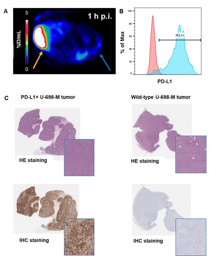
Ex vivo FACS analysis of dissected tumors confirmed the correlation of 68Ga-BMS-986192 uptake with PD-
L1 expression levels in tumor tissue (Figs. 5A and 5B). PD-L1 expression levels in transduced U-698-M
tumors was highly increased in comparison to non-transduced U-698-M wild-type xenografts, confirming
the generation of a stable PD-L1 expressing tumor cell line in vivo and PD-L1 mediated uptake of 68Ga-
BMS-986192.
Ex vivo histology and immunohistochemistry of the xenograft tissues also revealed high and
homogeneous PD-L1 expression in U-698-M PD-L1 positive tumors, while no PD-L1 expression was found
68
in U-698-M wildtype tissues (Fig. 5C). These results confirm the PD-L1 specific uptake of Ga-BMS-
986192 in µPET imaging in vivo and correlate with ex vivo flow cytometry analysis.
Biodistribution
The biodistribution data of 68Ga-BMS-986192 in PD-L1 positive-U-698-M and U-698-M wild-type tumor-
bearing mice (1h and 2h p.i.) are shown in Figure 6 and Supplemental Table 1. The data reflect the
68
results of µPET/CT imaging. Ga-BMS-986192 exhibited predominant renal clearance with only slight
uptake in the liver and non-targeted tissues. The significant uptake of 68Ga-BMS-986192 in PD-L1 positive
tumor xenografts is highly specific, as accumulation in PD-L1 negative tumors was less than blood pool
activity (Supplemental Table 1).
DISCUSSION
This study revealed that the high-affinity PD-L1 binding Adnectin protein BMS-986192, conjugated at the
68
C terminal position with DOTA via maleimide linkage (BXA-206362) can be labeled with Ga while
retaining high target binding affinity (IC50 2.0 ± 0.6 nM determined in a competitive radioligand binding
68
Ga-BMS-986192 for PD-L1 PET Imaging 12assay using PD-L1 positive U-698-M cells). The DOTA- maleimide moiety was conjugated to the cysteine
residue engineered into the C-terminus of the Adnectin (28,30). The same position within the Adnectin
scaffold has been used previously for site-specific conjugation of the 18F-labeled prosthetic group of 18F-
BMS-986192 without a significant effect on binding affinity (28). 19F-BMS-986192, unmodified and DOTA-
conjugated Adnectin displayed picomolar dissociation constants (KD) against human PD-L1 determined
by surface plasmon resonance (data not shown) (28). This indicates that conjugation of small molecules
to the C-terminus of Adnectin are well tolerated without observable effects on human PD-L1 binding.
The ease of tracer preparation, the quantitative radiochemical yields with high radiochemical purity
68
make the synthesis of Ga-BMS-986192 fully compatible with the everyday workflow in a clinical
radiopharmacy and are well suited for automated radiosynthesis via a module or a kit formulation.
Furthermore, the pharmacokinetics of 68Ga-BMS-986192 match very well the 68-minute physical half-life
68
of Ga as peak tumor uptake reached a plateau within 60 min post injection. Collectively, these
characteristics are highly encouraging for using 68Ga-BMS-986192 to image PD-L1 expression in humans.
Small animal PET and ex-vivo biodistribution studies demonstrated excellent targeting of PD-L1
expressing xenografts whereas tracer uptake was low in tumors without PD-L1 expression on
immunohistochemistry (Figs. 4 and 5). Specificity of tracer binding was further confirmed by co-injection
of an excess of unlabelled BMS-986192 which decreased tumor uptake of 68Ga-BMS-986192 by 80% (Fig.
68
6). As expected for a 10 kDa protein, Ga-BMS-986192 showed fast, renal clearance from the blood.
Similar to other small proteins, peptides and peptide-like molecules we observed also significant
retention of activity in the kidney at 1 and 2 h post injection. However, the activity concentration was
similar to clinically used PET imaging agents such as prostate specific membrane (PSMA) ligands (33,34).
Therefore, we do not expect renal uptake of 68Ga-BMS-986192 to limit its clinical use. Injection of an
excess of unlabelled adnectin significantly decreased renal uptake of 68Ga-BMS-986192. This is unlikely
68
Ga-BMS-986192 for PD-L1 PET Imaging 13due to blocking of PD-L1 binding since BMS986192 has only low affinity for murine PD-L1 (28).
Nevertheless, it indicates that renal uptake of radioactivity is a partially saturable process that could
potentially be reduced by injection of proteins or peptides that do not interfere with the binding of 68Ga-
BMS-986192 to its target. The activity concentration in the kidneys increased from 1 to 2 hour post
injection probably reflecting redistribution of the ligand from other tissues to the blood stream (Fig. 6).
18 68
A comparison of the preclinical results obtained for F-BMS986192 and Ga-BMS-986192
revealed similar specific activities as well as in vitro/in vivo behaviour of both tracers (28). Although
comparable protein and activity doses of 18F-BMS986192 and 68Ga-BMS-986192 were administered to
mice, a direct comparison of the tumor uptake of both radiolabelled ligands is not feasible as different
cell lines were utilized in the two studies, but both tracers showed similar pharmacokinetics with fast
renal clearance and similarly low background activity in all other organs. This is encouraging for the
clinical translation of 68Ga-BMS-986192 because PET imaging of PD-L1 expression with 18F-BMS-986192
has already been shown to be feasible in human pilot studies (29,35).
68
The following limitation of the study should be noted. Ga-BMS-986192 exclusively binds to
human PD-L1 with no relevant affinity towards its murine counterpart (data not shown). Tumor-to-
normal organ uptake ratios may therefore be lower in humans than in our mouse model. Furthermore,
we used a PD-L1 transduced cell line to study the binding of 68Ga-BMS-986192. The advantages of this
approach are that the parent cell line can be used as a negative control to assess specificity of ligand
binding. Furthermore, the stable expression of PD-L1 improves the reproducibility of these results to
ensure to have a tool for measurement PD-L1 expression of cancer cells, as endogenous cancer cell PD-
L1 expression is known to vary significantly over time (36). However, PD-L1 expression levels by PD-L1
transduced U698-M cells may be higher than in human tumors.
68
Ga-BMS-986192 for PD-L1 PET Imaging 14CONCLUSION
68
The novel PD-L1 imaging agent Ga-BMS-986192 shows similar PD-L1 targeting characteristics and
pharmacokinetic properties as 18F-BMS-986192 which has already been successfully used to image PD-L1
expression in cancer patients. In contrast to 18F-BMS-986192 which requires a two-step synthesis with
low radiochemical yields, radiolabeling of BMS-986192 with 68Ga is a straightforward one-step process
68
which is easy to automate. Ga-BMS-986192 therefore has the potential to significantly facilitate
preclinical and clinical imaging of PD-L1 expression.
Disclosure
David Leung, Wendy Hayes, Paul Morin, Adam Smith, David Donnelly, Daša Lipovšek, Sam Bonacorsi,
Daniel Cohen are employed by Bristol-Myers Squibb Co. 18F-BMS-986192 and 68Ga-BMS-986192 are the
subject of patent applications WO2016086021A1, WO2016086036A2, WO2017/210302 and
WO2017/210335. No other potential conflict of interest relevant to this article was reported.
ACKNOWLEDGEMENTS
We thank Sybille Reder, Markus Mittelhäuser and Hannes Rolbiesky for small-animal PET imaging and
Olga Seelbach and Marion Mielke for HE- and IHC staining. The current study was financially supported
by the Deutsche Forschungsgemeinschaft (SFB824; subproject C10, Z1 and Z2).
68
Ga-BMS-986192 for PD-L1 PET Imaging 15KEY POINTS
68
Question: Can Adnectins be efficiently radiolabeled with Ga to allow the in vivo
assessment and quantification of PD-L1 expression levels on tumor tissue
with PET?
Pertinent Findings: The expansion of the Adnectin based concept for PD-L1 expression PET
imaging to the requirements of 68Ga-chemistry enables fast and efficient
68
radiolabeling. Ga-BMS-986192 showed comparable in vitro and in vivo
18
PD-L1-targeting characteristics to its counterpart F-BMS986192. Both
tracers showed favourable pharmacokinetics with fast renal clearance
and similarly low background activity in non-targeted tissues.
Implications for Patient Care: The preclinical results are encouraging for the clinical translation of 68Ga-
18
BMS-986192 because PET imaging of PD-L1 expression with F-BMS-
986192 has already been shown to be feasible in human pilot studies.
68
Ga-BMS-986192 for PD-L1 PET Imaging 16REFERENCES
1. Gonzalez H, Hagerling C, Werb Z. Roles of the immune system in cancer: from tumor initiation to
metastatic progression. Genes Dev. 2018;32:1267-1284.
2. Topalian SL, Drake CG, Pardoll DM. Immune checkpoint blockade: a common denominator
approach to cancer therapy. Cancer Cell. 2015;27:450-461.
3. Riella LV, Paterson AM, Sharpe AH, Chandraker A. Role of the PD-1 pathway in the immune
response. Am J Transplant. 2012;12:2575-2587.
4. Keir ME, Butte MJ, Freeman GJ, Sharpe AH. PD-1 and its ligands in tolerance and immunity. Annu
Rev Immunol. 2008;26:677-704.
5. Konishi J, Yamazaki K, Azuma M, Kinoshita I, Dosaka-Akita H, Nishimura M. B7-H1 expression on
non-small cell lung cancer cells and its relationship with tumor-infiltrating lymphocytes and their
PD-1 expression. Clin Cancer Res. 2004;10:5094-5100.
6. Thompson RH, Gillett MD, Cheville JC, et al. Costimulatory B7-H1 in renal cell carcinoma patients:
Indicator of tumor aggressiveness and potential therapeutic target. ProcNatlAcadSciUSA.
2004;101:17174-17179.
7. Hino R, Kabashima K, Kato Y, et al. Tumor cell expression of programmed cell death-1 ligand 1 is
a prognostic factor for malignant melanoma. Cancer. 2010;116:1757-1766.
8. Freeman GJ, Long AJ, Iwai Y, et al. Engagement of the PD-1 immunoinhibitory receptor by a novel
B7 family member leads to negative regulation of lymphocyte activation. JExpMed.
2000;192:1027-1034.
9. Herbst RS, Baas P, Kim DW, et al. Pembrolizumab versus docetaxel for previously treated, PD-L1-
positive, advanced non-small-cell lung cancer: a randomised controlled trial. Lancet.
2016;387:1540-1550.
68
Ga-BMS-986192 for PD-L1 PET Imaging 1710. Weinstock M, McDermott D. Targeting PD-1/PD-L1 in the treatment of metastatic renal cell
carcinoma. TherAdvUrol. 2015;7:365-377.
11. Simeone E, Ascierto PA. Anti-PD-1 and PD-L1 antibodies in metastatic melanoma.
MelanomaManag. 2017;4:175-178.
12. Stenehjem DD, Tran D, Nkrumah MA, Gupta S. PD1/PDL1 inhibitors for the treatment of
advanced urothelial bladder cancer. OncoTargetsTher. 2018;11:5973-5989.
13. Alsaab HO, Sau S, Alzhrani R, et al. PD-1 and PD-L1 checkpoint signaling inhibition for cancer
immunotherapy: Mechanism, combinations, and clinical outcome. FrontPharmacol. 2017;8:561.
14. Chen L, Han X. Anti-PD-1/PD-L1 therapy of human cancer: past, present, and future. JClinInvest.
2015;125:3384-3391.
15. Cottrell TR, Taube JM. PD-L1 and emerging biomarkers in immune checkpoint blockade
TherapyCancer J. 2018;24:41-46.
16. Zou W, Wolchok JD, Chen L. PD-L1 (B7-H1) and PD-1 pathway blockade for cancer therapy:
Mechanisms, response biomarkers, and combinations. Sci TranslMed. 2016;8:328rv4.
17. Chakravarti N, Prieto VG. Predictive factors of activity of anti-programmed death-1/programmed
death ligand-1 drugs: immunohistochemistry analysis. TransLungCancerRes. 2015;4:743-751.
18. Kloten V, Lampignano R, Krahn T, Schlange T. Circulating tumor cell PD-L1 expression as
biomarker for therapeutic efficacy of immune checkpoint inhibition in NSCLC. Cells. 2019;8:809.
19. Yue C, Jiang Y, Li P, et al. Dynamic change of PD-L1 expression on circulating tumor cells in
advanced solid tumor patients undergoing PD-1 blockade therapy. Oncoimmunology.
2018;7:e1438111.
20. Madore J, Vilain RE, Menzies AM, et al. PD-L1 expression in melanoma shows marked
heterogeneity within and between patients: implications for anti-PD-1/PD-L 1 clinical trials.
Pigmentcel melanomaRes. 2015;28:245-253.
68
Ga-BMS-986192 for PD-L1 PET Imaging 1821. Niemeijer A, Leung D, Huisman M, et al. Whole body PD-1 and PD-L1 positron emission
tomography in patients with non-small-cell lung cancer. Naturecommun. 2018;9:1-5.
22. Ribas A, Hu-Lieskovan S. What does PD-L1 positive or negative mean?. JExpMed. 2016;213:2835-
2840.
23. Abdel-Rahman O. Correlation between PD-L1 expression and outcome of NSCLC patients treated
with anti-PD-1/PD-L1 agents: a meta-analysis. CritRevOncolHematol. 2016;101:75-85.
24. Broos K, Lecocq Q, Raes G, Devoogdt N, Keyaerts M, Breckpot K. Noninvasive imaging of the PD-
1:PD-L1 immune checkpoint: Embracing nuclear medicine for the benefit of personalized
immunotherapy. Theranostics. 2018;8:3559-3570.
25. Heskamp S, Hobo W, Molkenboer-Kuenen JD, et al. Noninvasive imaging of tumor PD-L1
expression using radiolabeled anti-PD-L1 antibodies. CancerRes. 2015;75:2928-2936.
26. Truillet C, Oh HLJ, Yeo SP, et al. Imaging PD-L1 expression with immunoPET. BioconjugChem.
2018;29:96-103.
27. Lipovšek D. Adnectins: engineered target-binding protein therapeutics. ProteinEngDesSel.
2011;24:3-9.
28. Donnelly DJ, Smith RA, Morin P, et al. Synthesis and biological evaluation of a novel 18F-labeled
adnectin as a PET radioligand for imaging PD-L1 expression. JNuclMed. 2018;59:529-535.
29. Niemeijer AN, Leung D, Huisman MC, et al. Whole body PD-1 and PD-L1 positron emission
tomography in patients with non-small-cell lung cancer. NatCommun. 2018;9:4664.
30. Morin PE, Donnelly D, Lipovsek D, et al. Bristol-Myers Squibb Company, USA. assignee. Novel PD-
L1-binding polypeptides for imaging. US patent , WO2016086021A1, 2016.
31. Audehm S, Glaser M, Pecoraro M, et al. Key features relevant to select antigens and TCR from
the MHC-mismatched repertoire to treat cancer. FrontImmunol. 2019;10:1485.
68
Ga-BMS-986192 for PD-L1 PET Imaging 1932. Mayer KE, Mall S, Yusufi N, et al. T-cell functionality testing is highly relevant to developing novel
immuno-tracers monitoring T cells in the context of immunotherapies and revealed CD7 as an
attractive target. Theranostics. 2018;8:6070-6087.
33. Weineisen M, Schottelius M, Simecek J, et al. 68Ga-and 177Lu-labeled PSMA I&T: optimization of
a PSMA-targeted theranostic concept and first proof-of-concept human studies. J Nucl Med.
2015;56: 1169-1176.
34. Robu S, Schmidt A, Eiber M, et al. Synthesis and preclinical evaluation of novel 18 F-labeled Glu-
urea-Glu-based PSMA inhibitors for prostate cancer imaging: a comparison with 18 F-DCFPyl and
18 F-PSMA-1007. EJNMMIRes. 2018;8:30.
35. Huisman M, Niemeijer AL, Windhorst B, et al. Quantification of PD-L1 expression with [18F]BMS-
986192 PET/CT in patients with advanced stage non-small-cell lung cancer. JNuclMed.
2020;61:1455-1460.
36. Yue C, Jiang Y, Li P, et al. Dynamic change of PD-L1 expression on circulating tumor cells in
advanced solid tumor patients undergoing PD-1 blockade therapy. Oncoimmunology.
2018;7:e1438111.
68
Ga-BMS-986192 for PD-L1 PET Imaging 20Figure 1 Generation of a stable PD-L1 expressing U-698-M cell line. Expression of GFP and PD-L1
of (A) U-698-M wild-type cells and (B) U-698-M cells transduced by the retroviral vector
MP71 containing PD-L1 and GFP separated by a P2A element. Representative dot-plots
of U-698-M wild-type and PD-L1 positive U-698-M cells are shown.
68
Ga-BMS-986192 for PD-L1 PET Imaging 2168
Figure 2 Binding affinity and specificity of Ga-BMS-986192 towards PD-L1 determined in
68
competitive radioligand binding assays. (A) Cell bound activity of Ga-BMS-986192 in
the presence of increasing concentrations of cold ligand (BXA-206362). (B) Cell bound
activity of 68Ga-BMS-986192 in the presence of cold ligand (0.1 nM) on PD-L1 positive (U-
698-M-PDL1+) and negative U-698-M cells.
68
Ga-BMS-986192 for PD-L1 PET Imaging 22Figure 3 Dynamic PET imaging in anesthetized mice. A. Maximum intensity projections (MIP) of a
dynamic µPET scan. Summation images of different time-frames (0-9 %ID/mL) of 68Ga-
BMS-986192 in PD-L1 positive U-698-M and U-698-M wild-type xenograft bearing mice
over an acquisition time of 90 min. B. Time-activity curves in %ID/mL for blood pool
(heart), kidneys, muscle, and PD-L1 positive tumor derived from dynamic µPET/CT data.
C. Axial Slice of a 68Ga-BMS-986192 PET/CT scan (1 h p.i.) in a PD-L1 positive U-698-M
(yellow arrow) and U-698-M wild-type (blue arrow) xenograft bearing NSG mouse (0-9
%ID/mL).
68
Ga-BMS-986192 for PD-L1 PET Imaging 23Figure 4 Static PET imaging examples and quantitative analysis. A. Maximum intensity
projections (MIP) of static µPET scans of 68Ga-BMS-986192 (5-6 MBq, 4.5 - 5.5 µg) 1 h p.i.
and 2h p.i. in PD-L1 positive U-698-M (yellow arrows) and U-698-M wild-type (blue
arrows) xenograft bearing NSG mice (0% - 9% ID/mL). Mouse 3: Coinjection of 68Ga-BMS-
986192 + Blocking with 9 mg/kg unlabeled Adnectin. B. ROI Quantification of the static
PET scans (%IA/mL mean).
68
Ga-BMS-986192 for PD-L1 PET Imaging 24Figure 5 Comparison of PET, Immunohistochemistry and FACS analysis of tumor xenografts. A.
Axial Slice of a 68Ga-BMS-986192 PET Scan (1 h p.i.) in a PD-L1 positive U-698-M (yellow
arrow) and U-698-M wild-type (blue arrow) xenograft bearing NSG mouse (0-5 %ID/ML).
B. Ex vivo FACS analysis of PD-L1 expression on wild-type (red) and PD-L1 transduced
(blue) U-698-M tumor cells. C. Ex vivo HE and anti-PD-L1 immunohistochemistry (IHC)
staining of PD-L1 positive and wild-type U-698-M xenograft tissues.
68
Ga-BMS-986192 for PD-L1 PET Imaging 25Figure 6 Biodistribution data of 68Ga-BMS-986192 in PD-L1 positive U-698-M and U-698-M wild-
type xenograft bearing mice. Data are expressed as %ID/g (mean±SD).
68
Ga-BMS-986192 for PD-L1 PET Imaging 26Graphical Abstract
68
Ga-BMS-986192 for PD-L1 PET Imaging 27SUPPLEMENTAL DATA
In Vitro Stability of 68Ga-BMS-986192
In vitro stability of 68Ga-BMS-986192 in human serum at 37 °C was determined by Radio-TLC and Radio-
HPLC up to 4 h (Supplemental Fig 1).
Supplemental Figure 1 | In vitro serum stability of 68Ga-Adnectin up to 4 h determined by Radio-TLC and
Radio-HPLC.
68
Ga-BMS-986192 as a PET Agent for Imaging PD-L1 ExpressionEx-Vivo Biodistribution of 68Ga-BMS-986192
Supplemental Table 1 Biodistribution of 68Ga-BMS-986192 in PD-L1 positive U-698-M and U-698-M
wild-type bearing NSG mice at 1h and 2h p.i.. Data are given in %ID/g are
means ± SD (n=4 animals per group and time point).
Organ 1 h p.i [% ID/g] 2 h p.i. [% ID/g]
Blood 1.0 ± 0.3 1.1 ± 0.3
Heart 0.5 ± 0.1 0.4 ± 0.1
Lung 0.8 ± 0.2 0.7 ± 0.2
Liver 1.9 ± 0.7 1.2 ± 0.2
Spleen 0.7 ± 0.2 0.5 ± 0.2
Pancreas 0.2 ± 0.1 0.2 ± 0.04
Stomach 0.3 ± 0.04 0.3 ± 0.1
Small intestine 0.3 ± 0.1 0.5 ± 0.1
Colon 0.3 ± 0.03 0.4 ± 0.2
Kidney 56.9 ± 9.2 88.9 ± 1.8
Adrenals 1.2 ± 1.0 2.0 ± 2.2
Muscle 0.2 ± 0.1 0.2 ± 0.1
Bone 0.3 ± 0.1 0.5 ± 0.1
Brain 0.04 ± 0.01 0.1 ± 0.04
Tumor PD-L1+ 9.0 ± 2.1 7.6 ± 0.4
Tumor wild-type 0.6 ± 0.1 0.7 ± 0.4
68
Ga-BMS-986192 as a PET Agent for Imaging PD-L1 ExpressionYou can also read