Endoplasmic Reticulum Stress Increases DUSP5 Expression via PERK-CHOP Pathway, Leading to Hepatocyte Death - MDPI
←
→
Page content transcription
If your browser does not render page correctly, please read the page content below
International Journal of
            Molecular Sciences

Article
Endoplasmic Reticulum Stress Increases DUSP5
Expression via PERK-CHOP Pathway, Leading to
Hepatocyte Death
Hye Jin Jo 1 , Jin Won Yang 2 , Ji Hye Park 1,3 , Eul Sig Choi 1,3 , Chae-Seok Lim 1 , Seoul Lee 1,3
and Chang Yeob Han 1, *
 1    Department of Pharmacology, School of Medicine, Wonkwang University, Iksan 54538, Jeonbuk, Korea
 2    College of Pharmacy, Woosuk University, Wanju 55338, Jeonbuk, Korea
 3    Brain Research Institute, Wonkwang University, Iksan 54538, Jeonbuk, Korea
 *    Correspondence: hancy17@wku.ac.kr; Tel.: +82-63-850-6776; Fax: +82-63-850-7262

 Received: 14 August 2019; Accepted: 3 September 2019; Published: 5 September 2019

 Abstract: Hepatocyte death is critical for the pathogenesis of liver disease progression, which is
 closely associated with endoplasmic reticulum (ER) stress responses. However, the molecular basis
 for ER stress-mediated hepatocyte injury remains largely unknown. This study investigated the effect
 of ER stress on dual-specificity phosphatase 5 (DUSP5) expression and its role in hepatocyte death.
 Analysis of Gene Expression Omnibus (GEO) database showed that hepatic DUSP5 levels increased
 in the patients with liver fibrosis, which was verified in mouse models of liver diseases with ER stress.
 DUSP5 expression was elevated in both fibrotic and acutely injured liver of mice treated with liver
 toxicants. Treatment of ER stress inducers enhanced DUSP5 expression in hepatocytes, which was
 validated in vivo condition. The induction of DUSP5 by ER stress was blocked by either treatment with
 a chemical inhibitor of the protein kinase RNA-like endoplasmic reticulum kinase (PERK) pathway,
 or knockdown of C/EBP homologous protein (CHOP), whereas it was not affected by the silencing of
 IRE1 or ATF6. In addition, DUSP5 overexpression decreased extracellular-signal-regulated kinase
 (ERK) phosphorylation, but increased cleaved caspase-3 levels. Moreover, the reduction of cell viability
 under ER stress condition was attenuated by DUSP5 knockdown. In conclusion, DUSP5 expression is
 elevated in hepatocytes by ER stress through the PERK-CHOP pathway, contributing to hepatocyte
 death possibly through ERK inhibition.

 Keywords: DUSP5; ER stress; PERK; CHOP; hepatocyte; cell death

1. Introduction
      Hepatocyte injury and dysfunction are crucial for the pathogenesis of liver disease progression.
Hepatocyte death is a hallmark of acute and chronic liver diseases originated from various etiology
including virus, drug, or metabolic stress, which contributes to the development of inflammation
and fibrosis [1–3]. Many clinical studies have been tried to discover novel therapeutic strategies
for liver diseases, regarding the intervention of parenchymal cell death in patients [2,3]. However,
the patterns and mechanisms of cell death are varying on different hepatotoxic stimuli and/or stages
of the diseases. Thus, the understanding of the cellular and molecular basis for hepatocyte injury in
certain conditions is important for the management of liver diseases.
      Endoplasmic reticulum (ER) stress is induced by the accumulation of unfolded or misfolded
proteins inside the ER, which activates the unfolded protein response (UPR) in the cells [4]. UPR is
an adaptive process for maintenance of cellular homeostasis, but uncontrolled ER stress responses
also account for cell damages [4]. Thus, excessive and/or sustained ER stress acts as one of

Int. J. Mol. Sci. 2019, 20, 4369; doi:10.3390/ijms20184369                       www.mdpi.com/journal/ijms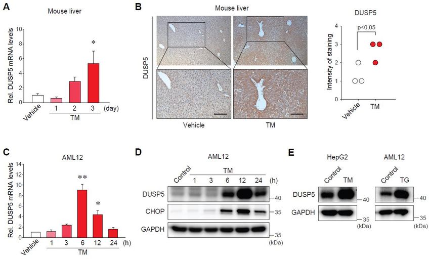
Int. J. Mol. Sci. 2019, 20, 4369 2 of 12
the major stimuli that lead to hepatocyte injury and subsequent death [5,6]. Indeed, the pathogenesis
of diverse liver diseases including nonalcoholic or alcoholic steatohepatitis, viral hepatitis,
cholestasis, and drug-induced liver injury is closely associated with cellular responses against ER
stress [6,7]. Nonetheless, the molecules responsible for ER stress-mediated hepatocyte injury remain
largely unknown.
Dual-specificity phosphatases (DUSPs) dephosphorylate their substrates at both serine/threonine
and tyrosine residues [8]. Several vital signaling molecules including mitogen-activated protein
kinases (MAPKs) have been discovered as the targets of DUSPs, and their functions are involved in
the regulation of biological processes such as immune responses and oncogenic transformation [8–10].
However, the potential roles of the specific DUSP and its regulatory basis of the gene expression, in
aspects of ER stress and hepatocyte injury, are elusive.
This study investigated the effects of ER stress on the gene expression of DUSP5 and its role in
hepatocyte function. Here, we report for the first time that ER stress increases DUSP5 expression
in hepatocytes and the induction of DUSP5 leads to ER stress-induced hepatocyte death. To prove
this, we analyzed Gene Expression Omnibus (GEO) database derived from patients with liver fibrosis
and carried out animal experiments using various liver injury models. In addition, in vivo and in vitro
models with ER stress were employed for the validation of the regulation of DUSP5 by ER stress.
Furthermore, we performed loss- or gain-of-function experiments using hepatocytes to discover that
protein kinase RNA-like endoplasmic reticulum kinase (PERK)-C/EBP homologous protein (CHOP)
pathway is responsible for DUSP5 expression under ER stress condition, and the DUSP5 induction
leads to hepatocyte death.
2. Results
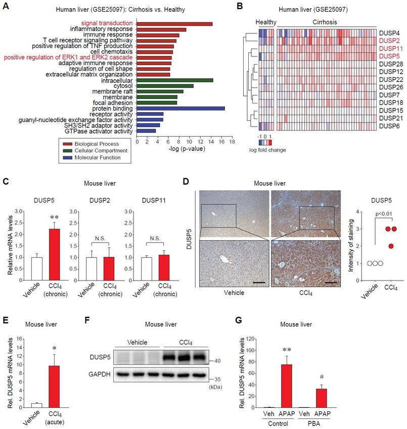
2.1. Dual-Specificity Phosphatase 5 (DUSP5) Expression Is Elevated in Patients and Mice with Liver Diseases
To find a novel potential target for liver diseases, we first analyzed the public GEO database
(GSE25097). Gene ontology (GO) analysis of the significantly upregulated genes in patients with
cirrhosis as compared to healthy subjects showed that the groups of genes involved in intracellular
signal transduction, such as the extracellular-signal-regulated kinase (ERK) pathway, were notably
changed in fibrotic liver (Figure 1A). It allowed us to pay attention to the possible roles of DUSPs
as MAPK phosphatases in the pathogenesis of liver injury. As a result of screening DUSPs in the dataset,
three DUSPs (i.e., DUSP5, DUSP2, DUSP11) were significantly increased in the liver of cirrhotic patients
(Figure 1B).
Next, we validated the alterations of those DUSPs in animal models of liver diseases associated
with ER stress. Among them, only the transcript levels of DUSP5 were significantly elevated in the liver
of mice treated with carbon tetrachloride (CCl4 ) for six weeks, as shown in Figure 1C, enabling us to
focus on DUSP5 in the following examinations. Immunohistochemistry showed the increase in DUSP5
protein levels (Figure 1D). The effect of acute liver injury on DUSP5 expression was further examined.
Consistently, both mRNA and protein levels of hepatic DUSP5 were dramatically raised in mice given
a single injection of CCl4 (Figure 1E,F). The increases in ER stress markers were confirmed in our
previous study [11]. Moreover, treatment of mice with acetaminophen (APAP) as a well-established
hepatotoxic drug augmented DUSP5 mRNA levels, which was significantly attenuated by pretreatment
with a chemical chaperone (Figure 1G). These results indicate that hepatic expression of DUSP5 is
upregulated in liver disease states, which might be connected with ER stress.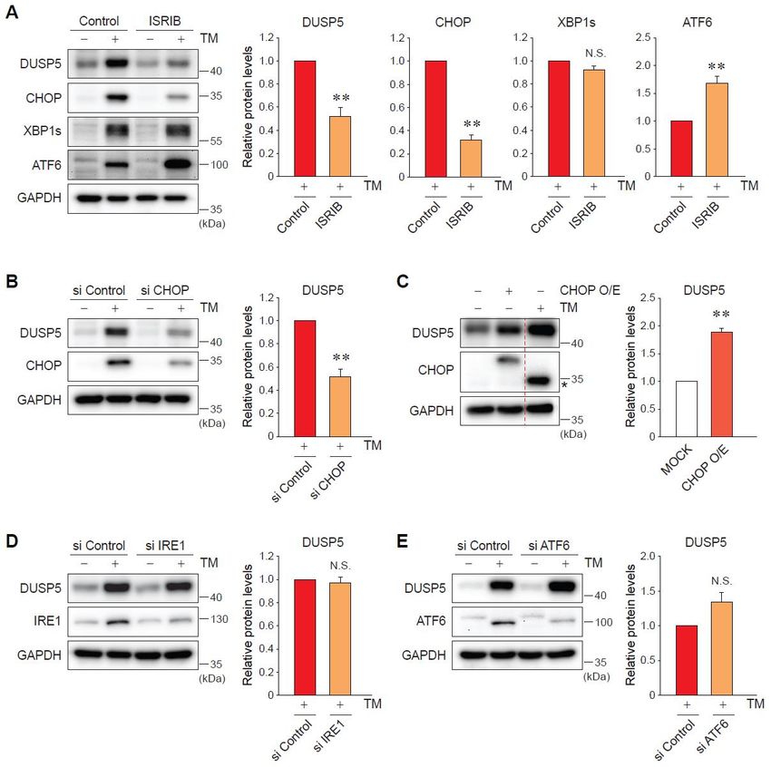
Int. J. Mol. Sci. 2019, 20, 4369 3 of 12
Int. J. Mol. Sci. 2019, 20, x FOR PEER REVIEW 3 of 12
Figure1.1.Dual-specificity
Figure Dual-specificityphosphatase
phosphatase 55(DUSP5)
(DUSP5)levels levelsare areelevated
elevatedin inpatients
patientsand
andmice
micewith
withliver
liver
injury (A) Gene ontology (GO) analysis using the DAVID bioinformatics
injury (A) Gene ontology (GO) analysis using the DAVID bioinformatics database (derived from database (derived from
GSE25097).
GSE25097).(B)(B)Heatmap
Heatmapof ofthe
theupregulated
upregulatedDUSPs DUSPsin inpatients
patientswithwithcirrhosis
cirrhosisversus
versushealthy
healthysubjects
subjects
(extracted
(extractedfrom
fromGSE25097).
GSE25097). (C)(C)
Quantitative
Quantitative reverse transcription-polymerase
reverse transcription-polymerase chainchain
reaction (qRT-PCR)
reaction (qRT-
assay
PCR) for hepatic
assay DUSPs.DUSPs.
for hepatic Mice were
Micetreated with vehicle
were treated or carbon
with vehicle or tetrachloride (CCl4 ) (0.6
carbon tetrachloride (CClmL/kg
4) (0.6
body
ml/kgweight, i.p., twice
body weight, i.p.,a twice
week,afor 6 weeks)
week, (n = 7 or
for 6 weeks) (n8/group). (D) Immunohistochemistry
= 7 or 8/group). (D) Immunohistochemistry (IHC)
for DUSP5
(IHC) in the liver
for DUSP5 in theof mice
liver treated
of mice as in panel
treated (C).panel
as in Scale(C).bar:Scale
50 µm. 50 m. Intensity
bar:Intensity of DUSP5ofstaining
DUSP5
was scored
staining from
was scored 4 (n =0 3/group).
0 to from (E) qRT-PCR
to 4 (n = 3/group). assay forassay
(E) qRT-PCR hepatic
for DUSP5. Mice were
hepatic DUSP5. Mice given
weresingle
given
injection with vehicle
single injection withorvehicle
CCl4 (0.6 ormL/kg
CCl4 body weight,body
(0.6 ml/kg i.p., 24 h) (n =i.p.,
weight, 3/group).
24 h)(F)(nImmunoblotting
= 3/group). (F)
for DUSP5 in the for
Immunoblotting liver of mice
DUSP5 in treated
the liverasofin panel
mice (E). (G)
treated qRT-PCR
as in panel (E).assay for hepatic
(G) qRT-PCR DUSP5.
assay Mice
for hepatic
were treated with 4-phenylbutyrate (PBA) (100 mg/kg, i.p.) for 2 h before the
DUSP5. Mice were treated with 4-phenylbutyrate (PBA) (100 mg/kg, i.p.) for 2 h before the injectioninjection with vehicle or
acetaminophen
with vehicle or(APAP) (500 mg/kg,
acetaminophen i.p., 6 h)
(APAP) (500(n = 5/group).
mg/kg, < 0.05
i.p., *6ph) (n = or ** p < 0.01
5/group). * pversus
< 0.05vehicle
or ** pgroup
< 0.01
(N.S.,
versusnot significant);
vehicle p < 0.05
group #(N.S., not versus APAP-treated
significant); # p < 0.05group.
versus APAP-treated group.
2.2. Endoplasmic Reticulum (ER) Stress Increases DUSP5 Expression in Hepatocytes
To investigate whether ER stress directly regulates DUSP5 expression, we used both animal and
cell models of ER stress. In the liver of mice treated with tunicamycin (TM) (a well-known ER stress
inducer), DUSP5 mRNA levels were gradually upregulated (Figure 2A). The induction of DUSP5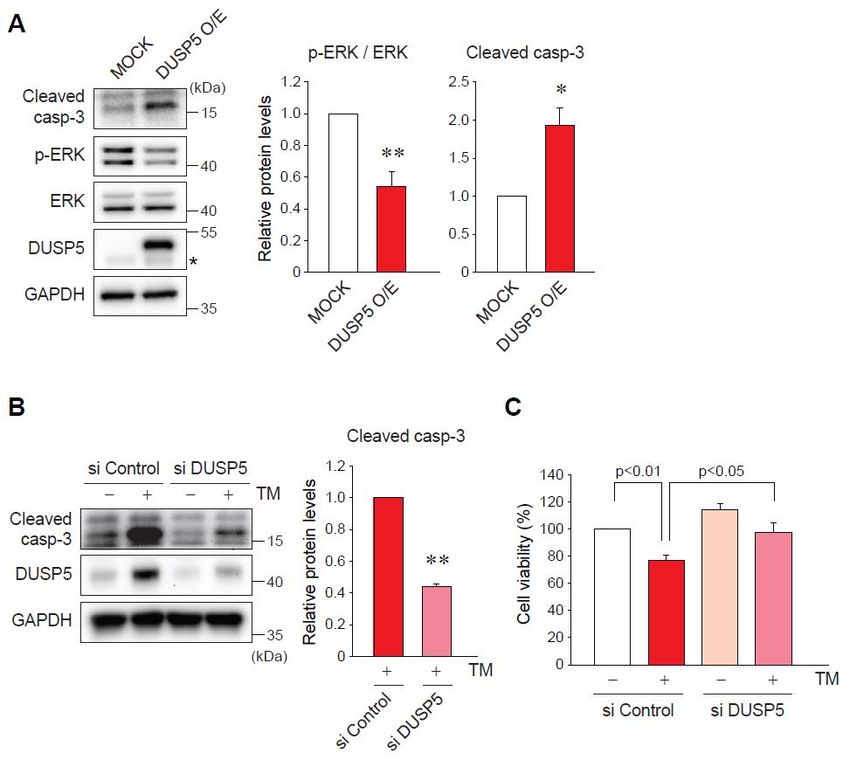
Int. J. Mol. Sci. 2019, 20, 4369                                                                                             4 of 12

2.2. Endoplasmic Reticulum (ER) Stress Increases DUSP5 Expression in Hepatocytes
       To investigate whether ER stress directly regulates DUSP5 expression, we used both animal
and
Int. J.cell
       Mol. models     of ER
            Sci. 2019, 20,     stress.
                           x FOR        In the liver of mice treated with tunicamycin (TM) (a well-known
                                 PEER REVIEW                                                                         4 ofER12
stress inducer), DUSP5 mRNA levels were gradually upregulated (Figure 2A). The induction of DUSP5
expression was
expression      was confirmed
                      confirmed by  by immunostaining
                                        immunostaining (Figure
                                                            (Figure 2B).
                                                                     2B). The
                                                                          The ability
                                                                              ability of
                                                                                      of TM
                                                                                          TM toto induce
                                                                                                  induce ER ER stress
                                                                                                               stress was
                                                                                                                        was
previously validated
previously      validated [11].
                              [11]. Consistently,
                                    Consistently, the
                                                    the transcript
                                                         transcriptlevels
                                                                     levelsof
                                                                            ofDUSP5
                                                                               DUSP5increased
                                                                                        increasedininAML12
                                                                                                         AML12cellscellsinina
a time-dependent manner (Figure 2C), which was in parallel with the protein expression (Figure 2D).
time-dependent         manner     (Figure   2C), which   was  in  parallel with the  protein   expression    (Figure    2D).
In addition,
In  addition, the the induction
                      induction of  of DUSP5
                                       DUSP5 by by TM
                                                   TM treatment
                                                       treatment waswas confirmed
                                                                         confirmed inin HepG2
                                                                                        HepG2 cellscells (Figure
                                                                                                         (Figure 2E,
                                                                                                                  2E, left).
                                                                                                                       left).
Moreover,treatment
Moreover,      treatmentof    ofAML12
                                AML12cellscells with
                                                with thapsigargin
                                                     thapsigargin (TG),
                                                                     (TG), another
                                                                           another ERER stress
                                                                                         stress inducer,
                                                                                                 inducer, had
                                                                                                            had the
                                                                                                                 the same
                                                                                                                      same
effect   (Figure    2E, right).  These   results demonstrate     that DUSP5   expression    is induced
effect (Figure 2E, right). These results demonstrate that DUSP5 expression is induced by ER stress        by  ER  stress   in
hepatocytes.
in hepatocytes.

      Figure
       Figure 2.    Endoplasmicreticulum
                2. Endoplasmic     reticulum(ER) (ER)stress
                                                       stress  increases
                                                             increases  thethe  expression
                                                                            expression        of DUSP5
                                                                                          of DUSP5         in hepatocytes.
                                                                                                       in hepatocytes.    (A)
      (A)
       qRT-PCR assay for hepatic DUSP5. Mice were treated with vehicle or tunicamycin (TM,(TM,
           qRT-PCR    assay for hepatic    DUSP5.    Mice   were  treated  with  vehicle  or tunicamycin           2 mg/kg,
                                                                                                              2 mg/kg,   i.p.)
      i.p.) forindicated
       for the  the indicated
                          timetime
                                points points  (n = 4/group).
                                         (n = 4/group).         * p
Int. J. Mol. Sci. 2019, 20, 4369 5 of 12
and ATF6, was verified (Figure 3A). CHOP is a key downstream molecule of PERK and contributes to
ER stress-mediated cell death [12]. Consistently, knockdown of CHOP successfully blocked the ability
ofInt.
TM to increase the levels of DUSP5 (Figure 3B). The overexpression of CHOP had the same effect
J. Mol. Sci. 2019, 20, x FOR PEER REVIEW 5 of 12
with TM treatment, upregulating DUSP5 expression (Figure 3C). In contrast, silencing of neither IRE1
nor ATF6 affected
Collectively, theseTM-induced DUSP5
data indicate expression
that CHOP (Figure 3D,E).
downstream Collectively,
of PERK signalingthese data for
accounts indicate that
ER-stress-
CHOP
mediated downstream of PERK signaling accounts for ER-stress-mediated DUSP5 induction.
DUSP5 induction.
Figure3.3.DUSP5
Figure DUSP5induction
inductionby byER ERstress
stressisismediated
mediatedby bythe
theprotein
proteinkinase
kinaseRNA-like
RNA-likeendoplasmic
endoplasmic
reticulum kinase (PERK)-C/EBP homologous protein (CHOP) pathway.
reticulum kinase (PERK)-C/EBP homologous protein (CHOP) pathway. (A) Immunoblotting for DUSP5. (A) Immunoblotting for
DUSP5.cells
AML12 AML12
werecells were pretreated
pretreated with 200 nM withof200 nM of integrated
integrated stress response
stress response inhibitor inhibitor
(ISRIB) for (ISRIB) for
30 min,
30 min,
and and continuously
continuously exposed toexposed
TM for 9toh.TM < 0.01
** pfor 9 h.versus
** p < 0.01 versus
TM alone (n = 3)group
TM alone
group (N.S., (n
not= significant).
3) (N.S., not
significant).
(B) AML12 cells (B)were
AML12 cells were
transfected with transfected with
control small control small
interfering RNAinterfering
(siRNA) orRNACHOP (siRNA)
siRNA or forCHOP
60 h,
followed by the treatment with TM for 9 h. ** p < 0.01 versus TM + si Control group (n = 4). (C) AML12
siRNA for 60 h, followed by the treatment with TM for 9 h. ** p < 0.01 versus TM + si Control group
(n =were
cells 4). (C) AML12 cells
transfected with were
MOCK transfected with MOCK or vector
or CHOP-overexpressing CHOP-overexpressing
(Myc-flag-taggedvector
CHOP)(Myc-flag-
for 24 h,
tagged
and thenCHOP) for 24group
one MOCK h, andwasthentreated
one MOCK
with TM groupfor was ** p < 0.01
9 h. treated withversus
TM forMOCK
9 h. ** group
p < 0.01 = 3).
(nversus
MOCK presents
Asterisk group (nthe= 3). Asterisk
band presents the
of endogenous band(D,
CHOP. of E)
endogenous
AML12 cells CHOP. (D, E) AML12
were transfected withcells were
control
transfected with control siRNA or each specific siRNA against inositol-requiring enzyme
siRNA or each specific siRNA against inositol-requiring enzyme 1 (IRE1) (D) or activating transcription 1 (IRE1) (D)
or activating
factor 6 (ATF6)transcription
(E) as in panelfactor = 3) (N.S.,
(B). 6(n(ATF6) (E) as
notinsignificant).
panel (B). (n = 3) (N.S., not significant).
2.4. DUSP5 Overexpression by ER Stress Induces Hepatocyte Death
DUSP5 acts as a phosphatase to target ERK, thereby inhibiting its activity [9,13]. Since ERK
signaling is one of the main pathways for cell survival [14], we hypothesized whether DUSP5 is
associated with hepatocyte death under ER stress condition. First, we checked the possible regulationInt. J. Mol. Sci. 2019, 20, 4369 6 of 12
2.4. DUSP5 Overexpression by ER Stress Induces Hepatocyte Death
DUSP5 acts as a phosphatase to target ERK, thereby inhibiting its activity [9,13]. Since ERK
signaling is one of the main pathways for cell survival [14], we hypothesized whether DUSP5 is
Int. J. Mol. Sci.
associated 2019,hepatocyte
with 20, x FOR PEER REVIEW
death 6 of of
under ER stress condition. First, we checked the possible regulation 12
ERK phosphorylation by DUSP5. As expected, the enforced expression of DUSP5 caused a decrease in
decrease in p-ERK levels in AML12 cells (Figure 4A). Then, we determined the role of DUSP5 in ER
p-ERK levels in AML12 cells (Figure 4A). Then, we determined the role of DUSP5 in ER stress-induced
stress-induced hepatocyte death. The activation and cleavage of caspase-3 is a key indicator of cell
hepatocyte death. The activation and cleavage of caspase-3 is a key indicator of cell death exerted
death exerted by ER stress [15]. The overexpression of DUSP5 raised cleaved caspase-3 expression
by ER stress [15]. The overexpression of DUSP5 raised cleaved caspase-3 expression (Figure 4A).
(Figure 4A). Consistently, treatment of AML12 cells with TM increased the levels of cleaved caspase-
Consistently, treatment of AML12 cells with TM increased the levels of cleaved caspase-3, which was
3, which was notably inhibited by DUSP5 knockdown (Figure 4B). In addition, MTT assays showed
notably inhibited by DUSP5 knockdown (Figure 4B). In addition, MTT assays showed that the decrease
that the decrease in cell viability by TM was significantly recovered by silencing of DUSP5 (Figure
in cell viability by TM was significantly recovered by silencing of DUSP5 (Figure 4C). Our results
4C). Our results suppose that DUSP5 functions as a mediator of ER stress-induced hepatocyte death
suppose that DUSP5 functions as a mediator of ER stress-induced hepatocyte death possibly through
possibly through ERK inhibition.
ERK inhibition.
Figure 4. DUSP5 inhibits extracellular-signal-regulated kinase (ERK) signal and leads to ER stress-induced
Figure 4. DUSP5 inhibits extracellular-signal-regulated kinase (ERK) signal and leads to ER
hepatocyte death. (A) Immunoblotting for cleaved caspase-3 and p-ERK. AML12 cell were transfected with
stress-induced hepatocyte death. (A) Immunoblotting for cleaved caspase-3 and p-ERK. AML12
MOCK or DUSP5-overexpressing vector (Myc-flag-tagged DUSP5) for 24 h. * p < 0.05 versus MOCK group
cell were transfected with MOCK or DUSP5-overexpressing vector (Myc-flag-tagged DUSP5) for 24 h.
(n = 3). Asterisk presents the band of endogenous DUSP5. (B) Immunoblotting for cleaved caspase-3 in
* p < 0.05 versus MOCK group (n = 3). Asterisk presents the band of endogenous DUSP5.
AML12 cells transfected with control or DUSP5 siRNA, followed by the treatment with TM. ** p < 0.01
(B) Immunoblotting for cleaved caspase-3 in AML12 cells transfected with control or DUSP5
versus TM + si Control group (n = 3). (C) Methylthiazolyldiphenyl-tetrazolium bromide (MTT) assay in
siRNA, followed by the treatment with TM. ** p < 0.01 versus TM + si Control group (n = 3).
AML12 cells treated with vehicle or TM for 24 h, after transfection with control or DUSP5 siRNA. Values
(C) Methylthiazolyldiphenyl-tetrazolium bromide (MTT) assay in AML12 cells treated with vehicle or
are the
TM means
for 24 ± standard
h, after errorwith
transfection of mean (SEM)
control of 4 separate
or DUSP5 siRNA. experiments (each
Values are the performed
means in triplicate).
± standard error of
mean (SEM) of 4 separate experiments (each performed in triplicate).
3. Discussion
Cellular integrity and homeostasis are maintained by the proper balance between the activation
and deactivation of signals, attributing to the coordinated regulation by diverse signaling molecules
and pathways. Thus, its imbalance and aberrant signal amplification is often observed in disease
states and may be critical for the pathogenesis. An important finding of the present study is theInt. J. Mol. Sci. 2019, 20, 4369 7 of 12
3. Discussion
Cellular integrity and homeostasis are maintained by the proper balance between the activation
and deactivation of signals, attributing to the coordinated regulation by diverse signaling molecules
and pathways. Thus, its imbalance and aberrant signal amplification is often observed in disease
states and may be critical for the pathogenesis. An important finding of the present study is
the identification of DUSP5 as a phosphatase changed in liver diseases and a novel potential marker of
liver injury. Our analysis of human GEO database and the experiments using animal models found
that the hepatic expression of DUSP5 increased in patients with liver cirrhosis, and in mice with liver
fibrosis at both transcript and protein levels. Of note, DUSP5 expression was profoundly enhanced
in acutely injured liver of mice exposed to liver toxicants, implying the possible detrimental roles
of DUSP5 in early pathogenic events during liver disease progression. In addition, the induction of
DUSP5 observed in the liver of APAP-treated mice was diminished by treatment with the chemical
chaperone, which indicates that DUSP5 overexpression can be involved in drug-induced liver injury in
association with ER stress. Recent reports suggested that several other DUSPs such as DUSP9, DUSP12,
and DUSP26 had the protective effects on the development of nonalcoholic steatohepatitis in mice,
and the expressions of those DUSPs decreased in the disease condition [16–18], supporting the concept
that the overexpressed DUSP5 may play a role in the process of diverse liver diseases at different stages.
Besides the pathophysiological roles of specific DUSPs, their cellular determinant and regulatory
basis are largely unknown. Recently, it has been suggested that DUSP5 expression is regulated by
serum response factor (SRF) and ETS like-1 protein (Elk-1) [19], and by microRNAs (i.e., miR-95
and miR-32-5p) [20,21]. Another key finding of this study is discovery of the direct regulation of DUSP5
gene expression by ER stress. Treatment of ER stress inducers upregulated the expression of DUSP5 in
hepatocytes, which was validated in the in vivo model using TM. Taken together with the induction of
DUSP5 expression in liver diseases, it seems that DUSP5 may contribute to the pathogenesis of ER
stress-associated liver disease progression. Moreover, we newly identified the PERK-CHOP pathway,
but not IRE1 and ATF6, as a signal for positive regulation of DUSP5 expression under ER stress condition.
The results from the experiments using the chemical inhibitor and siRNA-mediated knockdown of
UPR pathways revealed that CHOP downstream of PERK is responsible for the induction of DUSP5
by ER stress. CHOP overexpression alone even in the absence of ER stress stimulation efficaciously
induced DUSP5 expression, supporting the bona fide effect of CHOP on the gene regulation of DUSP5.
Our additional promoter analysis predicted that the CHOP-binding motifs [22,23] putatively exists
in the proximal region of human and mouse DUSP5 gene promoters (Supplementary Figure S1),
which needs to be clarified in further examinations. Our recent study showed that ER stress activated
the NACHT, LRR, and PYD domains-containing protein 3 (NLRP3) inflammasome via PERK-CHOP
signaling, which was inhibited by farnesoid X receptor [24]. Hence, the PERK-CHOP pathway may
contribute to hepatic dysfunction through the regulation of multiple gene expression in response to
ER stress.
Phosphorylation is a pivotal mode of intracellular signal transduction for the modulation of cell
fate, which is tightly regulated by both kinases and phosphatases. We demonstrated in this paper
that DUSP5 induction may have a role in ER stress-induced hepatocyte death probably through ERK
inhibition. In this study, the inhibitory effect of DUSP5 on ERK phosphorylation was also found
in a hepatocyte model, expanding our knowledge on the action of DUSP5 in the MAPK regulation.
Since ERK2 is involved in the stabilization of DUSP5 [25], a signaling loop between DUSP5 and ERK
may occur and probably affects cell viability. However, the complex effects of ERK on cell death has
also been suggested [14,26], although it is often required for cell survival. Thus, the possible role
of DUSP5 inhibition of ERK activity in ER stress-mediated hepatocyte death needs to be carefully
interpreted. Furthermore, the potential other substrate(s) of DUSP5 for the regulation of hepatocyte
functions should be recognized in future studies.
In the current study, we also found that DUSP5 overexpression caused the increase in
the apoptotic marker in hepatocytes, which was reciprocally changed with ERK activity. Moreover, ERInt. J. Mol. Sci. 2019, 20, 4369 8 of 12
stress-triggered hepatocyte death was attenuated by DUSP5 knockdown, suggesting the potential
effects of DUSP5 on hepatic injury. Recent studies showed that loss of DUSP5 protected
against hypertension-induced nephropathy in rates [27], and DUSP5 overexpression suppressed
endotoxin-induced inflammatory gene expression in macrophages and inhibited the activity of nuclear
factor-κB (NF-κB) in a phosphatase-activity independent manner [28]. Thus, the precise mechanisms
and roles of DUSP5 in cell death and liver disease progression require further examinations and in vivo
validation in future studies. The preventive effects of DUSP9, DUSP12, and DUSP26 on fatty liver
relied on the suppression of apoptosis signal-regulating kinase 1 (ASK1) or transforming growth factor
beta-activated kinase 1 (TAK1) [16–18]. Our previous study showed that the induction of pleckstrin
homology-like domain, family A, member 3 (PHLDA3) by ER stress led to hepatocyte death via
the repression of AKT [11]. Thus, the partial effect of DUSP5 knockdown on the recovery of cell viability
under ER stress condition seems to be attributed to the possible involvement of PHLDA3 and other
unknown molecule(s). Collectively, it is highly likely that ER stress-associated hepatocyte injury might
be mediated by the overexpression of DUSP5 and following inhibition of ERK, in conjunction with
the regulation of other signals including AKT.
In conclusion, DUSP5 expression levels are elevated in chronically and acutely injured liver,
and ER stress upregulates its gene expression through the PERK-CHOP pathway in hepatocytes.
The induction of DUSP5 is responsible for hepatocyte injury and death under ER stress condition,
which might be mediated at least in part by ERK inhibition. Our findings provide a novel insight into
molecular basis for ER stress-mediated cell death and related pathogenic events of liver diseases.
4. Materials and Methods
4.1. Materials
Anti-DUSP5 and anti-glyceraldehyde 3-phosphate dehydrogenase (GAPDH) antibodies were
obtained from Abcam (Cambridge, MA, USA) and Santa Cruz Biotechnology (Santa Cruz, CA, USA),
respectively. Antibodies directed against CHOP, spliced form of X-box-binding protein-1 (XBP1s),
IRE1, and ATF6, phosphorylated ERK, ERK or cleaved caspase-3 were purchased from Cell Signaling
Technology (Danvers, MA, USA). Horseradish peroxidase-linked anti-rabbit and anti-mouse IgGs
were also provided from Cell Signaling Technology. TM, ISRIB, and other reagents were supplied from
Sigma (St. Louis, MO, USA).
4.2. Bioinformatic Analysis
Hepatic gene expression data for patients with fibrosis were extracted from the Gene Expression
Omnibus (GSE25097). Differentially expressed genes (DEGs) were chosen as the genes with statistical
significance (p < 0.01 with a fold-change over 1.5) in cirrhotic patients as compared to healthy
subjects, and clustered by GO analysis using the Database for Annotation, Visualization and Integrated
Discovery (DAVID) 6.8 bioinformatics resource (https://david.ncifcrf.gov/), and the enriched terms were
presented as top ten biological process (BP) and each top five cellular compartment (CC) or molecular
function (MF) category. Upregulated DUSPs in cirrhosis patients were visualized using heatmap,
and the significantly changed (p < 0.05 with a fold-change over 1.5) DUSPs were marked in red.
4.3. Animal Treatments
The animal samples used in the present study were derived from the previous studies [11,29].
Animal experiments were performed according to the guidelines of the Institutional Animal Care
and Use Committee at Seoul National University. C57BL/6 mice were purchased from Charles River
Orient (Seoul, Korea). For chronic liver damages, six-week-old male mice were intraperitoneally
(i.p.) injected with vehicle or CCl4 (0.6 mL/kg) twice a week for 6 weeks. For acute liver injury,
eight-week-old male mice were given a single injection with vehicle or CCl4 (0.6 mL/kg, i.p.) for 24 h.
In another experiment, eight-week-old male mice were administered intraperitoneal injection of vehicleInt. J. Mol. Sci. 2019, 20, 4369 9 of 12
or APAP (500 mg/kg, 6 h) after pretreatment with vehicle or 4-phenylbutyrate (PBA, 100 mg/kg, 2 h).
For ER stress model, six-week-old male were given a single injection with vehicle or TM (2 mg/kg, i.p.)
for 24–72 h.
4.4. RNA Isolation and Real-Time Reverse Transcription-Polymerase Chain Reaction (RT-PCR) Assays
Total RNA was extracted using TRIzol (Thermo Fisher Scientific, Waltham, MA, USA), and 1 µg of
total RNA was reverse-transcribed to obtain cDNA. For quantitative analysis, the cDNA samples were
diluted 1:3 with RNase-free water and 2 µL of this dilution was used. Quantitative RT-PCR (qRT-PCR)
was performed using SYBR Premix Ex Taq II kit (Takara Bio, Shiga, Japan) and StepOne instrument
(Applied Biosystems, Thermo Fisher Scientific) according to the manufacturer’s instructions. A melting
curve of each PCR amplicon was determined to validate its accuracy. The relative levels of each
mRNAs were normalized to β-Actin and calculated using the 2−∆∆Ct method. Primer sequences
and annealing temperatures for PCR are provided in Supplementary Table S1.
4.5. Immunohistochemistry
Immunostaining was done similarly as described in the previous study [11].
Briefly, the paraffin-embedded tissue sections were deparaffinized with xylene and rehydrated
with alcohols series. The sections were interacted with anti-DUSP5 antibody (Thermo Fisher Scientific)
overnight at 4 ◦ C, followed by incubation with polymeric horseradish peroxidase (HRP)-linked
secondary antibody. The labeling was done using 3,30 -diaminobenzidine. The images were obtained
using light microscope (Olympus, Tokyo, Japan), and the areas were randomly selected in a consistent
and unbiased manner, and whole areas of each slide were examined to verify the quality of staining.
Among the multiple pictures, the representative image was shown. The staining intensities were
measured using ImageJ software.
4.6. Cell Culture
AML12 (non-transformed mouse hepatocyte-derived cell line) and HepG2 (human
hepatocyte-derived cell line) were purchased from American Type Culture Collection (ATCC)
(Rockville, MD, USA). AML12 cells were cultured in the Dulbecco’s Modified Eagle Medium
(DMEM)/F-12 containing 10% fetal bovine serum (FBS), insulin-transferrin-selenium X (ITSX),
dexamethasone (40 ng/mL), 100 units/mL penicillin, and 100 µg/mL streptomycin. HepG2 cells were
maintained in the DMEM containing 10% FBS, and the antibiotics. The cells with less than 20 passage
numbers were used.
4.7. Immunoblottings
Immunoblot analysis was carried out according to the previously published methods [11].
Cells were centrifuged at 3000× g for 3 min, and then lysed in lysis buffer in the ice for
30 min. The lysates were centrifuged at 10,000× g for 10 min to attain supernatants. Proteins of
interest in lysates were resolved using polyacrylamide gels and transferred to nitrocellulose or
poly(vinylidene fluoride) membrane. The bands were developed using enhanced chemiluminescence
(ECL) system (Millipore, Billerica, MA, USA). The band intensities were quantified using ImageJ
software and normalized to GAPDH.
4.8. Transient Transfection and Small Interfering RNA (siRNA) Knockdown
AML12 cells were transfected with MOCK or overexpression constructs (0.5 or 1 µg), or with
control siRNA or each specific siRNAs (100 pmol) using Lipofectamine 2000 (Invitrogen, Thermo
Fisher Scientific) according to the manufacturer’s instructions. Myc-flag-tagged CHOP (NM_007837)
and Myc-flag-tagged DUSP5 (NM_001085390) open reading frame (ORF) clones were obtained fromInt. J. Mol. Sci. 2019, 20, 4369 10 of 12
OriGene Technologies (Rockville, MD, USA). Scrambled siRNA (control) and the specific siRNAs direct
against CHOP, IRE1, ATF6, or DUSP5 were purchased from Thermo Fisher Scientific.
4.9. Methylthiazolyldiphenyl-tetrazolium bromide (MTT) Assay
TM-induced cytotoxicity was assessed, as previously described [11]. Briefly, AML12 cells were
plated at a density of 2 × 10⁵ cells per well in a 24-well plate to measure the degree of cell survival.
After treatment, viable cells were stained with MTT reagent (0.25 mg/mL, 1 h). Then, the media
were removed, and formazan crystals produced in the wells were dissolved in dimethylsulfoxide.
Absorbance was measured at 570 nm using a microplate reader (Molecular Devices, San Jose, CA, USA).
4.10. Statistical Analysis
Data were presented as mean ± standard error of mean (SEM) and statistically significance was
calculated by the Student’s t-test or by the one-way analysis of variance test with a post-hoc multiple
comparison procedure. Significant differences were considered at p < 0.05 or p < 0.01. Statistical
analyses were performed using IBM SPSS Statistics 24 software.
Supplementary Materials: Supplementary materials can be found at http://www.mdpi.com/1422-0067/20/18/
4369/s1. Figure S1. Putative CHOP-response elements (CHOP-REs) in human and mouse DUSP5 promoters.
Table S1. The primer sequences for qRT-PCR assays.
Author Contributions: Conceptualization, H.J.J. and C.Y.H.; investigation, H.J.J., J.W.Y., J.H.P. and C.Y.H.;
methodology, E.S.C.; resources, C.-S.L. and S.L.; writing—original draft preparation, H.J.J. and C.Y.H.;
writing—review and editing, C.Y.H.; supervision, C.Y.H.
Funding: This research was supported by Basic Science Research Program through the National Research
Foundation of Korea (NRF) funded by the Ministry of Education (2017R1D1A1B03028272).
Acknowledgments: We thank Sang Geon Kim (Seoul National University, Republic of Korea) for allowing use of
liver samples from the animal models of liver diseases or ER stress.
Conflicts of Interest: The authors declare no conflict of interest.
Abbreviations
APAP Acetaminophen
ATF6 Activating transcription factor 6
CCl4 Carbon tetrachloride
CHOP CCAAT-enhancer-binding protein homologous protein
DUSP Dual-specificity phosphatase
ERK Extracellular-signal-regulated kinase
ER stress Endoplasmic reticulum stress
GAPDH Glyceraldehyde 3-phosphate dehydrogenase
GEO Gene Expression Omnibus
GO Gene ontology
IRE1 Inositol-requiring enzyme 1
MAPK Mitogen-activated protein kinase
PBA 4-phenylbutyrate
PERK Protein kinase RNA-like endoplasmic reticulum kinase
TG Thapsigargin
TM Tunicamycin
UPR Unfolded protein response
XBP1 X-box-binding protein-1Int. J. Mol. Sci. 2019, 20, 4369 11 of 12
References
1. Malhi, H.; Gores, G.J. Cellular and molecular mechanisms of liver injury. Gastroenterology 2008, 134, 1641–1654.
[CrossRef]
2. Luedde, T.; Kaplowitz, N.; Schwabe, R.F. Cell death and cell death responses in liver disease: Mechanisms
and clinical relevance. Gastroenterology 2014, 147, 765–783.e4. [CrossRef]
3. Wattacheril, J.; Issa, D.; Sanyal, A. Nonalcoholic Steatohepatitis (NASH) and hepatic fibrosis: Emerging
therapies. Annu. Rev. Pharmacol. Toxicol. 2018, 58, 649–662. [CrossRef]
4. Hetz, C.; Chevet, E.; Harding, H.P. Targeting the unfolded protein response in disease. Nat. Rev. Drug Discov.
2013, 12, 703–719. [CrossRef]
5. Friedman, S.L.; Neuschwander-Tetri, B.A.; Rinella, M.; Sanyal, A.J. Mechanisms of NAFLD development
and therapeutic strategies. Nat. Med. 2018, 24, 908–922. [CrossRef]
6. Malhi, H.; Kaufman, R.J. Endoplasmic reticulum stress in liver disease. J. Hepatol. 2011, 54, 795–809.
[CrossRef]
7. Dara, L.; Ji, C.; Kaplowitz, N. The contribution of endoplasmic reticulum stress to liver diseases. Hepatology
2011, 53, 1752–1763. [CrossRef]
8. Huang, C.Y.; Tan, T.H. DUSPs, to MAP kinases and beyond. Cell Biosci. 2012, 2, 24. [CrossRef]
9. Chen, H.F.; Chuang, H.C.; Tan, T.H. Regulation of dual-specificity phosphatase (DUSP) ubiquitination
and protein stability. Int. J. Mol. Sci. 2019, 20, 2668. [CrossRef]
10. Lang, R.; Raffi, F.A.M. Dual-specificity phosphatases in immunity and infection: An update. Int. J. Mol. Sci.
2019, 20, 2710. [CrossRef]
11. Han, C.Y.; Lim, S.W.; Koo, J.H.; Kim, W.; Kim, S.G. PHLDA3 overexpression in hepatocytes by endoplasmic
reticulum stress via IRE1-Xbp1s pathway expedites liver injury. Gut 2016, 65, 1377–1388. [CrossRef]
12. Hetz, C.; Papa, F.R. The unfolded protein response and cell fate control. Mol. Cell 2018, 69, 169–181. [CrossRef]
13. Mandl, M.; Slack, D.N.; Keyse, S.M. Specific inactivation and nuclear anchoring of extracellular
signal-regulated kinase 2 by the inducible dual-specificity protein phosphatase DUSP5. Mol. Cell Biol. 2005,
25, 1830–1845. [CrossRef]
14. Lu, Z.; Xu, S. ERK1/2 MAP kinases in cell survival and apoptosis. IUBMB Life 2006, 58, 621–631. [CrossRef]
15. Iurlaro, R.; Munoz-Pinedo, C. Cell death induced by endoplasmic reticulum stress. FEBS J. 2016,
283, 2640–2652. [CrossRef]
16. Ye, P.; Xiang, M.; Liao, H.; Liu, J.; Luo, H.; Wang, Y.; Huang, L.; Chen, M.; Xia, J. Dual-specificity phosphatase
9 protects against nonalcoholic fatty liver disease in mice through ASK1 suppression. Hepatology 2019,
69, 76–93. [CrossRef]
17. Huang, Z.; Wu, L.M.; Zhang, J.L.; Sabri, A.; Wang, S.J.; Qin, G.J.; Guo, C.Q.; Wen, H.T.; Du, B.B.; Zhang, D.H.;
et al. Dual specificity phosphatase 12 regulates hepatic lipid metabolism through inhibition of the lipogenesis
and apoptosis signal-regulating kinase 1 pathways. Hepatology 2019. [CrossRef]
18. Ye, P.; Liu, J.; Xu, W.; Liu, D.; Ding, X.; Le, S.; Zhang, H.; Chen, S.; Chen, M.; Xia, J. Dual-specificity
phosphatase 26 protects against nonalcoholic fatty liver disease in mice through transforming growth factor
beta-activated kinase 1 suppression. Hepatology 2019, 69, 1946–1964. [CrossRef]
19. Buffet, C.; Catelli, M.G.; Hecale-Perlemoine, K.; Bricaire, L.; Garcia, C.; Gallet-Dierick, A.; Rodriguez, S.;
Cormier, F.; Groussin, L. Dual specificity phosphatase 5, a specific negative regulator of ERK signaling, is
Induced by serum response factor and Elk-1 transcription factor. PLoS ONE 2015, 10, e0145484. [CrossRef]
20. Du, M.; Zhuang, Y.; Tan, P.; Yu, Z.; Zhang, X.; Wang, A. microRNA-95 knockdown inhibits
epithelial-mesenchymal transition and cancer stem cell phenotype in gastric cancer cells through MAPK
pathway by upregulating DUSP5. J. Cell. Physiol. 2019. [CrossRef]
21. Yan, T.; Zhang, F.; Sun, C.; Sun, J.; Wang, Y.; Xu, X.; Shi, J.; Shi, G. miR-32-5p-mediated Dusp5 downregulation
contributes to neuropathic pain. Biochem. Biophys. Res. Commun. 2018, 495, 506–511. [CrossRef]
22. Han, J.; Back, S.H.; Hur, J.; Lin, Y.H.; Gildersleeve, R.; Shan, J.; Yuan, C.L.; Krokowski, D.; Wang, S.;
Hatzoglou, M.; et al. ER-stress-induced transcriptional regulation increases protein synthesis leading to cell
death. Nat. Cell Biol. 2013, 15, 481–490. [CrossRef]
23. Ohoka, N.; Yoshii, S.; Hattori, T.; Onozaki, K.; Hayashi, H. TRB3, a novel ER stress-inducible gene, is induced
via ATF4-CHOP pathway and is involved in cell death. EMBO J. 2005, 24, 1243–1255. [CrossRef]Int. J. Mol. Sci. 2019, 20, 4369 12 of 12
24. Han, C.Y.; Rho, H.S.; Kim, A.; Kim, T.H.; Jang, K.; Jun, D.W.; Kim, J.W.; Kim, B.; Kim, S.G. FXR inhibits
endoplasmic reticulum stress-induced NLRP3 inflammasome in hepatocytes and ameliorates liver injury.
Cell Rep. 2018, 24, 2985–2999. [CrossRef]
25. Kucharska, A.; Rushworth, L.K.; Staples, C.; Morrice, N.A.; Keyse, S.M. Regulation of the inducible nuclear
dual-specificity phosphatase DUSP5 by ERK MAPK. Cell. Signal. 2009, 21, 1794–1805. [CrossRef]
26. Cagnol, S.; Chambard, J.C. ERK and cell death: Mechanisms of ERK-induced cell death—Apoptosis,
autophagy and senescence. FEBS J. 2010, 277, 2–21. [CrossRef]
27. Zhang, C.; He, X.; Murphy, S.R.; Zhang, H.; Wang, S.; Ge, Y.; Gao, W.; Williams, J.M.; Geurts, A.M.; Roman, R.J.;
et al. Knockout of dual-specificity protein phosphatase 5 protects against hypertension-induced renal injury.
J. Pharmacol. Exp. Ther. 2019, 370, 206–217. [CrossRef]
28. Seo, H.; Cho, Y.C.; Ju, A.; Lee, S.; Park, B.C.; Park, S.G.; Kim, J.H.; Kim, K.; Cho, S. Dual-specificity phosphatase
5 acts as an anti-inflammatory regulator by inhibiting the ERK and NF-kappaB signaling pathways. Sci. Rep.
2017, 7, 17348. [CrossRef]
29. Han, C.Y.; Koo, J.H.; Kim, S.H.; Gardenghi, S.; Rivella, S.; Strnad, P.; Hwang, S.J.; Kim, S.G. Hepcidin
inhibits Smad3 phosphorylation in hepatic stellate cells by impeding ferroportin-mediated regulation of Akt.
Nat. Commun. 2016, 7, 13817. [CrossRef]
© 2019 by the authors. Licensee MDPI, Basel, Switzerland. This article is an open access
article distributed under the terms and conditions of the Creative Commons Attribution
(CC BY) license (http://creativecommons.org/licenses/by/4.0/).You can also read