Investigation of Strand-Selective Interaction of SNA-Modified siRNA with AGO2-MID - MDPI
←
→
Page content transcription
If your browser does not render page correctly, please read the page content below
International Journal of
            Molecular Sciences

Article
Investigation of Strand-Selective Interaction of
SNA-Modified siRNA with AGO2-MID
Yukiko Kamiya * , Yuuki Takeyama, Tomonari Mizuno, Fuminori Satoh and
Hiroyuki Asanuma *
 Department of Biomolecular Engineering, Graduate School of Engineering, Nagoya University, Furo-cho,
 Chikusa-ku, Nagoya 464-8603, Japan; takeyama.yuuki@g.mbox.nagoya-u.ac.jp (Y.T.);
 mizuno.tomonari@b.mbox.nagoya-u.ac.jp (T.M.); sato.fuminori@f.mbox.nagoya-u.ac.jp (F.S.)
 * Correspondence: yukikok@chembio.nagoya-u.ac.jp (Y.K.); asanuma@chembio.nagoya-u.ac.jp (H.A.);
   Tel.: +81-52-789-2552 (Y.K.); +81-52-789-2488 (H.A.)

 Received: 28 June 2020; Accepted: 20 July 2020; Published: 23 July 2020

 Abstract: Small interfering RNA (siRNA) has been recognized as a powerful gene-silencing tool. For
 therapeutic application, chemical modification is often required to improve the properties of siRNA,
 including its nuclease resistance, activity, off-target effects, and tissue distribution. Careful siRNA
 guide strand selection in the RNA-induced silencing complex (RISC) is important to increase the RNA
 interference (RNAi) activity as well as to reduce off-target effects. The passenger strand-mediated
 off-target activity was previously reduced and on-target activity was enhanced by substitution
 with acyclic artificial nucleic acid, namely serinol nucleic acid (SNA). In the present study, the
 reduction of off-target activity caused by the passenger strand was investigated by modifying
 siRNAs with SNA. The interactions of SNA-substituted mononucleotides, dinucleotides, and
 (2,2,6,6-tetramethylpiperidin-1-yl)oxyl (TEMPO)-labeled double-stranded RNA (dsRNA) with the
 MID domain of the Argonaute 2 (AGO2) protein, which plays a pivotal role in strand selection by
 accommodation of the 5’-terminus of siRNA, were comprehensively analyzed. The obtained nuclear
 magnetic resonance (NMR) data revealed that AGO2-MID selectively bound to the guide strand of
 siRNA due to the inhibitory effect of the SNA backbone located at the 5’ end of the passenger strand.

 Keywords: siRNA; RNAi; AGO2; MID; serinol nucleic acid; off-target effect; guide strand selection;
 RNA; TEMPO; NMR

1. Introduction
      Small interfering RNAs (siRNAs) are duplexes of approximately 23 base pairs composed of
passenger strands and guide strands. They promote gene silencing of messenger RNA (mRNA) targets
complementary to the guide strand via RNA interference (RNAi). Notably, synthetic siRNAs have
shown therapeutic potential. The first siRNA drug, namely patisiran, was approved by the Food
and Drug Administration (FDA) in 2018 [1]. Chemical modifications of siRNAs can improve their
resistance to nuclease digestion, increase potency, and reduce off-target effects [2,3].
      Translational inhibition mediated by siRNAs is executed by the RNA-induced silencing complex
(RISC), which comprises a siRNA guide strand and the Argonaute 2 (AGO2) protein. During the
formation of the complex, the strand with the 5’ end, which is involved in the thermodynamically
less stable region of base pairing, is selected for loading into RISC [4]. Strand selection is critical, as
base pairing between the strand loaded into RISC and the mRNA target determine the selectivity of
gene silencing. However, the control of the guide strand selection only by using RNA nucleotides is
insufficient; therefore, it is necessary to develop methods that would result in a reduction of off-target
activity mediated by the passenger strand-incorporated RISC. Various attempts have been made to

Int. J. Mol. Sci. 2020, 21, 5218; doi:10.3390/ijms21155218                        www.mdpi.com/journal/ijms
Int. J. Mol. Sci. 2020, 21, 5218                                                                    2 of 13

reduce the activity of passenger strand-incorporated RISC, even via unintended formation of the
complex, or alternatively, to improve the guide strand selectivity [2,3,5–11]. Nevertheless, further
developments are required to achieve precise control.
     Structural studies revealed that AGO2 is composed of globular domains, i.e., N, PAZ, MID, and
PIWI [12–16]. In the complex of micro RNA (miRNA) with full-length human AGO2, the 5’ end of
miRNA is bound to the MID domain, while the 3’ end is bound to the PAZ domain. The seed region
of miRNA is formed in an A-type helical geometry and is located in a narrow portion of the RNA
binding groove of PIWI. Importantly, the 5’-terminal phosphate and nucleotide are not involved in
pairing with the target mRNA. These moieties are located in a narrow binding pocket on the MID
domain of AGO2 and exhibit stacking interactions and extensive hydrogen bonds with the amino acid
residues of MID and PIWI. A previous study on the Y529E mutant showed that during the formation
of RISC (pre-RISC), the 5’-end binding pocket of MID plays an important function in binding of
miRNA to AGO2 [17]. Thus, to enhance the loading of the desired strand of siRNA into RISC, siRNAs
exhibiting various 5’-end modifications have been proposed. For instance, introduction of metabolically
stable (E)-5’-vinylphosphonate [18–20] or a 5’-triazol modifier, which was developed by computational
screening [21,22], at the 5’ end of the guide strand increased the activity of siRNA. Moreover, preventing
phosphorylation of the 5’-hydroxyl group of the passenger strand by Clp1 by 5’-O-methylation or
5’-morpholino modification reduced the formation of RISC with the passenger strand [23,24]. We
previously reported an acyclic nucleic acid, specifically serinol nucleic acid (SNA), which stably
hybridized with RNA [25–27]. In addition, we designed SNA-substituted siRNA, in which one residue
at each terminus of the passenger strand and at the 3’ terminus of the guide strand was substituted with
SNA (Figure 1) [28]. Consequently, the passenger strand-mediated off-target activity was effectively
reduced and the on-target activity was enhanced by a simple SNA substitution without any additional
modification of the 5’-hydroxyl group of siRNA. We expect that the SNA-substituted siRNA design
can be applied in the development of nucleic acid drugs to knock down any disease-related genes
and supplement miRNA. To make this design versatile, it is necessary to elucidate the mechanisms
by which the SNA substitution at the terminal positions of siRNA reduces the off-target effects. We
speculated that the SNA substitution inhibits the interaction of the passenger strand with AGO2-MID
due to the considerable differences between the structures of SNA and ribose, resulting in loading of
the guide strand into AGO2 (Figure 1). In the present study, we focused on the interactions between
AGO2-MID and SNA-modified RNAs. Specifically, we performed isothermal calorimetry (ITC) and
nuclear magnetic resonance (NMR) analyses to investigate the interactions between AGO2-MID
and SNA-substituted mononucleotides, dinucleotides, and (2,2,6,6-tetramethylpiperidin-1-yl)oxyl
(TEMPO)-labeled double-stranded RNAs (dsRNAs). We determined that AGO2-MID asymmetrically
binds to the guide strand of the SNA-modified siRNA, in which the passenger strand contains an SNA
moiety at the 5’ end.
Int. J. Mol. Sci. 2020, 21, 5218 3 of 13
Int. J. Mol. Sci. 2020, 21, x FOR PEER REVIEW 3 of 12
Figure 1. Schematic illustration of the proposed mechanism, by which serinol nucleic acid
Figure 1. Schematicsmall
(SNA)-substituted illustration of theRNA
interfering proposed
(siRNA)mechanism, by whichactivity.
reduces off-target serinol In
nucleic acid (SNA)-
pre-RNA-induced
substituted small interfering RNA (siRNA) reduces off-target activity. In pre-RNA-induced
silencing complex (pre-RISC), an interaction between the 5’ terminus of one strand and the MID silencing
domain
complex (pre-RISC), an interaction between the 5’ terminus of one strand and the MID domain
results in selection of that strand for the formation of RISC. Off-target effects can occur if the passengerresults
in selection
strand of that strand
is selected. for the formation
The presence of the SNAofmoiety
RISC. Off-target effects can
at the 5’ terminus of occur if the passenger
the passenger strand
strand inhibits
isthe
selected. The with
interaction presence of the 2SNA
Argonaute moiety at reducing
(AGO2)-MID, the 5’ terminus of the
off-target passenger strand inhibits the
effects.
interaction with Argonaute 2 (AGO2)-MID, reducing off-target effects.
2. Results and Discussion
2. Results and Discussion
2.1. Analyses of the Binding between AGO2-MID and Mono- and Dinucleotides
2.1. Analyses
We firstofanalyzed
the Binding between AGO2-MID
the binding and Mono-for
affinity of AGO2-MID andmono-
Dinucleotides
and dinucleotides using isothermal
calorimetry
We first(ITC). The affinity
analyzed of AGO2-MID
the binding affinityforofriboadenosine
AGO2-MID (A), for adenosine
mono- and monophosphate
dinucleotides (AMP),
using
SNA-A (sA), and phosphorylated SNA-A (p-sA) was
isothermal calorimetry (ITC). The affinity of AGO2-MID for riboadenosine compared (Figure 2 and S1 and(A),
Tableadenosine
1). It was
determined that(AMP), although AMP (sA),
bound to phosphorylated
AGO2-MID (Ka SNA-A 3 M−1 ), p-sA did not. The RNA
5.9 × 10(p-sA)
monophosphate SNA-A and was compared (Figure 2
dimer containing a 5’-phosphate group (p-rArA) exhibited
and S1 and Table 1). It was determined that although AMP bound to AGO2-MID approximately 40 times
(Ka higher
5.9 × 10affinity
3 M−1),
for AGO2-MID than AMP. The binding constant is within the range of
p-sA did not. The RNA dimer containing a 5’-phosphate group (p-rArA) exhibited approximately 40 typical protein-nucleotide
interactions.
times Interestingly,
higher affinity the 5’-phosphorylated
for AGO2-MID than AMP. TheSNA-RNA dimer is
binding constant (p-sArA)
within thewasrange
recognized by
of typical
AGO2-MID (K 7.2 × 10³ M−1 ), although the binding affinity was considerably lower than that for the
a
protein-nucleotide interactions. Interestingly, the 5’-phosphorylated SNA-RNA dimer (p-sArA) was
5’-phosphorylated
recognized by AGO2-MIDRNA dimer.(Ka 7.2The SNA
× 10 3 Mdimer with anthe
−1), although (S)-terminal phosphate
binding affinity was group also bound
considerably to
lower
AGO2-MID 3 −1
× 10 M ), indicating
than that for(Kthea 5.9
5’-phosphorylated RNA that dimer.the The
presence
SNA of the SNA
dimer withmoiety at the second
an (S)-terminal residue
phosphate
from the 5’ end was tolerable for interaction with AGO2-MID. On the other
group also bound to AGO2-MID (Ka 5.9 × 10 M ), indicating that the presence of the SNA moiety at
3 −1 hand, non-phosphorylated
dimers
the secondof SNA
residue or from
RNAthe did5’not
enddetectably
was tolerable bindfortointeraction
AGO2-MID (Figure
with S1). These
AGO2-MID. data
On the suggested
other hand,
that the phosphate group and the ribose backbone of the 5’ terminal residue
non-phosphorylated dimers of SNA or RNA did not detectably bind to AGO2-MID (Figure S1). These of siRNA cooperatively
contributed
data suggested to that
binding to AGO2-MID,
the phosphate groupwhich
and the is consistent with previous
ribose backbone reports [12–16].
of the 5’ terminal residue Moreover,
of siRNA
the results indicated
cooperatively that even
contributed the phosphorylated
to binding to AGO2-MID, SNA residue
which did not bind
is consistent withtoprevious
AGO2-MID. reports [12–
16]. Moreover, the results indicated that even the phosphorylated SNA residue did not bind to AGO2-
MID.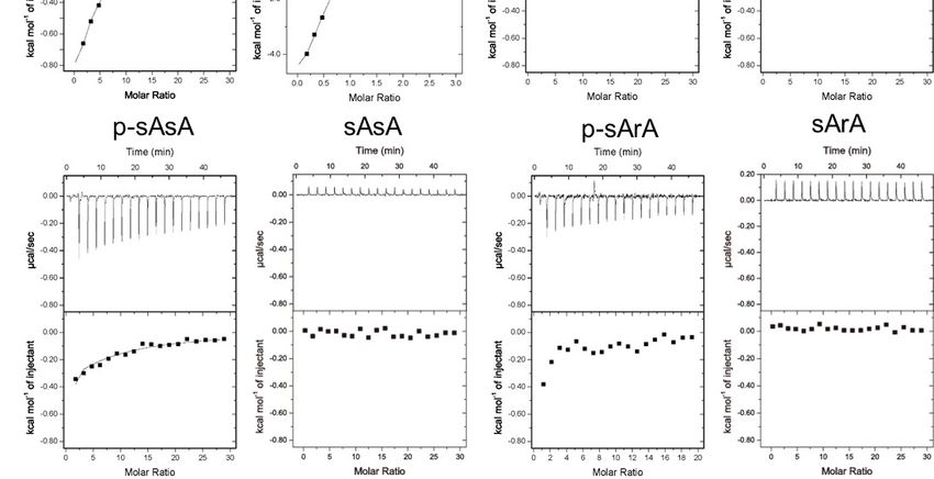
Int. Sci.
Int. J. Mol.  J. Mol. Sci.21,
                   2020,   2020, 21, 5218
                              x FOR   PEER REVIEW                                                                      4 of4 13
                                                                                                                              of 12

             Figure 2. Isothermal calorimetry (ITC) analyses of the binding between AGO2-MID and mono- or
      Figuredinucleotides.
              2. Isothermal(a)calorimetry  (ITC) analyses of the binding between AGO2-MID and mono- or
                                Chemical structures of RNA and SNA mononucleotides. (b) ITC profiles of the
      dinucleotides.   (a) Chemical
            binding between          structures
                                AGO2-MID          of RNA
                                            and mono-    andand    SNA mononucleotides.
                                                              dinucleotides with or without(b)  ITC profiles
                                                                                             terminal         of the
                                                                                                      phosphate
      binding   between    AGO2-MID     and   mono-    and  dinucleotides
                                                                ◦           with or  without   terminal
            groups. ITC experiments were performed at 25 C in 25 mM MES (pH 6.5), 200 mM NaCl, 1 mM      phosphate
      groups.  ITC experiments were performed
            tris(2-carboxyethyl)phosphine   (TCEP). at 25each
                                                     For  °C in   25 mM MES
                                                               experiment,    (pHAGO2-MID
                                                                           20 µM  6.5), 200 mM
                                                                                             wasNaCl,  1 mM
                                                                                                  loaded      tris(2-
                                                                                                         into the
            sample cell.
      carboxyethyl)phosphine     (TCEP). For each experiment, 20 μM AGO2-MID was loaded into the sample
      cell.           Table 1. K values for the interaction of AGO2-MID with mono- and dinucleotides.
                                  a

                     Mononucleot(s)ide
                 Table                        Ka (× 103 M
                       1. Ka values for the interaction of−1AGO2-MID
                                                             )   Dinucleotide        (× 103 M−1 ) 1
                                                                                  Kadinucleotides
                                                                     with mono- and
                              AMP                      5.9                 p-rArA                   230
                     Mononucleot(s)ide
                             p-sA               Ka (×n.d.103 M-1) Dinucleotide
                                                                          p-sArA          Ka (× 107.2
                                                                                                    3 M-1)

                               AMP
                              sA                        5.9
                                                     n.d.                p-rArA
                                                                          p-sAsA                2305.9
                               p-sA
             AMP is adenosine monophosphate; p-sA is 5’ n.d.             p-sArA
                                                        phosphorylated SNA                       7.2 n.d. indicates that
                                                                           adenosine; rA is adenosine.
             binding was not detected.
                                 sA                     n.d.             p-sAsA                  5.9
      AMP is adenosine monophosphate; p-sA is 5’ phosphorylated SNA adenosine; rA is adenosine. n.d.
            We subsequently analyzed the RNAi activity and strand selectivity of SNA-modified siRNA.
      indicates that binding was not detected.
       siRNAs modified with SNA at both 5’ and 3’ termini of the passenger strand and the 3’ terminus of the
       guide
      We      strand with analyzed
           subsequently    and without
                                     thea phosphate group
                                          RNAi activity   at the
                                                        and      5’ end
                                                              strand    were prepared
                                                                      selectivity       (P1s-1s/G1s-0ssiRNA.
                                                                                  of SNA-modified       and
siRNAs modified with SNA at both 5’ and 3’ termini of the passenger strand and the 3’ terminus of
the guide strand with and without a phosphate group at the 5’ end were prepared (P1s-1s/G1s-0s and
P1s-1s/G1s-0sp, respectively; Figure 3). siRNAs modified with SNA at both termini of both strands
and with a phosphate group at the 5’ end of the guide strand (P1s-1s/G1s-1sp) were also synthesized.
Additionally, unmodified siRNA (native P/G) was prepared. A luciferase reporter assay was
Int. J. Mol. Sci. 2020, 21, 5218 5 of 13
P1s-1s/G1s-0sp, respectively; Figure 3). siRNAs modified with SNA at both termini of both strands
and with a phosphate group at the 5’ end of the guide strand (P1s-1s/G1s-1sp) were also synthesized.
Int. J. Mol. Sci. 2020, 21,
Additionally, x FOR PEER
unmodified REVIEW
siRNA (native P/G) was prepared. A luciferase reporter assay was conducted 5 of 12
to analyze the RNAi activities originating from the passenger strand and guide strand-incorporated
incorporated
RISC. In aRISC. In a previously
previously reported
reported assay, assay, the
the pGL3-On pGL3-On
and pGL3-Off and pGL3-Off
plasmids, whichplasmids, whichbywere
were created
created by inserting the target sequence into the 3’-UTR of the firefly luciferase expression
inserting the target sequence into the 3’-UTR of the firefly luciferase expression vector in the sense vector in
the sense and antisense
and antisense directions,
directions, respectively,
respectively, were
were used toused to evaluate
evaluate silencingsilencing
induced induced by the
by the guide andguide
and passenger strands[28].
passenger strands [28].Moreover,
Moreover,according
according toto our
our previous
previous study
study [28],
[28], both
both strands
strands of native
of the the native
P/G exhibited RNAi activity. Compared with unmodified siRNA, the guide
P/G exhibited RNAi activity. Compared with unmodified siRNA, the guide strand of P1s-1s/G1s-0s, strand of P1s-1s/G1s-0s,
which which contained
contained an anSNASNA residueatatthe
residue the 3’
3’ end,
end,but
butnotnotthe 5’ end,
the showed
5’ end, showed increased RNAiRNAi
increased activity. In
activity.
contrast,
In contrast, thepassenger
the passengerstrand,
strand, in
in which
which both
bothtermini
terminiwereweremodified
modified with
withSNA,
SNA,displayed reduced
displayed reduced
activity (Figure 3). Notably, P1s-1s/G1s-0sp, which had analogous SNA modifications to P1s-1s/G1s-0s
activity (Figure 3). Notably, P1s-1s/G1s-0sp, which had analogous SNA modifications to P1s-1s/G1s-
and a 5’-phosphorylated guide strand, exhibited RNAi activity and guide strand selectivity equivalent
0s and a 5’-phosphorylated guide strand, exhibited RNAi activity and guide strand selectivity
to those of P1s-1s/G1s-0s (Figure 3).
equivalent to those of P1s-1s/G1s-0s (Figure 3).
Figure 3. RNA interference (RNAi) activities of SNA-modified siRNAs. (a) siRNAs used in the
Figure 3. RNA interference (RNAi) activities of SNA-modified siRNAs. (a) siRNAs used in the
luciferase assay. The upper strand is the passenger strand (P) and the lower strand is the guide strand
luciferase assay.
(G). Bold, The
italic upper strand
characters is the
indicate SNApassenger strand
and p denotes (P) and the(b)
5’ phosphate. lower
RNAistrand is the
activities guide
of the strand
guide
(G). Bold, italic characters indicate SNA and p denotes 5’ phosphate. (b) RNAi activities
strands (grey bars) and passenger strands (cyan bars) evaluated using reporter plasmids pGL3-On and of the guide
strands (grey bars)
pGL3-Off, and passenger
respectively. Plotted arestrands
means (cyan (n = 3)
and SDbars) evaluated using luciferase
of firefly/Renilla reporter signal
plasmids pGL3-On
relative to
and pGL3-Off,
the untreatedrespectively. < 0.01, ***p
sample. **p Plotted areInt. J. Mol. Sci. 2020, 21, 5218 6 of 13
2.2. TEMPO Modification of RNA for NMR Analyses
We then performed NMR analyses to obtain direct evidence of the strand selective interaction
between dsRNA and AGO2-MID. To this end, the interaction between AGO2-MID and an RNA duplex
modified with TEMPO was evaluated. It is known that an unpaired electron on the nitroxide group of
TEMPO influences the peak intensity of NMR signals due to paramagnetic relaxation enhancement
(PRE), which is related to r−6 [30]. Thus, TEMPO has been widely utilized in NMR analyses to provide
Int. J. Mol. Sci. 2020,
long-distance 21, x FOR PEER
information onREVIEW 6 of In
the conformation and interactions of various biomolecules [31–35]. 12
this study, we attempted to evaluate the strand selectivity of AGO2-MID using a TEMPO-modified
biomolecules [31–35]. In this study, we attempted to evaluate the strand selectivity of AGO2-MID
RNA duplexes.
using a TEMPO-modified RNA duplexes.
The nitroxide radical on TEMPO is readily degraded during the detritylation and oxidation
The nitroxide radical on TEMPO is readily degraded during the detritylation and oxidation steps
steps involved in the solid-support-based oligonucleotide synthesis [36,37]. TEMPO was previously
involved in the solid-support-based oligonucleotide synthesis [36,37]. TEMPO was previously
effectively incorporated into RNA using optimized protocols of detritylation and oxidation in the
effectively incorporated into RNA using optimized protocols of detritylation and oxidation in the
presence of dichloroacetic acid and tert-butyl hydroperoxide, respectively [37]. Methods, in which the
presence of dichloroacetic acid and tert-butyl hydroperoxide, respectively [37]. Methods, in which
nitroxide group was incorporated at terminal positions of oligonucleotides for post-synthesis spin
the nitroxide group was incorporated at terminal positions of oligonucleotides for post-synthesis spin
labeling of artificial ribose, nucleobase, or backbone moieties, have also been reported [38–43]. We
labeling of artificial ribose, nucleobase, or backbone moieties, have also been reported [38–43]. We
initially attempted to prepare the TEMPO-modified oligonucleotide utilizing an amidite monomer of
initially attempted to prepare the TEMPO-modified oligonucleotide utilizing an amidite monomer of
D-threoninol conjugated to TEMPO. Despite various modifications of the detritylation and oxidation
D-threoninol conjugated to TEMPO. Despite various modifications of the detritylation and oxidation
steps during solid-phase synthesis, the nitroxide group was degraded after several cycles as a result of a
steps during solid-phase synthesis, the nitroxide group was degraded after several cycles as a result
reduction reaction (Figure S2a–c). Consequently, we introduced the TEMPO moiety in a post-synthetic
of a reduction reaction (Figure S2a-c). Consequently, we introduced the TEMPO moiety in a post-
manner. We first synthesized an amidite monomer of Fmoc-protected D-threoninol (D-thr-Fmoc)
synthetic manner. We first synthesized an amidite monomer of Fmoc-protected D-threoninol (D-thr-
(Figure 4 and Scheme S1). Following conventional solid-phase oligonucleotide synthesis using
Fmoc) (Figure 4 and Scheme S1). Following conventional solid-phase oligonucleotide synthesis using
D-thr-Fmoc, we conducted simultaneous deprotection of the Fmoc group and conjugation of TEMPO
D-thr-Fmoc, we conducted simultaneous deprotection of the Fmoc group and conjugation of TEMPO
(Figure 4, step b). After the removal of RNA from controlled pore glass (CPG) and subsequent
(Figure 4, step b). After the removal of RNA from controlled pore glass (CPG) and subsequent
deprotection, the obtained RNAs were purified to yield TEMPO-modified RNA (Figure 4 and S2d).
deprotection, the obtained RNAs were purified to yield TEMPO-modified RNA (Figure 4 and S2d).
Figure 4. Scheme showing introduction of the (2,2,6,6-tetramethylpiperidin-1-yl)oxyl (TEMPO)
Figure 4. Scheme showing introduction of the (2,2,6,6-tetramethylpiperidin-1-yl)oxyl (TEMPO)
moiety following solid-phase synthesis of RNA. (a) An Fmoc-protected D-threoninol was
moiety following solid-phase synthesis of RNA. (a) An Fmoc-protected D-threoninol was
incorporated during solid-phase synthesis of RNA. (b) Deprotection of Fmoc and coupling of
incorporated during solid-phase synthesis of RNA. (b) Deprotection of Fmoc and coupling of 4-
4-carboxy-TEMPO was carried out simultaneously. RNA-conjugated controlled pore glass (CPG)
carboxy-TEMPO was carried out simultaneously. RNA-conjugated controlled pore glass (CPG) was
was dispersed in dimethylformamide (DMF) and incubated with 4-carboxy-TEMPO (100 eq., TCI),
dispersed in dimethylformamide (DMF) and incubated
1-ethyl-3-(3-dimethylaminopropyl)carbodiimide (EDC) (250with 4-carboxy-TEMPO (100
eq.), hydroxybenzotriazole eq., (250
(HOBt) TCI), 1-
eq.),
ethyl-3-(3-dimethylaminopropyl)carbodiimide
◦ (EDC) (250 eq.), hydroxybenzotriazole (HOBt)
and triethylamine (2.8 eq.) at 40 C for 12–24 h. (c) The removal of RNA from CPG and deprotections were (250
eq.), and triethylamine
performed (2.8 eq.)
using conventional at 40 °C
methods. TheforRNA 12–24
wash.purified
(c) Thebyremoval of RNA
reverse-phase from CPG and
high-performance
deprotections were performed
liquid chromatography (HPLC). using conventional methods. The RNA was purified by reverse-phase
high-performance liquid chromatography (HPLC).
2.3. Investigation of Strand Selection by AGO2-MID
2.3. Investigation of Strand Selection by AGO2-MID
The passenger strands were functionalized with TEMPO at either the 5’ or 3’ end and with an
SNAThe passenger
residue strands were
at the opposite end. functionalized with TEMPO
The obtained strands at either the
were hybridized to 5’ or 3’ end
a guide andmodified
strand with an
SNA residue at the opposite end. The obtained strands were hybridized to a guide strand modified
with SNA at the 3’ end (P1s-Te/G1s-0s and PTe-1s/G1s-0s; Figure S3). To determine whether the
TEMPO-modified siRNAs were able to induce gene silencing, we performed a luciferase reporter
assay. Similar to the terminally SNA-modified siRNA P1s-1s/G1s-0s, P1s-Te/G1s-0s and PTe-1s/G1s-
0s showed increased guide strand activity and significantly decreased passenger strand activityInt. J. Mol. Sci. 2020, 21, 5218 7 of 13
with SNA at the 3’ end (P1s-Te/G1s-0s and PTe-1s/G1s-0s; Figure S3). To determine whether the
TEMPO-modified siRNAs were able to induce gene silencing, we performed a luciferase reporter assay.
Similar to the terminally SNA-modified siRNA P1s-1s/G1s-0s, P1s-Te/G1s-0s and PTe-1s/G1s-0s showed
increased guide strand activity and significantly decreased passenger strand activity compared to the
native siRNA (Figure S3).
To analyze the interactions between SNA-modified siRNA mimics and AGO2-MID by NMR, we
prepared two short TEMPO-modified 10-bp RNA duplexes with SNA modifications (Figure 5a). In one
duplex, the passenger strand was modified with SNA at the 3’ end and TEMPO at the 5’ end (RNAa-5Te),
Int.
whileJ. Mol.
theSci.guide
2020, 21, x FORwas
strand PEER5’-phosphorylated
REVIEW and modified with SNA at the 3’ end (RNAb). 7Inofthe 12
second duplex, the passenger strand was modified with SNA at the 5’ end and TEMPO at the 3’ end
(RNAb).
(RNAa-3Te), In the second
whereas theduplex, the passenger
guide strand was RNAb. strand was modified
It is known with SNA selectively
that if AGO2-MID at the 5’ end and
interacts
TEMPO at the 3’ end (RNAa-3Te), whereas the guide strand was RNAb. It
with the 5’ terminus of the guide strand, the NMR signals corresponding to AGO2-MID should be is known that if AGO2-
MID selectively
broadened uponinteracts with thewith
the interaction 5’ terminus of the guidedue
RNAa-3Te/RNAb strand, the NMR
to strong PRE signals
(Figurecorresponding
5b). However,to if
AGO2-MID should be broadened upon the interaction with RNAa-3Te/RNAb
AGO2-MID interacts with the passenger strand, the NMR signals of AGO2-MID should be broadened due to strong PRE
(Figure
upon the 5b). However,with
interaction if AGO2-MID interacts with the passenger strand, the NMR signals of AGO2-
RNAa-5Te/RNAb.
MID should be broadened upon the interaction with RNAa-5Te/RNAb.
Figure 5. (a) RNAs used in the nuclear magnetic resonance (NMR) analyses. Te indicates TEMPO
Figure 5. (a) RNAs used in the nuclear magnetic resonance (NMR) analyses. Te indicates TEMPO
incorporated using a D-threoninol backbone. Bold, italic characters indicate SNA and p denotes the
incorporated using a D-threoninol backbone. Bold, italic characters indicate SNA and p denotes the
5’-phosphate group. (b) Schematic illustration of the NMR analyses for investigation of strand selection
5’-phosphate group. (b) Schematic illustration of the NMR analyses for investigation of strand
by AGO2-MID. (c and d) 1 H-15 N heteronuclear single-quantum coherence (HSQC) NMR spectra of
selection by AGO2-MID. (c and d) 1H-15N heteronuclear single-quantum coherence (HSQC) NMR
AGO2-MID (0.06 mM) with (c) RNAa-5Te/RNAb (0.4 mM) or (d) RNAa-3Te/RNAb (0.4 mM) in the
spectra of AGO2-MID (0.06 mM) with (c) RNAa-5Te/RNAb (0.4 mM) or (d) RNAa-3Te/RNAb (0.4
absence (magenta or blue) and presence (green or red) of 2 mM ascorbic acid.
mM) in the absence (magenta or blue) and presence (green or red) of 2 mM ascorbic acid.
We first compared the 1 H-15 N heteronuclear single-quantum coherence (HSQC) spectra of
We
AGO2-MID firstwith
compared the 1TEMPO-labeled
and without H-15N heteronuclear
RNA single-quantum coherence
duplexes in the presence (HSQC) acid,
of ascorbic spectra of
which
AGO2-MID with and without TEMPO-labeled RNA duplexes in the presence of ascorbic acid,
reduced the nitroxide radical. Chemical shift perturbations of several NMR signals corresponding which
reduced the nitroxide
to the AGO2-MID radical.
residues Chemical
were shiftwhen
observed perturbations of several
the spectrum NMR
without RNAsignals
was corresponding to
compared to that
the AGO2-MID residues were observed when the spectrum without RNA was compared to that with
RNA (Figure S4). Notably, the perturbed chemical shifts were consistent with those previously
reported for spectra of AGO2-MID with and without UMP [11–13]. We subsequently compared the
spectra of AGO2-MID complexed with TEMPO-labeled RNA in the absence and presence of ascorbic
acid. While NMR signals in the 1H-15N HSQC spectra of AGO2-MID in the presence of RNAa-
3Te/RNAb were considerably broadened, much less broadening was observed in the presence ofInt. J. Mol. Sci. 2020, 21, 5218 8 of 13
with RNA (Figure S4). Notably, the perturbed chemical shifts were consistent with those previously
reported for spectra of AGO2-MID with and without UMP [11–13]. We subsequently compared
the spectra of AGO2-MID complexed with TEMPO-labeled RNA in the absence and presence of
ascorbic acid. While NMR signals in the 1 H-15 N HSQC spectra of AGO2-MID in the presence of
RNAa-3Te/RNAb were considerably broadened, much less broadening was observed in the presence
of RNAa-5Te/RNAb (Figure 5c,d and S5). It is noteworthy that the broadened signals disappeared in
the 2D-spectra. The acquired NMR data clearly indicated that the TEMPO moiety in RNAa-3Te was
located in close proximity to AGO2-MID. This conclusion was based on the observation that some
of the signals attributed to AGO2-MID in the 2D spectrum of RNAa-3Te/RNAb disappeared in the
absence of ascorbic acid. Moreover, the disappearance of the signals was marginal in the spectra of
RNAa-5Te/RNAb, in which the TEMPO moiety was away from AGO2-MID. Thus, the obtained results
revealed that AGO2-MID preferentially interacted with the 5’ terminus of RNAb over the 5’ terminus
of RNAa, the residue of which was composed of an acyclic backbone.
In conclusion, we successfully synthesized TEMPO-modified RNA, enabling NMR analyses,
which revealed siRNA strand selectivity of AGO2-MID. Conjugation of TEMPO after solid-phase
synthesis of RNA was necessary to prevent the reduction of the nitroxide moiety. A similar strategy
could be used to post-synthetically introduce a TEMPO moiety at any desired position within an
oligonucleotide. Moreover, other functional groups that would be unstable under conditions used for
solid-phase oligonucleotide synthesis could be also introduced using this approach.
The conducted experiments demonstrated that modification of the 5’ end of the passenger
strand of siRNA facilitates an asymmetric interaction between AGO2-MID and siRNA, significantly
enhancing the selectivity for the desired guide strand. Based on the NMR and ITC data, we concluded
that the SNA substitution at the 5’ end of the passenger strand considerably decreased the binding
affinity of AGO2 for the passenger strand, resulting in selective incorporation of the guide strand
into RISC. The outcomes of the present study provide valuable mechanistic insight into how the SNA
modification at the 5’-terminus of the passenger strand of siRNA reduces off-target effects. The findings
demonstrated herein provide a platform for further application of SNA modification in the design of
therapeutic siRNAs.
3. Materials and Methods
3.1. Syntheses of Dinucleotides and Oligonucleotides
The reagents for the synthesis of the phosphoramidite monomer of Fmoc-protected D-threoninol
were purchased from Tokyo Kasei Co., Ltd. (Tokyo, Japan), Wako Pure Chemical Industries, Ltd.
(Osaka, Japan), and Sigma-Aldrich, (Saint Louis, MO, USA). The reagents for the oligomer syntheses
and Poly Pak II cartridges were obtained from Glen Research (Sterling, VA, USA) and ChemGenes
(Wilmington, MA, USA). The columns for HPLC purification were purchased from Kanto Chemical
Co., Ltd (Tokyo, Japan). The RNA oligonucleotides and oligonucleotides composed of SNA and RNA
were acquired from Hokkaido System Science Co., Ltd. (Sapporo, Japan).
Phosphoramidite SNA monomers were synthesized according to a previously reported
method [25,26]. Serinol-adenine (sA) was prepared by the deprotection of the 4,40 -dimethoxytrityl
(DMT) group in DMT-serinol-adenine using trifluoroacetic acid. Dinucleotides (sAsA, rAsA,
and sArA) were synthesized using SNA phosphoramidite and RNA monomers employing the
ABI-3400 DNA synthesizer (Applied Biosystems, Waltham, MA, USA). Phosphorylation of sA
and dinucleotides was conducted on solid-phase using phosphorylation reagent II (Glen Research,
Sterling, VA, USA). D-Threoninol-Fmoc was introduced during solid-phase synthesis. For the
deprotection of Fmoc and conjugation of TEMPO, RNA-conjugated CPG was dispersed in
dimethylformamide (DMF) and incubated in the presence of 4-carboxy-TEMPO (100 eq., TCI),
1-ethyl-3-(3-dimethylaminopropyl)carbodiimide (EDC) (250 eq.), hydroxybenzotriazole (HOBt)
(250 eq.), and triethylamine (2.8 eq.) at 40 ◦ C for 12–24 h. During this step, some elimination of RNAInt. J. Mol. Sci. 2020, 21, 5218 9 of 13
from CPG was observed; therefore, the reaction time should not be exceeded. CPG was washed with
DMF and CHCl3 and then dried. Subsequently, the RNA oligonucleotide was removed from CPG. The
deprotection was conducted by incubating the oligonucleotide with a NH4 OH/EtOH (3:1, v/v) solution
at 55 ◦ C for 8 h. After drying under vacuum, RNA was dissolved in 1 M tetra-n-butylammonium
fluoride in tetrahydrofuran (THF) and stirred at 25 ◦ C for 24 h for 2’ deprotection. The products
were purified using diethylaminoethyl cellulose (DEAE) and reverse-phase high-performance liquid
chromatography (HPLC) and analyzed by electrospray ionization mass spectrometry (ESI-MS) or
matrix-assisted laser desorption/ionization-time-of-flight mass spectrometry (MALDI-TOF-MS) using
the Autoflex II apparatus (Bruker Daltonics, Billerica, MA, USA).
3.2. ESI-MS Data
p-sA Obsd. 345.085 (Calcd. for C10 H3 N5 O4 346.0791); sA Obsd. 265.115 (Calcd. for C10 H14 N6 O3
266.1127); p-rArA Obsd. 675.131 (Calcd. for C20 H26 N10 O13 P2 676.4325); p-sArA Obsd. 674.143 (Calcd.
for C20 H27 N11 O12 P2 675.1316); p-sAsA Obsd. 673.1476 (Calcd. for C20 H27 N12 O11 P2 674.1403); rArA
Obsd. 595.165 (Calcd. for C20 H25 N10 O10 P 596.4538); sAsA Obsd. 593.196 (Calcd. for C20 H27 N12 O8 P
594.18124); sArA Obsd. 594.180 (Calcd. for C20 H26 N11 O10 P 595.1653).
3.3. MALDI-TOF-MS Data
P1s-Te: Obsd. m/z 7186.66 (Calcd. for P1s-Te: m/z 7187.08); PTe-1s: Obsd. m/z 7184.43 (Calcd. for
PTe-1s: m/z 7178.08); RNAa-5Te: Obsd. m/z 3181.48 (Calcd. for RNAa-5Te: m/z 3175.60); RNAa-3Te:
Obsd. m/z 3192.59 (Calcd. for RNAa-3Te: m/z 3185.94).
3.4. Protein Expression
DNA fragment encoding human AGO2-MID (432-578) was cloned between the BamHI and
SalI sites of the pCold I plasmid (Takara, Kusatsu, Japan), in which the SUMO-TEV coding region
was inserted between the NdeI and BamHI sites. Hexahistidine-tagged SUMO-TEV-hAGO2-MID
was expressed in the Escherichia coli BL21(DE3)pLysS strain induced with 0.5 mM isopropyl
β-D-thiogalactopyranoside. For NMR analyses, the protein was expressed in the M9 minimal
medium with appropriate [15 N] NH4 Cl (Chembridge isotope laboratories Inc., Tewksbury, MA, USA)).
The protein was purified using a Ni2+ -NTA high-performance column (GE Healthcare, Chicago, IL,
USA). The hexahistidine-modified SUMO was cleaved by incubation with proTEV (Promega, Madison,
WI, USA) and further purified employing a Ni2+ -NTA high-performance column and a cation exchange
column (GE Healthcare, Chicago, IL, USA).
3.5. Cell Culture
The 293FT cells were cultured in Dulbecco’s modified Eagle’s medium supplemented with 10%
fetal bovine serum, 80 µg/mL penicillin, and 90 µg/mL streptomycin. The cells were cultured at 37 ◦ C
with 5% CO2 in humidified air.
3.6. Dual Luciferase Assay
Reporter plasmids pGL3-On and pGL3-Off or pGlo-On and pGlo-Off were used for the dual
luciferase assay [27,28]. The target sequence in pGL3-On and pGlo-On was 5’-AACCTTACCCAC
CTCATGTATCT-3’ and that in pGL3-Rv and pGlo-Off was the complementary sequence 5’-AGAT
ACATGAGGTGGGTAAGGT-3’. The target sequences were inserted into the 3’-UTR region of the
gene encoding firefly luciferase in pGL3. Co-transfections of the 293FT cells with siRNA (final
concentrations 17 nM), 100 ng of pGL3 or pGlo, and 0.5 ng of the Renilla luciferase expression vector
were performed using Lipofectamine 2000 (Invitrogen, Waltham, MA, USA) in 96-well plates according
to the manufacturer’s instructions. Following incubation at 37 ◦ C for 48 h, 75 µL of the medium was
removed, and 75 µL of the Dual-Glo regent (Promega, Madison, WI, USA) was added. Firefly luciferaseInt. J. Mol. Sci. 2020, 21, 5218 10 of 13
luminescence was measured on a Multi-label Plate Reader (EnSpire, Perkin Elmer, Waltham, MA,
USA). Subsequently, 75 µL of the Dual-Glo Stop & Glo reagent was added, and the Renilla luciferase
luminescence was measured.
3.7. Calorimetric Analysis
The ITC experiments were performed at 25 ◦ C using the iTC200 calorimeter (GE Healthcare,
Chicago, IL, USA). AGO2-MID was dialyzed against 25 mM MES (pH 6.5), 200 mM NaCl, and 1 mM
tris(2-carboxyethyl)phosphine (TCEP). 20 µM AGO2-MID was first loaded into the sample cell and
then 2 µL of adenosine (3 mM), AMP (3 mM), sA (3 mM), p-sA (3 mM), rArA (3 mM), p-rArA (0.3 mM),
sArA (3 mM), p-sArA (2 mM), rAsA (3 mM), sAsA (3 mM), or p-sAsA (3 mM) was added every 150 s.
3.8. NMR Analyses
AGO2-MID (0.06 mM) was mixed with TEMPO-labeled RNA duplex (0.4 mM) dissolved in 25 mM
MES (pH 6.5), 100 mM NaCl, and 10% (v/v) D2 O in the absence or presence of ascorbic acid (2 mM).
The 1 H-15 N HSPQC spectra were acquired at 293 K using the Avance600 spectrometer equipped
with a cryo-probe (Bruker BioSpin, Billerica, MA). The data were processed using the Topspin and
Sparky software.
Supplementary Materials: Supplementary materials can be found at http://www.mdpi.com/1422-0067/21/15/
5218/s1. Figure S1: ITC analyses of binding of AGO2-MID with RNA mono and dinucleotides. Figure S2:
MALDI-TOF-MS analyses of crude DNA or RNA oligomers containing D-threoninol-conjugated TEMPO. Figure S3:
RNAi activities of TEMPO- and SNA-modified siRNAs. Figure S4: 1H-15N HSQC spectra of AGO2-MID complexed
with RNA. Figure S5: 1H-15N HSQC spectra of AGO2-MID complexed with TEMPO-RNA in the absence or
presence of ascorbic acid. Scheme S1: Synthesis of phosphoramidite monomer of Fmoc-conjugated D-threoninol.
Author Contributions: Conceptualization, Y.K.; methodology, Y.K.; preparation of protein and oligonucleotides,
Y.K., Y.T., T.M., and F.S.; Luciferase, ITC and NMR analyses, Y.K., Y.T., and T.M.; writing—original draft preparation,
Y.K.; writing—review and editing, Y.K. and H.A.; visualization, Y.K. funding acquisition, Y.K. and H.A. All authors
have read and agreed to the published version of the manuscript.
Funding: This research was funded in part by the Grants-in-Aid for Scientific Research (Grant Numbers 20K05745
to Y.K.) and AMED (Grant Number 19am0401007 to H.A.).
Acknowledgments: We thank Yuichiro Aiba (Nagoya University) for providing the SUMO-TEV plasmid and
Takashi Tenno (Nagoya University) for his help in NMR measurements. The authors wish to acknowledge the
Division for Medical Research Engineering, Nagoya University Graduate School of Medicine for allowing use of
the iTC200 calorimeter.
Conflicts of Interest: The authors declare no conflict of interest.
Abbreviations
siRNA Small interfering RNA
AGO Argonaute
RISC RNA-induced silencing complex
NMR nuclear magnetic resonance
TEMPO 2,2,6,6-tetramethylpiperidine 1-oxyl
SNA Serinol nucleic acid
RISC RNA-induced silencing complex
RNAi RNA interference
AMP Adenosine monophosphate
TCEP Tris(2-carboxyethyl)phosphine hydrochloride
ITC Isothermal titration calorimetry
PRE paramagnetic relaxation enhancement
HSQC heteronuclear single-quantum coherenceInt. J. Mol. Sci. 2020, 21, 5218 11 of 13
References
1. Adams, D.; Gonzalez-Duarte, A.; O’Riordan, W.D.; Yang, C.C.; Ueda, M.; Kristen, A.V.; Tournev, I.;
Schmidt, H.H.; Coelho, T.; Berk, J.L.; et al. Patisiran, an RNAi Therapeutic, for Hereditary Transthyretin
Amyloidosis. N. Engl. J. Med. 2018, 379, 11–21. [CrossRef] [PubMed]
2. Khvorova, A.; Watts, J.K. The chemical evolution of oligonucleotide therapies of clinical utility. Nat. Biotechnol.
2017, 35, 238–248. [CrossRef] [PubMed]
3. Shen, X.; Corey, D.R. Chemistry, mechanism and clinical status of antisense oligonucleotides and duplex
RNAs. Nucleic Acids Res. 2018, 46, 1584–1600. [CrossRef] [PubMed]
4. Schwarz, D.S.; Hutvagner, G.; Du, T.; Xu, Z.; Aronin, N.; Zamore, P.D. Asymmetry in the assembly of the
RNAi enzyme complex. Cell 2003, 115, 199–208. [CrossRef]
5. Vaish, N.; Chen, F.; Seth, S.; Fosnaugh, K.; Liu, Y.; Adami, R.; Brown, T.; Chen, Y.; Harvie, P.; Johns, R.; et al.
Improved specificity of gene silencing by siRNAs containing unlocked nucleobase analogs. Nucleic Acids Res.
2011, 39, 1823–1832. [CrossRef]
6. Snead, N.M.; Escamilla-Powers, J.R.; Rossi, J.J.; McCaffrey, A.P. 5’ Unlocked Nucleic Acid Modification
Improves siRNA Targeting. Mol. Ther. Nucleic Acids 2013, 2, e103. [CrossRef]
7. Amarzguioui, M.; Holen, T.; Babaie, E.; Prydz, H. Tolerance for mutations and chemical modifications in a
siRNA. Nucleic Acids Res. 2003, 31, 589–595. [CrossRef]
8. Chiu, Y.L.; Rana, T.M. siRNA function in RNAi: A chemical modification analysis. RNA 2003, 9, 1034–1048.
[CrossRef]
9. Watts, J.K.; Choubdar, N.; Sadalapure, K.; Robert, F.; Wahba, A.S.; Pelletier, J.; Pinto, B.M.; Damha, M.J.
2’-fluoro-4’-thioarabino-modified oligonucleotides: Conformational switches linked to siRNA activity.
Nucleic Acids Res. 2007, 35, 1441–1451. [CrossRef]
10. Takahashi, M.; Nagai, C.; Hatakeyama, H.; Minakawa, N.; Harashima, H.; Matsuda, A. Intracellular stability
of 2’-OMe-4’-thioribonucleoside modified siRNA leads to long-term RNAi effect. Nucleic Acids Res. 2012, 40,
5787–5793. [CrossRef]
11. Deleavey, G.F.; Frank, F.; Hassler, M.; Wisnovsky, S.; Nagar, B.; Damha, M.J. The 5’ binding MID domain of
human Argonaute2 tolerates chemically modified nucleotide analogues. Nucleic Acid Ther. 2013, 23, 81–87.
[CrossRef] [PubMed]
12. Frank, F.; Sonenberg, N.; Nagar, B. Structural basis for 5’-nucleotide base-specific recognition of guide RNA
by human AGO2. Nature 2010, 465, 818–822. [CrossRef] [PubMed]
13. Frank, F.; Fabian, M.R.; Stepinski, J.; Jemielity, J.; Darzynkiewicz, E.; Sonenberg, N.; Nagar, B. Structural
analysis of 5’-mRNA-cap interactions with the human AGO2 MID domain. EMBO Rep. 2011, 12, 415–420.
[CrossRef]
14. Schirle, N.T.; MacRae, I.J. The crystal structure of human Argonaute2. Science 2012, 336, 1037–1040. [CrossRef]
15. Elkayam, E.; Kuhn, C.D.; Tocilj, A.; Haase, A.D.; Greene, E.M.; Hannon, G.J.; Joshua-Tor, L. The structure of
human argonaute-2 in complex with miR-20a. Cell 2012, 150, 100–110. [CrossRef] [PubMed]
16. Schirle, N.T.; Sheu-Gruttadauria, J.; MacRae, I.J. Structural basis for microRNA targeting. Science 2014, 346,
608–613. [CrossRef]
17. Rüdel, S.; Wang, Y.; Lenobel, R.; Körner, R.; Hsiao, H.H.; Urlaub, H.; Patel, D.; Meister, G. Phosphorylation of
human Argonaute proteins affects small RNA binding. Nucleic Acids Res. 2011, 39, 2330–2343. [CrossRef]
18. Prakash, T.P.; Lima, W.F.; Murray, H.M.; Li, W.; Kinberger, G.A.; Chappell, A.E.; Gaus, H.; Seth, P.P.; Bhat, B.;
Crooke, S.T.; et al. Identification of metabolically stable 5’-phosphate analogs that support single-stranded
siRNA activity. Nucleic Acids Res. 2015, 43, 2993–3011. [CrossRef]
19. Elkayam, E.; Parmar, R.; Brown, C.R.; Willoughby, J.L.; Theile, C.S.; Manoharan, M.; Joshua-Tor, L. siRNA
carrying an (E)-vinylphosphonate moiety at the 5 end of the guide strand augments gene silencing by
enhanced binding to human Argonaute-2. Nucleic Acids Res. 2017, 45, 3528–3536. [CrossRef]
20. Schirle, N.T.; Kinberger, G.A.; Murray, H.F.; Lima, W.F.; Prakash, T.P.; MacRae, I.J. Structural Analysis
of Human Argonaute-2 Bound to a Modified siRNA Guide. J. Am. Chem. Soc. 2016, 138, 8694–8697.
[CrossRef]
21. Onizuka, K.; Harrison, J.G.; Ball-Jones, A.A.; Ibarra-Soza, J.M.; Zheng, Y.; Ly, D.; Lam, W.; Mac, S.; Tantillo, D.J.;
Beal, P.A. Short interfering RNA guide strand modifiers from computational screening. J. Am. Chem. Soc.
2013, 135, 17069–17077. [CrossRef] [PubMed]Int. J. Mol. Sci. 2020, 21, 5218 12 of 13
22. Suter, S.R.; Sheu-Gruttadauria, J.; Schirle, N.T.; Valenzuela, R.; Ball-Jones, A.A.; Onizuka, K.; MacRae, I.J.;
Beal, P.A. Structure-Guided Control of siRNA Off-Target Effects. J. Am. Chem. Soc. 2016, 138, 8667–8669.
[CrossRef] [PubMed]
23. Chen, P.Y.; Weinmann, L.; Gaidatzis, D.; Pei, Y.; Zavolan, M.; Tuschl, T.; Meister, G. Strand-specific
5’-O-methylation of siRNA duplexes controls guide strand selection and targeting specificity. RNA 2008, 14,
263–274. [CrossRef] [PubMed]
24. Kumar, P.; Parmar, R.G.; Brown, C.R.; Willoughby, J.L.S.; Foster, D.J.; Babu, I.R.; Schofield, S.; Jadhav, V.;
Charisse, K.; Nair, J.K.; et al. 5’-Morpholino modification of the sense strand of an siRNA makes it a more
effective passenger. Chem. Commun. 2019, 55, 5139–5142. [CrossRef]
25. Kashida, H.; Murayama, K.; Toda, T.; Asanuma, H. Control of the chirality and helicity of oligomers of
serinol nucleic acid (SNA) by sequence design. Angew. Chem. 2011, 50, 1285–1288. [CrossRef]
26. Murayama, K.; Tanaka, Y.; Toda, T.; Kashida, H.; Asanuma, H. Highly stable duplex formation by artificial
nucleic acids acyclic threoninol nucleic acid (aTNA) and serinol nucleic acid (SNA) with acyclic scaffolds.
Chem. Eur. J. 2013, 19, 14151–14158. [CrossRef]
27. Kamiya, Y.; Donoshita, Y.; Kamimoto, H.; Murayama, K.; Ariyoshi, J.; Asanuma, H. Introduction of
2,6-Diaminopurines into Serinol Nucleic Acid Improves Anti-miRNA Performance. ChemBioChem 2017, 18,
1917–1922. [CrossRef]
28. Kamiya, Y.; Takai, J.; Ito, H.; Murayama, K.; Kashida, H.; Asanuma, H. Enhancement of stability and
activity of siRNA by terminal substitution with serinol nucleic acid (SNA). ChemBioChem 2014, 15, 2549–2555.
[CrossRef]
29. Weitzer, S.; Martinez, J. The human RNA kinase hClp1 is active on 3’ transfer RNA exons and short interfering
RNAs. Nature 2007, 447, 222–226. [CrossRef]
30. Kosen, P.A. Spin labeling of proteins. Methods Enzymol. 1989, 177, 86–121.
31. Ramos, A.; Varani, G. A new method to detect long-range protein-RNA contacts: NMR detection of
electron-proton relaxation induced by nitroxide spin-labeled RNA. J. Am. Chem. Soc. 1998, 120, 10992–10993.
[CrossRef]
32. Liu, S.; Venot, A.; Meng, L.; Tian, F.; Moremen, K.W.; Boons, G.J.; Prestegard, J.H. Spin-labeled analogs
of CMP-NeuAc as NMR probes of the alpha-2,6-sialyltransferase ST6Gal I. Chem. Biol. 2007, 14, 409–418.
[CrossRef] [PubMed]
33. Su, X.C.; Otting, G. Paramagnetic labelling of proteins and oligonucleotides for NMR. J. Biomol. NMR 2010,
46, 101–112. [CrossRef]
34. Yagi-Utsumi, M.; Kameda, T.; Yamaguchi, Y.; Kato, K. NMR characterization of the interactions between
lyso-GM1 aqueous micelles and amyloid beta. FEBS Lett. 2010, 584, 831–836. [CrossRef] [PubMed]
35. Jahnke, W. Spin labels as a tool to identify and characterize protein-ligand interactions by NMR spectroscopy.
ChemBioChem 2002, 3, 167–173. [CrossRef]
36. Rozantsev, E.G.; Sholle, V.D. Synthesis and Reactions of Stable Nitroxyl Radicals I. Synthesis. Synthesis 1971,
4, 190–202. [CrossRef]
37. Shelke, S.A.; Sigurdsson, S.T. Site-Directed Spin Labelling of Nucleic Acids. Eur. J. Org. Chem. 2012, 2012,
2291–2301. [CrossRef]
38. Edwards, T.E.; Okonogi, T.M.; Robinson, B.H.; Sigurdsson, S.T. Site-specific incorporation of nitroxide
spin-labels into internal sites of the TAR RNA; structure-dependent dynamics of RNA by EPR spectroscopy.
J. Am. Chem. Soc. 2001, 123, 1527–1528. [CrossRef]
39. Ding, P.; Wunnicke, D.; Steinhoff, H.J.; Seela, F. Site-directed spin-labeling of DNA by the azide-alkyne
‘click’ reaction: Nanometer distance measurements on 7-deaza-2’-deoxyadenosine and 2’-deoxyuridine
nitroxide conjugates spatially separated or linked to a ‘dA-dT’ base pair. Chem. Eur. J. 2010, 16, 14385–14396.
[CrossRef]
40. Weinrich, T.; Jaumann, E.A.; Scheffer, U.; Prisner, T.F.; Gobel, M.W. A Cytidine Phosphoramidite with
Protected Nitroxide Spin Label: Synthesis of a Full-Length TAR RNA and Investigation by In-Line Probing
and EPR Spectroscopy. Chem. Eur. J. 2018, 24, 6202–6207. [CrossRef]
41. Kerzhner, M.; Matsuoka, H.; Wuebben, C.; Famulok, M.; Schiemann, O. High-Yield Spin Labeling of Long
RNAs for Electron Paramagnetic Resonance Spectroscopy. Biochemistry 2018, 57, 2923–2931. [CrossRef]
[PubMed]Int. J. Mol. Sci. 2020, 21, 5218 13 of 13
42. Shepherd, N.E.; Gamsjaegera, R.; Vandevenne, M.; Cubeddua, L.; Mackay, J.P. Site directed nitroxide spin
labeling of oligonucleotides for NMR and EPR studies. Tetrahedron 2015, 71, 813–819. [CrossRef]
43. Wunderlich, C.H.; Huber, R.G.; Spitzer, R.; Liedl, K.R.; Kloiber, K.; Kreutz, C. A novel paramagnetic relaxation
enhancement tag for nucleic acids: A tool to study structure and dynamics of RNA. ACS Chem. Biol. 2013, 8,
2697–2706. [CrossRef] [PubMed]
© 2020 by the authors. Licensee MDPI, Basel, Switzerland. This article is an open access
article distributed under the terms and conditions of the Creative Commons Attribution
(CC BY) license (http://creativecommons.org/licenses/by/4.0/).You can also read