Fungal Cell Wall-Graphene Oxide Microcomposite Membrane for Organic Solvent Nanofiltration
←
→
Page content transcription
If your browser does not render page correctly, please read the page content below
Research Article
                                                                                                                              www.afm-journal.de

Fungal Cell Wall-Graphene Oxide Microcomposite
Membrane for Organic Solvent Nanofiltration
Liyuan Zhang,* Mengchen Zhang, Gongping Liu,* Wanqin Jin, and Xiaoyan Li

                                                                                                    orderly stacking GO laminates serve as
High permeability and selectivity have long been pursued in membrane sepa-                          selective sieves to block the molecules
ration technology. However, this purpose remains a paramount challenge for                          with size larger than the interlayer dis-
molecular separations mainly limited by the trade-off between permeance and                         tance.[14–20] Substantial progresses have
-selectivity. Here, a bio-utilization strategy based on deep understanding of                       been achieved, as revealed in previous
                                                                                                    research.[21–28] However, it is still great
bio-features to fabricate a cell wall-graphene oxide microcomposite mem-
                                                                                                    challenge to develop GO-based OSN
brane for organic solvent nanofiltration is rationally designed. The membrane                       membrane with high permeance, prom-
displays a unique configuration with alternating stacking of cell wall layers                       ising selectivity, and superior stability. On
and ultrathin graphene oxide layers. Moreover, the interactions between the                         one hand, the long and tortuous pathways
cell wall and graphene oxide as well as between the membrane and solvent                            impose high transportation resistance,
                                                                                                    which decreases permeance.[29–31] On
are mainly revealed by all atom molecular dynamics to uncover the possible
                                                                                                    the other hand, the unfavorable swelling
working principle of the membrane. Specifically, the strong graphene oxide-                         effects of GO laminates in organic sol-
cell wall interaction and anti-swelling behavior of the cell wall together restrict                 vents disintegrate precise laminar struc-
the expansion of the graphene oxide layer to promise high selectivity. Mean-                        ture, which deteriorates the rejection
while, the well-developed porosity of the cell wall allows a high throughput of                     performance.[32–34] Moreover, increasing
various solvents through the membrane, showing excellent rejection for small                        the rejection performance would com-
                                                                                                    monly decrease the permeance and vice
molecules and solvent permeance as high as 56 L m2 h1 bar−1. The proposed
                                                                                                    versa, which is the classic permeance-
cell wall microcomposite 2D structure could encourage the practical applica-                        selectivity trade off.
tions of GO-based membranes.                                                                           Microorganisms       contain     sophisti-
                                                                                                    cated structures after millions of years’
                                                                                                    evolution. Understanding the biological
                                                                             structures and their unique functions could provide unex-
1. Introduction
                                                                             pected solution to the practical problems.[35] As a typical
Separation of chemicals from organic solvents is generally                   example, fungus is a classical microorganism that has cell
required in many substantial areas, such as pharmaceutical and               wall as a protective shield and a critical permeability barrier.[36]
dye industries.[1] Organic solvent nanofiltration (OSN) mem-                 The fungal cell wall is composed of biomacromolecules and
brane technology is a promising option for such a purpose.[2–5]              possesses abundant nanoscale channels with mean size esti-
Recently, graphene oxide (GO) is becoming important building                 mated to be within 10 nm.[37,38] These channels enable effec-
block to fabricate the performing OSN membranes.[6–13] The                   tive mass transfer from outer environment to the inner cell to
                                                                             maintain the metabolisms, which is also potentially beneficial
                                                                             for a high throughput in membrane.[36,39] Moreover, due to
Dr. L. Zhang, Prof. X. Li                                                    the inter-crosslinking of macromolecules, the fungal cell wall
Department of Civil Engineering
The University of Hong Kong                                                  is of superb mechanical stability and anti-swelling behavior
Pokfulam Road, Hong Kong 999077, P. R. China                                 in common organic solvents (e.g., alcohol).[40–43] No wonder,
E-mail: atomic_liyuan@yeah.net                                               the fracture and swelling of membrane would be restricted
Dr. M. Zhang, Prof. G. Liu, Prof. W. Jin                                     to maintain a certain interlayer spacing with incorporation of
College of Chemical Engineering                                              cell wall. Additionally, cell wall could not only self-assemble
State Key Laboratory of Materials-Oriented Chemical Engineering
Nanjing Tech University
                                                                             into macroscopic film,[43] but also capture various nanoscale
30 Puzhu Road(S), Nanjing 211816, P. R. China                                materials by adhesion onto its surface.[41] Building on the deep
E-mail: gpliu@njtech.edu.cn                                                  understanding, the bio-utilization by applying fungal cell wall
Dr. M. Zhang                                                                 as building block to build the GO-based membrane might be a
School of Biotechnology and Health Sciences                                  rational strategy to overcome the critical restrictive permeance-
Wuyi University                                                              selectivity trade-off and stability issue.[44]
Jiangmen 529020, P. R. China
                                                                                In this paper, we designed a fungal cell wall-GO microcom-
      The ORCID identification number(s) for the author(s) of this article
      can be found under https://doi.org/10.1002/adfm.202100110.
                                                                             posite membrane (briefly CW-GO microcomposite membrane)
                                                                             for highly efficient OSN. The GO-CW interactions induced the
DOI: 10.1002/adfm.202100110                                                  formation of unique alternative stacking structure of cell wall

Adv. Funct. Mater. 2021, 31, 2100110                               2100110 (1 of 9)                                        © 2021 Wiley-VCH GmbH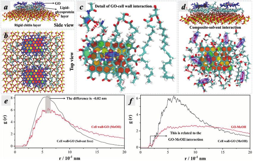
www.advancedsciencenews.com                                                                                                        www.afm-journal.de

Scheme 1. The configuration and potential working principles of CW-GO microcomposite membrane: a) CW-GO microcomposite membrane with
∙∙∙GO/CW∙∙∙ alternative stacking structure; b) cell wall restricting GO layer expansion to promise a high selectivity; and c) cell wall possessing well-
developed nanoscale channels to allow a high permeance.

layer and GO layer (Scheme 1a). Within this microcomposite                    contacted with the GO solution, indicating the formation of the
configuration, the cell wall with anti-swelling behavior pro-                 CW-GO microcomposite. Actually, the GO nanosheets wrapped
hibits the GO interlayer expansion to promise a high selec-                   several cell walls and the ultrathin GO layer ultimately covered
tivity (Scheme 1b). Moreover, the cell wall with well-developed               the surface of cell walls (Figure S5, Supporting Information).
nanoscale channels creates the membrane transport short-                      The CW-GO microcomposites were purified by repeated cen-
cuts to allow a high permeance (Scheme 1c). In typical, the                   trifugation and water washing to remove excessive unbonded
CW-GO microcomposite membrane exhibited a competitive                         GO, and then dispersed in water for further use. Noticeably, the
dye rejection rate (>93%) and permeance in organic solvents                   centrifugation runs with 4000 rpm, which could not force the
[≈56 L m2 h1 bar−1 (LMH bar−1)].                                              dispersed GO nanosheets fall down with the microcomposite.
                                                                              Therefore, even excess GO would not affect the formation of
                                                                              microcomposite with ultrathin GO layer attaching into the
2. Results                                                                    cell wall. However, decrease of the GO dosage would produce
                                                                              incomplete GO layer on the cell wall. The concentration of the
2.1. Fabrication of CW-GO Microcomposite Membrane                             microcomposites is ≈8.3 mg mL−1 and the mass concentration
                                                                              of GO on cell wall is 0.12 g/g (Materials S2, Supporting Infor-
The cell wall suspension was prepared by collecting and                       mation). The CW-GO microcomposite membrane was fabri-
purifying the hyphae from fungal pellets (Materials S1 and                    cated by assembling the CW-GO microcomposites via vacuum
Figure S1, Supporting Information). As shown in Figure S2,                    filtration method (Figure S3, Supporting Information). The
Supporting Information, the pure cell wall exhibited belt-like                sample is named as CW-GO-x, where the x represents the ratio
morphology with thickness of ≈100 nm. Noticeably, owing to                    of microcomposites dosage (mg) to PVDF film diameter (4 cm).
the multifunctional biomacromolecules, various nanomate-                          The membrane could be readily peeled off after drying.
rials dispersed in aqueous solution could readily adhere to the               As shown in Figure 1a, the membrane is transparent, and
surface of fungal cell wall.[45–47] The fabrication process of the            noticeably the light brown color is resulted from the GO. The
CW-GO microcomposite membrane is provided in Figure S3,                       pure cell wall membrane tends to be colorless transparent
Supporting Information. When the GO solution was mixed                        (Figure S5, Supporting Information). Moreover, the CW-GO
with the cell wall suspension, the GO nanosheets attached to                  microcomposite membrane is flexible (Figure 1b), which can be
the surface of fungal cell wall to form the CW-GO microcom-                   bended without fracture. This indirectly indicates its superior
posite. Figure S4, Supporting Information, displays that the                  mechanical stability, which is conducive to the OSN applica-
white cell wall suspension changed to light brown as soon as it               tions. Moreover, as compared with the previous research using

Adv. Funct. Mater. 2021, 31, 2100110                              2100110 (2 of 9)                                              © 2021 Wiley-VCH GmbH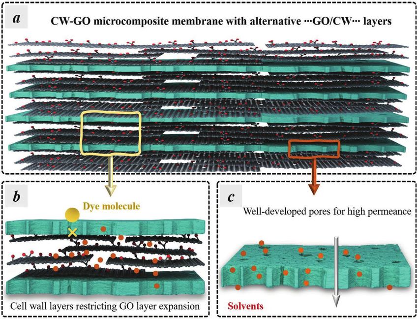
www.advancedsciencenews.com www.afm-journal.de
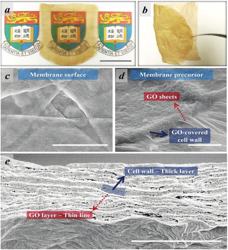
Figure 1. a,b) Photo images of the CW-GO microcomposite membrane (scale bar: 1 cm). SEM images of the CW-GO-1: c) low magnification by top
view; d) high magnification by top view; and e) cross section of the hybrid membrane [scale bar: 3 µm for (c), 1 µm for (d), and 2 µm for (e)].
synthetic polymers or inorganic materials,[6,8,11,48–50] the current and SEM analysis, each of the stacked cell walls should be cov-
method is environmentally friend and sustainable. ered with GO sheets. This sufficiently indicated an alternative
GO-CW-GO stacking configuration of the membrane.
The cross-section of the membrane GO-CW-1 is shown in
2.2. Characterization of CW-GO Microcomposite Membrane Figure 1e. The thickness can be measured from the cross-sec-
tion SEM image, which is ≈1448 nm. The thickness of pristine
SEM technique was used to investigate the microstructure of GO membrane with identical GO mass for GO-CW-1 is ≈40 nm
the CW-GO microcomposite membrane by taking CW-GO-1 (Figure S6, Supporting Information). Typically, the membrane
as an example. As given in Figure 1c, the high-magnification exhibited a microcomposite alternative stacking configura-
image revealed an intact surface of the membrane with cell wall tion, which is in line with the above analysis. In general, the
microbelts stacking with each other. Interestingly, as shown in thickness of the single cell wall and GO is ≈100 nm (Figure S2,
Figure 1d, the SEM image of the single microcomposite sug- Supporting Information) and ≈1 nm,[51] respectively. From the
gested the adhesion of GO sheets onto the cell wall. Moreover, cross-section image, there are around 14 layers of cell wall,
GO sheets could cover two parallelly adjacent cell walls. The gap which is equal to a thickness of total cell wall layers of ≈1400 nm
between the GO sheet and cell wall as in Figure 1d disappeared in membrane. In turn, the thickness of the total GO layers is
after the formation of CW-GO microcomposite membrane ≈48 nm, which is remarkably close to the results of Figure S6,
(Figure 1c) by vacuum filtration. This indicated an ultimate con- Supporting Information, above. For each GO layer, its thick-
tact between the cell wall and GO, which should be driven by ness is ≈3.4 nm. This exhibits an ultrathin feature of each GO
the atmospheric pressure. Based on the fabrication procedures layer. This interesting alternative GO-CW-GO microcomposite
Adv. Funct. Mater. 2021, 31, 2100110 2100110 (3 of 9) © 2021 Wiley-VCH GmbH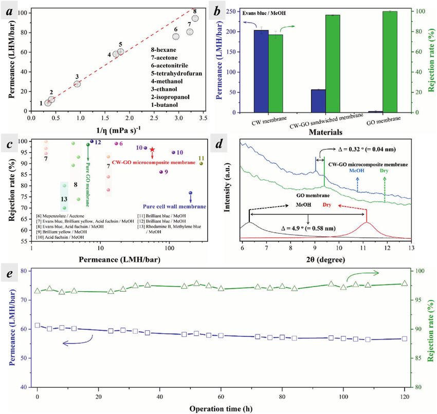
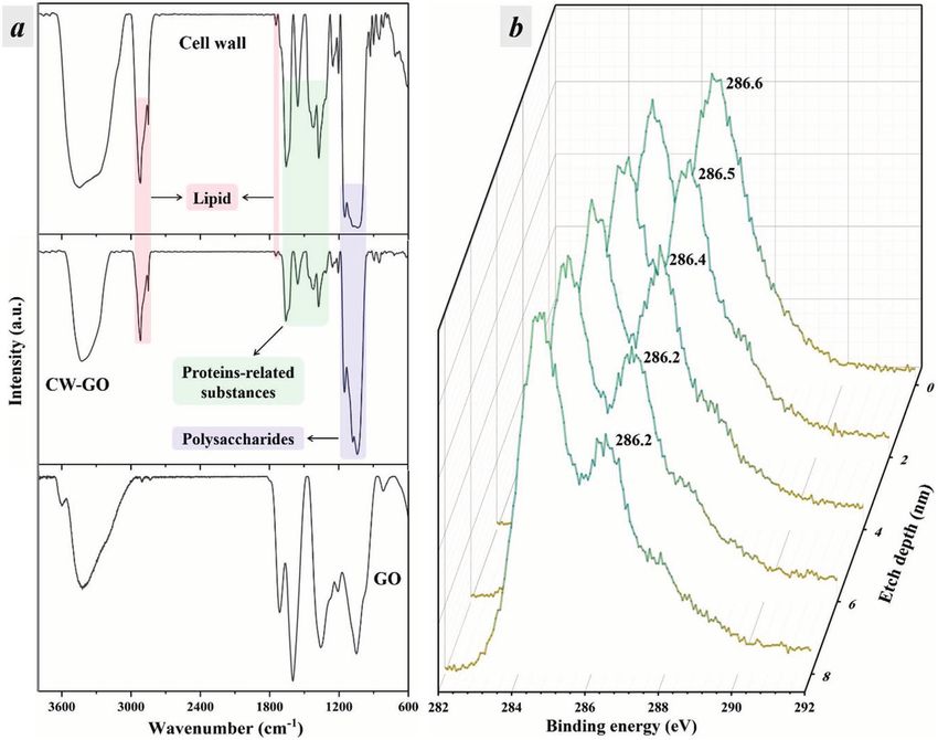
www.advancedsciencenews.com www.afm-journal.de Figure 2. Characterizations of the materials: a) the FTIR spectra of cell wall, CW-GO microcomposite membrane, and GO; and b) the XPS depth profiling of the C1s spectra of CW-GO microcomposite membrane. Note: the sputtering rate is ≈12 nm min−1 and every turn of etching takes ≈10 s. structure with well-developed nano channels within cell wall FTIR analysis that the non-covalent bonding should be the and ultrathin feature of GO layer might favor a high perme- major interaction manners for cell wall and GO. Moreover, the ance for the solvent in OSN process. Moreover, the opening of XPS detection in the depth direction was conducted to under- interlayer spacing of GO layers might also be restricted by the stand configuration of the CW-GO microcomposite membrane. solvent-resisted cell walls. Totally speaking, the membrane pos- As given in right part of Figure 2, the peak related to the CO sibly possesses high permeance and selectivity in OSN applica- and CO for the raw CW-GO microcomposite membrane was tions. In addition, the membrane prepared with various dosage at ≈286.8 eV. However, the binding energy decreased with the of microcomposite possessed similar morphology (Figure S7, increase of the depth. Typically, when the depth reached to 6 nm, Supporting Information). the peak position decreased to ≈286.2 eV, which is nearly iden- The interaction between cell wall and GO was investigated tical to that of the pure CW membrane (Figure S9, Supporting by FITR (Figure 2). The analysis of the IR peaks is given in Information). The result suggests that the thickness of the GO Figure S9, Supporting Information. In FTIR, transmittance layer is between 4 and 6 nm, which is close to the calculated mode was applied, and the IR light went through the whole value above. Similar conclusion could be draw from the analysis hybrid membrane. As seen in Figure 2, the IR signals of the of Raman spectra (Figure S10, Supporting Information). hybrid membrane are nearly identical to that of cell wall. This could be ascribed to the dominated amount of cell wall in the membrane. It must be noted that GO could chemically react 2.3. OSN Performance of CW-GO Microcomposite Membrane with cell wall at >80 °C,[42] but in this research, the tempera- ture was maintained at ≈25 °C, which could not cause obvious The CW-GO microcomposite membrane is permeable to all chemical reaction. In other words, it could be confirmed by tested organic solvents, and the permeance of the CW-GO Adv. Funct. Mater. 2021, 31, 2100110 2100110 (4 of 9) © 2021 Wiley-VCH GmbH
www.advancedsciencenews.com                                                                                              www.afm-journal.de

Figure 3. Performance of the CW-GO microcomposite membrane with pressure of 2 bar for every experiment: a) permeance of CW-GO-1 in different
solvents; b) permeance and rejection rate of membrane made of various pure materials; c) comparison between CW-GO microcomposite membrane
and diverse GO-based materials reported in the aspects of permeance and rejection rate (the aerobic number represents the reference number);
d) XRD patterns of CW-GO microcomposite membrane and pure GO membrane in dry and wet (MeOH as solvent) states; and e) permeance and
rejection rate of CW-GO-1 in a long-term OSN process.

microcomposite membrane showed a predominant dependence                the decrease of solvent viscosity, the permeance increased from
on the viscosity of solvent (Table S1, Supporting Information).        8.3 LMH bar−1 for 1-butanol to 94.4 LMH bar−1 for n-hexane.
It has been reported that for a pressure-driven flow, the solvent      Such nearly linear relationship between the solvent viscosity
transport through two parallel GO nanosheets can be described          and the membrane permeance can be exemplified by the
by the Hagen–Poiseuille equation for viscous flow assuming a           Hagen–Poiseuille equation.[12] This is in accordance with pre-
no-slip boundary condition.[12] It is clear from the Figure 3a that    vious reports, indicating the viscous nature of the solvents when
the permeance behavior for the CW-GO microcomposite mem-               flowing through the CW-GO microcomposite membrane.[12,52]
brane is very good fit with the Hagen–Poiseuille equation, that           The separation performance of membranes for OSN was
is, the permeance has a linear correlation with the reciprocal         then investigated using a methanol solution of Evans blue. As
of the viscosity, independent of the organic solvents used. With       compared in Figure 3b, the permeance for the pure cell wall

Adv. Funct. Mater. 2021, 31, 2100110                         2100110 (5 of 9)                                         © 2021 Wiley-VCH GmbH
www.advancedsciencenews.com                                                                                         www.afm-journal.de

membrane is as high as 203.4 LMH bar−1, but the rejection            membrane does not change obviously in NMP (≈0.98 nm)
rate is 96%. These results demonstrated that the      of interlayer distance by 0.04 nm in MeOH does not deterio-
cell wall is the major contributor for the high permeance of         rate the rejection performance of the CW-GO microcomposite
solvents. Meanwhile, the GO layers are the determining com-          membrane. In contrast, when immersing the pure GO mem-
ponents to promise a high selectivity to reject the dyes. We fur-    brane in MeOH, it suffered from significant enlargement of
ther evaluated the effect of membrane thickness on their OSN         GO interlayer distance by ≈0.58 nm. This should be caused by
performance. As seen in Figure S11, Supporting Information,          the severe solvation of GO by MeOH, which indirectly demon-
the methanol permeance sharply dropped and the rejection rate        strated the high stability of CW-GO microcomposite membrane
slightly improved with the increase of membrane thickness.           in organic solvents. On the other hand, the peak position of
Note that the relative thin CW-GO membranes have uniform,            the dried CW-GO microcomposite membrane is ≈1.5° smaller
dense, and ordered alternative stacking structure, following an      than that of the dried GO membrane. That is to say, CW-GO
inversely proportional law between membrane permeance and            microcomposite membrane exhibited a relatively large inter-
membrane thickness. Whereas the relative thick CW-GO mem-            layer spacing, which might be originated from the wrinkled
branes have top CW-GO layers in random order,[53] resulting          texture of GO layers (Figure 2c,d). Furthermore, the stability
in the higher membrane permeance than the predicted value            was revealed by a long-term running experiment. As given in
by the Hagen–Poiseuille flow model (Table S2, Supporting             Figure 3e, the CW-GO microcomposite membrane retained a
Information). Moreover, another several dyes with different          steady and high OSN performance over a duration of 120 h.
molecular weights (including Sudan I, Methylene blue, Rhoda-         Such outstanding membrane stability is desirable to meet the
mine B, Substantive red, Titan yellow, Brilliant blue, and Rose      industrial requirements.
Bengal) were tested to verify the applicability of membranes
for OSN (Figure S12, Supporting Information). The CW-GO
microcomposite membrane exhibited methanol permeance                 3. Discussion
of 54–64 LMH bar−1, and rejected more than 90% of organic
dye molecules with molecular weights ranging from 248.3 g to         3.1. Cell Wall-GO Interactions
1017.6 g mol−1, showing good molecular sieving properties.
    Based on the above results, the CW-GO microcomposite             Non-covalent interactions between cell wall and GO were con-
membrane fully utilizes the highly permeable cell wall chan-         firmed with FTIR and XPS above. However, further information
nels and the highly selective GO channels. As a result, the sol-     cannot be obtained by the analysis of these characterizations.
vent permeance of the CW-GO microcomposite membrane is               Here all atom molecular dynamics were taken to understand
an order of magnitude higher than that of pristine GO mem-           the cell wall-GO interactions. Based on the well-recognized
brane, while its dye rejection performance is improved by more       fungal model, the chitin is in the inner section of cell wall,
than 25% as compared with the pure cell wall membrane. As            which acts as mechanical support for the cell and would not
illustrated in Figure 3c, our optimized CW-GO microcom-              expose to the outer environment. Other components like the
posite membrane simultaneously excels in both permeance              lipids and glycoproteins are on the outer section of cell wall,
and rejection properties. This displays highly competitive OSN       and they are cross-linked with each other to form a very stable
performance over the most-reported GO-based membranes, of            structure. Here, a solid SiO2 plate was used to represent the
which the information is summarized in Table S3, Supporting          rigid chitin and on its surface the molecular fractions of lipids
Information.                                                         and glycoproteins were attached to simulate the cell wall (Mate-
    More importantly, sufficient stability is the prerequisite for   rials S4, Supporting Information). Two GO nanosheets were
practical OSN applications.[54] Here, XRD technique was applied      introduced onto the cell wall in one time.
to investigate the stability of the CW-GO microcomposite                As shown in Figure 4a,b, lipids and glycoproteins were lying
membrane.[55] As shown in Figure 3d, the dried cell wall-GO          down on the rigid substrate. After the introduction of GO, their
membrane has a diffraction peak at ≈9.37°, which indicates an        configuration had no obvious change. Meanwhile, the GO plane
interlayer distance of ≈0.94 nm between the stacked GO sheets.       laid down on the cell wall surface with an ultimate contact,
Note: there is only one broad peak (≈18°) in the XRD pattern         which suggested a strong interaction (Figure 4c,e). Specifically,
of the pure cell wall (Figure S13, Supporting Information),          the hydroxy and amino groups from glycoproteins interacted
which indicates its amorphous nature. However, the diffrac-          with the oxygen on GO to form hydrogen bonds (Figure 4c).
tion peak gently declines to ≈9.05° as the CW-GO microcom-           As experimentally confirmed previously, lipids could play with
posite membrane is in organic solvent (methanol), suggesting         the conjugated structure of GO through hydrophobic interac-
a slight increase of interlayer distance to ≈0.98 nm. Mean-          tion.[56] That is to say, hydrophobic lipid component and hydro-
while, the interlayer distance of the CW-GO microcomposite           philic component in cell wall could all strongly interact with the

Adv. Funct. Mater. 2021, 31, 2100110                       2100110 (6 of 9)                                      © 2021 Wiley-VCH GmbH
www.advancedsciencenews.com www.afm-journal.de
Figure 4. Interaction between cell wall and GO by all atom molecular dynamics: a) side view and b) top view of the configuration illustrating the inter-
action between GO and cell wall; c) the yellow dash circle marking a typical hydrogen bonding between GO and lipid-glycoprotein layer; interaction of
solvent with cell wall-GO microcomposites; d) side view of the GO-cell wall microcomposites in the presence of methanol (the methanol molecules
were not shown here) and the hydrogen bonding from GO-methanol and cell wall-methanol interactions marked by blue dash circles; and e,f) radial
distribution functions of cell wall-GO, microcomposite membrane-solvent and GO-solvent structures.
amphiphilic GO through hydrophobic interaction and hydrogen solvent. As revealed above, the cell wall-GO interaction was
bonding. In addition, the short-range van der Waals force and merely based on non-covalent binding. Based on the calculation
long-range electrostatic force also play additional roles for the in Figures 4d and 4f, methanol could interact with the GO and
cell wall-GO interactions. Basically, these strong interactions cell wall, respectively. The hydrogen bonding is still the major
are the basis for the formation of CW-GO microcomposites. force for their interactions, as marked by blue dash circles.
However, methanol could not dissociate the already-formed
bonding between cell wall and GO. In other words, the non-
3.2. Solvation Behaviors covalent cell wall-GO interactions were strong enough to avoid
the GO departure from cell wall induced by solvent. Thus, the
As concluded above, the pure cell wall membrane has a rather GO could be stabilized by the cell wall even in the presence of
poor capability to reject the dyes, but the dye rejection of the organic solvent. The solvation behavior of CW-GO microcom-
pristine GO membrane is superior. The GO layers in the posite membrane in other solvents was studied and is shown
CW-GO microcomposite membrane played a dominant role in Figures S14 and S17, Supporting Information. Basically, the
in dye rejection. Moreover, according to the characterizations ultimate cell wall-GO contact might already create the sieving
above, each GO layer between the two cell walls (due to stacking channel to prevent the undesired molecules from passing
structure) most possibly contained an average of 3–4 GO sheets. through the membrane. Moreover, based on the simulations
Therefore, in each GO layer the majority of GO sheets mainly of microcomposite in solvents, it could be indirectly confirmed
interacted with the two adjacent cell walls (Scheme 1a). Conse- that the cell wall-GO interactions are stronger than the respec-
quently, understanding the solvation behavior of cell wall-GO tive cell wall-solvents interactions and GO-solvents interactions.
microcomposite was important to elucidate the high perfor- This explains the rapid formation of CW-GO microcompos-
mance of the membrane for dye rejection. ites as soon as the cell wall dispersion was mixed with the GO
Herein, all atom molecular dynamics were further applied dispersion.
to simulate the interaction between organic solvent and cell Although the top and bottom GO sheets could be stabi-
wall-GO microcomposites. The methanol was used as a typical lized by the cell wall to sieve the molecule, the middle parts
Adv. Funct. Mater. 2021, 31, 2100110 2100110 (7 of 9) © 2021 Wiley-VCH GmbHwww.advancedsciencenews.com www.afm-journal.de
between these two sheets might swell in the organic solvents. 5. Experimental Section
This would expand the interlayer spacing to possibly attenuate
Preparation of the CW-GO Microcomposite Membrane: The preparation
the rejection performance.[55] Nonetheless, based on the above
of the raw materials is given in Materials S1 and S2, Supporting
results (Figure 3d,e), the CW-GO microcomposite membrane Information. The cell wall-GO dispersion in water was the sole precursor
actually had a high stability. To explain this phenomenon, to prepare the CW-GO microcomposite membrane. Vacuum filtration
the swelling of the cell wall was investigated. As shown in route was applied to fabricate the membrane. Specifically, PVDF
Figure S15, Supporting Information, the cell wall exhibited membrane was used as substrate to support the formation of CW-GO
an inherently anti-swelling behavior towards the organic sol- microcomposite membrane due to its smooth surface. The cell wall-GO
microcomposites dispersion (8.3 mg mL−1) was poured into the filtration
vents. In contrast, the pure GO membrane swelled heavily
vessel with PVDF membrane on. Then, vacuum was on to remove the
in organic solvent as revealed by XRD above (Figure 3d). The water in microcomposites dispersion. The process was fast, which cost
interlayer distance of the dry GO membrane increased aggres- ≈5 min. In a typical formula, 0.5 ml of dispersion was used to obtain the
sively from ≈0.82 nm to ≈1.4 nm. That is to say, the cell wall hybrid membrane on an area with effective diameter ≈4 cm.
could not only stabilize the GO sheets directly interacting with As soon as the formation of the CW-GO microcomposite membrane,
it, but also further restrict the expansion of the inner section the whole membranes were taken into convection oven that has
of the GO layer. These together promised high rejection of been preheated to 105 °C. The edge of the CW-GO microcomposite
membrane would automatically separate from the PVDF membrane.
dye. Actually, the anti-swelling behavior of CW-GO microcom- Then, a careful hand-peeling could remove the hybrid membrane
posite membrane is similar to the pure cell wall membrane from the substrate membrane. On the other hand, there is no need
(Figure S16, Supporting Information), which indirectly veri- to separate the hybrid membrane from the substrate membrane since
fied this opinion. the combined membranes could be directly used in dye rejection. The
Apart from the promising rejection rate, the high perme- separation of CW-GO microcomposite membrane is to illustrate its
ance of the hybrid membrane was another attracting indicator superior mechanical stability.
Performance Evaluation: Membrane performance is tested using
for the potential practical applications. In general, the contin-
nanofiltration process under 2 bar by a home-made filtration device at
uous stacking of GO nanosheets would form a continuously room temperature. The effective area of the membranes is 17.35 cm2.
long and tortuous pathway for the solvent passing through The solvent flux (F, L m−2 h−1) was evaluated with various organic
the laminar GO membrane,[17] which usually cause a signifi- solvents (i.e., butanol, isopropanol, ethanol, methanol, tetrahydrofuran,
cant mass transfer resistance and a low membrane perme- acetonitrile, acetone, hexane), and the dye rejection ratio (R, %) was
ance. In our case, the membrane configuration is designed as determined using different dyes (i.e., Evan blue, titan yellow, substantive
red) with the concentration of 10 mg L−1. Each data was obtained by
a CW layer-GO layer alternative stacking mode (Figure S18,
a new membrane sample after the filtration system reaching a steady
Supporting Information). The GO layer in the CW-GO micro- state. The concentrations of feed and permeate solutions were measured
composite membrane only consisted of 3–4 layers of GO by using UV–vis spectrophotometer. During the long-term filtration
nanosheets, and each GO layer did not contact each other process, a constant-flux pump was employed to pump feed solution into
directly but was separated by the cell wall. The pathway in the membrane module at a cross-flow rate of 50 mL min−1.
such ultrathin GO layer would become much shorter for sol- Solvent flux and dye rejection are calculated as follows:
vent transportation. Moreover, the porous cell wall layer has V
an exceptionally low resistance for solvent penetration, which F = (1)
A×t
can be confirmed by the ultrahigh flux for pure cell wall mem-
brane. Therefore, the solvents could rapidly pass through cp
the non-continuous ultrathin GO layer and cell wall layer, R = 1− × 100% (2)
cf
resulting in a remarkably high permeance of the CW-GO
microcomposite membrane.[36]
where V is the volume of permeate collected (L), A is the membrane
effective area (m2), t is the permeation time (h), and cp and cf are the
concentrations of the permeate and feed solution, respectively.
4. Conclusion
In summary, we have developed a superior GO-based mem-
brane by incorporating the unique features of fungal cell wall. Supporting Information
The membrane was composed of an alternative stacking of Supporting Information is available from the Wiley Online Library or
cell wall layer and ultrathin GO layer. The strong cell wall-GO from the author.
interaction and the anti-swelling property of cell wall together
restrict the expansion of interlayer space of GO, which blocks
the targeted molecules away from going through the mem-
brane. Meanwhile, the high porosity of the fungal cell wall
Acknowledgements
directly improves the permeability of membrane. Moreover, L.Z. and M.Z. contributed equally to this work. Authors thank the
the membrane reported here has not only the high selectivity financial supports from National Natural Science Foundation of
and permeability, but also the long-term stability in OSN pro- China (No. 22008182, 22038006, 21922805), the Guangdong Basic
and Applied Basic Research Foundation (No. 2019A1515111191), and
cess. The CW-GO microcomposite membrane holds brilliant
the Guangdong Ordinary University Youth Innovative Talents Project
prospect boosting the practical applications of GO-based mem- (No. 2019KQNCX162), the Department of Education of Guangdong
brane in OSN and providing interesting insight for membrane Province (Nos. 2020ZDZX2015, 2020KSYS004), and the Natural Science
design. Foundation of Guangdong Province of China (General Program).
Adv. Funct. Mater. 2021, 31, 2100110 2100110 (8 of 9) © 2021 Wiley-VCH GmbHwww.advancedsciencenews.com www.afm-journal.de
Conflict of Interest [22] G. Liu, W. Jin, N. Xu, Chem. Soc. Rev. 2015, 44, 5016.
[23] G. Liu, W. Jin, N. Xu, Angew. Chem., Int. Ed. 2016, 55, 13384.
The authors declare no conflict of interest. [24] L. Prozorovska, P. R. Kidambi, Adv. Mater. 2018, 30, 1801179.
[25] S. Wang, L. Yang, G. He, B. Shi, Y. Li, H. Wu, R. Zhang, S. Nunes,
Z. Jiang, Chem. Soc. Rev. 2020, 49, 1071.
Data Availability Statement [26] J. Zhu, J. Hou, A. Uliana, Y. Zhang, M. Tian, B. Van der Bruggen,
J. Mater. Chem. A 2018, 6, 3773.
Research data are not shared. [27] J. Shen, G. Liu, Y. Han, W. Jin, Nat. Rev. Mater. 2021, https://doi.
org/10.1038/s41578-020-00268-7.
[28] L. Cheng, G. Liu, J. Zhao, W. Jin, Acc. Mater. Res. 2021, 2, 114.
Keywords [29] Y. Kang, Y. Xia, H. Wang, X. Zhang, Adv. Funct. Mater. 2019, 29,
1902014.
cell wall, graphene oxide, membranes, molecular dynamics, organic
[30] R. R. Nair, H. A. Wu, P. N. Jayaram, I. V. Grigorieva, A. K. Geim,
solvent nanofiltration
Science 2012, 335, 442.
Received: January 7, 2021 [31] P. Su, F. Wang, Z. Li, C. Y. Tang, W. Li, J. Mater. Chem. A 2020, 8,
Revised: March 12, 2021 15319.
Published online: [32] C. N. Yeh, K. Raidongia, J. Shao, Q. H. Yang, J. Huang, Nat. Chem.
2014, 7, 166.
[33] M. Zhang, Y. Mao, G. Liu, G. Liu, Y. Fan, W. Jin, Angew. Chem., Int.
Ed. 2020, 59, 1689.
[1] P. Vandezande, L. E. Gevers, I. F. Vankelecom, Chem. Soc. Rev. 2008, [34] S. Zheng, Q. Tu, J. J. Urban, S. Li, B. Mi, ACS Nano 2017, 11, 6440.
37, 365. [35] J. Zhu, Y. Shan, T. Wang, H. Sun, Z. Zhao, L. Mei, Z. Fan, Z. Xu,
[2] S. Karan, S. Samitsu, X. Peng, K. Kurashima, I. Ichinose, Science I. Shakir, Y. Huang, B. Lu, X. Duan, Nat. Commun. 2016, 7, 13432.
2012, 335, 444. [36] L. Walker, P. Sood, M. D. Lenardon, G. Milne, J. Olson, G. Jensen,
[3] P. Marchetti, M. F. Jimenez Solomon, G. Szekely, A. G. Livingston, J. Wolf, A. Casadevall, J. Adler-Moore, N. A. R. Gow, mBIO 2018, 9,
Chem. Rev. 2014, 114, 10735. e02383.
[4] E. M. Rundquist, C. J. Pink, A. G. Livingston, Green Chem. 2012, 14, [37] J. G. De Nobel, C. Dijkers, E. Hooijberg, F. M. Klis, J. Gen. Microbiol.
2197. 1989, 135, 2077.
[5] S. Karan, Z. Jiang, A. G. Livingston, Science 2015, 348, 1347. [38] E. J. Conway, M. Downey, Biochem. J. 1950, 47, 347.
[6] F. Fei, L. Cseri, G. Szekely, C. F. Blanford, ACS Appl. Mater. Interfaces [39] R. R. Lew, Fungal Genet. Biol. 1998, 24, 69.
2018, 10, 16140. [40] L. Chai, J. Wang, H. Wang, L. Zhang, W. Yu, L. Mai, Nano Energy
[7] T. Gao, L. Huang, C. Li, G. Xu, G. Shi, Carbon 2017, 124, 263. 2015, 17, 224.
[8] T. Gao, H. Wu, L. Tao, L. Qu, C. Li, J. Mater. Chem. A 2018, 6, 19563. [41] H. Wang, X. Li, L. Chai, L. Zhang, Chem. Commun. 2015, 51,
[9] L. Huang, J. Chen, T. Gao, M. Zhang, Y. Li, L. Dai, L. Qu, G. Shi, 8524.
Adv. Mater. 2016, 28, 8669. [42] L. Zhang, X. Li, M. Wang, Y. He, L. Chai, J. Huang, H. Wang, X. Wu,
[10] L. Nie, K. Goh, Y. Wang, J. Lee, Y. Huang, H. E. Karahan, K. Zhou, Y. Lai, ACS Appl. Mater. Interfaces 2016, 8, 34638.
M. D. Guiver, T.-H. Bae, Sci. Adv. 2020, 6, eaaz9184. [43] L. Zhang, Y. Wang, B. Peng, W. Yu, H. Wang, T. Wang, B. Deng,
[11] S. Wang, D. Mahalingam, B. Sutisna, S. P. Nunes, J. Mater. Chem. L. Chai, K. Zhang, J. Wang, Green Chem. 2014, 16, 3926.
A 2019, 7, 11673. [44] H. B. Park, J. Kamcev, L. M. Robeson, M. Elimelech, B. D. Freeman,
[12] Q. Yang, Y. Su, C. Chi, C. T. Cherian, K. Huang, V. G. Kravets, Science 2017, 356, eaab0530.
F. C. Wang, J. C. Zhang, A. Pratt, A. N. Grigorenko, F. Guinea, [45] W. Li, Y. Zhang, Z. Xu, Q. Meng, Z. Fan, S. Ye, G. Zhang, Angew.
A. K. Geim, R. R. Nair, Nat. Mater. 2017, 16, 1198. Chem., Int. Ed. 2016, 55, 955.
[13] S. Zheng, Q. Tu, M. Wang, J. J. Urban, B. Mi, ACS Nano 2020, 14, [46] N. C. Bigall, M. Reitzig, W. Naumann, P. Simon, K. H. van Pee,
6013. A. Eychmuller, Angew. Chem., Int. Ed. 2008, 47, 7876.
[14] A. Akbari, S. E. Meragawi, S. T. Martin, B. Corry, E. Shamsaei, [47] Z. Li, S. W. Chung, J. M. Nam, D. S. Ginger, C. A. Mirkin, Angew.
C. D. Easton, D. Bhattacharyya, M. Majumder, ACS Appl. Mater. Chem., Int. Ed. 2003, 42, 2306.
Interfaces 2018, 10, 2067. [48] H. Dou, M. Xu, B. Jiang, G. Wen, L. Zhao, B. Wang, A. Yu, Z. Bai,
[15] J. Abraham, K. S. Vasu, C. D. Williams, K. Gopinadhan, Y. Su, Y. Sun, L. Zhang, Z. Chen, Z. Jiang, Adv. Funct. Mater. 2019, 29,
C. T. Cherian, J. Dix, E. Prestat, S. J. Haigh, I. V. Grigorieva, 1905229.
P. Carbone, A. K. Geim, R. R. Nair, Nat. Nanotechnol. 2017, 12, 546. [49] D. Hua, T.-S. Chung, Carbon 2017, 122, 604.
[16] L. Chen, G. Shi, J. Shen, B. Peng, B. Zhang, Y. Wang, F. Bian, [50] Y. Li, C. Li, S. Li, B. Su, L. Han, B. Mandal, J. Mater. Chem. A 2019,
J. Wang, D. Li, Z. Qian, G. Xu, G. Liu, J. Zeng, L. Zhang, Y. Yang, 7, 13315.
G. Zhou, M. Wu, W. Jin, J. Li, H. Fang, Nature 2017, 550, 380. [51] O. C. Compton, S. T. Nguyen, Small 2010, 6, 711.
[17] R. K. Joshi, P. Carbone, F. C. Wang, V. G. Kravets, Y. Su, [52] X. Wu, X. Cui, W. Wu, J. Wang, Y. Li, Z. Jiang, Angew. Chem., Int. Ed.
I. V. Grigorieva, H. A. Wu, A. K. Geim, R. R. Nair, Science 2014, 343, 2019, 58, 18524.
752. [53] C.-H. Tsou, Q.-F. An, S.-C. Lo, M. De Guzman, W.-S. Hung,
[18] B. Mi, Science 2014, 343, 740. C.-C. Hu, K.-R. Lee, J.-Y. Lai, J. Membr. Sci. 2015, 477, 93.
[19] M. Zhang, K. Guan, Y. Ji, G. Liu, W. Jin, N. Xu, Nat. Commun. 2019, [54] Y. Li, S. Li, J. Zhu, A. Volodine, B. Van der Bruggen, Chem. Sci. 2020,
10, 1253. 11, 4263.
[20] Y. Kang, R. Qiu, M. Jian, P. Wang, Y. Xia, B. Motevalli, W. Zhao, [55] A. Iakunkov, J. Sun, A. Rebrikova, M. Korobov, A. Klechikov,
Z. Tian, J. Z. Liu, H. Wang, H. Liu, X. Zhang, Adv. Funct. Mater. A. Vorobiev, N. Boulanger, A. V. Talyzin, J. Mater. Chem. A 2019, 7,
2020, 30, 2003159. 11331.
[21] H. Huang, Y. Ying, X. Peng, J. Mater. Chem. A 2014, 2, 13772. [56] L. Wu, L. Zeng, X. Jiang, J. Am. Chem. Soc. 2015, 137, 10052.
Adv. Funct. Mater. 2021, 31, 2100110 2100110 (9 of 9) © 2021 Wiley-VCH GmbHYou can also read