Genes involved in pancreatic islet cell rejuvenation
←
→
Page content transcription
If your browser does not render page correctly, please read the page content below
Review Article

Indian J Med Res 137, April 2013, pp 695-703

Genes involved in pancreatic islet cell rejuvenation

Vinay S. Bansal, C. Prasanna Raja, Krishnan Venkataraman & M.A. Vijayalakshmi

Centre of Bio-Separation Technology (CBST), VIT University, Vellore, India

Received October 17, 2011

               Pancreas plays an important role in maintaining the glucose homeostasis. The deterioration of β-cells
               in the pancreas is a crucial factor in the progression of diabetes mellitus; therefore, the restoration
               of β-cell mass and its function is of vital importance for effective therapeutic strategies. The precise
               mechanism for increase in functional β-cell mass is still unknown. This review focuses on the importance
               of certain genes which are involved in the rejuvenation of pancreas. These genes are divided according to
               their functions into three categories: participate either in proliferation (mitotic division of differentiated
               β-cells), neogenesis/transdifferentiation (development from precursor cells) or inhibition of β-cell
               apoptosis (programmed cell death). The rate of β-cell rejuvenation is the balance among the rates of
               β-cell proliferation, neogenesis and apoptosis. Understanding these genes and their pathways may lead
               to the discovery of new drugs, target based gene delivery and development of safer antidiabetic drugs.

Key words Apoptosis - pancreatic genes - proliferation - rejuvenation - transdifferentiation

Introduction                                                              Type 2 diabetes mellitus (T2DM) develops from a
                                                                          combination of genetic and acquired factors (such as
     Diabetes is a major cause of health concern in
                                                                          changes in metabolic homeostasis) that impair β-cell
the world and is growing in epidemic proportions. It
                                                                          function on one side, and tissue insulin sensitivity on the
is assumed that in the next ten years it will become
                                                                          other2,3. Normally, β-cell mass can adapt to changes in
number one disease of the world1. Type-1 diabetes
                                                                          metabolic homeostasis. Recurrence of these changes in
mellitus (T1DM) is an autoimmune disease while type
                                                                          metabolism creates a stress on pancreas often predating
2 is mostly a lifestyle disease. Majority of people suffer
                                                                          the on-set of T2DM by many years. This pancreatic
mainly due to type-2 diabetes and is responsible for
                                                                          stress causes β-cell mass expansion, through enhanced
the current diabetes explosion. The detection early
                                                                          proliferation and neogenesis. The progression from
markers for the disease and its prevention is an active
                                                                          this stress condition to a state of diabetes is inevitably
area of research to develop target based novel drugs.
                                                                          associated with a decrease in the β-cell mass2-4. This
Dysfunctional pancreas in diabetes                                        β-cell loss arises due to an increase in β-cell apoptosis,
    Insulin, a key polypeptide hormone secreted by the                    which clearly outweighs replication and neogenesis.
pancreas, targets several tissues for the utilization of                      The war against diabetes through the development
glucose and thus maintains the glucose homeostasis.                       of new drugs is an ongoing continuous process5. With

                                                                    695
696 INDIAN J MED RES, april 2013
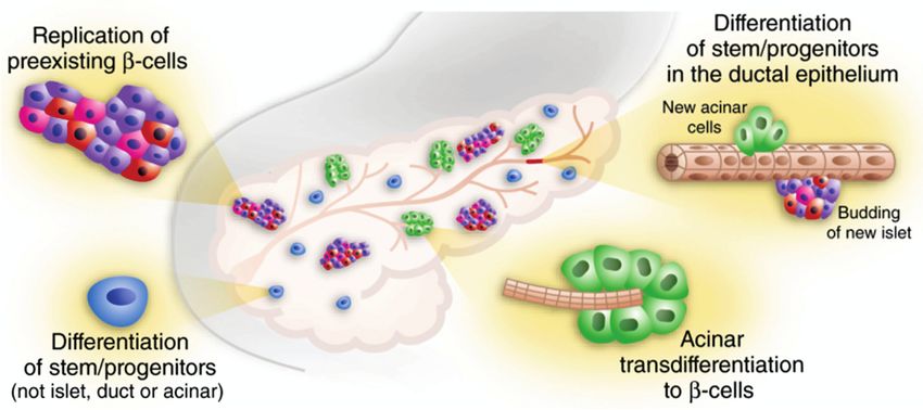
Fig. Pancreas as the source of new β-cells. New β-cells can arise from (1) Replication of pre-existing β-cells (2) Differentiaition of stem/
progenitors or Transdifferentiation of acinar cells (3) Prevention of apoptosis (not shown in Figure) also contributes for regeneration
of pancreas in which gene/gene products play in conjunction either with (1) or (2). All these mechanism may contribute synergistically for
the regeneration of pancreas. See Tables 1, 2 and 3 for gene/gene products involved in the above processes for pancreatic rejuvenation.
Reproduced with permission from Nature Publishing Group, London, UK6.
the technological advancement, efforts are being made have confirmed that human β-cells proliferate and give
to rejuvenate the pancreatic cells or create artificial rise to a population of progenitor/stem cell. Various
pancreas. Pancreatic rejuvenation can happen either due genes and transcription factors are involved in this
to proliferation of existing β-cells or differentiation of process viz. Reg (Regenerating islets derived proteins),
progenitor cells to β-cells6 (figure), or due to decrease Sox9, Hnf-6, NeuroD1, Neurogenin-3 and Netrin-1
in β-cell apoptosis. (Table I). Besides these genes, certain peptides or their
analogues such as glucagon like peptide-1/exendin-4 are
β-Cell proliferation also involved in islet regeneration. These observations
Islets regeneration refers to an increase in β-cell are confirmed by using dipeptidyl peptidase (DPP) IV
mass by proliferation and replication of existing islet inhibitor sitagliptin in mice15.
cells. Several mouse studies7-9 here shown that β-cells So far, five REG proteins have been reported in
do not proliferate, however, lineage tracing studies10-14 humans that belong to Reg gene family. Some of the
Table I. Genes involved in beta cell proliferation
Genes proteins Functions References
Reg gene family Increase islets cell size and density, Regeneration of pancreas 16, 22, 24, 25, 27
(RegI, II, IIIα, IIIβ, IIIγ)
Sox9 Stimulates proliferation and survival of pluripotent progenitors. 28
Hnf-6 Essential for maintenance of Ngn3 expression. 30, 32
Neurog3 (Ngn-3) Initiates endocrine differentiation and activates NeuroD1. 34
or
Neurogenin-3
NeuroD1/BETA-2 It is required for normal pancreatic development and glucose homeostasis. 35
Netrin-1 Involves in islets regeneration. 36BANSAL et al: BETA CELLS REJUVENATION 697
members of this family have been implicated in β-cell endocrine development. HNF6 is expressed in early
replication and/or neogenesis as shown in in vivo pancreatogenesis in all endodermally derived cells,
studies using transgenic and knockout mice16. These but is not detected in differentiated endocrine cells
also preserve the β-cell mass in autoimmune type at late-gestation29. Hnf-6 null mice embryos showed
1 diabetes17. This Reg family of genes are expressed impaired endocrine differentiation and perturbed duct
in both young and old mice that were subjected to morphogenesis during embryogenesis30. In addition
partial pancreatectomy18. In isolated rat islets, Reg1 to defects in endocrine development, Hnf-6 null
mRNA levels were significantly increased by glucose, embryos showed defects in duct development31. Loss
amino acids, foetal serum or specific growth factors of Hnf-6 from Ngn-3 expressing cells did not affect
such as insulin, growth hormone and platelet-derived β-cell function or glucose homeostasis suggesting
growth factors (PDGF)19. PDGF receptor signalling that Hnf-6 is dispensable for later events of endocrine
controls age-dependent β-cell proliferation in mouse differentiation. These data confirm that HNF6 has both
and human pancreatic islet cells20. Disruption of RegI early and late functions in the developing pancreas
gene resulted in a significantly decreased rate of DNA and is essential for maintenance of Ngn-3 expression
synthesis and diminished β-cell hyperplasia in response and proper pancreatic duct morphology32. NeuroD1, a
to obesity, confirming the role of endogenous RegI in downstream target of Ngn-3, carries on the endocrine
islets cell growth21. A study conducted by Huszarik et differentiation programme initiated by Ngn3 and
al22 showed upregulation of RegII during diabetogenic participates in the maintenance of the differentiated
process and also after adjuvant therapy in NOD mice. phenotype of the mature islet cells33. During pancreatic
While all Reg family mRNAs can be detected from endocrine development, Ngn-3 acts early to determine
total pancreas, RegII and RegIIIα genes have been endocrine cell fate, while NeuroD1 directs endocrine
detected in pancreatic islet cells as confirmed by cell differentiation34. At early stage of life, mice lacking
immunofluorescence23 and RegIIIα expression was a functional NeuroD1 (also called as BETA2) gene
remarkably increased during pregnancy in rats24. Mice exhibit a striking reduction in the number of insulin-
overexpressing RegIII β was resistant to streptozotocin producing β-cells and failed to develop mature islets
induced diabetes mellitus25. RegIIIγ, another member with a marked hyperglycaemia. Attempts to rescue the
of Reg family of genes is also found to be involved in diabetic phenotype by administration of insulin were
regeneration of pancreas. REG III protein was found unsuccessful, suggesting that the mutant animals were
to be expressed only in regenerating islets and not in unable to respond to insulin, have become insulin
normal rat pancreas26 and its gene expression level resistant, or perhaps contained additional defects35.
induced 10-100 folds on day 3 of pancreatectomy27. Thus BETA-2 is required for the expansion of the
These data suggest that there is a strong link between pancreatic β-cells population, as well as other islet
Reg gene family and rejuvenation of pancreatic islets. cell types which are involved in the development of
endocrine cells into islets of Langerhans34.
Transcription factors in β-cell proliferation
Netrins are laminin-like diffusible chemotactic
Certain transcription factors (Sox9, Hnf-6, Ngn-3 proteins involved in pancreatic morphogenesis and
and NeuroD1) are also found to be involved in the play a role in the regulation of duct-cell and foetal islet
proliferation of β-cells. SOX9 is the first specific cell migration. In adult rat pancreas, Netrin-1 mRNA
marker and maintenance factor of multi-potential was practically undetectable. After duct ligation, its
progenitors during pancreatic organogenesis. SOX9, expression was very low in the head part of the pancreas
in the embryonic pancreas stimulates proliferation whereas it was strongly upregulated in the tail part at
and prevents apoptosis of pluripotent progenitor cells. 3rd, 5th and 7th day of post-ligation with the maximum
It controls pancreatic progenitor cell maintenance by expression on day 536. Netrin-1 mRNA was found to
modulating Notch signal transduction. The phenotypic be expressed by islet cells and exocrine cells with
alterations in the Sox9-deficient pancreas shows ductal characteristics. These observations suggest that
a striking resemblance to the pancreatic defects Netrin-1 plays a role in pancreatic morphogenesis, both
associated with mutations in components of the prenatally and in the regenerating adult rat pancreas.
Notch signalling pathway, thus establishing a possible
link between Sox9 and the notch signal transduction Transdifferentiation of pancreas
pathway for stem cell maintenance28. The hepatocyte Islet neogenesis specifically refers to an increase in
nuclear factor 6 (Hnf-6), homeodomain-containing β-cell mass via transdifferentiation of adult pancreatic
transcription factor, is an important regulator of stem cells, putatively found in the ductal epithelium698 INDIAN J MED RES, april 2013
or acinar tissue. Trans-differentiation involves in the is restricted to β-cells and known to be important in
conversion of alpha or delta cells of the pancreas the embryonic development of pancreas45. It has been
into insulin producing β-cells. Various genes/proteins observed that MafA expression is decreased during the
contribute to this process. These include INGAP, diabetic condition46.
Gastrin, MafA, Pdx-1, Foxa2, Nkx2.2, Nkx6.1, Pax4,
etc. (Table II). Transcription factors in pancreatic neogenesis
INGAP (islets neogenesis associated protein) is a There are several transcription factors involved
member of the C-lectin protein family that serves as the both in neogenesis and replication. However, it is
initiator of a cascade of events that culminates in islet not clear whether these work alone or in combination
neogenesis and can reverse diabetes in streptozotocin- with other transcription factors in a coordinated
induced diabetic C57BL/6J mice37. These studies were manner. The list of the transcription factors and their
further confirmed in beagle dogs38. There was also a role in transdifferentiation is summarized in Table II.
significant increase in insulin gene expression in the Pancreatic duodenal homeobox-1 (Pdx-1), a homeobox
INGAP treated animals. INGAP is also found in human transcription factor, besides being involved as a
pancreas during pathological states involving islet regulator of pancreatic development (the differentiation
neogenesis which further suggests that INGAP is of and gene expression in the β-cell)47, Pdx1 also turns out
primary importance in the process of islet neogenesis39. to be a major player in the maintenance of an adequate
Gastrin, a classical gut hormone secreted by G cells pool of healthy β-cells in adults48,49. It maintains the
in the stomach lining is found to stimulate pancreatic homeostasis between β-cell neogenesis and apoptosis.
β-cell neogenesis. Intravenous infusion of gastrin into In mice with a 50 per cent reduction in Pdx1, the
the ligated duct cells, resulted in a doubling of the isolated islets showed more susceptibility to apoptosis
β-cell mass in rats40, due to high expression of gastrin/ at basal glucose concentrations along with impaired
cholecystokinin (CCK) B receptors in duct ligated ability to maintain β-cell mass with age. Its expression
cells41. These observations were confirmed using EGF is shown to be down-regulated during hyperglycaemic
plus gastrin combinatorial therapy, which showed an condition50. The survival functions of Pdx1 may be
increase in insulin positive cells in human islet42. In mediated by insulin/IGF signaling acting through the
another study using GLP-1 and gastrin in NOD mice, forkhead transcription factor Foxo1 (forkhead/winged
there was a significant reduction in blood glucose helix transcription factor). Foxa2 (formerly known
due to increase in pancreatic β-cell mass and insulin as Hnf-3) is a key regulator of foregut development
content43. MafA a member of Maf subfamily of leucine that plays an essential role in the cell type-specific
zippers is capable of strongly activating the insulin transcription of the Pdx-1 gene in the pancreas51. On
promoter. Maf factors play important roles in cellular deletion of Foxa2 in mice, there is a significant down-
differentiation of exocrine cells to β-cells44. MafA regulation of Pdx-1 mRNA52. This shows that Foxa2
Table II. Genes involved in pancreatic neogenesis
Genes Functions References
INGAP An initiator of islet neogenesis. 39
Gastrin Induces islet β-cell neogenesis from pancreatic exocrine duct cells. 40
MafA. Reprogrammes differentiated pancreatic exocrine cells into cells that closely 44
resemble β-cells.
Pdx-1 Initiates endocrine neogenesis. 47
Foxa2 A major upstream regulator of Pdx-1. 52
(HNF-3 Beta)
Nkx2.2 Regulator of pancreatic endocrine differentiation. 53
Nkx6.1 Maintaining & expanding population of β-cell precursors as these progress 58
from precursors to differentiated β-cell.
Pax4 Expressed in endocrine cell progenitors and directs formation of β and delta 60
cells.BANSAL et al: BETA CELLS REJUVENATION 699
is an essential upstream factor that regulates Pdx-1 β-cell lineages. Mice deficient in Pax4 fail to develop
mRNA levels in β-cells. beta and delta cells within the pancreas suggesting
that Pax4 expression commits selected endocrine
Nkx2.2 is another essential pancreatic transcription
precursors towards the beta and delta cell lineage59.
factor that affects the expression of ghrelin during
The Pax4 genes are expressed during the embryonic
pancreatic development53. Nkx2.2-null mice lose all
pancreatic development, but later on these are restricted
β-cells and the majority of the α-cells; and the islet
to β-cells alone. Inactivation of Pax4 in mice results
is predominantly populated by gherlin expressing
in the improper maturation of alpha- and β-cells60.
cells. The discovery that ghrelin cells often replace
This indicates the role of PAX4 in the maintenance of
the other endocrine populations in the pancreas54
progenitors and in maturation of β-cells.
suggests a lineage relationship between the ghrelin
producing epsilon cells and other hormone-producing Inhibition of β-cell apoptosis
populations55. In another study, disruption of the
Under normal circumstances, apoptosis is highly
Nkx2.2 gene has shown to lead to the accumulation
regulated to maintain normal physiological function
of incomplete differentiated β-cells that express some
of the cells. In diabetes, during excess stress, the
β-cell markers, but not insulin56. This illustrates the role
pancreatic cells not only undergo apoptosis but also
of Nkx2.2 in pancreatic endocrine cell differentiation.
become necrotic and are unable to secrete insulin.
The phenotypic effects of Nkx2.2 mutant mice may
A study conducted by Butler et al61 indicated that
in part result from the loss of other homeodomain
increased apoptosis rather than decreased neogenesis/
transcription factor Nkx6.1. In pancreas, Nkx6.1 also
proliferation might be the main mechanism leading
follows an expression pattern similar to that of Nkx2.2;
to reduced β-cell mass in T2DM. Thus, decrease in
but restricted to the β-cells alone57. Deletion of the
the rate of apoptosis itself, may increase the β-cell
Nkx6.1 gene in mice caused a marked reduction of
mass via proliferation. Several genes/proteins are
β-cells. In the same study, with the double knockout
involved in pancreatic apoptosis and their functions are
animal model (Nkx2.2/Nkx6.1) showed the same
summarized in Table III.
phenotype as of Nkx2.2 single mutant58. This shows that
Nkx6.1 functions downstream of Nkx2.2 in pancreatic Perforin (pore forming protein) is a cytolytic
development. The paired homeobox transcription factor protein, initiating apoptosis by inducing minimal
Pax4 appears to be a strong candidate for specifying cell membrane damage while effectively releasing
Table III. Genes involved in inhibition of beta cell apoptosis
Genes/proteins Functions References
Pdx-1 Possesses anti-apoptotic activity that helps facilitate the 48
maintenance of β-cell mass.
Granzymes A serine protease, that activates Bid (a pro-apoptotic factor) 66
essential for death-receptor induced apoptosis of islets.
Caspase-3 Principle executioner of apoptosis. 69
BIRC5 (Survivin) Bifunctional role, controlling both proliferation and inhibition 73
of apoptosis
Bcl-2 Survival genes that suppresses apoptosis. 75
C-Myc Induces extensive apoptosis in pancreatic β-cells. 76
Thioredoxin-interacting protein It is a pro-apoptotic factor that plays an essential role in 82
(TXNIP) glucose toxicity-induced β-cell apoptosis.
Huntingtin-interacting protein 14 Prevents apoptosis and regulates insulin secretion in β-cells. 83
(HIP-14)
Bace-2 Negative regulator of 84
β-cell mass.700 INDIAN J MED RES, april 2013
granzymes from the endosomal compartment into the proliferation78. In chronic hyperglycemia, an increase
cytosol62. Of the granzyme family, granzyme A and B in the expression of Myc was observed in pancreas79. A
are the most common in human and mouse63. Granzyme thioredoxin-interacting protein (TXNIP), a regulatory
A induces single-strand DNA breaks, while granzyme protein, is involved in the inhibition of thioredoxin
B cleaves specific substrates including caspases and and thereby modulates the cellular redox state and
the pro-apoptotic molecule Bid64. Activated Bid is promotes oxidative stress80. Its overexpression in
targeted to the mitochondria where it sequesters anti- β-cells is found to induce apoptosis81, and the process
apoptotic members of the Bcl-2 family, allowing the involves the activation of intrinsic mitochondrial
oligomerization of Bax and/or Bak which mediates pathway, while the Endoplasmic reticulum (ER)-
loss of mitochondrial outer membrane potential, mediated cell death remains unaffected. It was
release of cytochrome C and causes irreversible demonstrated that their mRNA expression levels are
apoptosis65. During hyperglycaemic condition its elevated during the diabetic conditions82. In a recent
expression is elevated in β-cells, thus increasing the study, huntingtin-interacting protein 14 (HIP-14) is
rate of apoptosis51. Deficiency in Bid prevents β-cells
found to posses anti-apoptotic property of β-cells.
from undergoing mitochondrial apoptotic pathway66,67.
This also plays an important role in glucose-stimulated
Caspase-3 has been extensively studied in various
insulin secretion83. Bace2 (Beta site amyloid precursor
tissues due to its role as the principal executioner of
apoptosis68. As such, Caspase-3 is an attractive target protein cleaving enzyme 2), a proteolytic enzyme acts
to inhibit apoptosis in diseased conditions including as a negative regulator of β-cell mass via inhibiting
diabetes69. Caspase-3 null (Casp3_/_) mice were found Tmem27 expression. Overexpression of Tmem27 or
to be protected from developing diabetes in a multiple- ablation of Bace2 leads to an increased β-cell mass by
low-dose streptozotocin autoimmune diabetic model70. inhibiting apoptosis84.
This illustrates the importance of Caspase-3 in β-cell Future prespective
death and its activity is found to be increased during
the diabetic conditions. An attractive regulator of Several lifestyle diseases such as T2DM, obesity
β-cell replication and survival after birth is Survivin. are increasing significantly. It has been predicted that
This protein blocks the functions of Caspases in the in near future diabetes will overtake all the current
mitochondria-dependent cell death pathway, protecting infectious and non-infectious diseases. In spite of
cells from apoptosis71. Deletion of Survivin within the rapid progress in our understanding regarding the
mice endocrine pancreas results in diabetes manifested pathophysiology of diabetes, challenges remain high
by hyperglycaemia and polyuria. Exogenous expression due to increased complexity of the disease contributed
of Survivin in streptozotocin -induced diabetic model by genetic and environmental factors. In future, there
protects the β-cells from apoptosis72. Thus, it may play need to be more emphasis on the prognosis and better
a role in the replication and/or survival of matured treatment for diabetes associated diseases, and on the
β-cells73. Surprisingly it is modestly upregulated discovery of reliable biomarker(s) for its early detection.
in diabetic patients74 which may help to assuage Development of new technologies i.e. rejuvenation of
diabetes. the pancreas by small molecules or gene targeted drug
Beside survivin, there are other proteins present delivery, pancreatic transplantation, stem cell therapy
in pancreas which prevent apoptosis. BCL-xL, an and creating artificial pancreas, etc. will be the mainstay
anti-apoptotic protein coded by the “survival gene”, of the diabetes research. Thus, it becomes essential to
is involved in the inhibition of apoptosis. Marked have better understanding of the various pathways at
overexpression of Bcl-xL, resulted in a severe defect the molecular level that are involved in pancreatic islet
in insulin secretion and hyperglycaemia in transgenic cell rejuvenation.
mice75. Under conditions of stress, β-cells require
BCL-xL to maintain their survival in vivo48. Myc References
is a potent inducer of both β-cell proliferation and 1. Shaw JE, Sicree RA, Zimmet PZ. Global estimates of the
apoptosis in vitro76. Myc sensitizes cells to a variety prevalence of diabetes for 2010 and 2030. Diabetes Res Clin
of apoptotic triggers rather than directly inducing Pract 2010; 87 : 4-14.
apoptosis by itself77. Sustained Myc activation leads 2. Kahn SE. The relative contributions of insulin resistance and
to initial hyperplasia and increased apoptosis later on, β-cell dysfunction to the pathophysiology of Type 2 diabetes.
suggesting that apoptosis ultimately predominates over Diabetologia 2003; 46 : 3-19.BANSAL et al: BETA CELLS REJUVENATION 701
3. Farrannini E, Mari A. β-cell function and its relation to insulin nutrient and non-nutrient growth factors. Diabetologia 1992;
action in humans: a critical appraisal. Diabetologia 2004; 47 35 : 238-42.
: 943-56. 20. Chen H, Gu X, Liu Y, Wang J, Wirt SE, Bottino R, et al. PDGF
4. Buchanan TA. Pancreatic β-cell loss and preservation in Type signalling controls age-dependent proliferation in pancreatic
2 diabetes. Clini Ther 2003; 25 : 32-46. β-cells. Nature 2011; 478 : 349-57.
5. Balasubramanyam M, Mohan V. Orally active insulin mimics: 21. Unno M, Nata K, Noguchi N, Narushima Y, Akiyama T, Ikeda
where do we stand now? J Biosci 2001; 26 : 383-90. T, et al. Production and characterization of Reg knockout
6. Bonner-Weir S, Weir GC. New source of pancreatic β-cells. mice:reduced proliferation of pancreatic β-cells in Reg
Nat Biotechnol 2005; 23 : 857-61. knockout mice. Diabetes 2002; 51 : S478-83.
7. Chase LG, Ulloa-Montoya F, Kidder BL, Verfaillie CM. Islet- 22. Huszarik K, Wright B, Keller C, Nikoopour E, Krougly O,
derived fibroblast-like cells are not derived via epithelial- Lee-Chan E, et al. Adjuvant immunotherapy increases β-cell
regenerative factor Reg2 in the pancreas of diabetic mice.
mesenchymal transition from Pdx-1 or Insulin-Positive cells.
J Immunol 2010; 185 : 5120-9.
Diabetes 2007; 56 : 3-7.
23. Gurr W, Shaw M, Li Y, Sherwin R. Reg II is a β-cell protein
8. Atouf F, Park CH, Pechhold K, Malancha Ta, Choi Y,
and autoantigen in diabetes of NOD mice. Diabetes 2007; 56
Lumelsky NL. No evidence for mouse pancreatic β-cell
: 34-40.
epithelial-mesenchymal transition in vitro. Diabetes 2007; 56
: 699-702. 24. Xue Y, Liu C, Xu Y, Yuan Q, Xu K, Mao X, et al. Study
on pancreatic islet adaptation and gene expression during
9. Morton RA, Geras-Raaka E, Wilson LM, Raaka BM,
pregnancy in rats. Endocrine 2010; 37 : 83-97.
Gershengorn MC. Endocrine precursor cells from mouse islets
are not generated by epithelial-to-mesenchymal transition of 25. Xiong X, Wang X, Li B, Chowdhury S, Lu Y, Srikant CB, et
mature β-cells. Mol Cell Endocrinol 2007; 270 : 87-93. al. Pancreatic islet-specific overexpression of Reg3β protein
induced the expression of pro-islet genes and protected the
10. Joglekar MC, Hardikar AA. Epithelial-to-mesenchymal
mice against streptozotocin induced diabetes mellitus. Am J
transition in pancreatic islet β-cells. Cell Cycle 2010; 9 :
Physiol Endocrinol Metab 2011; 300 : E669-80.
4077-9.
26. Terazono K, Yamamoto H, Takasawa S, Shiga K, Yonemura Y,
11. Joglekar MV, Joglekar VM, Joglekar VM, Hardikarc AA.
Tochino Y, et al. A novel gene activated in regenerating islets.
Human fetal pancreatic insulin-producing cells proliferate I.
J Biol Chem 1988; 263 : 2111-4.
J Endocrinol 2009; 201 : 27-36.
27. Joglekar MV, Parekh VS, Mehta S, Bhonde RR, Hardikar AA.
12. Joglekar MV, Patil D, Joglekar VM, Rao GV, Reddy DN,
Micro RNA profiling of developing and regenerating pancreas
Mitnala S, et al. The miR-30 family microRNAs confer
reveal post-transcriptional regulation of neurogenin3. Devel
epithelial phenotype to human pancreatic cells. Islets 2009;
Biol 2007; 311 : 603-12.
1 : 137-47.
28. Seymour PA, Freude KK, Tran MN, Mayes EE, Jensen J, Kist
13. Russ HA, Ravassard P, Kerr-Conte J, Pattou F, Efrat S.
R, et al. SOX9 is required for maintenance of the pancreatic
Epithelial-mesenchymal transition in cells expanded in vitro
progenitor cell pool. Proc Natl Acad Sci USA 2006; 104 :
from lineage-traced adult human pancreatic β-cells. Plos One
1865-70.
2009; 4 : 1-8.
29. Landry C, Clotman F, Hioki T, Oda H, Picard JJ, Lemaigre
14. Bar Y, Russ HA, Knoller S, Ouziel-Yahalom L, Efrat S.
FP, et al. HNF-6 is expressed in endoderm derivatives and
HES-1 is involved in adaptation of adult human β-cells to
nervous system of the mouse embryo and participates to
proliferation in vitro. Diabetes 2008; 57 : 2413-20.
the cross-regulatory network of liver-enriched transcription
15. Mu J, Woods J, Zhou YP, Roy RS, Li Z, Zycband E, et al. factors. Devel Biol 1997; 192 : 247-57.
Chronic inhibition of dipeptidyl peptidase-4 with a sitagliptin
30. Jacquemin P, Durviaux SM, Jensen J, Godfraind C, Gradwohl
analog preserves pancreatic β-cell mass and function in a
G, Guillemot F, et al. Transcription factor hepatocyte nuclear
rodent model of type 2 diabetes. Diabetes 2006; 55 : 1695-
factor 6 regulates pancreatic endocrine cell differentiation and
74.
controls expression of the proendocrine gene ngn3. Mol Cell
16. Liu J, Cui W, Li B, Lu Y. Possible roles of Reg family of Biol 2000; 20 : 4445-54.
proteins in pancreatic islet cell growth. Endoc Metab Immune 31. Pierreux CE, Poll AV, Kemp CR, Clotman F, Maestro MA,
Disord-Drug Targets 2008; 8 : 1-10. Cordi S, et al. The transcription factor hepatocyte nuclear
17. Singh B, Nikoopour E, Huszarik K, Elliott JF, Jevnikar factor-6 controls the development of pancreatic ducts in the
AM. Immunomodulation and regeneration of islet β-cells mouse. Gastroentrology 2006; 130 : 532-41.
by cytokines in autoimmune type 1 diabetes. J Interferon 32. Zhang H, Ables ET, Pope CF, Washington MK, Hipkens S,
Cytokine Res 2011; 31 : 711-9. Means AL, et al. Multiple, temporal-specific roles for HNF6
18. Rankin MM, Kushner JA. Aging induces a distinct gene in pancreatic endocrine and ductal differentiation. Mech Dev
expression program in mouse islets. Islets 2010; 2 : 345-52. 2009; 126 : 958-73.
19. Francis PJ, Southgate JL, Wilkin TJ, Bone AJ. Expression of 33. Jensen J. Gene regulatory factors in pancreatic development.
an islet regenerating (reg) gene in isolated rat islets: effects of Devel Dyna 2004; 229 : 176-200.702 INDIAN J MED RES, april 2013
34. Gasa R, Mrejen C, Lynn FC, Cox P, Sanchez L, Yang KY, 48. Johnson JD, Ahmed NT, Luciani DS, Han Z, Tran H, Fujita J,
et al. Induction of pancreatic islet cell differentiation by et al. Increased islet apoptosis in Pdx1+/- mice. J Clin Invest
the neurogenin-neuroD cascade. Differentiation 2008; 76 : 2003; 111 : 1147-60.
381-91. 49. Bernardo AS, Hay CW, Docherty K. Pancreatic transcription
35. Naya FJ, Huang H, Qiu Y, Mutoh H, Demayo FJ, Leiter AB, factors and their role in the birth, life and survival of the
et al. Diabetes, defective pancreatic morphogenesis, and pancreatic β-cell. Mol Cell Endocrinol 2008; 294 : 1-9.
abnormal enteroendocrine differentiation in BETA2/neurod-
50. Robertson RP, Harmon JS. Diabetes, glucose toxicity, and
deficient mice. Genes Devel 1997; 11 : 2323-34.
oxidative stress: A case of double jeopardy for the pancreatic
36. De Breuck S, Lardon J, Rooman I, Bouwens L. Netrin-1 islet β-cell. Free Radic Biol Med 2006; 41 : 177-84.
expression in fetal and regenerating rat pancreas and its effect
51. Sharma S, Jhala US, Johnson T, Ferreri K, Leonard J,
on the migration of human pancreatic duct and porcine islet
Montminy M. Hormonal regulation of an islet-specific
precursor cells. Diabetologia 2003; 46 : 926-33.
enhancer in the pancreatic homeobox gene STF-1. Mol Cell
37. Rosenberg L, Lipsett M, Yoon JW, Prentki M, Wang R, Jun Biol 1997; 17 : 2598-604.
HS, et al. A pentadecapeptide fragment of islet neogenesis-
52. Lee CS, Sund NJ, Vatamaniuk MZ, Matschinsky FM, Stoffers
associated protein increases β-cell mass and reverses diabetes
DA, Kaestner KH. Foxa2 controls Pdx1 gene expression in
in C57BL/6J mice. Ann Surg 2004; 240 : 875-84.
pancreatic β-cells in vivo. Diabetes 2002; 51 : 2546-51.
38. Pittenger GL, Taylor-Fishwick DA, Johns RH, Burcus
53. Hill JT, Chao CS, Anderson KR, Kaufman F, Johnson CW,
N, Kosuri S, Vinik AI. Intramuscular injection of islet
Sussel L. Nkx2.2 activates the ghrelin promoter in pancreatic
neogenesis-associated protein peptide stimulates pancreatic
islet neogenesis in healthy dogs. Pancrea 2007; 34 : 103-11. islet cells. Mol Endocrinol 2010; 24 : 381-90.
39. Pittenger GL, Taylor-Fishwick D, Vinik AI. The role of islet 54. Prado CL, Pugh-Bernard AE, Elghazi L, Sosa-Pineda B,
neogeneis-associated protein (INGAP) in pancreatic islet Sussel L. Ghrelin cells replace insulin-producing β-cell in two
neogenesis. Curr Protein Pept Sci 2009; 10 : 37-45. mouse models of pancreas development. Proc Natl Acad Sci
USA 2004; 101 : 2924-9.
40. Rooman I, Lardon J, Bouwens L. Gastrin stimulates β-cell
neogenesis and increases islet mass from transdifferentiated 55. Hill JT, Mastracci TL,Vinton C, Doyle ML, Anderson KR,
but not from normal exocrine pancreas tissue. Diabetes 2002; Loomis ZL, et al. Ghrelin is dispensable for embryonic
51 : 686-90. pancreatic islet development and differentiation. Regul Pept
2009; 157 : 51-6.
41. Rooman I, Lardon J, Flamez D, Schuit F, Bouwens L.
Mitogenic effect of gastrin and expression of gastrin receptors 56. Qiua M, Shimamurab K, Sussel L, Chen S, Rubenstein J.
in duct-like cells of rat pancreas. Gastroenterology 2001; 121 Control of anteroposterior and dorsoventral domains of
: 940-9. Nkx-6.1 gene expression relative to other Nkx genes during
vertebrate CNS development. Mech Dev 1998; 72 : 77-88.
42. Suarez-Pinzon WL, Lakey JR, Brand SJ, Rabinovitch A.
Combination therapy with epidermal growth factor and gastrin 57. Oster A, Jensen J, Serup P, Galante P, Madsen OD, Larsson
induces neogenesis of human islet β-cells from pancreatic LI. Rat endocrine pancreatic development in relation to two
duct cells and an increase in functional β-cells mass. J Clin homeobox gene products (Pdx-1 and Nkx 6). J Histochem
Endocrinol Metab 2005; 90 : 3401-9. Cytochem 1998; 46 : 707-15.
43. Suarez-Pinzon WL, Power RF, Yan Y, Wasserfall C, Atkinson 58. Sander M, Sussel L, Conners J, Scheel D, Kalamaras J, Cruz
M, Rabinovitch A. Combination therapy with glucagon-like FD, et al. Homeobox gene Nkx6.1 lies downstream of Nkx2.2
peptide-1 and gastrin restores normoglycemia in diabetic in the major pathway of β-cell formation in the pancreas.
NOD mice. Diabetes 2008; 57 : 3281-8. Development 2000; 127 : 5533-40.
44. Zhou Q, Brown J, Kanarek A, Rajagopal J, Melton DA. In 59. Ritz-Laser B, Estreicher A, Gauthier BR, Mamin A, Edlund
vivo reprogramming of adult pancreatic exocrine cells to H, Philippe J. The pancreatic β-cell specific transcription
β-cells. Nature 2008; 455 : 627-33. factor Pax-4 inhibits glucagon gene expression through Pax-6.
Diabetologia 2002; 45 : 97-107.
45. Olbrot M, Rud J, Moss LG, Sharma A. Identification of
β-cell-specific insulin gene transcription factor RIPE3b1 as 60. Sosa-Pineda B, Chowdry K, Torres M, Oliver G, Gruss P. The
mammalian MafA. Proc Natl Acad Sci USA 2002; 99 : 6737- Pax-4 gene is essential for differentiation of insulin-producing
42. β-cells in mammalian pancreas. Nature 1997; 386 : 399-402.
46. Matsuoka T, Kaneto H, Miyatsuka T, Yamamoto T, Yamamoto 61. Butler AE, Janson J, Bonner-Weir S, Ritzel R, Rizza RA,
K, Kato K, et al. Regulation of MafA expression in pancreatic Butler PC. β-cell deficit and increased β-cell apoptosis in
β-cells in db/db mice with diabetes. Diabetes 2010; 59 : 1709- humans with type 2 diabetes. Diabetes 2003; 52 : 102-10.
20. 62. Browne KA, Blink E, Sutton VR, Froelich CJ, Jans DA,
47. Taniguchi H, Yamato E, Tashiro F, Ikegami H, Ogihara Trapani JA. Cytosolic delivery of Granzyme B by Bacterial
T, Miyazaki J. β-cell neogenesis induced by adenovirus- Toxins: Evidence that endosomal disruption, in addition to
mediated gene delivery of transcription factor pdx-1 into transmembrane pore formation, is an important function of
mouse pancreas. Gene Therapy 2003; 10 : 15-23. perforin. Mol Cell Biol 1999; 19 : 8604-15.BANSAL et al: BETA CELLS REJUVENATION 703
63. Chowdhury D, Lieberman J. Death by thousand cuts: obtained by laser capture microdissection from subjects with
Granzyme pathways of programmed cell death. Annu Rev type 2 diabetes. PLos One 2010; 5 : e11499.
Immunol 2008; 26 : 389-420.
75. Yun-Ping Z, Pena JC, Roe MW, Mittal A, Levisetti M, Baldwin
64. Sutton VR, Davis JE, Cancilla M, Johnstone RW, Ruefli AA, AC, et al. Overexpression of Bcl-xl in β-cells prevents cell
Karin Sedelies, et al. Browne and Joseph A. Trapani. Initiation death but impairs mitochondrial signal for insulin secretion.
of apoptosis by granzyme B requires direct cleavage of Bid, Am J Physiol Endocrinol Metab 2000; 278 : E340-51.
but not direct Granzyme B-mediated caspase activation. J Exp
Med 2000; 192 : 1403-13. 76. Pelengaris S, Rudolph B, Littlewood T. Action of Myc in vivo
- proliferation and apoptosis. Curr Opin Genet Dev 2000; 10
65. Strasser A. The role of BH3-only proteins in the immune
: 100-5.
system. Nat Rev Immunol 2005; 5 : 189-200.
77. Prendergast GC. Mechanisms of apoptosis by c-Myc.
66. McKenzie MD, Carrington EM, Kaufmann T, Strasser A,
Huang DCS, Kay TWH, et al. Proapoptotic BH3-only protein Oncogene 1999; 18 : 2967-87.
Bid is essential for death receptor-induced apoptosis of 78. Laybutt DR, Weir GC, Kaneto H, Lebet J, Palmiter RD, Sharma
pancreatic β-cells. Diabetes 2008; 57 : 1284-92. A, et al. Overexpression of c-Myc in β-cells of transgenic mice
67. Estella E, McKenzie MD, Catterall T, Sutton VR, Bird PI, causes proliferation and apoptosis, downregulation of insulin
Trapani JA, et al. Granzyme B-mediated death of pancreatic gene expression, and diabetes. Diabetes 2002; 51 : 1793-804.
β-cells requires the proapoptotic BH3-only molecule Bid. 79. Jonas JC, Laybutt DR, Steil GM, Trivedi N, Pertusa JG, Van
Diabetes 2006; 55 : 2212-9. de Casteele M, et al. High glucose stimulates early response
68. Fernandes-Alnemri T, Litwack G, Alnemri ES. CPP32, a novel gene c-Myc expression in rat pancreatic β-cells. J Biol Chem
human apoptotic protein with homology to Caenorhabditis 2001; 276 : 35375-81.
elegans cell death protein Ced-3 and mammalian interleukin-l
β-converting enzyme. J Biol Chem 1994; 269 : 30761-4. 80. Patwari P, Higgins LJ, Chutkow WA, Yoshioka J, Lee RT. The
interaction of thioredoxin with Txnip. Evidence for formation
69. Choi D, Woo M. Executioners of apoptosis in pancreatic of a mixed disulfide by disulfide exchange. J Biol Chem 2006;
β-cells: not just for cell death. Am J Physiol Endocrinol Metab 281 : 21884-91.
2010; 298 : E735-41.
81. Minn AH, Hafele C, Shalev A. Thioredoxin-interacting protein
70. Liadis N, Murakami K, Eweida M, Elford AR, Sheu L,
is stimulated by glucose through a carbohydrate response
Gaisano HY, et al. Caspase-3-dependent β-cell apoptosis in
the initiation of autoimmune diabetes mellitus. Mol Cell Biol element and induces β-cell apoptosis. Endocrinology 2005;
2005; 25 : 3620-9. 145 : 2397-405.
71. Schimmer AD. Inhibitor of apoptosis proteins: translating 82. Chen J, Saxena G, Mungrue IN, Lusis AJ, Shalev A.
basic knowledge into clinical practice. Cancer Res 2004; 64 Thioredoxin-interacting protein: a critical link between
: 7183-90. glucose toxicity and β-cell apoptosis. Diabetes 2008; 57 :
938-44.
72. Dohi T, Salz W, Costa M, Ariyan C, Basadonna GP, Altieri DC.
Inhibition of apoptosis by survivin improves transplantation 83. Berchtold LA, Størling ZM, Ortisc F, Lage K, Bang-Berthelsen
of pancreatic islets for treatment of diabetes in mice. EMBO C, Bergholdt R, et al. Huntingtin-interacting protein 14 is a
Rep 2006; 7 : 438-43. type 1 diabetes candidate protein regulating insulin secretion
73. Jiang Y, Nishimura W, Devor-Henneman D, Kusewitt D, Wang and β-cell apoptosis. Proc Natl Acad Sci USA 2011; 108 :
H, Holloway MP, et al. Postnatal expansion of the pancreatic E681-8.
β-cell mass is dependent on Survivin. Diabetes 2008; 57 : 84. Esterházy D, Stützer I, Wang H, Rechsteiner MP, Beauchamp
2718-27. J, Döbeli H, et al. Bace2 is a β-cell enriched protease that
74. Marselli L, Thorne J, Dahiya S, Sgroi DC, Sharma A, Bonner- regulates pancreatic β-cell function and mass. Cell Metabolism
Weir S, et al. Gene expression profiles of β-cell enriched tissue 2011; 14 : 365-77.
Reprint requests: Dr Vinay S. Bansal, Centre of Bio-Separation Technology (CBST), VIT University,
Vellore 632 014, India
e-mail: vsb55@yahoo.comYou can also read