Original Article The activation of GPER inhibits cells proliferation, invasion and EMT of triple-negative breast cancer via CD151/miR-199a-3p bio-axis
←
→
Page content transcription
If your browser does not render page correctly, please read the page content below
Am J Transl Res 2020;12(1):32-44
www.ajtr.org /ISSN:1943-8141/AJTR0100831

Original Article
The activation of GPER inhibits cells proliferation,
invasion and EMT of triple-negative breast cancer
via CD151/miR-199a-3p bio-axis
Ruiyan Huang1, Junbai Li1, Feng Pan2, Baofan Zhang1, Yufeng Yao3

Departments of 1Chemotherapy, 2Oncology, Wenzhou Central Hospital, Wenhzou 232500, Zhejiang, P. R. China;
3
  Department of General Surgery, Jiangsu Cancer Hospital & Jiangsu Institute of Cancer Research & The Affiliated
Cancer Hospital of Nanjing Medical University, Nanjing 210009, Jiangsu, P. R. China
Received August 12, 2019; Accepted January 2, 2020; Epub January 15, 2020; Published January 30, 2020

Abstract: Background: Triple-negative breast cancer (TNBC) is an aggressive breast cancer subtype. G protein cou-
pled receptor (GPER), the key player in the intercellular signaling communication, has been verified to participate
in tumorigenesis. The present study aims to explore the effects of GPER on cell proliferation, invasion and EMT
through CD151/miR-199a-3p bio-axis in TNBC cells. Methods: Total proteins were isolated from TNBC cell lines
and GPER expression was determined using western blot assay. CCK-8 assay was used to detect cell viability after
being treated with GPER activation. Western blotting and immunofluorescence were applied to measure the level of
proteins associated with cell proliferation, angiogenesis and EMT, as well as the Hippo signal pathway. The level of
miR-199a-3p and transfection efficiency were evaluated by reverse transcriptase quantitative PCR (RT-qPCR) after
being transfected with miR-199a-3p mimics. Cell migration and invasion of TNBC cells were assessed by wound
healing and transwell assays. Moreover, luciferase reporter assay was conducted to verify the relationship between
CD151 and miR-199a-3p. Results: GPER activation treatment suppressed MDA-MB-231 cell viability, proliferation,
migration, invasion, angiogenesis and EMT process. The expression of E-cadherin was increased, but N-cadherin,
Vimentin, VEGFA, AngII and CD151 were decreased after GPER activation treatment. Conversely, inhibition of GPER
indeed up-regulated CD151 expression. In addition, overexpression of miR-199a-3p supressed cell proliferation, mi-
gration, invasion and angiogenesis, as well as EMT process and the Hippo signal pathway. Conclusion: Collectively,
the activation of GPER inhibits cells proliferation, invasion and EMT of triple-negative breast cancer via CD151/
miR-199a-3p bio-axis. This study provides a novel intervention target for the treatment of breast cancer cells and a
fresh idea for the clinical therapy of breast cancer.

Keywords: Triple-negative breast cancer, G protein coupled receptor, miRNA-199a-3p, CD151, hippo pathway

Introduction                                                  aggressive breast cancer subtype, TNBC is
                                                              genetically heterogeneous, which challenges
Breast cancer is an extraordinarily diverse dis-              the identification of clinically effective molecu-
ease, including manifestations, morphology,                   lar makers and treatments.
molecular structure, and response to treat-
ment. According to different criteria, breast                 Estrogens regulates breast cancer progression
cancer can be divided into different subtypes,                mainly by binding to and activating the estrogen
among which triple negative breast cancer                     receptor (ER) α and ERβ to regulate the expres-
(TNBC) is a special clinical pathological sub-                sion of genes related to cell proliferation, migra-
type. TNBC refers to breast cancer with nega-                 tion and viability [2]. The G protein estrogen
tive expressions of estrogen receptor (ER), pro-              receptor (GPR30/GPER) can also mediate the
gesterone receptor (PR), and human epidermal                  action of estrogens in both normal and malig-
growth factor receptor 2 (HER-2), expressing                  nant cell contexts [3]. Further, ligand-activated
cytokeratin 5/6 (CK5/6) and/or either epider-                 GPER induces a network of signal transduction
mal growth factor receptor (EGFR) [1]. As an                  pathways including epidermal growth factor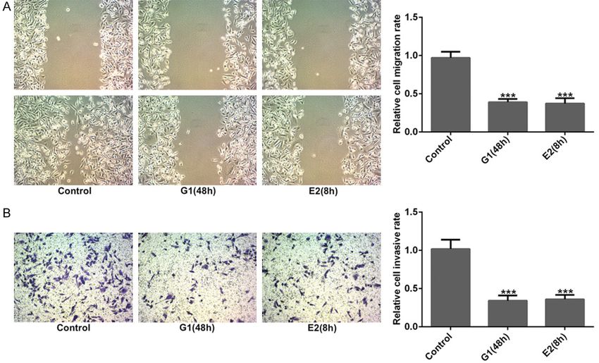
The effect of GPER on TNBC via CD151/miR-199a-3p

receptor (EGFR), intracellular cyclic AMP, calci-    SFA-1 or Tspan-24, can be expressed in many
um mobilization, MAPK and PI3K [4]. It has           cell types and considered to comprise molecu-
been reported that GPER was detected in tis-         lar facilitators [17]. The mRNA and protein lev-
sues of 132 patients with TNBC and its expres-       els of CD151 are highly expressed in breast
sion level was negatively correlated with high-      cancer, colon cancer and hepatocellular carci-
level tumors, showing that the lower the expres-     noma [18]. Moreover, studies have shown that
sion level of GPER was, the worse the prognosis      the expression change of CD151 is markedly
became [5, 6]. These all suggest that GPER is a      correlated with the growth process, invasion
positive prognostic factor in TNBC.                  and migration of cancers [19]. Other studies
                                                     have reported that CD151 is highly expressed
Epithelial-mesenchymal transition (EMT) pro-         in ER positive and TNBC cells and can promote
cess mainly includes the decrease of intercel-       the proliferation, invasion and migration of
lular connection and adhesion, the enhance-          breast cancer cells through targeted binding
ment of cell vitality and the changes of various     with miR-124 [20]. Therefore, this study aims to
related molecules, and it can be determined by       explore whether the activation of GPER in TNBC
the loss of E-cadherin along with the up-regula-     cells can suppress the process of TNBC cells by
tion of N-cadherin, Fibronectin and Vimentin         inhibiting the expression of CD151 binding to
[7]. Previous studies have also reported that        miR-199a-3p.
the activation of GPER inhibits EMT, migration
and angiogenesis of TNBC cells via NF-κB sig-        It still remains unclear that whether the activa-
naling pathway. More importantly, the role of        tion of GPER inhibits cells proliferation, inva-
GPER has been highlighted in nervous, cardio-        sion and EMT of triple-negative breast cancer
vascular and immune systems as well as the           via CD151/miR-199a-3p bio-axis, thus, more
inflammatory processes [8, 9]. For instance,         researches are needed. The regulatory role of
GPER was shown to be involved in thymic atro-        GPER in the expression of miR-199a-3p/CD151
phy and thymocyte apoptosis induced by estro-        are also investigated to reveal the possible
gens and GPER agonist G-1 in a knockout mice         internal molecular mechanisms and signaling
in vivo [10]. Interestingly, GPER expression has     pathways. This finding will provide new theoreti-
been associated with poor clinical-pathological      cal basis for in-depth exploration of the breast
features in breast, endometrial and ovarian          cancer treatment.
cancer patients.
                                                     Materiel and methods
MicroRNAs (miRNAs), about 18~22 nucleo-
                                                     Cell culture and treatment
tides, are small non-coding RNA molecules
[11]. They regulate the expression of targeted       Three TNBC cell lines (HCC1806, HCC1937,
genes by directly binding the 3’-untranslated        MDA-MB-231) and normal breast epithelial cell
regions (3’-UTR) of corresponding messenger          lines (HMEC-184) were cultured in RPMI 1640
RNAs (mRNAs) [12]. miRNAs participate in the         media (Gibco, Life Technologies, Carlsbad, CA,
pathogenesis of various biological behaviors,        USA) supplemented with 10% fetal bovine
such as suppressing or promoting tumors. As a        serum (FBS, Gibco) and 1% penicillin-strepto-
tumor suppressive factor, miRNA-199a-3p              mycin solution (Gibco). Cultures were main-
(miR-199a-3p) is down-regulated in multiple          tained in a humidified incubator with 5% CO2 at
cancer tissues and cells, including hepatocel-       37°C.
lular carcinoma [13], osteosarcoma [14] and
papillary thyroid carcinoma [15]. Highly             17β-Estradiol (E2) was purchased from Sigma-
expressed in hair follicles and in some tumor        Aldrich, and solubilized in ethanol. G-1(1-[4-
cells, miR-199a-3p participated in tumor pro-        (-6-bromobenzol [1, 3] diodo-5-yl)-3a,4,5,9b
gression. However, it is significantly under         tetrahidro3H5cyclopenta[c]quinolin-8yl]-etha-
expressed in hepatocellular carcinoma and            none) was obtained from Tocris Bioscience
bladder cancer and regulates cell proliferation      (Bristol, UK), which was solubilized in ethanol.
and migration. In addition, miR-199a-3p pro-         G-1 and E2 inducers have been reported to
motes cell proliferation and survival of endo-       belong to the GPER agonists for up-regulating
thelial cells as well as breast cancer cells [16].   GPER expression. Cultured in regular growth
CD151, also known as GP-27, MER-2, PETA-3,           medium, MDA-MB-231 cells were switched to

33                                                               Am J Transl Res 2020;12(1):32-44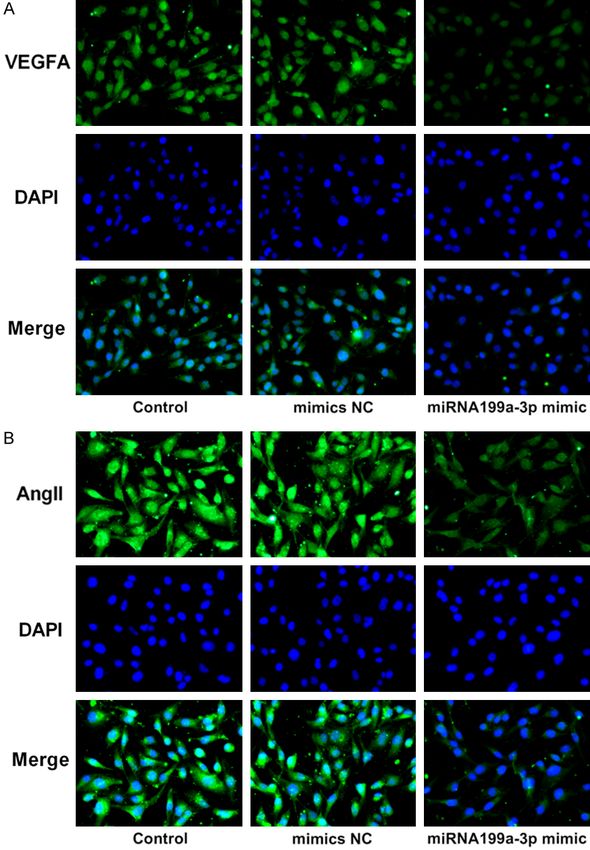
The effect of GPER on TNBC via CD151/miR-199a-3p

medium without serum and phenol red for 24 h,       tom cells were fixed with 95% ethanol, stained
and then treated with E2 (10 nM) for 6 h and        with 0.1% crystal violet. Additionally, photo-
8 h or with G-1 (1 μM) for 24 h and 48 h.           graphs were taken in three independent fields
Experiments Grouping, Control, G-1 (24 h), G-1      for each well.
(48 h), E2 (6 h) and E2 (8 h) groups.
                                                    Immunofluorescence assay
Cell transfection
                                                    MDA-MB-231 cells were seeded at 10% con-
Cell transfection was performed to up-regulate      fluence onto small glass coverslips placed
the expression of miR-199a-5p in MDA-MB-231         in 24-well plates. After different treatments
cells. miR-199a-5p mimics and its negative          were performed, the coverslips were removed,
control (NC) were both designed and synthe-         washed with phosphate-buffered saline (PBS)
sized by GenePharma Corporation (Shanghai,          three times, and fixed with 4% paraformalde-
China). The plasmids along with miR-199a-5p         hyde in PBS for 20 minutes. Pushing through
mimics or scramble were transfected into            the cytomembrane (0.1% Triton, 0.1% sodium
MDA-MB-231 cells with Lipofectamine 2000            citrate for 10 minutes) and blocking in 5% goat
reagents (Invitrogen, USA) according to the         serum for 1 h were followed by incubation with
manufacturer’s instructions. Transfection effi-     VEGFA and Ang II primary antibodies at 4°C
ciency was confirmed with qRT-PCR.                  overnight. After washing with PBS, cells were
                                                    incubated with a 1:500 dilution of a fluorescent
Cell viability assay                                tag (Alexa Fluor 488; Thermo Fisher Scientific,
                                                    Inc.) and conjugated with secondary antibodies
Cell viability after corresponding treatments       in dark for 30 minutes. Next, cells were treated
was assessed using cell counting kit-8 (CCK-        with DAPI (1:10,000, Invitrogen) for 5 minutes,
8) assay (Beyotime Biotechnology, Shanghai,         and then covered with an antifade mounting
China). Briefly, after with or without treatment,   medium and placed onto microscope slides.
MDA-MB-231 cells were seeded into 96-well           Finally, representative images were captured
plate (Thermo Fisher Scientific, Inc.) with cell    by Olympus FV1000 Digital laser scanning
concentration of 1 × 10⁴ cells/well. Then, 10 μl    microscopy.
CCK-8 solution was added into each well and
the cells were maintained in humidified incuba-     Quantitative reverse transcription PCR (qRT-
tor for 1 h at 37°C. Then, the absorbance at        PCR)
490 nm was measured using a microplate
reader (Bio-Tek Instruments, USA). Cell viability   Total RNA was extracted with TRIZOL reagent
(%) was calculated using the average absor-         (Invitrogen) in accordance with the manufac-
bance of different treatment groups/average         turer’s instructions. qRT-PCR was conducted to
absorbance of control groups × 100%.                measure the expression level of miR-199a-3p
                                                    in MDA-MB-231 cells after relevant treatment
Wound healing assay and transwell assay             or transfection. The cDNA was synthesized
                                                    using the PrimeScript RT reagent Kit (TaKaRa).
Cells were put into a 6-well plate. After cells     Furthermore, quantitative PCR was performed
received different treatments, a 200-μl sterile     using SYBR Premix Ex Taq (TaKaRa) to detect
pipette tip was used to create scratches. Sub-      the expression of miR-199a-3p and U6. The
sequently, cells were washed twice with PBS         expression of U6 was used as internal control.
and placed in DMEM without FBS. Photographs         All experiments were performed in triplicate.
were captured at 0 h and after 24 h using a         The data was analysed using 2-ΔΔCt method.
microscope (Carl Zeiss), and the data was ana-
lysed with Image pro-plus software.                 Western blotting

Transwell membranes was coated with Matrigel        Western blotting was employed to assess the
(BD Biosciences, San Jose, CA). 1 × 10⁴ cells in    protein expression of genes involved in cell pro-
150 μl serum-free medium were added to the          liferation and angiogenesis, EMT process and
upper chamber, while the medium in the lower        hippo pathways in MDA-MB-231 cells after rel-
chamber was kept with 10% FBS. After 24 h,          evant transfection or treatment. Briefly, total
the cells in the top well was removed. The bot-     proteins were isolated using RIPA lysis buffer

34                                                              Am J Transl Res 2020;12(1):32-44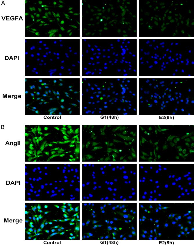
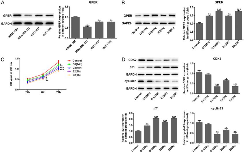
The effect of GPER on TNBC via CD151/miR-199a-3p

Figure 1. Expression of GPER and the effects of its activation on cell viability of MDA-MB-231 cells. A. Western blot-
ting indicated that the relative GPER expression in three TNBC cell lines (HCC1806, HCC1937 and MDA-MB-231)
was downregulated significantly when compared with the normal breast epithelial cell line (HMEC-184). **P
The effect of GPER on TNBC via CD151/miR-199a-3p

Figure 2. Activation of GPER suppressed cell migration and invasion. After G-1 (1 μM for 48 h) or E2 (10 nM for 8 h)
treatment, the relative migration (A) and invasion (B) of MDA-MB-231 cells were reduced sharply when compared
with the control group from the results of wound healing and transwell assays. ***P
The effect of GPER on TNBC via CD151/miR-199a-3p

                                                                                 pared with HMEC-184 cells,
                                                                                 in particular, the downward
                                                                                 trend was most significant in
                                                                                 MDA-MB-231 cells (P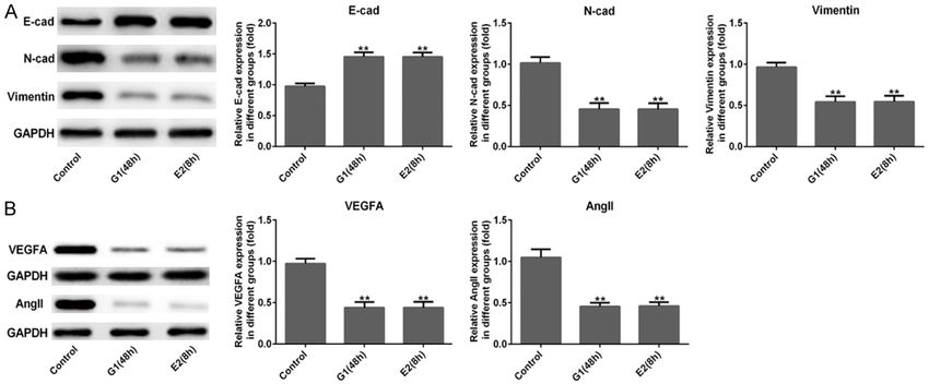
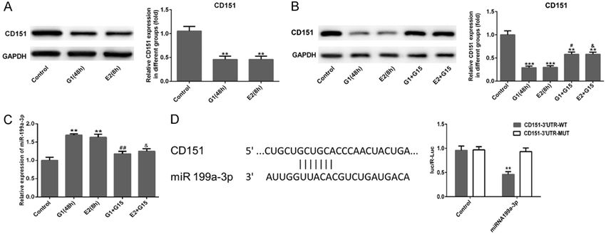
The effect of GPER on TNBC via CD151/miR-199a-3p

Figure 5. Activation of GPER suppressed CD151 expression and elevated miR-199a-3p. (A) The relative expres-
sion of CD151 in MDA-MB-231 cells was downregulated significantly after G-1 (1 μM for 48 h) or E2 (10 nM for 8
h) treatment, and (B) their expression were elevated after G15 (100 nM for 6 h) treatment following G-1 (1 μM for
48 h) or E2 (10 nM for 8 h) treatment using western blot assay. (C) qRT-PCR was used to measure the expression
level of miR-199a-3p in MDA-MB-231 cells in different groups. (D) The TargetScan database revealed the putative
binding site of CD151 in the 3’-UTR of miR-199a-3p. The relative luciferase activity was reduced when cells were co-
transfected with miR-199a-3p and CD151-3’-UTR (WT). ***P
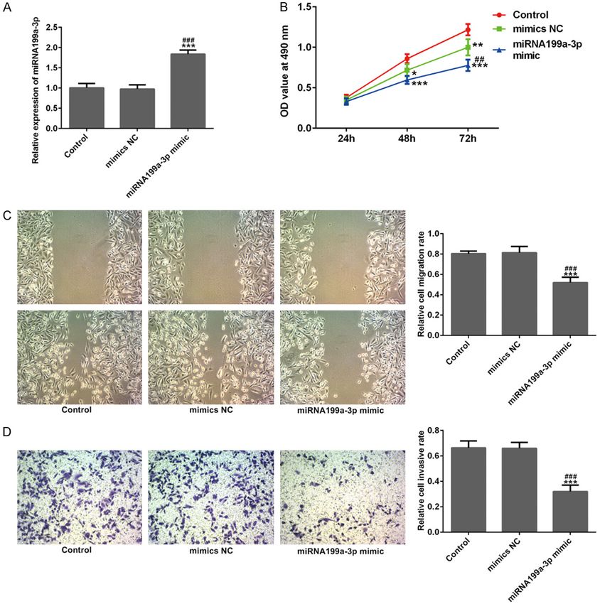
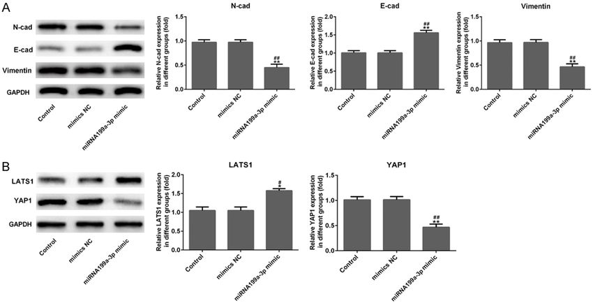
The effect of GPER on TNBC via CD151/miR-199a-3p

Figure 6. miR-199a-3p suppressed cell viability, migration and invasion. A. After miR-199a-3p mimic transfection,
the relative expression level of miR-199a-3p was overexpressed in MDA-MB-231 cells using quantitative RT-PCR.
B-D. After miR-199a-3p mimic transfection, relative cell viability, migration and invasion of MDA-MB-231 cells were
inhibited significantly from the results of CCK-8, wound healing and Transwell assays. ***P
The effect of GPER on TNBC via CD151/miR-199a-3p

                                                                                  These findings suggested that
                                                                                  miR-199a-3p might suppress
                                                                                  Hippo signaling pathway in
                                                                                  TNBC cells by up-regulating
                                                                                  LATS1 and inhibiting YAP1.

                                                                                  Discussion

                                                                                  In this research, we mainly
                                                                                  revealed the inhibiting effects
                                                                                  of activation of GPER and
                                                                                  overexpression of miR-199a-
                                                                                  3p on TNBC cell proliferation,
                                                                                  migration, invasion and an-
                                                                                  giogenesis, as well as EMT
                                                                                  process. GPER was under-ex-
                                                                                  pressed in TNBC cells, espe-
                                                                                  cially in MDA-MB-231 cells.
                                                                                  The expression level of and
                                                                                  CD151 was decreased and
                                                                                  miR-199a-3p was increased
                                                                                  in MDA-MB-231 cells after
                                                                                  GPER overexpression with G-1
                                                                                  or E2 treatment. Further, over-
                                                                                  expression of miR-199a-3p,
                                                                                  the same as activation of
                                                                                  GPER, significantly suppress-
                                                                                  ed cell proliferation, migra-
                                                                                  tion, invasion, angiogenesis
                                                                                  and EMT processes in MDA-
                                                                                  MB-231 cells through inhibit-
                                                                                  ing Hippo signal pathway.
                                                                                  Meanwhile, there is a target-
                                                                                  ing correlation between miR-
                                                                                  199a-3p and CD151 regulat-
                                                                                  ing the progression of TNBC
Figure 7. miR-199a-3p s suppressed the protein expression related to angio-       cells.
genesis. Representative images of immunofluorescence staining indicated
the expression of VEGFA (A) and Ang II (B) were downregulated in MDA-
                                                                               Breast cancer is one of the
MB-231 cells after miR-199a-3p mimic transfection. Magnification, × 200.
                                                                               most commonly diagnosed
                                                                               cancers in women, but there
invasion, EMT and angiogenesis of MDA-MB-                    is no targeted therapies for TNBC at present;
231 cells.                                                   Chemotherapy regimens, along with their
                                                             adverse effects, remain the mainstay of treat-
miR-199a-3p inactivated Hippo signaling                      ment in most TNBC patients. Thus, finding a
pathway in MDA-MB-231 cells by upregulating                  targeted biological agent for TNBC is supposed
LATS1 and inhibiting YAP1                                    to make a paradigm shift in the treatment of
                                                             these patients [21]. GPER is expressed exten-
Finally, we evaluated the effect of miR-199a-                sively in TNBC clinical specimens and positively
3p mimic on Hippo signaling pathway using                    associated with high recurrence of TNBCs [22].
western blotting. Figure 8B showed that miR-                 In addition, GPER mediates a specific gene sig-
199a-3p mimic remarkably up-regulated the                    nature related to cell growth, migration and
expression of LATS1 (P
The effect of GPER on TNBC via CD151/miR-199a-3p Figure 8. miR-199a-3p inhibited cell epithelial-mesenchymal transition (EMT) and regulated Hippo signaling path- way. A. Results of western blotting showed that the protein expression levels of N-cad and Vimentin were decreased distinctly in MDA-MB-231 cells were evaluated using after miR-199a-3p mimic transfection with a marked increase in E-cad expression. B. After miR-199a-3p mimic transfection in MDA-MB-231 cells, overexpression of miR-199a-3p increased the protein expression of LATS1 and suppressed the YAP1 expression, which are the key factors in the Hippo pathway. *P
The effect of GPER on TNBC via CD151/miR-199a-3p

Over the past decade, the Hippo pathway has           Address correspondence to: Dr. Yufeng Yao, Depart-
been shown to play a critical role in controlling     ment of General Surgery, Jiangsu Cancer Hospital,
organ size via regulating cell proliferation and      No. 42 Baiziting Road, Nanjing 210009, Jiangsu,
apoptosis [32]. As a major downstream effec-          P. R. China. Tel: +86-0258-3284725; E-mail: yaoyf-
tor of Hippo pathway, it is not surprising that       1122@163.com
Yes-associated protein (YAP) functions as an
                                                      References
oncogene, and since its expression is strikingly
increased in perivascular cells, which are            [1]  Cheang MC, Voduc D, Bajdik C, Leung S,
believed to own the properties of cancer stem              McKinney S, Chia SK, Perou CM and Nielsen
cells [33]. The core kinase cascade of the                 TO. Basal-like breast cancer defined by five bio-
Hippo pathway consists of upstream Ste20-                  markers has superior prognostic value than
like protein kinases (STKs) (commonly known                triple-negative phenotype. Clin Cancer Res
as MST1/2) and the large tumor suppressor                  2008; 14: 1368-1376.
kinases 1/2 (LATS1/2) [34]. LATS1/2 kinase            [2] Jia M, Dahlman-Wright K and Gustafsson JA.
can directly phosphorylate and inactivate YAP              Estrogen receptor alpha and beta in health
and the Hippo signaling cascade is regulated               and disease. Best Pract Res Clin Endocrinol
by numerous upstream signals which include                 Metab 2015; 29: 557-568.
G-protein coupled receptors (GPCRs) [35], Wnt         [3] Lappano R and Maggiolini M. G protein-cou-
signaling [36], as well as microRNAs [37]. In              pled receptors: novel targets for drug discovery
this research, we found that overexpression of             in cancer. Nat Rev Drug Discov 2011; 10: 47-
miR-199a-3p distinctly inhibited the Hippo                 60.
                                                      [4] Maggiolini M and Picard D. The unfolding sto-
pathway via up-regulating LATS1 and down-reg-
                                                           ries of GPR30, a new membrane-bound estro-
ulating YAP1 in MDA-MB-231 cells (Figure 8).               gen receptor. J Endocrinol 2010; 204: 105-
Conclusion                                                 114.
                                                      [5] Chen ZJ, Wei W, Jiang GM, Liu H, Wei WD, Yang
The present study revealed that the exact                  X, Wu YM, Liu H, Wong CK, Du J and Wang HS.
anti-tumor effects of GPER and miR-199a-3p                 Activation of GPER suppresses epithelial mes-
overexpression on TNBC cells. Briefly, activa-             enchymal transition of triple negative breast
                                                           cancer cells via NF-kappaB signals. Mol Oncol
tion of GPER suppressed TNBC MDA-MB-231
                                                           2016; 10: 775-788.
cell proliferation, migration, invasion and angio-
                                                      [6] Liang S, Chen Z, Jiang G, Zhou Y, Liu Q, Su Q,
genesis, as well as EMT process, by regulating             Wei W, Du J and Wang H. Activation of GPER
miR-199a-3p/CD151 axis, inactivating Hippo                 suppresses migration and angiogenesis of tri-
signaling pathway. This finding may provide new            ple negative breast cancer via inhibition of NF-
theoretical basis for deeply exploring the tar-            kappaB/IL-6 signals. Cancer Lett 2017; 386:
geted therapies for Triple-Negative Breast                 12-23.
Cancer.                                               [7] Nieto MA, Huang RY, Jackson RA and Thiery JP.
                                                           Emt: 2016. Cell 2016; 166: 21-45.
Acknowledgements                                      [8] De Marco P, Bartella V, Vivacqua A, Lappano R,
                                                           Santolla MF, Morcavallo A, Pezzi V, Belfiore A
We thank Director Mei-long Hu in the
                                                           and Maggiolini M. Insulin-like growth factor-I
Chemoradiotherapy Department of the First                  regulates GPER expression and function in
Hospital Affiliated to Wenzhou Medical College,            cancer cells. Oncogene 2013; 32: 678-688.
and Director Hui Dai in the Hangzhou Tumor            [9] Albanito L, Sisci D, Aquila S, Brunelli E,
Hospital, for their inspiration in article, sugges-        Vivacqua A, Madeo A, Lappano R, Pandey DP,
tions and supports of experimental work. This              Picard D, Mauro L, Ando S and Maggiolini M.
work is supported by the Jiangsu Provincial                Epidermal growth factor induces G protein-
Medical Youth Talent, the Project of Invigorating          coupled receptor 30 expression in estrogen
Health Care through Science, Technology and                receptor-negative breast cancer cells. Endocri-
Education (QNRC2016661); Project of Jiangsu                nology 2008; 149: 3799-3808.
provincial Six Talent Peaks in China (WSN-057)        [10] Curtis C, Shah SP, Chin SF, Turashvili G, Rueda
and Jiangsu Provincial Talent on Maternal and              OM, Dunning MJ, Speed D, Lynch AG, Sama-
Child Health (FRC201759).                                  rajiwa S, Yuan Y, Graf S, Ha G, Haffari G,
                                                           Bashashati A, Russell R, McKinney S, Group M,
Disclosure of conflict of interest                         Langerod A, Green A, Provenzano E, Wishart G,
                                                           Pinder S, Watson P, Markowetz F, Murphy L,
None.                                                      Ellis I, Purushotham A, Borresen-Dale AL,

42                                                                  Am J Transl Res 2020;12(1):32-44
The effect of GPER on TNBC via CD151/miR-199a-3p
Brenton JD, Tavare S, Caldas C and Aparicio S. suppresses breast cancer cell growth and mo-
The genomic and transcriptomic architecture tility by targeting CD151. Cell Physiol Biochem
of 2,000 breast tumours reveals novel sub- 2013; 31: 823-832.
groups. Nature 2012; 486: 346-352. [21] Nakhjavani M, Palethorpe HM, Tomita Y, Smith
[11] Chen L, Zhang A, Li Y, Zhang K, Han L, Du W, E, Price TJ, Yool AJ, Pei JV, Townsend AR and
Yan W, Li R, Wang Y, Wang K, Pu P, Jiang T, Hardingham JE. Stereoselective anti-cancer
Jiang C and Kang C. MiR-24 regulates the activities of ginsenoside Rg3 on triple negative
proliferation and invasion of glioma by ST7L breast cancer cell models. Pharmaceuticals
via beta-catenin/Tcf-4 signaling. Cancer Lett (Basel) 2019; 12.
2013; 329: 174-180. [22] Steiman J, Peralta EA, Louis S and Kamel O.
[12] Chen L, Zhang W, Yan W, Han L, Zhang K, Shi Z, Biology of the estrogen receptor, GPR30, in
Zhang J, Wang Y, Li Y, Yu S, Pu P, Jiang C, Jiang triple negative breast cancer. Am J Surg 2013;
T and Kang C. The putative tumor suppressor 206: 698-703.
miR-524-5p directly targets Jagged-1 and Hes- [23] Pandey DP, Lappano R, Albanito L, Madeo A,
1 in glioma. Carcinogenesis 2012; 33: 2276- Maggiolini M and Picard D. Estrogenic GPR30
2282. signalling induces proliferation and migration
[13] Hou J, Lin L, Zhou W, Wang Z, Ding G, Dong Q, of breast cancer cells through CTGF. EMBO J
Qin L, Wu X, Zheng Y, Yang Y, Tian W, Zhang Q, 2009; 28: 523-532.
Wang C, Zhang Q, Zhuang SM, Zheng L, Liang [24] Filardo EJ, Graeber CT, Quinn JA, Resnick MB,
A, Tao W and Cao X. Identification of miR- Giri D, DeLellis RA, Steinhoff MM and Sabo E.
Nomes in human liver and hepatocellular car- Distribution of GPR30, a seven membrane-
cinoma reveals miR-199a/b-3p as therapeutic spanning estrogen receptor, in primary breast
target for hepatocellular carcinoma. Cancer cancer and its association with clinicopatho-
Cell 2011; 19: 232-243. logic determinants of tumor progression. Clin
[14] Duan Z, Choy E, Harmon D, Liu X, Susa M, Cancer Res 2006; 12: 6359-6366.
Mankin H and Hornicek F. MicroRNA-199a-3p [25] Smith HO, Leslie KK, Singh M, Qualls CR,
is downregulated in human osteosarcoma and Revankar CM, Joste NE and Prossnitz ER.
regulates cell proliferation and migration. Mol GPR30: a novel indicator of poor survival for
Cancer Ther 2011; 10: 1337-1345. endometrial carcinoma. Am J Obstet Gynecol
[15] Minna E, Romeo P, De Cecco L, Dugo M, 2007; 196: 386, e381-389; discussion 386
Cassinelli G, Pilotti S, Degl’Innocenti D, Lanzi e389-311.
C, Casalini P, Pierotti MA, Greco A and Borrello [26] Smith HO, Arias-Pulido H, Kuo DY, Howard T,
MG. miR-199a-3p displays tumor suppressor Qualls CR, Lee SJ, Verschraegen CF, Hathaway
functions in papillary thyroid carcinoma. HJ, Joste NE and Prossnitz ER. GPR30 predicts
Oncotarget 2014; 5: 2513-2528. poor survival for ovarian cancer. Gynecol Oncol
[16] Shatseva T, Lee DY, Deng Z and Yang BB. 2009; 114: 465-471.
MicroRNA miR-199a-3p regulates cell prolifer-
[27] Vivacqua A, Romeo E, De Marco P, De
ation and survival by targeting caveolin-2. J
Francesco EM, Abonante S and Maggiolini M.
Cell Sci 2011; 124: 2826-2836.
GPER mediates the Egr-1 expression induced
[17] Zhang Z, Wang F, Li Q, Zhang H, Cui Y, Ma C,
by 17beta-estradiol and 4-hydroxitamoxifen in
Zhu J, Gu X and Sun Z. CD151 knockdown
breast and endometrial cancer cells. Breast
inhibits osteosarcoma metastasis through
Cancer Res Treat 2012; 133: 1025-1035.
the GSK-3beta/beta-catenin/MMP9 pathway.
Oncol Rep 2016; 35: 1764-1770. [28] Zhan Y, Brown C, Maynard E, Anshelevich A, Ni
[18] Yang XH, Richardson AL, Torres-Arzayus MI, W, Ho IC and Oettgen P. Ets-1 is a critical regu-
Zhou P, Sharma C, Kazarov AR, Andzelm MM, lator of Ang II-mediated vascular inflammation
Strominger JL, Brown M and Hemler ME. and remodeling. J Clin Invest 2005; 115:
CD151 accelerates breast cancer by regulat- 2508-2516.
ing alpha 6 integrin function, signaling, and [29] Risau W and Flamme I. Vasculogenesis. Annu
molecular organization. Cancer Res 2008; 68: Rev Cell Dev Biol 1995; 11: 73-91.
3204-3213. [30] Sotiropoulou G, Pampalakis G, Lianidou E and
[19] Novitskaya V, Romanska H, Dawoud M, Jones Mourelatos Z. Emerging roles of microRNAs as
JL and Berditchevski F. Tetraspanin CD151 molecular switches in the integrated circuit of
regulates growth of mammary epithelial cells the cancer cell. RNA 2009; 15: 1443-1461.
in three-dimensional extracellular matrix: im- [31] Kwon MJ, Park S, Choi JY, Oh E, Kim YJ, Park
plication for mammary ductal carcinoma in YH, Cho EY, Kwon MJ, Nam SJ, Im YH, Shin YK
situ. Cancer Res 2010; 70: 4698-4708. and Choi YL. Clinical significance of CD151
[20] Han ZB, Yang Z, Chi Y, Zhang L, Wang Y, Ji Y, overexpression in subtypes of invasive breast
Wang J, Zhao H and Han ZC. MicroRNA-124 cancer. Br J Cancer 2012; 106: 923-930.
43 Am J Transl Res 2020;12(1):32-44The effect of GPER on TNBC via CD151/miR-199a-3p
[32] Zygulska AL, Krzemieniecki K and Pierzchalski [36] Varelas X, Miller BW, Sopko R, Song S,
P. Hippo pathway-brief overview of its rele- Gregorieff A, Fellouse FA, Sakuma R, Pawson T,
vance in cancer. J Physiol Pharmacol 2017; Hunziker W, McNeill H, Wrana JL and Attisano
68: 311-335. L. The Hippo pathway regulates Wnt/beta-
[33] Zhao B, Li L, Lei Q and Guan KL. The Hippo-YAP catenin signaling. Dev Cell 2010; 18: 579-591.
pathway in organ size control and tumorigene- [37] Mitamura T, Watari H, Wang L, Kanno H,
sis: an updated version. Genes Dev 2010; 24: Kitagawa M, Hassan MK, Kimura T, Tanino
862-874. M, Nishihara H, Tanaka S and Sakuragi N. mi-
[34] Meng Z, Moroishi T, Mottier-Pavie V, Plouffe croRNA 31 functions as an endometrial cancer
SW, Hansen CG, Hong AW, Park HW, Mo JS, Lu oncogene by suppressing Hippo tumor sup-
W, Lu S, Flores F, Yu FX, Halder G and Guan KL. pressor pathway. Mol Cancer 2014; 13: 97.
MAP4K family kinases act in parallel to
MST1/2 to activate LATS1/2 in the Hippo path-
way. Nat Commun 2015; 6: 8357.
[35] Yu FX, Zhao B, Panupinthu N, Jewell JL, Lian I,
Wang LH, Zhao J, Yuan H, Tumaneng K, Li H, Fu
XD, Mills GB and Guan KL. Regulation of the
Hippo-YAP pathway by G-protein-coupled re-
ceptor signaling. Cell 2012; 150: 780-791.
44 Am J Transl Res 2020;12(1):32-44You can also read