Coelomic Fluid of Eisenia fetida Ameliorates Cetuximab to Reduce K-Ras and Vimentin Expression through Promoting RUNX3 in an AOM/DSS-Induced ...
←
→
Page content transcription
If your browser does not render page correctly, please read the page content below
Hindawi
Evidence-Based Complementary and Alternative Medicine
Volume 2020, Article ID 9418520, 14 pages
https://doi.org/10.1155/2020/9418520

Research Article
Coelomic Fluid of Eisenia fetida Ameliorates Cetuximab to Reduce
K-Ras and Vimentin Expression through Promoting RUNX3 in an
AOM/DSS-Induced Colitis Associated Colon Cancer

          Sofy Permana ,1 Reyudzky Putri Fityanti,2 Eviana Norahmawati,3 Agustin Iskandar,4
          Erika Desy Anggraini Mulyadi,5 and Agustina Tri Endharti4,6
          1
            Department of Biology, Faculty of Mathematics and Natural Sciences, Universitas Brawijaya, Malang, Indonesia
          2
            Master Program in Biomedical Sciences, Faculty of Medicine, Universitas Brawijaya, Malang, Indonesia
          3
            Department of Pathology Anatomy, Faculty of Medicine, Universitas Brawijaya, Malang, Indonesia
          4
            Department of Parasitology, Faculty of Medicine, Universitas Brawijaya, Malang, Indonesia
          5
            Undergraduate Program of Biology, Faculty of Mathematics and Natural Sciences, Universitas Brawijaya, Malang, Indonesia
          6
            Biomedical Central Laboratory, Faculty of Medicine, Universitas Brawijaya, Malang, Indonesia

          Correspondence should be addressed to Sofy Permana; sofy-bio@ub.ac.id

          Received 25 February 2020; Revised 2 May 2020; Accepted 12 June 2020; Published 20 July 2020

          Academic Editor: Ho Lin

          Copyright © 2020 Sofy Permana et al. This is an open access article distributed under the Creative Commons Attribution License,
          which permits unrestricted use, distribution, and reproduction in any medium, provided the original work is properly cited.

          Ulcerative colitis is a major risk factor that increases the occurrence of colorectal cancer. In colorectal cancer due to colitis,
          intestinal inflammation plays an important role which causes DNA damage. The aim of this study is to investigate the anticancer
          effect of coelomic fluid of Eisenia fetida (CFEF) and cetuximab combinations. Colitis associated colon cancer was induced in
          BALB/c mice by DSS/AOM. The mice were randomly divided into six groups: group 1 received vehicle (control), groups 2–6
          received DSS/AOM, groups 3–5 received cetuximab + CFEF (30, 60, or 120 mg/kgBW), and group 6 received CFEF only. After the
          12th week of treatments, the colon tissues were removed for histological examination and immune-fluorescence. Intestinal
          Epithelial Cells (CECs) were analyzed by flow cytometer. Administration of CFEF significantly decreased the severity of DSS/
          AOM-induced CAC in a dose-dependent manner. The combinations of CFEF-cetuximab were revealed by histological change.
          The CFEF significantly reduced the severity scores (P < 0.05). The combinations of CFEF-cetuximab significantly inhibited K-Ras
          and vimentin expressions, whereas the percentage of RUNX3 significantly increased in CECs. The increasing of RUNX3 could
          prevent EMT, so that it can decrease K-Ras and vimentin to suppressed cell invasion and migration by CFEF. Our results suggest
          that CFEF has the therapeutic potential to CAC.

1. Introduction                                                         to the nucleus via MAPK pathway. In the MAPK pathway,
                                                                        K-Ras plays a role in regulating cell proliferation and cell
Colorectal cancer is the most common malignant tumor                    survival. Thus, K-Ras is considered to have a significant
with high prevalence and low 5-year survival [1, 2]. Ul-                potential therapeutic value [3–5]. In addition, tumor bud-
cerative colitis is a major risk factor that increases the oc-          ding is also included in colorectal cancer [4, 5]. Tumor
currence of colorectal cancer. In colorectal cancer due to              budding is a part of epithelial mesenchymal transition
colitis, intestinal inflammation plays an important role                 (EMT). In the EMT process, there is an upregulation of
which causes DNA damage, increased cell proliferation,                  vimentin. Impaired EMT activation has been known to play
decreased tumor suppressors, and also apoptosis [3–5].                  a role in cancer metastases [5, 6].
These things occur through signal transduction from Epi-                    The migration, as one of the factors involved in cancer
dermal Growth Factor Receptor (EGFR) bound with ligands                 metastasis, is a process known as EMT. The EMT phenotype
2                                                                   Evidence-Based Complementary and Alternative Medicine

is characterized by the loss of cell-to-cell adhesion and          coelomic fluid than other species [22]. The use of coelomic
remodeling of the epithelial molecule E-cadherin and               fluid as a therapy only causes side-effects that can be ignored.
mesenchyme markers such as vimentin. EMT has been                  Also because there have never been reports of the side-effects
shown to prevent overexpression of Runt-Related Tran-              from therapies using coelomic fluid, coelomic fluid may be
scription Factor-3 (RUNX3), which facilitates metastasis,          consumed continuously [24, 25].
cell invasion migration, and loss of RUNX3 in epithelial cells         Considering the interest in the possible anticancer
[7–9]. Runt-Related Transcription Factor-3 is a transcription      properties of coelomic fluid, the main goal of this investi-
factor known for its tumor suppressor activity and more            gation was to evaluate the actions of coelomic fluid from
recently has been implicated in cancer metastasis. RUNX3 is        Eisenia fetida on mice model colitis associated colorectal
an interpretation factor known for its tumor suppressor            cancer. Nevertheless, to our knowledge, evidence testifying
activity and lately has been involved in malignant growth          the antiproliferative property of CFEF in CAC is still limited.
metastasis. The Runt-Related Transcription Factor-3                In this study, we demonstrated the antimetastasis effects and
(RUNX3), which plays a significant part in cell proliferation       studied associated mechanisms of continuous CFEF inter-
[8–13], has been shown to play a tumor suppressor role in          vention on an AOM/DSS mouse model.
several types of cancers and its expression levels are
downregulated in cancer. It is currently clear that EMT has        2. Materials and Methods
implications on cancer metastasis by triggering the loss of
cell-cell adhesion to facilitate the invasion of cancer cell.      2.1. Mice. Female BALB/c mice aged 8–10 weeks were
However, the mechanism of RUNX3-mediated suppression               housed in laboratory of animal, Faculty of Medicine, Uni-
of cancer metastasis remains unclear and the role of RUNX3         versitas Brawijaya, were approved pathogen-free barrier
in colorectal cancer has not yet been well studied [11–14].        facility at constant temperature, and were kept in controlled
Further biochemical studies need to be followed up to in-          conditions of humidity (50 ± 10%), light (12-hour light/dark
vestigate the mechanistic principles involved in RUNX3-            cycle), and temperature (25 ± 2°C). All the studies and an-
mediated inhibition of EMT in colitis associated colon             imal protocols were performed in accordance with the in-
cancer. In the development of EMT, cancer cells lose the           stitutional guidelines by The Institutional Animal Care and
properties of epithelial cells and develop mesenchymal cell        Use Committee (IAUCUC) by University of Brawijaya
properties, including overexpression of vimentin [12–16].          Ethical Committee (no. 1130-KEP-UB).
The potential mechanisms of cetuximab combination me-
diated EMT are not well understood. Cetuximab is a target
therapy for colorectal cancer that works in the EGFR ex-           2.2. Coelomic Fluid Collection. Heat and cold shock methods
tracellular domain by inhibition of K-Ras [11, 14].                were used to collect coelomic fluid (Endharti et al.) [8].
     Cetuximab is a target therapy for colorectal cancer that      Briefly, the same quantities of Eisenia fetida (±20 g) were
works in the EGFR extracellular domain by inhibiting               place in plastic box with wire mesh filter, rinsed with distilled
MAPK pathway. Although colorectal cancer patients with             water to remove adhering materials or particles, and then
metastasis have been treated with cetuximab, unfortunately,        dried by using tissue paper. The gut was cleaned of organic
cetuximab can cause many side-effects such as diarrhea,             matter after 48 hours as it was fed on filter paper. They were
nausea, vomiting, and skin rashes [17–19].                         thoroughly washed with distilled water and then put in glass
     According to Endharti et al. [8–10], the combination of       funnel, using hot water (40°C–50°C) in a glass beaker to give
coelomic fluid with 5-fluorouracil has anticancer potential for      heat shock and earthworms were shocked by the use of ice
colorectal cancer. Coelomic fluid in earthworms is secreted         cubes in a plastic box. Instead, the procedure was within
through dorsal pores in their skin. This fluid have been shown      three minutes to resolve the shock effect and give cold shock
to have antioxidant, antibacterial, anti-inflammatory, and          in a similar manner. Coelomic fluid Eisenia fetida (CFEF)
antitumor activities [18, 19]. Some of the active compounds in     was collected into tubes and stored in aliquots at −20°C.
coelomic fluid are lectins, lysenin, phenoloxidase, antibac-
terial peptides polysaccharides, fibrinolytic enzymes, and          2.3. The Precipitation of Coelomic Fluid. Coelomic fluid was
proteases (Endharti et al.) [8–10]. It is also established that    precipitated according to Endharti et al. [8–10]. Briefly,
coelomic fluid has a cytotoxic and antiproliferative effect on       coelomic fluid was precipitated by adding of 2 mM am-
cancer activity that may increase apoptosis in HT-29 cells         monium sulfate. The pellet collected by centrifugation was
(Permana et al.) [9]. Eisenia fetida is a type of earthworm that   resuspended in 20 mM Tris-HCl (pH8.0) followed by cen-
is easily maintained and it can produce more coelomic fluid         trifugation. Then, the pellet was resuspended in cold acetone
than other types of earthworms [16–19]. This coelomic fluid         by adding the acetone slowly, followed by 20-minute in-
has also found several bioactive compounds, exhibiting a           cubation. The remaining acetone was removed from the
variety of biological functions [17–20].                           pellet by centrifugation. Protein concentrations were mea-
     Coelomic fluid is also known to have a cytotoxic and           sured using NanoDrop spectrophotometer.
antiproliferative effect on cancer activity that can increase
apoptosis in HT-29 cell lines [18–20]. The combination of
coelomic fluid with 5-fluorouracil has anticancer potential          2.4. Induction of AOM/DSS-Induced Colitis Associated Colon
for colorectal cancer [21–25]. Eisenia fetida is a species of      Cancer (CAC). Colitis associated colon cancer model was
earthworm that is easily maintained and it can produce more        induced as previously described by Endharti et al. [8–10].
Evidence-Based Complementary and Alternative Medicine                                                                          3

Briefly, mice were intraperitoneal injected with a single dose      hour at room temperature. Slides were mounted with 4′,6-
of 10 mg/kg azoxymethane (AOM; Sigma-Aldrich, USA) on              diamidino-2-phenylindole (DAPI, BioLegend, USA) (1 : 1000)
day 1. Colon cancer was induced by cyclical DSS treatment,         for 5 minutes. Slides were observed under fluorescence mi-
which consisted of 1 week of 3% DSS followed by 7 days of          croscope (OLYMPUS 1X71). The contrast and/or brightness
untreated water. One week after the AOM injection, mice            adjustment was applied evenly over the whole field of the
were given four cycles of 3% dextran sulfate sodium (DSS)          image. Image analysis was performed using FIJI/ImageJ-2
(Sigma-Aldrich, USA) and followed by one week of regular           software (Bethesda, MD, USA).
water. Mice housed under specific-pathogen-free conditions
were divided into seven groups: group 1 (vehicle). Groups 2–7
were given AOM and 3% DSS. Groups 3–6 were treated with            2.7. Isolation of Colon Epithelial Cells (CECs). Primary CECs
cetuximab (10 mg/kg BW). Groups 4–6 were also treated with         were isolated using a protocol adapted from Endharti et al.
CFEF at 30, 60, and 120 mg/g/BW, respectively. Group 7 was         [8–10]. Briefly, mice duodenal and colonic tissues were re-
only given CFEF 120 mg/g/BW. Cetuximab and CFEF were               moved from mice flushed of luminal contents by removing the
injected intraperitoneally once a week during the four cycles      longitudinal muscle layer and washed using wash buffer
of DSS treatment. One week after AOM injection, mice were          (Hank’s Balanced Salt Solution (HBSS), Mg2+ and Ca2+ free
given four cycles of DSS in their drinking water and then          (Gibco), 100 U penicillin-streptomycin (Gibco), and 25 μg
distilled water until the end of the experiment. Mice were         ml−1 amphotericin B (Gibco). Intestinal tissues were cut into
sequentially killed randomly at the end of the 10th week, and      small pieces, suspended in 50 ml wash buffer, and inverted
at least six mice were killed for each group at each time point.   vigorously ten times, and the contents were allowed to settle
Colon tissues were collected for analysis by immunofluo-            for 1 minute. The supernatant was removed and a further four
rescence and flow cytometry. The colitis development was            times washed off the settled contents. After the fifth wash, the
detected using Fecal Occult Blood Test (FOBT).                     settled contents were removed and suspended in wash buffer.
                                                                   Intestinal tissues were digested in 50 ml of a digestion buffer
                                                                   (75 U ml−1 collagenase type XI-0.5 mM DTT (Sigma-Aldrich),
2.5. Immunohistochemistry for IL-6 Expression. IL-6 ex-            4% FBS (Gibco)) in Dulbecco’s Modification of Eagles Me-
pression in colon tissues was inspected using a standard           dium (DMEM) (Gibco). The tissue-containing digestion buffer
immunohistochemistry method. Immunostaining was done               was placed in an incubator of 37°C and allowed to shake for 3
on serial sections as described previously by Zeng et al. [11].    hours at 200 rpm. The effluent was centrifuged at 200 ×g, for 5
Briefly, the tissues were fixed in 10% formaldehyde and              minutes, at 4°C. The remaining pellet containing isolated CECs
embedded in paraffin and then cut into 4 μm sections. Tissue         was suspended digestion buffer. This process was repeated four
sections were, respectively, deparaffinized in xylene and            times. The resultant pooled supernatants were pelleted by
rehydrated by grade alcohols. The tissue was incubated             centrifugation (1000 ×g) for 30 minutes at 4°C. The pellet of
overnight at 4°C with primary antibody specific for IL-6            CECs were washed and resuspended in FACS buffer.
(Santa Cruz, CA, USA, dilution 1 : 200). The tissue sections
were then incubated with biotin-labeled goat anti-mouse
antibody (Sigma, USA) followed by exposure to avidin-              2.8. The Percentage of RUNX3 Using Flow Cytometry. The
peroxidase complex (Sigma, USA). Staining was developed            CECs (1 × 10⁶) cells were placed in 1.5 mL tube and resus-
with diaminobenzidine (DAB, Sigma) substrate and sections          pended in FACS buffer containing monoclonal antibodies
were counterstained with hematoxylin.                              anti-MMP2 conjugated with FITC (Bioscience) or anti-
                                                                   RUNX3 conjugated with PE (Bioscience) and incubated for
                                                                   20 minutes at 4°C. After the staining, cells were washed twice
2.6. Immunofluorescence for K-Ras, Vimentin, E-Cadherin,            and analyzed the following day on the FACS Calibur (BD
and β-Catenin Expressions. Immunofluorescence was done              Biosciences). Data were analyzed using Cell Quest-Pro
on serial sections as described previously by Permana et al.       software (Becton, Dickinson, USA).
[10]. Immunofluorescent analysis of colon tissue stained with
antivimentin antibody (biolegend), anti-K-Ras antibody
(MyBioSource), anti-E-cadherin antibody (Santa Cruz), and          2.9. Statistical Analysis. One-way ANOVA was used to
anti-β-catenin antibody (Santa Cruz). In brief, colon tissue       analyze the data. The data outcome was defined as a
was made into histopathology slide and deparaffinized. Then          mean ± standard deviation and the statistical significance of
slides were incubated in chamber with buffer citrate for 5          a distinction regarded significant with P < 0.05 between each
minutes of 300 volts for antigen retrieval. Tissue sections were   group. The statistical assessment was carried out using SPSS
washed with 0.1% Triton-X 100 in PBS for 5 minutes and             software.
followed with incubation of 1% BSA for 30 minutes. After
washing, tissue sections were incubated with the following         3. Results
each specific primary antibody for vimentin (BioLegend,
USA, 1 : 200), K-Ras (MyBioSource, USA, 1 : 100), E-cadherin       3.1. CFEF Ameliorated DSS/AOM-Induced Colitis Associated
(Santa Cruz, Biotechnology., Inc. USA, 1 : 100), and β-catenin     Colon Cancer. We first analyzed severity of colitis to eval-
(Santa Cruz, Biotechnology. Inc. USA, 1 : 200), respectively,      uate early impacts of CFEF. Colon length ratio in control
for 2 hours; each tissue section was followed by incubation        and DSS/AOM-induced colitis associated colon cancer
with the secondary antibodies anti-mouse FITC (1 : 100) for 1      (Figure 1(a)). Mice were significantly protected against DSS/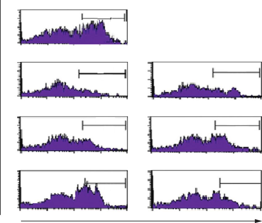
4                                                                        Evidence-Based Complementary and Alternative Medicine

    CTX             Normal   –      +      +     +       +       –                                     20
    CFEF µg/gBW              –      –      30    60      120     120                                   18                      ∗
                                                                                                                                        ∗∗
                                                                                                       16
                                                                                                       14

                                                                             Colon length (cm)
                                                                                                       12
                                                                                                       10
                                                                                                        8
                                                                                                        6
                                                                                                        4
                                                                                                        2
                                                                                                        0
                                                                       CTX                                  Normal   –   +         +         +    +     –
                                                                       CFEF µg/gBW                                   –   –         30        60   120   +
                                  (a)                                                                                    (b)
          Normal
                                                                                                       70                               ∗∗

                                                                                                       60

                                                                             IL-6 expression (cells)
                                                                                                       50

                                                                                                       40
          CTX (–)                CTX (+)         CTX + CFEF 30
                                                                                                       30

                                                                                                       20

                                                                                                       10

       CTX + CFEF 60         CTX + CFEF 120           CFEF 120                                          0
                                                                       CTX                                  Normal   –   +         +         +    +     –
                                                                       CFEF µg/gBW                                   –   –         30        60   120   120

                                  (c)                                                                                    (d)

Figure 1: Coelomic fluid of Eisenia fetida (CFEF) ameliorated cetuximab AOM/DSS-induced colitis associated colon cancer. (a) We first
analyzed severity of colitis to evaluate early impacts of CFEF. Colon length ratio in control and DSS/AOM-induced colitis associated colon
cancer. (b) Mice were significantly protected against DSS/AOM-induced colitis associated colon cancer compared with control mice
(P < 0.05). (c) Immunohistochemical staining of IL-6 antibody of colon tissue. (d) The number of IL-6 expression of the colons was
significantly lower in CFEF-cetuximab-treated mice than control mice after AOM/DSS administration (P < 0.05). Results shown are
mean + SD, with n � 6 replicates in each group. ∗ P < 0.05, ∗∗ P < 0.001.

AOM-induced colitis associated colon cancer compared                    3.2. The Activity of the Enzyme Myeloperoxidase (MPO) Was
with control mice (P < 0.05) (Figure 1(b)). The expression of           Used to Evaluate Infiltration of Neutrophils. MPO levels were
IL-6 showed that the AOM/DSS-treated mice with CFEF                     measured from lysed colon cells (Figure 2(a)). The ex-
administration had significantly less inflammation com-                   pression of cell proliferation was inhibited by CFEF-
pared to the AOM/DSS-treated mice (Figure 1(c)). CFEF-                  cetuximab in colon tissue. The expressions of proliferation
cetuximab-treated mice showed significant protection                     were analyzed using flow cytometry. Representative result
against 3% DSS/AOM-induced colitis associated colon                     from each group inhibited proliferation in CFEF-cetuximab
cancer (P < 0.05) (Figure 1(d)). AOM/DSS-induced mice                   combination groups (Figure 2(b)). The percentage of BrdU
were significantly induced colon inflammation compared                    positive cells indicated cell proliferation. The combination
with control mice (P < 0.05). Data showed that the AOM/                 CFEF-cetuximab therapy groups have lower cell prolifera-
DSS-treated mice with CFEF administration had signifi-                   tion than cetuximab single therapy group (Figure 2(c)).
cantly less inflammation compared to the AOM/DSS-treated                 Results shown were mean ± SD with n � 6 replicated in each
mice.                                                                   group by ∗ P < 0.05, ∗∗ P < 0.001.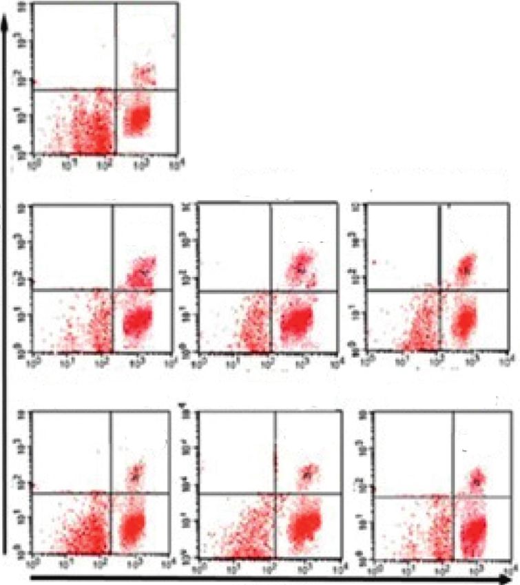
Evidence-Based Complementary and Alternative Medicine 5
14000 ∗∗
12000
MPO level (ng/mL)
10000
8000
6000
4000
2000
0
CTX Normal – + + + + –
CFEF µg/gBW – – 30 60 120 +
(a)
CTX (–) CTX (+) CTX + CFEF 30 mg/mL
20 20 20
80.5
15 15 72.5 15 48.6
M1
M1 M1
10 10 10
5 5 5
0 0 0
100 101 102 103 104 100 101 102 103 104 100 101 102 103 104
Count
CTX + CFEF 60mg/mL CTX + CFEF 120mg/mL CFEF 120 mg/mL
20 20 20
36.2
15 32.3 15 22.4 15
M1
M1 M1
10 10 10
5 5 5
0 0 0
0 1 2 3 4 0 1 2 3 4
10 10 10 10 10 10 10 10 10 10 100 101 102 103 104
BrDU
(b)
100 ∗∗
80
Proliferation (%)
60
40
20
0
CTX – + + + + –
CFEF µg/gBW – – 30 60 120 +
(c)
Figure 2: CFEF-cetuximab decreased MPO levels and proliferation in CECs cell. (a) MPO levels were measured from lysed colon cells. (b)
Representative of CECs proliferation after therapies with cetuximab and various concentrations of CFEF (30, 60, and 120 mg/g BW) in each
histogram were shown inside the panels. Percentages of CECs proliferation in each histogram are shown. (c) The percentage of CECs
proliferation was showed by BrdU positive cells and analyzed using flow cytometry (FACS Calibur, Becton Dickinson). Results shown are
mean + SD, with n � 6 replicates in each group. ∗ P < 0.05, ∗∗ P < 0.001.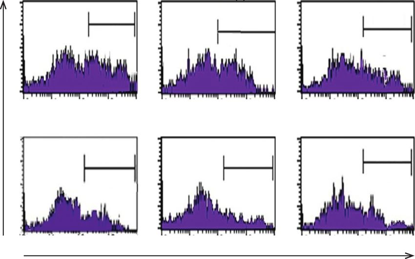
6                                                              Evidence-Based Complementary and Alternative Medicine

3.3. CFEF Plays a Major Role in Reducing Activated K-Ras          To explore whether CFEF could regulate the improve-
Mediated Inflammation during Tumor genesis. To analyze         ment of tumor suppressor in CECs, the RUNX3 protein
the oncogene expression is involved in modulating re-         expression from DSS/AOM-induced mice was characterized
sponses to CFEF effects of inflammation. K-Ras expression       by flow cytometry. The percentage of RUNX3 between
was determined by using immune-fluorescence. As shown in       control and treatment groups was significantly increased,
Figure 3, the combination of cetuximab and CFEF exert an      P < 0.05, and for CFEF, P < 0.05 (Figure 5). In this study,
ameliorate anticancer activity on AOM/DSS-induced mice.       there was a significant increase in the percentage of RUNX3.
The representative images shown in Figure 3 demonstrated a    There is a clear direct relationship, indicating that an increase
clear decreased K-Ras expression after treatment with CFEF    in the dosage of CFEF increased the percentage of protein.
alone or in combinations with CFEF and cetuximab. When
the combination of CFEF and cetuximab was applied, the
percentage of FAK expression was lower than cetuximab or      3.7. RUNX3 is a Regulator of MMP-2 and MMP-9 for
CFEF alone (P < 0.05). Treatment with cetuximab only          Invasiveness. To determine how RUNX3 inhibits cell mi-
slightly reduced the expression of K-Ras (P < 0.05). These    gration and invasion, we focused on clarifying the rela-
data indicated that K-Ras expression was decreased via        tionship between RUNX3 and MMPs, which have been
CFEF and cetuximab combinations. CFEF at three doses          reported to participate in tumor progression. We found that
significantly inhibited K-Ras expression compared to the       RUNX3 overexpression significantly inhibited the expres-
control (Figure 3). These results indicate that CFEF can      sion of MMP-2 and MMP-9 in CECs (Figures 6(a) and 6(b)).
inhibit protein oncogene that was found to be particularly    As shown in Figure 6, the percentages of MMP-2 and MMP-
important in tumor progression.                               9 were increased. Hence, we hypothesized that MMP-2 and
                                                              MMP-9 were affected by the invasive effects. As shown in
                                                              Figure 6, the cells treated with CFEF-cetuximab showed
3.4. Combination of Cetuximab and CFEF Restored Mem-          significantly lower invasiveness. Together, these data dem-
brane Integrity by Inhibition of Vimentin Expression. To      onstrated that RUNX3 inhibits metastasis through MMP-2
explore that CFEF have role in EMT process of cancer cell     and MMP-9. To investigate that RUNX3 has dependent
migration, invasion, and metastasis via vimentin, the colon   mechanism of MMP2 and MMP9, we showed that RUNX2
tissue was observed by using vimentin staining performed by   regulates MMP2 and MMP9 expressions. We proved that
immune-fluorescence in colon tissues. In DSS-AOM only          percentage of both (RUNX3+ MMP-2+) and (RUNX3+
group, the expression of vimentin decreased compare with      MMP-9+) cells significantly diminished in CECs on com-
that of combination-treated group. Treatment with cetux-      bination therapy groups (7(a)–7(d)). Together, these data
imab only slightly reduced the expression of vimentin         demonstrated that RUNX3 inhibits metastasis through
(P < 0.05). These data indicated that vimentin expression     MMP-2 and MMP-9.
was decreased via CFEF and cetuximab combinations. CFEF
at three doses significantly inhibited vimentin expression     4. Discussion
compared to the control (Figure 4).
                                                              Colorectal cancer is a malignant tumor that is the leading
                                                              cause of death worldwide. These malignant tumors originate
3.5. Combination of Cetuximab and CFEF Induced by             from the colon or rectum epithelium that penetrates the
β-Catenin and E-Cadherin Expression. We further examined      mucosal muscular layer. In colorectal cancer due to ulcer-
the effects of CFEF with or without combinations to the        ative colitis, DNA damage occurs that leads to increased cell
expression of β-catenin and E-cadherin. As shown in           proliferation, inhibition of tumor suppressors, and inhibi-
Figures 5(a)–5(d), CFEF enhanced the protein expression of    tion of apoptosis [7–10]. AOM can cause mutagenesis, so it
E-cadherin. Treatment of combination of cetuximab and         will affect intracellular pathways such as K-Ras [10–12].
CFEF significantly increased the protein expression of             Chronic inflammation triggers cellular events that can
β-catenin and E-cadherin. This study proved clear decreased   promote malignant transformation of cells and carcino-
β-catenin and E-cadherin expressions after treatment with     genesis. Epithelial mesenchymal transition (EMT) in human
CFEF alone or in combination with CFEF and cetuximab.         intestinal tumor progression is associated with the upre-
When the combination of CFEF and cetuximab was applied,       gulation of the intermediate filament protein vimentin. Since
both expressions of β-catenin and E-cadherin were higher      vimentin is integral for the structural integrity of the cell
than cetuximab or CFEF alone (P < 0.05).                      [24, 25, 27] and adhesion signaling [8, 25], downregulating
                                                              vimentin expression is sufficient to alter cell morphology
                                                              [27, 28] as well as inhibiting cell motility and invasion [28].
3.6. Combination of Cetuximab and CFEF Increases Per-         This finding indicated that in colitis associated colon cancer
centage of RUNX3 in DSS/AOM-Induced Mice. RUNX3 is            undergoing EMT. There is growing evidence here that
important proteins involved in cell cycle regulation [26].    E-cadherin plays a crucial role in human cancer invasion and
Therefore, we wanted to determine if CFEF-cetuximab           metastasis [24, 25, 27, 28]. The stimulation of E-cadherin
treatments influenced the abundance of RUNX3 in cancer         while simultaneously attenuating expression of vimentin
cells. To do this, we analyzed cells that were expressed by   and MMP, suggests blocking of EMT and inhibiting the
RUNX3 by using flow cytometry.                                 viability and motility.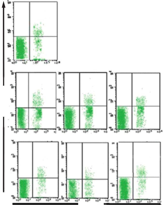
Evidence-Based Complementary and Alternative Medicine                                                                                                7

                                                                                   CTX +                 CTX +            CTX +
                  Normal          CTX (–)                           CTX (+)                                                         CFEF 120
                                                                                   CFEF 30               CFEF 60         CFEF 120
        MERGE
        DAPI
        K-RAS

                                                                                  (a)
                                                            30000                               ∗

                                                            25000
                                         K-Ras expression

                                                            20000

                                                            15000

                                                            10000

                                                             5000

                                                                0
                                   CTX                              Normal    –    +        +       +       +      –
                                   CFEF mg/gBW                                –    –       30       60     120     120
                                                                                  (b)

Figure 3: K-Ras expressions were reduced by CFEF-cetuximab in colon tissue. The expression of K-Ras after therapies with cetuximab and
various concentrations of CFEF (30, 60, and 120 mg/g BW) were analyzed using immunofluorescence. The expression of K-Ras reduced in
colon tissue by the decreasing of fluorescence intensity. (a) Representative images of K-Ras expression showed slightly lower on the
combination therapy group. (b) The percentage of K-Ras expression decreased on combination therapy groups compared with cetuximab
single group. Results shown are mean ± SD, with n � 6 replicates in each group. ∗ P < 0.05, ∗∗ P < 0.001.

    One of the targeted therapies for colorectal cancer is                              EGFR to induce cell apoptosis [25]. A previous in vitro and in
cetuximab which works by inhibiting the growth of cancer                                vivo study has shown that earthworm fibrinolytic enzyme
cells by targeting EGFR, so that there is inhibition in the                             component A from Eisenia fetida could effectively inhibit the
MAPK pathway and decreased K-Ras expression. In this                                    proliferation of MCF-7 cells. Another study showed that
study, cetuximab was combined with coelomic fluid from                                   earthworm fibrinolytic enzyme has significant antitumor
Eisenia fetida. Coelomic fluid has been known to be used as a                            activity by inhibiting the expression of matrix metal-
therapy for several diseases because it has anti-inflammatory,                           loproteinase-2 (MMP-2) [30]. MMP-2 (gelatinase A) and
antioxidant, antibacterial, and antitumor effects [7]. Biological                        MMP-9 (gelatinase B) are both cancer associated. Gelatinase
substances contained coelomic fluid including lectin [8] and                             cleave many targets that regulate key signaling in cell growth,
earthworm fibrinolytic enzyme also known as lumbrokinase                                 migration, invasion, inflammation, and angiogenesis [31]. So
[26, 29, 30]. Lectins have been proven to be used as anticancer                         it is possible that there might be a change in MMP expression
in the MCF-7 cell line via the MAPK pathway by targeting                                via activation of RUNX3 signaling [30]. Downregulation of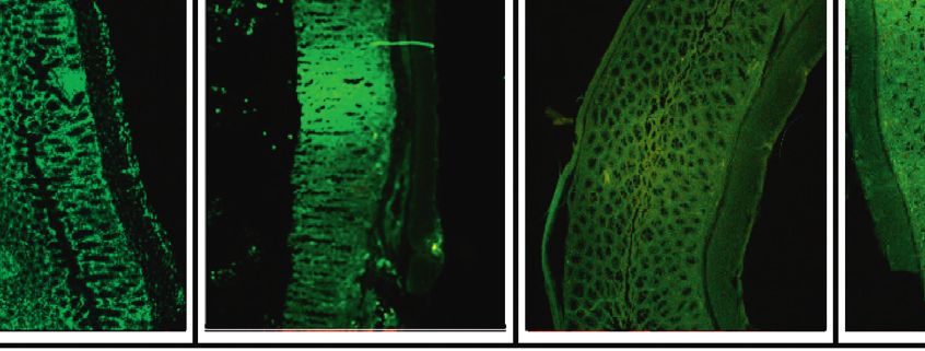
8                                                                                             Evidence-Based Complementary and Alternative Medicine

                                                                                           CTX +            CTX +            CTX +
                    Normal        CTX (–)                            CTX (+)                                                             CFEF 120
                                                                                           CFEF 30          CFEF 60         CFEF 120
         MERGE
         DAPI
         VIMENTIN

                                                                                       (a)
                                                                                   ∗                       ∗∗
                                                            20000

                                                            15000
                                      Vimentin expression

                                                            10000

                                                             5000

                                                                0
                                    CTX                             Normal     –       +        +      +        +     –
                                   CFEF mg/gBW                                 –       –        30    60        120   120
                                                                                       (b)

Figure 4: Vimentin expressions were reduced by CFEF-cetuximab in colon tissue. The expression of vimentin after therapies with
cetuximab and various concentrations of CFEF (30, 60, and 120 mg/g BW) were analyzed using immunofluorescence. The expression of
vimentin reduced in colon tissue by the decreasing of fluorescence intensity. (a) Representative images of vimentin expression showed
slightly lower on the combination therapy group. (b) The percentage of vimentin expression decreased on combination therapy groups
compared with cetuximab single group. Results shown are mean ± SD, with n � 6 replicates in each group. ∗ P < 0.05, ∗∗ P < 0.001.

β-catenin expression can cause a decrease in vimentin ex-                                    the things that is approved for cancer metastases is impaired
pression, and this is also in accordance with our results                                    activation of EMT. This shows that targeting EMT can be a
(Figures 2 and 5). Thus, the combination of cetuximab and                                    promising therapeutic strategy for patients with colorectal
CFEF can work synergistically.                                                               cancer [20]. This is also consistent with the results shown by
    K-Ras is also an effector molecule responsible for signal                                 increasing of vimentin expression in DSS/AOM-induced
transduction from EGFR. EGFR plays an important role in                                      mice. The improving of vimentin can occur through path-
controlling the proliferation of cancer cells and also me-                                   ways involving β-catenin and TGF-β. β-Catenin increases
tastasis [19]. An increase in K-Ras will increase proliferation                              EMT through activation of the vimentin signal pathway
and metastasis. This is consistent with the results that K-Ras                               [25–28, 32]. Epithelial mesenchymal transformation during
increased significantly in DSS/AOM-induced mice. One of                                       development and oncogenesis is an essential biological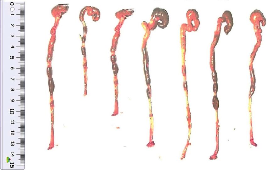
Evidence-Based Complementary and Alternative Medicine                                                                                      9

                                                                                   CTX +             CTX +            CTX +
                    Normal   CTX (–)                                CTX (+)                                                     CFEF 120
                                                                                   CFEF 30           CFEF 60         CFEF 120
       MERGE
       DAPI
       E-Cadherin

                                                                                  (a)
                                                            16000                        ∗∗
                                                            14000
                                    E-Cadherin expression

                                                            12000
                                                            10000
                                                             8000
                                                             6000
                                                             4000
                                                             2000
                                                                0
                              CTX                                   Normal    –    +     +      +       +      –
                              CFEF mg/gBW                                     –    –    30      60     120     120
                                                                                  (b)
                                                                         Figure 5: Continued.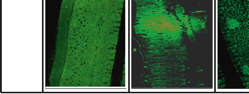
10                                                                                           Evidence-Based Complementary and Alternative Medicine

                                                                                       CTX +              CTX +            CTX +
                    Normal        CTX (–)                               CTX (+)                                                       CFEF 120
                                                                                       CFEF 30            CFEF 60         CFEF 120
        MERGE
        DAPI
        β-Catenin

                                                                                      (c)
                                                                16000                               ∗∗
                                                                14000
                                                                12000
                                         β-Catenin expression

                                                                10000
                                                                8000
                                                                6000
                                                                4000
                                                                2000
                                                                   0
                                   CTX                                  Normal    –    +        +     +      +      –
                                   CFEF mg/gBW                                    –    –       30    60     120     120
                                                                                      (d)

Figure 5: E-Cadherin and β-catenin expressions were reduced by CFEF-cetuximab in colon tissue. The expression of E-cadherin and
β-catenin after therapies with cetuximab and various concentrations of CFEF (30, 60, and 120 mg/g BW) were analyzed using immu-
nofluorescence. (a) Representative images of E-cadherin expression showed slightly higher on the combination therapy group. (b) The
percentage of E-cadherin expression increased on the combination therapy groups compared with cetuximab single group. (c) Repre-
sentative images of β-catenin expression showed higher on the combination therapy group. (d) The percentage of β-catenin expression
increased on combination therapy groups compared with cetuximab single group. Results shown are mean ± SD, with n � 6 replicates in
each group. ∗ P < 0.05, ∗∗ P < 0.001.

process. Downregulating E-cadherin and raising the mes-                                     In this study, we found that overexpression of RUNX3
enchymal marker vimentin are main transitional factors                                      significantly inhibited motility and invasiveness in CAC
[24, 25, 27].                                                                               cells.
    RUNX3 has been shown to inhibit EMT, which en-                                              Our finding indicated that overexpression of RUNX3
courages metastasis; by regulating these signaling pathways,                                could prevent EMT [27, 28, 32]. We examined EMT markers
RUNX3 plays a critical role in the regulation of tumor cell                                 at the protein levels in the RUNX3-overexpressing cells.
migration, invasion, and proliferation from epithelial to                                   Cancers provide evidence that expression of RUNX3 in
mesenchymal [25, 28]. RUNX3 is ideally inactivated in lung                                  metastatic tissue has decreased significantly. This indicates
adenocarcinoma induced by K-Ras, suggesting its potential                                   that RUNX3 plays an important role in tumorigenesis and
role as a tumor suppressor in lung adenocarcinoma [29, 32].                                 progression. The expression of protein RUNX3 was found to
Evidence-Based Complementary and Alternative Medicine 11
Normal
20
21
15
Counts
10
5
0
100 101 102 103 104
CTX (–) CTX (+)
20 20
15 54 15 43
Counts
Counts
10 M1 10 M1
5 5
0 0
100 101 102 103 104 100 101 102 103 104
Count
CTX + CFEF 30 mg/gBW CTX + CFEF 60 mg/gBW
20 20
33 27
15 15
M1 M1
Counts
Counts
10 10
5 5
0 0
100 101 102 103 104 100 101 102 103 104
CTX + CFEF 120 mg/gBW CFEF 120mg/gBW
20 20
15 19 15 26
Counts
Counts
10 M1 10 M1
5 5
0 0
100 101 102 103 104 100 101 102 103 104
RUNX3
(a)
70 ∗
∗∗
60
50
RUNX3 (%)
40
30
20
10
0
CTX Normal – + + + + –
CFEF mg/gBW – – 30 60 120 120
(b)
Figure 6: The percentage of RUNX3 was enhanced by CFEF-cetuximab in CECs cells. (a) Representative percentage of RUNX3 after
therapies with cetuximab and various concentrations of CFEF (30, 60, and 120 mg/g BW) were analyzed using flow cytometry (FACS
Calibur, Becton Dickinson) in each histogram as shown. Cells were gated from double positive cells (K-Ras+ and vimentin+ populations) and
those populations that express RUNX3 were analyzed. (b) The percentage of RUNX3+ cell enhanced on combination therapy groups
compared with cetuximab single group. Results shown are mean ± SD, with n � 6 replicates in each group. ∗ P < 0.05, ∗∗ P < 0.001.
be substantially associated with decreased survival of CRC invasiveness. Finally, functional experiments revealed that
patients [25, 27, 28, 32]. RUNX3 overexpression is associated restoration of RUNX3 in CAC cells suppressed cell invasion
with decreased breast cancer cell invasiveness [26–29, 32]. The and migration and regulated EMT-vimentin by CFEF.
overexpressing RUNX3 cells had reduced invasive potential of RUNX3 overexpression is associated with decreased
the cells. RUNX3 is important in regulation of motility and breast cancer cell invasiveness [26–29, 32]. The12 Evidence-Based Complementary and Alternative Medicine
Normal 40 ∗
∗∗
15.1 30
MMP-2 (%)
20
10
CTX (–) CTX (+) CTX + CFEF 30mg/gBW
0
32.8 26.6 23.9 CTX Normal – + + + + –
MMP-2 (%)
CFEF µg/gBW – – 30 60 120 120
CTX + CFEF 60 mg/gBW CTX + CFEF 120mg/gBW CFEF 120mg/gBW
18.6 13.4 19.2
RUNX3
(a) (b)
Normal 40 ∗
∗∗
12.9
30
MMP-9 (%)
20
CTX (–) CTX (+) CTX + CFEF 30 mg/gBW 10
28.9 23.4 18.7
0
MMP-9 (%)
CTX Normal – + + + + –
CFEF µg/gBW – – 30 60 120 120
CTX + CFEF 60 mg/gBW CTX + CFEF 120mg/gBW CFEF 120mg/gBW
13.5 12.3 11.9
RUNX3
(c) (d)
Figure 7: CFEF-cetuximab combination therapies were dependent RUNX3 to reduce MMP-2 and MMP-9 percentage in CECs cell. (a)
Representative percentage of MMP2 after therapies with cetuximab and various concentrations of CFEF (30, 60, and 120 mg/g BW) in each
histogram was shown and analyzed using flow cytometry (FACS Calibur, Becton Dickinson). Cells were gated from double positive cells (K-Ras+
and vimentin+ populations) and those populations that express RUNX3 were analyzed. Percentage of RUNX3+ MMP-2+ cell in each quadrant is
shown inside the panels. (b) The percentage of RUNX3+ MMP-2+ cell reduced in CECs on combination therapy groups compared with
cetuximab single group. (c) Representative percentage of RUNX3+ MMP-9+ cell reduced in CECs after combination therapy. Percentage of
RUNX3+ MMP-9+ cell in each quadrant is shown inside the panels. (d) The percentage of RUNX3+ MMP-9+ cell decreased on combination
therapy groups compared with cetuximab single group. Results shown are mean ± SD, with n � 6 replicates in each group. ∗ P < 0.05, ∗∗ P < 0.001.Evidence-Based Complementary and Alternative Medicine 13
overexpressing RUNX3 cells had reduced invasive potential of expression by immune-fluorescence. EN and AI made
the cells. RUNX3 is important in the regulation of motility histopathology of colon and analyzed of histopathology data.
and invasiveness. Finally, functional experiments revealed EDAM isolated protein and purify of coelomic fluid and
that restoration of RUNX3 in CAC cells suppressed cell in- FOBT. SP, ATE, RY, EDAM, and AI prepared, edited, and
vasion and migration and regulated EMT-vimentin by CFEF. reviewed the manuscript. All the authors read and approved
The present findings indicated that CFEF may serve as a the final manuscript. All the authors contributed to the final
novel agent for colon cancer treatment. The in vivo effects of approval of the version to be published.
anticancer as well as the potential therapeutic effectiveness of
CFEF are worth further exploring. The earthworm crude Acknowledgments
extract is shown to have the ability to kill cancer cells directly
in vitro [8, 9, 33] and to prevent the incidence and growth of The authors are grateful to Heni Endarwati, S.Si, Ami
tumor in vivo [10, 33, 34]. In addition, earthworm proteases Maghfironi, S.Si, and Suci Megasari, S.Si., M.P., for the
have been shown to improve the therapy effects by both technical assistance during this research.
radiations therapy and chemotherapy.
Recently, a glycosylated component is separated from References
the earthworm E. fetida by Liu et al. [30], which has relations
with apoptosis of tumor cells. It is identified to be a plasmin [1] X.-L. Li, J. Zhou, Z. R. Chen et al., “p53mutations in colorectal
and also a plasminogen activator. The earthworm protease cancer- molecular pathogenesis and pharmacological reac-
possesses obvious antitumor activity. It has been found that tivation,” World Journal of Gastroenterology, vol. 21, no. 1,
the earthworm protease can induce apoptosis of hepatoma pp. 84–93, 2015.
cells and downregulate the expression of matrix of matrix [2] A. T. Endharti, A. Wulandari, A. Listyana et al., “Dendrophtoe
pentandra (L.) Miq extract effectively inhibits inflammation,
metalloproteinase.
proliferation and induce p53 expression on colitis-associated
colon cancer,” BMC Complementary and Alternative Medi-
5. Conclusion cine, vol. 16, no. 374, 2016.
[3] M. Porru, L. Pompili, C. Caruso, A. Biroccio, and C. Leonetti,
In conclusion, this study showed that the combination of the “Targeting K-RAS in metastatic colorectal cancer: current
CFEF and cetuximab is able to inhibit proliferation by re- strategies and emerging opportunities,” Journal of Experi-
ducing K-Ras and vimentin expression in colorectal cancer mental & Clinical Cancer Research, vol. 37, no. 1, 57 pages,
in vivo. This finding maybe offers a promising method for a 2018.
new anticancer therapy concept, especially for colorectal [4] S. J. Cho and S. Kakar, “Tumor Budding in Colorectal Car-
cancer treatment. cinoma, translating a morphologic score into clinically
meaningful result,” Archives of Pathology & Laboratory
Abbreviation Medicine, vol. 142, no. 8, pp. 952–957, 2018.
[5] M. A. Nieto, “Epithelial plasticity: a common theme in em-
AOM: Azoxymethane bryonic and cancer cells,” Science, vol. 342, no. 6159, 2013.
CAC: Colitis associated colon cancer [6] C. G. Leichman, S. L. McDonough, S. R. Smalley et al.,
“Cetuximab combined with induction Oxaliplatin and
CFEF: Coelomic fluid Eisenia fetida
Capecitabine, followed by neoadjuvant chemoradiation for
EGFR: Epidermal growth factor receptor locally advanced rectal cancer: SWOG 0713,” Clinical Colo-
DSS: Dextran sulfate sodium rectal Cancer, vol. 17, no. 1, pp. e121–e125, 2017.
EMT: Epithelial mesenchymal transition [7] A. T. Endharti, A. D. Baskoro, and E. Norahmawati, “Ther-
CECs: Intestinal epithelial cells apeutic effect of soluble worm protein acting as immune
K-Ras: Kirsten-rat sarcoma viral oncogene homolog regulatory on colitis,” Asian Pacific Journal of Tropical Bio-
MAPK: Mitogen-activated protein kinase medicine, vol. 7, no. 1, pp. 70–77, 2017.
RUNX3: Runt-Related Transcription Factor-3. [8] A. T. Endharti, Y. Purnamasari, R. Primasari, S. Poeranto, and
S. Permana, “Coelomic fluid of Lumbricus rubellus syner-
Data Availability gistically enhance cytotoxic effect of fluorouracil through
modulation of focal adhesion kinase and p21 in HT-29 cancer
The data used to support the findings of this study are in- cell line,” The Scientific World Journal, vol. 2019, Article ID
cluded within the article. 5632859, 9 pages, 2019.
[9] S. Permana, Pearlindah, Z. Sholihah, A. Iskandar, H. Susanti,
and A. Tri Endharti, “Cytotoxic effects and anti-proliferative
Conflicts of Interest cancer activity of coelomic fluid from Lumbricus rubellus
promotes apoptosis and reduces G2/M phase progression in
The authors declare that there are no conflicts of interest. HT-29 cells,” Journal of Applied Pharmaceutical Science,
vol. 8, no. 11, pp. 28–34, 2018.
Authors’ Contributions [10] S. Permana, R. P. Hadi, E. Norahmawati, and A. Tri Endharti,
“Coelomic fluid of Lumbricus rubellus enhances anti-pro-
SP performed conception and design of experiments, also lioniferative effect of 5-fluorouracil by modulating focal ad-
wrote the manuscript, and purified the protein. ATE ana- hesion kinase express and IL-1β of colorectal cancer in mice,”
lyzed and interpreted flow cytometry; RPF collected coe- Journal of Applied Pharmaceutical Science, vol. 9, no. 8,
lomic fluid, maintained mice, and performed protein pp. 41–46, 2019.14 Evidence-Based Complementary and Alternative Medicine
[11] J. Zeng, Z. H. Tang, S. Liu, and S. Guo, “Clinicopathological Alternative Medicine, vol. 2020, Article ID 5978131, 7 pages,
significance of overexpression of interleukin-6 in colorectal 2020.
cancer,” World Journal of Gastroenterology, vol. 23, no. 10, [27] C. Y. Liu, H. H. Lin, M. J. Tang, and Y. Wang, “Vimentin
pp. 1780–1786, 2017. contributes to epithelial-mesenchymal transition cancer cell
[12] C. Hong, S. Takahashi, M. Imamura et al., “Earthworm fi- mechanics by mediating cytoskeletal organization and focal
brinolytic enzyme: anti-tumor activity on human hepatoma adhesion maturation,” Oncotarget, vol. 6, no. 18,
cells in vitro and in vivo,” Chinese Medical Journal, vol. 120, pp. 15966–15983, 2015.
no. 10, pp. 898–904, 2007. [28] S. Wu, Y. Du, J. Beckford, and H. Alachkar, “Upregulation of
[13] D. Augustine, R. S. Rao, J. Anbu, and K. N. Chidambara the EMT marker vimentin is associated with poor clinical
Murthy, “In vitro antiproliferative effect of earthworm coe- outcome in acute myeloid leukemia,” Journal of Translational
lomic fluid of Eudrilus Eugine, Eisenia foetida, and Perionyx Medicine, vol. 16, no. 1, p. 170, 2018.
excavatus on squamous cell carcinoma-9 cell line: a pilot [29] S. J. Chen, Y. T. Chen, L. J. Zeng et al., “Bmi1 combines with
study,” Pharmacognosy Research, vol. 9, no. 5, 2017. oncogenic KRAS to induce malignant transformation of
[14] Z. Deng, S. Gao, X. Xiao et al., “The effect of earthworm human pancreatic duct cells in vitro,” Tumor Biology, vol. 37,
extract on mice S180 tumor growth and apoptosis,” Bio- no. 8, pp. 11299–11309, 2016.
medicine & Pharmacotheraphy, vol. 115, 2019. [30] C. M. Liu, X. T. Chen, Y. Y. Pan et al., “Antitumor studies of
[15] S. R. Hamilton, F. T. Bosman, P. Boffetta et al., “Carcinoma of earthworm fibrinolytic enzyme component A from Eisenia
the colon and rectum,” in WHO Classification of Tumours of foetida on breast cancer cell line MCF-7,” Indian Journal of
the Digestive System, F. T. Bosman, F. Carneiro, R. H. Hruban, Pharmaceutical Sciences, vol. 79, no. 3, pp. 361–368, 2017.
and N. D. Theise, Eds., pp. 134–146, IARC, Lyon, France, 4th [31] B. Bauvois, “New facets of matrix metalloproteinases MMP-2
edition, 2010. and MMP-9 as cell surface transducers: outside-in signaling
[16] J. Chen and X.-F. Huang, “The signal pathways in azoxy- and relationship to tumor progression,” Biochimica et Bio-
methane-induced colon cancer and preventive implications,” physica Acta (BBA)-Reviews on Cancer, vol. 1825, no. 1,
Cancer Biology & Therapy, vol. 8, no. 14, pp. 1313–1317, 2009. pp. 29–36, 2012.
[17] B. Parang, C. W. Barrett, and C. S. Williams, “AOM/DSS [32] Z. Wang, A. Divanyan, F. L. Jourd’heuil et al., “Vimentin
model of colitis-associated cancer,” Methods in Molecular expression is required for the development of EMT-related
Biology, vol. 1422, pp. 297–307, 2016. renal fibrosis following unilateral ureteral obstruction in
[18] J. M. Williams, C. A. Duckworth, K. Vowell, M. D. Burkitt, mice,” American Journal of Physiology-Renal Physiology,
and D. Mark Pritchard, “Intestinal preparation techniques for vol. 315, no. 4, pp. F769–F780, 2018.
histological analysis in the mouse,” Current Protocols in [33] A. T. Endharti and S. Permana, “Extract from mango mis-
Mouse Biology, vol. 6, no. 2, pp. 148–168, 2016. tletoes Dendrophthoe pentandra ameliorates TNBS induced
[19] K. Knickelbein and L. Zhang, “Mutant KRAS as a critical colitis by regulating CD4+ T cells in mesenteric lymph nodes,”
determinant of the therapeutic response of colorectal cancer,” BMC Complementary and Alternative Medicine, vol. 17, no. 1,
Genes & Diseases, vol. 2, no. 1, pp. 4–12, 2015. p. 468, 2017.
[20] S. Gurzu, C. Silveanu, A. Fetyko, V. Butiurca, Z. Kovacs, and [34] A. T. Endharti and S. Permana, “T-Bet is dependent on stat4
I. Jung, “Systematic review of the old and new concepts in the inhibiting acute colitis but not stat-1 using L4 somatic antigen
epithelial-mesenchymal transition of colorectal cancer,” of Heligmosomoides polygyrus,” The Scientific World Journal,
World Journal of Gastroenterology, vol. 22, no. 30, vol. 2018, Article ID 8571920, 9 pages, 2018.
pp. 6764–6775, 2016.
[21] L. Xu, W.-H. Cui, W.-C. Zhou et al., “Activation of Wnt/
β-catenin signalling is required for TGF-β/Smad2/3 signalling
during myofibroblast proliferation,” Journal of Cellular and
Molecular Medicine, vol. 21, no. 8, pp. 1545–1554, 2017.
[22] P. Engelmann, Y. Hayashi, K. Bodó et al., “Phenotypic and
functional characterization of earthworm coelomocyte sub-
sets: linking light scatter-based cell typing and imaging of the
sorted populations,” Developmental & Comparative Immu-
nology, vol. 65, pp. 41–52, 2016.
[23] L. Ouyang, Y. Chen, X. Y. Wang et al., “Polygonatum
odoratum lectin induces apoptosis and autophagy via tar-
geting EGFR-mediated Ras-Raf-MEK-ERK pathway in hu-
man MCF-7 breast cancer cells,” Phytomedicine, vol. 21,
no. 12, pp. 1658–1665, 2014.
[24] S. K. Bhutia, P. K. Panda, N. Sinha et al., “Plant lectins in
cancer therapeutics: targeting apoptosis and autophagy-de-
pendent cell death,” Pharmacological Research, vol. 144,
pp. 8–18, 2019.
[25] K. Mori, T. Uchida, T. Yoshie et al., “A mitochondrial ROS
pathway controls matrix metalloproteinase 9 levels and in-
vasive properties in RAS-activated cancer cells,” The FEBS
Journal, vol. 286, no. 3, pp. 459–478, 2019.
[26] J. Xue, X. Wu, and M. Qu, “RUNX3 Inhibits the Invasion and
Metastasis of Human Colon Cancer HT-29 Cells by Upre-
gulating MMP-2/9,” Evidence-Based Complementary andYou can also read