Phosphorylation of Pit-1 by cyclin-dependent kinase 5 at serine 126 is associated with cell proliferation and poor prognosis in prolactinomas
←
→
Page content transcription
If your browser does not render page correctly, please read the page content below
Open Chemistry 2021; 19: 785–793
Research Article
Weiyan Xie#, Qiuyue Fang#, Jing Guo, Lei Gong, Chuzhong Li, Yazhuo Zhang*
Phosphorylation of Pit-1 by cyclin-dependent
kinase 5 at serine 126 is associated with cell
proliferation and poor prognosis in
prolactinomas
https://doi.org/10.1515/chem-2021-0070 prolactinomas. Our data indicated that Ser126-Pit-1 is spe-
received November 22, 2020; accepted June 18, 2021 cifically phosphorylated by CDK5 and high-level pSer-126-
Abstract: Pit-1 (POU1F1) is a POU-homeodomain tran- Pit-1 can promote cell proliferation and PRL secretion. In
scription factor, and it is one of the most important addition, a higher level of pSer-126-Pit-1 correlates with a
tissue-specific transcription factors in pituitary develop- worse prognosis in patients with prolactinoma. Our results
ment. Cyclin-dependent kinase 5 (CDK5) is a protein show that CDK5 mediated Ser126-Pit-1 phosphorylation and
kinase that can phosphorylate many key transcription regulated prolactinoma progression and PRL secretion.
factors, but the mechanism under which CDK5 phosphory- Keywords: Pit-1, CDK5, prolactinoma, phosphorylation,
lates Pit-1 is unclear. To investigate whether CDK5 can proliferation
regulate cell proliferation and promote hormone secretion
through phosphorylation of Ser126-Pit-1 in prolactinomas,
we generated an antibody that specifically recognizes phos-
phorylated serine at position 126 of Pit-1 (Ser126-Pit-1). We 1 Background
used western blotting to detect the level of Pit-1 phosphory-
lation and observed the proliferation and apoptosis of GH3 Pituitary adenomas (PAs) constitute approximately 15%
cells with different levels of Pit-1 phosphorylation by clone of all intracranial neoplasms. Prolactinoma is the most
formation experiments, cell viability assays, and flow cyto- common subtype of hormone-secreting pituitary tumors,
metry. ELISA was used to measure the level of PRL in the accounting for approximately 45–50% of cases. Due to the
supernatant of GH3 cells. Tissue microarrays and immuno- dysfunctional production of hormones, patients with pro-
histochemistry were used to evaluate the expression of lactinomas often suffer from severe disorders affecting
the phosphorylation level of Ser126-Pit-1 (pSer126-Pit-1) in growth and development. However, the mechanism of the
biological behavior of some prolactinomas has not yet been
fully defined.
# Weiyan Xie and Qiuyue Fang contributed equally to this work. Pit-1 is a POU-homeodomain transcription factor,
and it was described in the pituitary gland. Pit-1 is
expressed exclusively in somatotrophs, lactotrophs, and
* Corresponding author: Yazhuo Zhang, Beijing Neurosurgical thyrotrophs, and it is necessary for the establishment and
Institute, Capital Medical University, No.119 South 4th Ring West maintenance of these differentiated cell types, as well as
Road, Fengtai District, Beijing, China; Department of Neurosurgery, for the proliferation of somatotrophs and lactotrophs [1].
Beijing Tiantan Hospital, Capital Medical University, Beijing, China;
It is also expressed in breast, pancreatic, and prostate
Beijing Institute for Brain Disorders Brain Tumor Center, China
National Clinical Research Center for Neurological Diseases, Key cancer, and its overexpression promotes tumor growth
Laboratory of Central Nervous System Injury Research, Beijing, and metastasis [2–4]. In the process of signal transduc-
China, e-mail: zyztxzz@126.com, tel: +86-010-5997-8000, tion regulation, the level of Pit-1 phosphorylation and the
fax: +86-010-5997-8000 duration of activity regulated by its phosphorylation state
Weiyan Xie, Qiuyue Fang, Jing Guo, Lei Gong, Chuzhong Li:
are very important. Studies have shown that Pit-1 phos-
Department of Cell Biology, Beijing Neurosurgical Institute, Capital
Medical University, No.119 South 4th Ring West Road, Fengtai
phorylation plays an important role in regulating the
District, Beijing, China expression of the target gene. The most important phos-
ORCID: Yazhuo Zhang 0000-0002-8583-2580 phorylation sites found on Pit-1 are serine 115 (S115) and
Open Access. © 2021 Weiyan Xie et al., published by De Gruyter. This work is licensed under the Creative Commons Attribution 4.0
International License.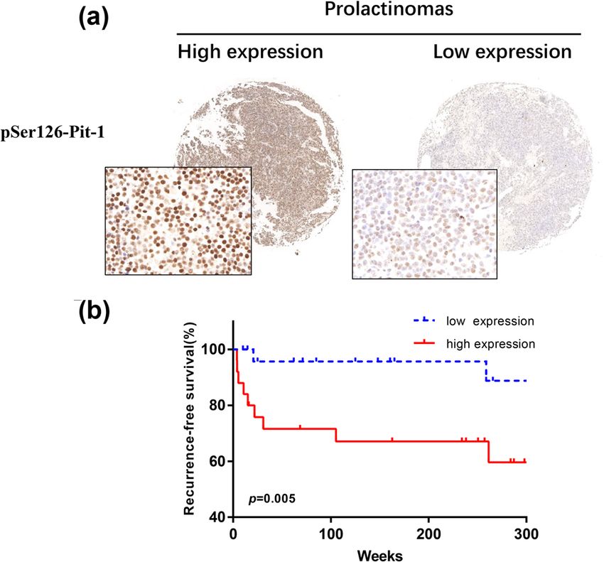
786 Weiyan Xie et al.
threonine 229 (T220). Augustijn and others have shown Table 1: Clinical and pathological characteristics of the patients
that PKA, PKC, and cell cycle-dependent kinases can
phosphorylate Pit-1 at the T220 site of the homologous Characteristics Patients
domain [5]. Phosphorylation or mutation of this site will Numbers 48
change its relationship with Ets-1 binding segment RIII Age (mean ± SD, years) 39.3 ± 10.7, 14–62
(AA190-257), which reduces the ability of Pit-1 to bind Sex (M/F) 22/26
to PRL and the TSH promoter, and it activates cAMP, Macroadenoma (%) 23 (47.9%)
Microadenoma (%) 4 (8.3%)
resulting in Pit-1 participation in the regulation of cell
Giant adenoma (%) 21 (43.8%)
proliferation, apoptosis, and tumorigenesis [6–8]. Mean follow-up, years (mean ± SD, range) 4.8 ± 1.17, 2.5–7
Cyclin-dependent kinase 5 (CDK5) is a vital member
of the serine/threonine kinase family; CDK5 activity is F, female; M, male.
highest in the central nervous system and participates
in a variety of neural system functional activities, including from the date of surgery to the date of tumor recurrence.
neuron migration, neuron apoptosis, survival, and synaptic Patients were collected at the date of the last neuroimaging
plasticity [9]. Its deregulation is directly involved in diverse follow-up. Normal human anterior pituitaries of people who
pathological events, such as enhanced neurodegenerative died of non-neurological or non-endocrine diseases were
and neuropsychiatric disorders and cancer [10]. CDK5 is not obtained from a donation program. The Ethics Committee
activated by cyclins but by its activators p35 and p39, whose of Beijing Tiantan Hospital study approved the protocol,
constitutive expression is largely restricted to cells of neural and all patients signed the informed consent forms. The
crest origin [11,12]. Our group found for the first time that project ethics approval Number is KY2019-136-01. The patient
CDK5 exists in normal human pituitary and PAs and found characteristics are summarized in Table 1.
that CDK5 can promote the growth and invasion of PAs, but
the true regulatory mechanism of CDK5 in pituitary tumors
is far from clear [13,14]. Specifically, it is unknown whether 2.2 Tumor samples and tissue microarray
CDK5 is involved in the regulation of hormone synthesis, construction
secretion, and apoptosis in prolactinoma, which requires
in-depth research. Formalin-fixed paraffin-embedded tissue blocks were sec-
In the present study, the role of CDK5 was further tioned and stained with hematoxylin and eosin (H&E).
studied regarding its regulation pSer126-Pit-1. We explored Three 2.0 mm diameter core biopsies were selected from
the function of pSer126-Pit-1 on the proliferation, hormone the paraffin-embedded tissue blocks and transferred to
secretion, and apoptosis of prolactinomas in vitro. Then, tissue microarrays (TMAs) using a Mini core Tissue Arrayer
we investigated the clinical significance of the expression (Mitogen, UK). Tissue microarrays were cut into 4 μm sec-
level of pSer126-Pit-1 using surgical specimens. tions using a serial microtome, and the samples were ran-
domly ordered and anonymized on the TMA slides. To
minimize loss of antigenicity, the microarray slides were
processed within 1 week of cutting.
2 Materials and methods
2.1 Patients 2.3 IHC techniques and antibodies
In the study, we retrospectively reviewed 48 patients who In advance of IHC, TMA slides were stained with H&E and
had undergone pituitary surgery at Beijing Tiantan Hospital evaluated for quality and tumor content. TMAs were pro-
between 2008 and 2012. All patients had plasma prolactin cessed in a Leica BOND-III (Leica Biosystems, Germany)
(PRL) levels >200 ng/mL and positive immunostaining for automated, random, and continuous-access slide staining
PRL. Medical therapy was interrupted at least 2 months system that simultaneously performed several IHC assays.
before surgery. Tumor size was determined by MRI, and A Bond Polymer Refine Detection System (Leica Biosystems,
tumors were classified as microadenomas (1 and 4 cm). The mean postoperative follow-up was 4.8 years each antibody, and TMAs were stained for each antibody
(range: 2.5–7 years). Recurrence-free survival was measured in the same run to avoid interassay variability. The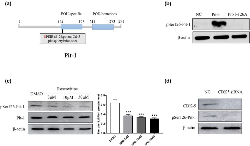
Phosphorylation of Pit-1 promotes cell proliferation 787
immunostained slides were examined for expression using to each well of the 96-well plate and cultured for 3 h in an
an Aperio AT2 digital scanner (Leica Biosystems, Germany). incubator. The optical density was measured at 450 nm,
Primary antibodies anti-pSer126-Pit-1 (4 μg/mL, Abmart) and a proliferation curve based on time and absorbance
were commercially developed using standard methods by was generated.
the injection of specific Pit-1-phosphothreonine peptide Ac-
VVL(pS) PSHGIE-amide into a rabbit at the Abmart anti-
body production facility, Shanghai, China. The optimal titer 2.7 Colony formation test
of primary antibodies had been determined in previous
experiments. The percentage of immunostaining and the The treated cell lines were seeded into six-well plates at
staining intensity (0, negative; 1+, weak; 2+, moderate; 1,000 cells per well and incubated for 2 weeks. After
and 3+, strong) was recorded, and an H-score was calcu- incubation, the cells were fixed in 4% paraformaldehyde
lated as follows: for 15 min and stained in 1 mL of a 0.1% crystal violet
H-score = (% cells 1 +) + 2(% cells 2 +) solution for 30 min. The culture plate was photographed.
Visible colonies in each well were quantified by ImageJ
+ 3(% cells 3 +).
software.
Based on the H-score, pSer126-Pit-1 staining in the
tissue sections was categorized as low (H-score of ≤168)
or high (H-score >168). 2.8 ELISA
PRL protein levels were determined using a rat PRL ELISA
kit from BioVision (K4688‐100) according to the manufac-
2.4 Cell culture
turer’s instructions. GH3 cells were harvested 72 h after treat-
ment with plasmid. The total protein content of the cells was
Rat pituitary cells (GH3) were obtained from the China
determined for standardization of PRL production with a BCA
Infrastructure of Cell Line Resources (Beijing, China) and
protein assay kit (Pierce Biotechnology). The culture superna-
cultured in 35 mm dishes, we use ATCC‐formulated F‐12K
tants were collected and normalized to the cell numbers.
medium (Invitrogen) containing 2.5% fetal bovine serum
(Gibco) and 15% horse serum (Gibco) in a 37°C incubator
with a humidified atmosphere of 95% air and 5% CO2. The
2.9 Cell apoptosis assay
culture medium was replaced every other day.
Cell apoptosis was determined using Annexin V-FITC/PI
kits (BD Biosciences, Franklin Lakes, NJ). Cells were
2.5 Plasmid construction and CDK5 inhibitor
seeded for 48 h after transiently transfected with the indi-
cated plasmids. Then, the cells were harvested and stained
A CDK5 siRNA (SR507441) and CDK5 expression plasmid
with annexin V-FITC and PI according to the instructions of
(NM_004935) constructs were purchased from OriGene
the manufacturer. Cells were analyzed using BD Accuri™C6
Technologies (Rockville, MD, USA). Mutant Pit-1 (GFP-
(BD Biosciences). Data analysis was performed using CFlow®
Ser126A-Pit-1) and Pit-1 were generated by GenScript Biotech
software (BD Biosciences).
(Nanjing, China). All constructs were confirmed by DNA
sequencing (Shanghai Shenggong Bio, China). Roscovitine
was obtained from Sigma-Aldrich (R7772; St. Louis, MO, USA).
2.10 Electrophoretic mobility shift
assay (EMSA)
2.6 Cell counting kit-8 (CCK-8) assay
We used a 5′-biotinylated oligonucleotide as a probe. The
Cells were seeded in 96-well plates at the density of 1 × probes were incubated with the recombinant protein at
104 cells per well in 100 μL of cell culture medium for 24 h room temperature for 30 min. The entire reaction mixture
and were then transiently transfected with the indicated was run on a nondenaturing 0.5 × TBE 6% polyacryl-
plasmids, and short interfering RNA cell viability was amide gel at 60 V for 1 h at 4°C, and then the mixture
measured using the CCK-8 assay kit (Dojindo, Japan). was transferred onto Biodyne® B nylon membranes (Pall
Following incubation, 10 μL of CCK-8 solution was added Corporation). Signals were visualized with reagents included788 Weiyan Xie et al.
in the kit and with a ChemiDoc XRS system (Bio-Rad anti-CDK5 (ab40773, 1/200) were obtained from Abcam
Laboratories, USA). (Cambridge, MA, USA). Anti-Pit-1 (sc-393943, 1/100) was
sourced from Santa Cruz Biotechnology (Dallas, TX, USA). The
optimal titer of primary antibodies had been determined in
previous experiments.
2.11 Luciferase reporter assay
GH3 cells were cultured at a density of 2 × 10⁴ cells per
well in 96-well culture plates. The cells were transfected 2.13 Statistical analysis
with 0.2 μg of dual-luciferase reporter construct p1, or
they were cotransfected with 0.2 μg of the luciferase All statistical analyses were performed using GraphPad
reporter construct p2 and the internal control vector Prism 7.00 statistical software. Experimental data are
pRL-TK, pRL-SV40, or pRL-CMV (Promega, Madison, reported as the mean ± SD (standard deviation) of at least
WI) at a ratio of 20:1 (reporter construct: control vector); three independent experiments, as indicated in the
transfections were performed using LipofectamineTM 2000 respective figure legends and methods. Statistical ana-
(Invitrogen, Carlsbad, CA) according to the manufacturer’s lysis was performed by one-way ANOVA or Student’s
instructions. Five hours posttransfection, the transfection t-test. A P-valuePhosphorylation of Pit-1 promotes cell proliferation 789
Figure 1: Both CDK5 inhibition and depletion affect the phosphorylation level of Ser126-Pit-1. (a) Bioinformatics analysis reveals that Ser126
Pit-1 was the only typical CDK5 potential phosphorylation site. (b) GH3 pituitary cells were transfected with WT-Pit-1 or Pit-1-126A. Western
blotting shows that the anti-pS126-Pit-1 antibody can specifically recognize the phosphorylation of Ser126-Pit-1. (c). Western blot (left) and
densitometry analysis (right) showed that S126-Pit-1 phosphorylation was reduced by roscovitine treatment at the indicated concentra-
tions. The protein levels of Pit-1 was not clearly different. β-Actin served as a loading control. (d) Western blot showed that Ser126-Pit-1
phosphorylation was reduced by CDK5 siRNA treatment at the indicated concentrations. β-Actin served as a loading control. **, P < 0.01;
***, P < 0.001. The bar represents the mean ± SD.
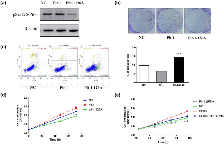
protein level of Pit-1 was not obviously changed; however, with MT-Pit-1 (Figure 2a). We detected cell proliferation
as the concentration of roscovitine increased in a certain with a colony formation assay, and the results are pre-
dose range, the phosphorylation level of Ser126-Pit-1 gra- sented as the percentage of clones formed as a function of
dually decreased. We transfected GH3 cells with short time. After 2 weeks of incubation, we clearly observed
interfering RNA (siRNA) targeting CDK5 mRNA, and the that the proportion of clones formed in GH3 cells in the
knockdown efficiency was verified by the western blot. WT-Pit-1 groups was higher than that in the MT-Pit-1
As shown in Figure 1d, the phosphorylation level of groups (Figure 2b). Similarly, we found that cell viability
Ser126-Pit-1 was decreased by CDK5 knockdown in GH3 was significantly higher in the WT-Pit-1 groups than
cells. In particular, we found that both CDK5 inhibition it was in the MT-Pit-1 groups (Figure 2d), suggesting
and depletion significantly decreased pSer126-Pit-1. that Ser126A-Pit-1 suppresses GH3 cell proliferation. To
determine whether apoptosis was a contributing factor in
cell survival inhibition, we performed flow cytometric
analysis of cells pretreated with WT-Pit-1 and MT-Pit-1.
3.3 pSer126-Pit-1 promotes GH3 cell Apoptosis assays showed that the apoptosis rates of GH3
proliferation and PRL secretion cells increased in the MT-Pit-1 groups compared with the
WT-Pit-1 groups after incubation for 72 h (Figure 2c). In
To determine the influence of the phosphorylation level order to confirm that whether CDK5 affects cell prolifera-
of Pit-1 on cell proliferation and apoptosis, we transfected tion via p-Ser126-Pit-1 dominantly, GH3 cells were co-
GH3 cells with WT-Pit-1 and Ser126A-Pit-1 (MT-Pit-1). transfected with CDK5 and Pit-1 siRNA to assess whether
Western blot results showed that pSer126-Pit-1 in GH3 the effect of CDK5 could be reversed by Pit-1 siRNA. The
cells with WT-Pit-1is significantly higher than GH3 cells results indicated that the cell viability in CDK5 groups790 Weiyan Xie et al.
Figure 2: pSer126-Pit-1 promotes GH3 cell proliferation and suppresses apoptosis. (a) Western blot showed pSer126-Pit-1 in GH3 cell with
WT-Pit-1 or Pit-1-126A. (b) The colony formation test shows that the percentage of colonies formed was reduced in the Pit-1-126A groups
compared with that of the Pit-1 groups. (c) The apoptosis rates of the Pit-1-126A groups were significantly higher than those of the Pit-1
groups. (d) Higher cell viability was observed in the Pit-1 groups. (e) The effect of CDK5 could be reversed by Pit-1 siRNA. **, P < 0.01; ***, P <
0.001. The bar represents the mean ± SD.
was significantly higher than CDK5 + Pit-1 siRNA groups, of PRL in the supernatant of GH3 cells. The ELISA results
and the cell viability of Pit-1 siRNA decreased (Figure 2e). show that the level of PRL was significantly higher in the Pit-
These data suggest that CDK5-mediated pSer126-Pit reg- 1 groups than it was in the Ser126A-Pit-1 groups, which
ulates cell proliferation. indicates that high pSer126-Pit-1 promoted PRL synthesis
Pit-1 binds to the proximal PRL promoter and induces and secretion in GH3 cells (Figure 3c).
PRL expression [15,16]. To explore whether a mutation of
the Ser126 phosphorylation site in Pit-1 would affect the
ability of Pit-1 to bind to the PRL promoter, we performed
an electrophoretic mobility shift assay (EMSA) using 3.4 Ser126-Pit-1 phosphorylation in human
nuclear extracts from GH3 cells. As shown in Figure 3a, prolactinoma tissue
Pit-1 could bind to the PRL promoter as expected, but when
Ser 126 was mutated, the binding was almost completely To investigate the clinical significance of Ser126-Pit-1 phosphor-
abolished. HEK293 cells that transiently coexpressed the ylation, we carried out IHC staining of human prolactinoma
PRL promoter, WT-Pit-1, or Ser126A-Pit-1 were analyzed by tissue from 48 patients with the pSer126-Pit-1 phosphoantibody.
luciferase reporter assay. WT-Pit-1 demonstrated stronger Based on the IHC staining (Figure 4a), we divided human
PRL transcriptional activation than Ser126A-Pit-1 (Figure prolactinoma tissue into the pSer126-Pit-1 high expres-
3b). These results indicated that Ser126 in Pit-1 is the key sion group (mean H-score: 195) and the pSer126-Pit-1
site for the combination of Pit-1 with the PRL promoter. To low expression group (mean H-score: 138). The prognostic
determine whether pSer126-Pit-1 affects the ability of GH3 value of pSer126-Pit-1 for recurrence-free survival in prolac-
cells to secrete PRL, an ELISA was used to measure the level tinoma patients was evaluated by comparing the patientsPhosphorylation of Pit-1 promotes cell proliferation 791
Figure 3: WT-Pit-1 promotes PRL secretion and induces PRL expression. (a) Pit-1-PRL complexes were detected. (b) Ser126A-Pit-1 signifi-
cantly inhibited the luciferase activity of the PRL 3′UTR relative to the levels seen with Pit-1. (c) The PRL level of the Pit-1 groups was higher
than that of the Pit-1-126A groups. **, P < 0.01; ***, P < 0.001. The bar represents the mean ± SD.
with low and high pSer126-Pit-1 expression. According to regulation of transcriptional activity through phosphor-
Kaplan–Meier survival analysis, patients with high pSer126- ylation of key transcription factors, such as STAT3 (signal
Pit-1 expression had a distinctly shorter recurrence-free sur- transducer and activator of transcription 3), MEF2 (myo-
vival time than those with low pSer126-Pit-1 expression cyte enhancer factor 2), and mSds3 [17,18]. CDK5 can
(Figure 4b). These data suggest that pSer126-Pit-1 might serve phosphorylate serine 727 of STAT3 and regulate its tran-
as a prognostic biomarker for predicting the outcome of scriptional activity, which affects the downstream expression
prolactinoma. of c-fos, junB, and Foxp3 and regulates T cell development
[19,20]. Although CDK5 has been investigated in some
types of cancers, the functional role of CDK5 in the prolif-
eration and apoptosis of PA cells remains to be elucidated.
4 Discussion
The data presented here have shown that both inhibition
and depletion of CDK5 reduce pSer-126-Pit-1 in GH3 cells,
and the high level of phosphorylated Pit-1 can promote
cell proliferation and inhibit apoptosis. Meanwhile, Ser126
in Pit-1 is the key site enabling the association of Pit-1
with the PRL promoter. We found that a higher level
of pSer-126-Pit-1 correlates with a worse prognosis in
patients. These results suggested that CDK5 phosphory-
lates Ser126-Pit-1 to regulate prolactinoma progression
and PRL secretion.
CDK5 is a unique member of the cell cycle-dependent
kinase family. It plays a key regulatory role in the nervous
system’s transcriptional activity. It is specifically acti-
vated in human prolactinomas, and the pituitary has
high levels of Pit-1. However, whether CDK5 affects the
physiological function of Pit-1 by phosphorylation is
unknown. CDK5 belongs to the serine/threonine kinase
family and has specific sequence requirements for its
phosphorylation substrate. It can only phosphorylate Figure 4: pSer126-Pit-1 is associated with poor prognosis. (a)
serine or threonine sites containing S/TPXX (K/R/H) con- Representative images of pSer126-Pit-1 staining of a tissue micro-
served sequences. In response to changes in the external array, including high pSer126-Pit-1 levels (left panel) and low
pSer126-Pit-1 levels in prolactinomas (right panel). Insets show
environment or hormone levels, CDK5 will undergo trans-
200× magnifications of the low-power images, scale bar, 50 μm. (b)
location from the cytoplasm to the nucleus and will phos- Significant differences in recurrence-free survival are shown based
phorylate different transcription factor substrates. Previous on pSer126-Pit-1 expression status in 48 patients after surgical
studies have shown that CDK5 can participate in the removal of prolactinomas, *, P < 0.05.792 Weiyan Xie et al.
In our previous study, we found that CDK5 activity was in vitro. Therefore, Pit-1 phosphorylation at Ser126 may
upregulated in PAs and was associated with p35 [13,14,21]. play a critical role in prolactinoma progression, and it
Here, we have shown that both proliferation and apoptosis could be used in the prediction of the poor prognosis of
of pituitary cells were regulated by pSer126-Pit-1. After we prolactinomas. Additionally, our findings showed that
treated cells with the CDK5 inhibitor and CDK5 siRNA, CDK5 inhibitors could directly or indirectly block cell
CDK5 activity and expression levels were reduced, which proliferation in prolactinomas.
decreased the level of Ser126-Pit-1 phosphorylation. Our
results reveal that CDK5 can exert its biological function Acknowledgments: We thank Mr Sen Cheng and Mr Bin Li
by specifically regulating Ser126-Pit-1 phosphorylation. for revising the manuscript.
Pit-1 contains 291 amino acids, including an N-terminal
transcription activation domain, a POU-specific domain Funding information: The work was supported by grants
(POU-specific), and a POU homology domain (POU home- from the National Natural Science Foundation of China
odomain). POU-specific domains and POU-homeodomains (81672495, 81771489).
are high-affinity DNA binding domains, and they also
interact with other transcriptional regulators. Ser126 is Author contributions: W.X. and Y.Z. conceived the pro-
located at the beginning of the POU-specific domain. ject. Q.F. and W.X. designed the experiments, analyzed
Ser126-Pit-1 phosphorylation can change its affinity for the data, and wrote the manuscript. C.L. and J.G. assisted
DNA and affect its DNA binding site. After CDK5 speci- with the management of clinical data and specimens.
fically and appropriately phosphorylates Pit-1 in the L.G. performed the experiments. All authors read and
nucleus, Pit-1 may be overactivated to produce exces- approved the manuscript.
sive GH or PRL. Studies have shown that the R271W
mutation of Pit-1 converts arginine to tryptophan at Conflict of interest: Authors declare no conflict of interest.
position 271 in the C-terminal POU-H domain, leading
to the loss of a positive charge in the basic amino acid Data availability statement: The authors can confirm that
region. R271W-mutated Pit-1 can bind to DNA, thus com- all relevant data and materials are available upon request
petitively inhibiting wild-type Pit-1 [21]. According to from the authors.,
our experimental results, a mutation in the Ser126 site
in Pit-1 impacts its effective binding to the PRL promoter Ethics approval: The present study was approved by the
and reduces CDK5 phosphorylation of Pit-1. Ethics Committee of Beijing Tiantan Hospital, Capital
By interacting with different types of transcription Medical University (Beijing, China).
factors, Pit-1 can also regulate different signaling path-
ways, such as PKA (protein kinase A) and PKC (protein
kinase C), and Ras signaling pathways, thereby targeting
the regulation of these pathways could occur via regula-
tion of the Pit-1 promoter [22,23]. Previously, some
References
research groups found that the Pit-1 gene was related to
[1] Tatsumi K, Amino N. PIT1 abnormality. Growth Horm IGF Res.
pituitary dysplasia and PRL, GH, and TSH secretion
1999;9(Suppl B):18–22. discussion 3 .
defects. The absence of the Pit-1 gene leads to complete [2] Martinez-Ordonez A, Seoane S, Cabezas P, Eiro N, Sendon-
PRL and GH secretion defects [24–27]. Our results have Lago J, Macia M, et al. Breast cancer metastasis to liver and
confirmed that in comparison to the mutant Pit-1, WT-Pit- lung is facilitated by Pit-1-CXCL12-CXCR4 axis. Oncogene.
1 more stably binds to the PRL promoter and induces PRL 2018;37(11):1430–44.
[3] Feldmann G, Mishra A, Hong SM, Bisht S, Strock CJ, Ball DW,
expression, thus leading to excessive hormone secretion
et al. Inhibiting the cyclin-dependent kinase CDK5 blocks
and enabling cells to grow aggressively and invade sur-
pancreatic cancer formation and progression through the
rounding tissues. suppression of Ras-Ral signaling. Cancer Res.
2010;70(11):4460–9.
[4] Wissing MD, Dadon T, Kim E, Piontek KB, Shim JS, Kaelber NS,
et al. Small-molecule screening of PC3 prostate cancer cells
5 Conclusion identifies tilorone dihydrochloride to selectively inhibit cell
growth based on cyclin-dependent kinase 5 expression. Oncol
Rep. 2014;32(1):419–24.
CDK5 phosphorylates Pit-1 at Ser126 in GH3 cells, which is [5] Augustijn KD, Duval DL, Wechselberger R, Kaptein R, Gutierrez-
a step that is required for cell proliferation and apoptosis Hartmann A, van der Vliet PC. Structural characterization of thePhosphorylation of Pit-1 promotes cell proliferation 793
PIT-1/ETS-1 interaction: PIT-1 phosphorylation regulates PIT-1/ sion in human lactotroph and somatotroph pituitary adenomas
ETS-1 binding. Proc Natl Acad Sci U S A. is correlated to D2 receptor gene expression. J Clin Endocrinol
2002;99(20):12657–62. Metab. 1996;81(9):3390–6.
[6] Jean A, Gutierrez-Hartmann A, Duval DL. A Pit-1 threonine 220 [17] Hsu FN, Chen MC, Lin KC, Peng YT, Li PC, Lin E, et al. Cyclin-
phosphomimic reduces binding to monomeric DNA sites to dependent kinase 5 modulates STAT3 and androgen receptor
inhibit Ras and estrogen stimulation of the prolactin gene activation through phosphorylation of Ser(7)(2)(7) on STAT3 in
promoter. Mol Endocrinol. 2010;24(1):91–103. prostate cancer cells. Am J Physiol Endocrinol Metab.
[7] Ben-Batalla I, Seoane S, Macia M, Garcia-Caballero T, 2013;305(8):E975–86.
Gonzalez LO, Vizoso F, et al. The Pit-1/Pou1f1 transcription [18] Lin H, Chen MC, Chiu CY, Song YM, Lin SY. Cdk5 regulates
factor regulates and correlates with prolactin expression in STAT3 activation and cell proliferation in medullary thyroid
human breast cell lines and tumors. Endocr Relat Cancer. carcinoma cells. J Biol Chem. 2007;282(5):2776–84.
2010;17(1):73–85. [19] Lam E, Choi SH, Pareek TK, Kim BG, Letterio JJ. Cyclin-depen-
[8] Gil-Puig C, Seoane S, Blanco M, Macia M, Garcia-Caballero T, dent kinase 5 represses Foxp3 gene expression and Treg
Segura C, et al. Pit-1 is expressed in normal and tumorous development through specific phosphorylation of Stat3 at
human breast and regulates GH secretion and cell prolifera- Serine 727. Mol Immunol. 2015;67(2 Pt B):317–24.
tion. Eur J Endocrinol. 2005;153(2):335–44. [20] Fu AK, Fu WY, Ng AK, Chien WW, Ng YP, Wang JH, et al. Cyclin-
[9] Ghose A, Shashidhara LS. Cyclin beyond the cell cycle: new dependent kinase 5 phosphorylates signal transducer and
partners at the synapse. Dev Cell. 2011;21(4):601–2. activator of transcription 3 and regulates its transcriptional
[10] Cortes N, Guzman-Martinez L, Andrade V, Gonzalez A, activity. Proc Natl Acad Sci U S A. 2004;101(17):6728–33.
Maccioni RB, CDK5 A, et al. and Its Multiple Roles [21] Kishimoto M, Okimura Y, Fumoto M, Iguchi G, Iida K, Kaji H,
in the Nervous System. J Alzheimers Dis. et al. The R271W mutant form of Pit-1 does not act as a domi-
2019;68(3):843–55. nant inhibitor of Pit-1 action to activate the promoters of GH
[11] Mishiba T, Tanaka M, Mita N, He X, Sasamoto K, Itohara S, and prolactin genes. Eur J Endocrinol. 2003;148(6):619–25.
et al. Cdk5/p35 functions as a crucial regulator of spatial [22] Wasylyk B, Hagman J, Gutierrez-Hartmann A. Ets transcription
learning and memory. Mol Brain. 2014;7:82. factors: nuclear effectors of the Ras-MAP-kinase signaling
[12] Asada A, Saito T, Hisanaga S. Phosphorylation of p35 and p39 pathway. Trends Biochem Sci. 1998;23(6):213–6.
by Cdk5 determines the subcellular location of the holokinase [23] Xu L, Lavinsky RM, Dasen JS, Flynn SE, McInerney EM,
in a phosphorylation-site-specific manner. J Cell Sci. Mullen TM, et al. Signal-specific co-activator domain require-
2012;125(Pt 14):3421–9. ments for Pit-1 activation. Nature. 1998;395(6699):301–6.
[13] Xie W, Wang H, He Y, Li D, Gong L, Zhang Y. CDK5 and its [24] Ohta K, Nobukuni Y, Mitsubuchi H, Fujimoto S, Matsuo N,
activator P35 in normal pituitary and in pituitary adenomas: Inagaki H, et al. Mutations in the Pit-1 gene in children with
relationship to VEGF expression. Int J Biol Sci. combined pituitary hormone deficiency. Biochem Biophys Res
2014;10(2):192–9. Commun. 1992;189(2):851–5.
[14] Xie W, Liu C, Wu D, Li Z, Li C, Zhang Y. Phosphorylation of [25] Pfaffle RW, DiMattia GE, Parks JS, Brown MR, Wit JM, Jansen M,
kinase insert domain receptor by cyclin-dependent kinase 5 at et al. Mutation of the POU-specific domain of Pit-1 and hypo-
serine 229 is associated with invasive behavior and poor pituitarism without pituitary hypoplasia. Science.
prognosis in prolactin pituitary adenomas. Oncotarget. 1992;257(5073):1118–21.
2016;7(32):50883–94. [26] Radovick S, Nations M, Du Y, Berg LA, Weintraub BD,
[15] Pellegrini I, Barlier A, Gunz G, Figarella-Branger D, Enjalbert A, Wondisford FE. A mutation in the POU-homeodomain of Pit-1
Grisoli F, et al. Pit-1 gene expression in the human pituitary responsible for combined pituitary hormone deficiency.
and pituitary adenomas. J Clin Endocrinol Metab. Science. 1992;257(5073):1115–8.
1994;79(1):189–96. [27] Tatsumi K, Miyai K, Notomi T, Kaibe K, Amino N, Mizuno Y,
[16] Pellegrini-Bouiller I, Morange-Ramos I, Barlier A, Gunz G, et al. Cretinism with combined hormone deficiency caused by
Figarella-Branger D, Cortet-Rudelli C, et al. Pit-1 gene expres- a mutation in the PIT1 gene. Nat Genet. 1992;1(1):56–8.You can also read