Hypoxia-Related Gene FUT11 Promotes Pancreatic Cancer Progression by Maintaining the Stability of PDK1 - Frontiers
←
→
Page content transcription
If your browser does not render page correctly, please read the page content below
ORIGINAL RESEARCH
                                                                                                                                               published: 17 June 2021
                                                                                                                                        doi: 10.3389/fonc.2021.675991

                                              Hypoxia-Related Gene FUT11
                                              Promotes Pancreatic Cancer
                                              Progression by Maintaining the
                                              Stability of PDK1
                                              Wenpeng Cao 1*, Zhirui Zeng 2†, Runsang Pan 3†, Hao Wu 4, Xiangyan Zhang 5, Hui Chen 5,
                                              Yingjie Nie 6, Zijiang Yu 1* and Shan Lei 2*
                                              1Department of Anatomy, School of Basic Medicine, Guizhou Medical University, Guiyang, China, 2 Department of
                                              Physiology, School of Basic Medicine, Guizhou Medical University, Guiyang, China, 3 Department of Orthopedics, Guiyang
                                              Maternal and Child Health Care Hospital, Guiyang, China, 4 Department of Pediatric Surgery, The Affiliated Hospital of
                                              Guizhou Medical University, Guiyang, China, 5 NHC Key Laboratory of Pulmonary, Guizhou Provincial People’s Hospital,
                             Edited by:       Guiyang, China, 6 The Clinical Lab Center, Guizhou Provincial People’s Hospital, Guiyang, China
                              Wei Zhao,
         City University of Hong Kong,
                            Hong Kong         Background: Hypoxia is associated with the development of pancreatic cancer (PC).
                        Reviewed by:          However, genes associated with hypoxia response and their regulatory mechanism in PC
                       Tongzheng LIU,         cells were unclear. The current study aims to investigate the role of the hypoxia associated
                Jinan University, China
                        Chenxi Zhang,         gene fucosyltransferase 11 (FUT11) in the progression of PC.
         Nanjing Chest Hospital, China
                                              Methods: In the preliminary study, bioinformatics analysis predicted FUT11 as a key
                  *Correspondence:
                                              hypoxia associated gene in PC. The expression of FUT11 in PC was evaluated using
                       Wenpeng Cao
                1006074061@qq.com             quantitative real-time PCR (qRT-PCR), Western blot and immunohistochemistry. The
                          Zijiang Yu          effects of FUT11 on PC cells proliferation and migration under normoxia and hypoxia were
                 893767473@qq.com
                           Shan Lei           evaluated using Cell Counting Kit 8, 5-ethynyl-2’-deoxyuridine (EDU) assay, colony
                1109974497@qq.com             formation assay and transwell assay. The effects of FUT11 in vivo was examined in
     †
      These authors have contributed          mouse tumor models of liver metastasis and subcutaneous xenograft. Furthermore,
                 equally to this work
                                              Western blot, luciferase assay and immunoprecipitation were performed to explore the
                 Specialty section:
                                              regulatory relationship among FUT11, hypoxia-inducible factor 1a (HIF1a) and pyruvate
       This article was submitted to          dehydrogenase kinase 1 (PDK1) in PC.
    Molecular and Cellular Oncology,
              a section of the journal        Results: FUT11 was markedly increased of PC cells with hypoxia, upregulated in the PC
               Frontiers in Oncology          clinical tissues, and predicted a poor outcome of PC patients. Inhibition of FUT11 reduced
           Received: 04 March 2021            PC cell growth and migratory ability of PC cells under normoxia and hypoxia conditions in
            Accepted: 27 May 2021
                                              vitro, and growth and tumor cell metastasis in vivo. FUT11 bound to PDK1 and regulated
           Published: 17 June 2021
                                              the expression PDK1 under normoxia and hypoxia. FUT11 interacted with PDK1 and
                              Citation:
              Cao W, Zeng Z, Pan R,           decreased the ubiquitination of PDK1, lead to the activation of AKT/mTOR signaling
     Wu H, Zhang X, Chen H, Nie Y,            pathway. FUT11 knockdown significantly increased the degradation of PDK1 under
     Yu Z and Lei S (2021) Hypoxia-
     Related Gene FUT11 Promotes              hypoxia, while treatment with MG132 can relieve the degradation of PDK1 induced by
   Pancreatic Cancer Progression by           FUT11 knockdown. Overexpression of PDK1 in PC cells under hypoxia conditions
    Maintaining the Stability of PDK1.
                                              reversed the suppressive impacts of FUT11 knockdown on PC cell growth and
            Front. Oncol. 11:675991.
     doi: 10.3389/fonc.2021.675991            migration. In addition, HIF1a bound to the promoter of FUT11 and increased its

Frontiers in Oncology | www.frontiersin.org                                         1                                         June 2021 | Volume 11 | Article 675991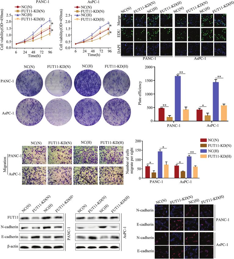
Cao et al.                                                                                                          FUT11 Promoted Pancreatic Cancer

                                              expression, as well as co-expressed with FUT11 in PC tissues. Furthermore,
                                              overexpression of FUT11 partially rescued the suppressive effects of HIF1a knockdown
                                              on PC cell growth and migration in hypoxia condition.
                                              Conclusion: Our data implicate that hypoxia-induced FUT11 contributes to proliferation
                                              and metastasis of PC by maintaining the stability of PDK1, thus mediating activation of
                                              AKT/mTOR signaling pathway, and suggest that FUT11 could be a novel and effective
                                              target for the treatment of pancreatic cancer.
                                              Keywords: pancreatic cancer, hypoxia, fucosyltransferase 11, hypoxia-inducible factor 1a, pyruvate
                                              dehydrogenase kinase 1

BACKGROUND                                                                          In this study, we found that the FUT11 was a direct target
                                                                                 gene of HIF1a by bioinformatics analysis and it was up-regulated
Pancreatic cancer (PC) has high morbidity and mortality                          in PC cells under hypoxia. FUT11 promoted the proliferation
worldwide (1). Although the treatment for PC such as surgery,                    and metastasis of PC cells via maintaining the stability of
targeted therapy and chemotherapy had been improved, the                         pyruvate dehydrogenase kinase 1(PDK1) under hypoxia. Our
number of PC related mortality is still increasing every year                    study indicates FUT11 could be a therapeutic target for the
(2). Therefore, it is a pressing need to uncover the molecular                   treatment of PC.
mechanism involved in PC and explore the potential biomarkers
for diagnosis and as novel targets for treatment of PC.
    The tumor microenvironment plays a vital role in the
development of tumors and is closely related to the efficacy of                   METHODS
tumor treatment. Targeting the tumor environment is a
therapeutic strategy for cancer treatment (3). Generally,
                                                                                 Bioinformatics Analysis
                                                                                 We downloaded the Gene expression profile GSE67549 and
hypoxic microenvironment of tumors up-regulates a series of
                                                                                 GSE9350 from the Gene Expression Omnibus database (GEO,
hypoxic-responsive genes, and induces the proliferation,
                                                                                 https://www.ncbi.nlm.nih.gov/gds). GSE67549 contained 9
migration, drug resistance and other biological events of cancer
                                                                                 normoxic PC cell samples and 9 hypoxic PC cell samples,
cells (4). Hypoxia inducible factor 1-alpha (HIF1a) is a main
                                                                                 while GSE9350 contained 2 normoxic PC cell samples and 2
regulator of transcriptional response to hypoxia in cancer cells.
                                                                                 hypoxic PC cell samples. Differential expression genes were
HIF1a up-regulates a number of genes that support tumor cells
                                                                                 identified using the cut-off as Log2 fold change (FC) >1 and P
to adopt to the hypoxic microenvironment (5). HIF1a
                                                                                 value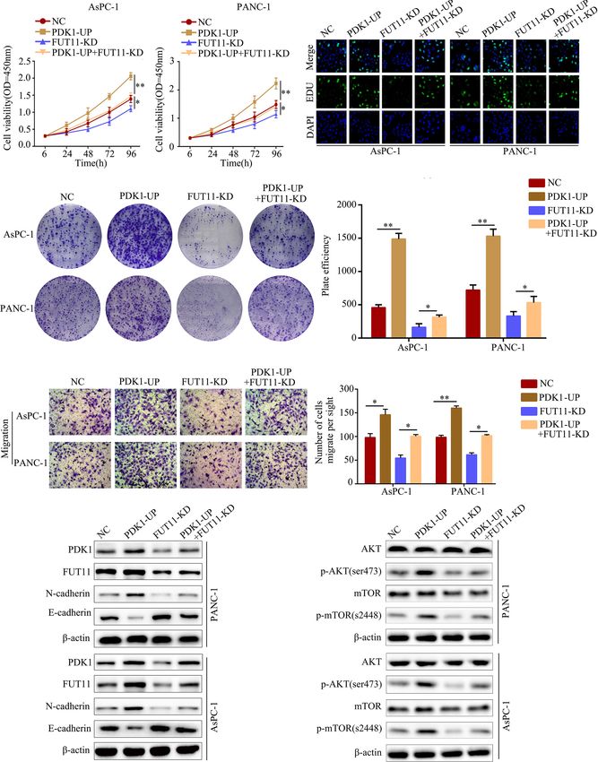
Cao et al.                                                                                                   FUT11 Promoted Pancreatic Cancer

5% CO2, while the condition of hypoxia was set to 1%O2, 94%N2           Western Blotting
and 5%CO2. Oligonucleotides targeting FUT11 (GUUAGAGA                   The proteins in PC cells and tissues were extracted using RIPA
CCACUGUAUCUGC) were cloned into the pLKO.1 vector                       reagent contain 5% PMSF protease inhibitor. The BCA method
(GenePharma, Shanghai, China). Full‐length PDK1 coding                  was performed to examine the protein concentration of each
sequence was subcloned into the lentiviral vector pCD315B-1             sample. Proteins (30mg/per line) were added and separated by
(System Biosciences, Beijing, China). The small interfering RNA         10% SDS-PAGE for 120 min. Then, the proteins were transferred
(siRNA) targeting HIF1a were obtained from JIMA (Shanghai,              into the PVDF membranes (Millipore, USA) with 0.45mm pore
China). To construct the stable cell lines with target gene             diameter, which was then blocked in 5% BSA for 30min and
overexpression or knockdown, 1 mg/mL puromycin (Sigma,                  incubated with primary antibodies including FUT11 (Abcam,
USA) was added to culture medium after transfection with                cat. no. ab121411, dilution, 1:500), N-cadherin (CST, cat. no.
lentivirus for 48h to continuously screen the stable cells for          14215, dilution, 1:1000), E-cadherin (CST, cat. no. 3195, dilution,
10 days.                                                                1:1000), PDK1 (Santa, cat. no. 4A11F5, dilution, 1:1000), AKT
                                                                        (CST, cat. no. 9272, dilution, 1:1000), p-AKT (CST, cat. no. 9271,
Quantitative Real-Time PCR (qRT-PCR)                                    dilution, 1:1000), mTOR (CST, cat. no. 2972, dilution, 1:1000), p-
Total RNA in PC tissues and cells was separated using TRIZOL            mTOR (CST, cat. no. 2971, dilution, 1:1000), HIF1a (CST, cat.
reagents (Beyotime Biotechnology, Hangzhou, China) and                  no. 36169, dilution, 1:1000) and b-actin (CST, cat. no. 3700,
diluted into DNase/RNase-free water. After quantification,               dilution, 1:1000) for 12h in 4˚C. High sensitivity ECL reagent
total RNA (2µg per sample) was reversely transcribed into               was used to visualize the blots in MultiImager and the relative
cDNA using RevertAid First Strand cDNA Synthesis Kit                    expression of protein was calculated using Image J. b-actin was
(Fermentas, USA). Finally, quantitative real-time PCR (qRT-             set as reference for FUT11, N-cadherin, E-cadherin, PDK1
PCR) was conducted to determine the expression level of target          and HIF1a.
genes using SYBR™ Green PCR Master Mix (Solarbio, Wuhan,
China). b-actin was used as the internal control. The primer            Transwell Assay
sequences in our study were purchased from Tianyi Huiyuan               For transwell migration assay, a total of 1×10⁵ cells were
(Wuhan, China) and shown in Supplementary Table 1.                      suspended using 200ml DMEM medium without FBS and
                                                                        seeded into the upper transwell chambers (Becton, Dickinson
Cell Counting Kit-8 (CCK-8) Assay                                       and Company, USA). Total 600ml DMEM medium contained
PANC-1 and AsPC-1 cells were plated in a 96-well plate in               10% FBS was placed in the lower transwell chambers. After 24h,
sextuplicate with 3×10³ cells/well. Briefly, 100ml DMEM medium           migratory cells were fixed with paraformaldehyde and stained
containing 10ml CCK-8 regent (Boster, Wuhan, China) was                 using 0.5% crystal violet. Finally, the migratory cells were
added to each well in 24h, 48h, 72h and 96h. The light                  counted and photos were taken.
absorbance of each well was detected at 450nm.
                                                                        In Vivo Assay
5-ethynyl-2’-deoxyuridine (EDU) Assay                                   For subcutaneous tumor xenograft model, 10 female BALB/c
The EDU assay was carried out using a BeyoClick™ EdU-488                nude mice were obtained from the animal central of Guizhou
Proliferation Detection Kit (Beyotime, Suzhou, China). In brief,        Medical University (Guizhou, China). After adaptive feeding, a
PC cells were cultured in 6-well plates and were allowed to             total of 1×10⁶ PANC-1 cells with FUT11 knockdown and
adhere. The primary culture medium was removed and fresh                negative control cells were subcutaneously injected into the
medium was added. Then, 10mM EDU was added into each well               upper-right flank of BALB/c mice (n = 5 in each group). The
and cells were cultured in 37°C for 2.5h. After that, cells were        health status of mice was monitored every day. The tumor
fixed in 4% paraformaldehyde (Beyotime, Suzhou, China) for               volume was monitored once a week and determined as
15 min and permeabilization using 0.3% Triton X-100 (Boster,            followed: (mm3) = (Long×Width2)/2. After 5 weeks, the mice
Wuhan, China) for 8 min. Then, 500ml Apollo dyeing reaction             were sacrificed and tumor tissues collected. The protein level of
buffer was added for 40 min in the dark. After staining,                KI67 and PCNA in tumor tissues was determined using
the nuclei were stained using DAPI for 10 min. The EDU                  immunohistochemical staining. The liver metastatic tumor
staining was observed under a fluorescence microscope (Zeiss,            model was established by injecting the FUT11 knockdown and
Oberkochen, Germany).                                                   negative control PANC-1 cells into the spleen capsule. FUT11
                                                                        knockdown and negative control group PANC-1 cells (1×10⁷
Colony Formation Assay                                                  cells) were injected into the spleen of BALB/c mice (n=5 in each
Cells with a density of 2000 cells/well were seeded into 6-well         group). Animal health and behavior after injecting were
plates and cultured in DMEM media containing 10% FBS. After             monitored each day. While the mouse had the features of hard
24h, intervention factor was added and cultured for 2 weeks.            breath and limitation of motion, mice were sacrificed in order to
After fixation in 4% paraformaldehyde for 15 min, 1% crystal             reduce animal suffering, and the liver tissues were dissected and
violet was used to stain cell colonies. Cell colonies was counted       used to count the metastatic foci. While mice in one group were
and photographed.                                                       all sacrificed, the animal experiment was terminated and the rest

Frontiers in Oncology | www.frontiersin.org                         3                                   June 2021 | Volume 11 | Article 675991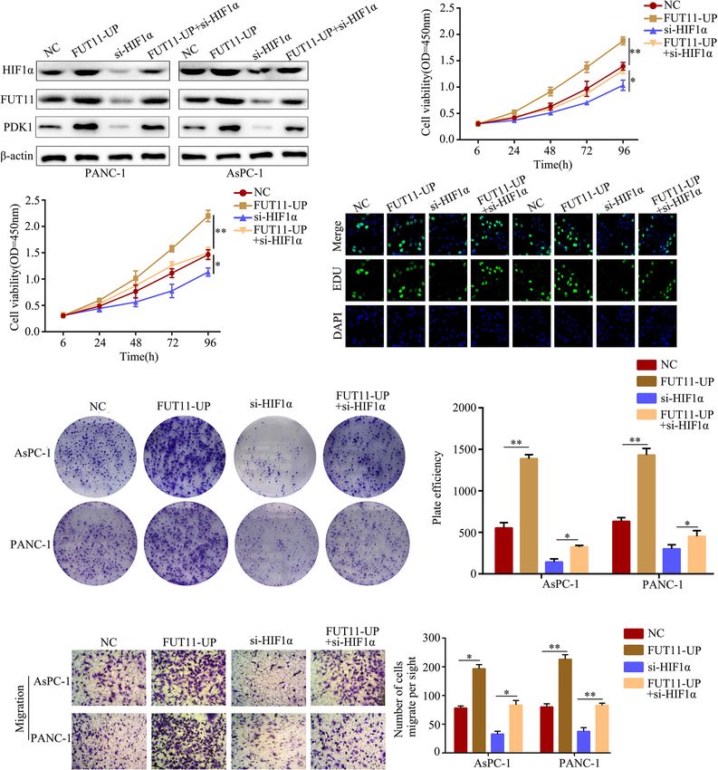
Cao et al.                                                                                                 FUT11 Promoted Pancreatic Cancer

of mice were all euthanasia. Finally, unpair-t test was used to         Luciferase Assay
determine the significant between this group according to the            After predicting the binding site of HIF1a (also named hypoxia
number of metastasis foci. HE staining was also used to detect          response element, HRE) in the promoter of FUT11 using online
the condition of metastatic foci in the liver. All procedures of        database JASPAR (http://jaspar.binf.ku.dk/), dual luciferase
animal studies were approved by the Ethics Committee of                 reporter assay was performed to verify the bind. Full‐length
Guizhou Medical University and followed the legal mandates              FUT11 promoter sequence and corresponding truncated
and national guidelines for the care and maintenance of                 fragment without HRE were carried into the psi‐basic
laboratory animals.                                                     luciferase reporter vector (Promega, USA). Finally, a total of
                                                                        1×10⁴ PANC-1 and AsPC-1 cells were plated into 24-well plate
Immunofluorescence Staining                                              and cultured overnight at 37°C. Then, both of luciferase reporter
Cells were fixed with 4% paraformaldehyde (Solarbio, Wuhan,              vectors contained full‐length FUT11 promoter sequence and
China) for 15min. Then, the samples were incubated with anti-           corresponding truncated fragment without HRE, and the si‐
FUT11 (dilution, 1:100), anti-N-cadherin (dilution, 1:100), anti-       HIF1a/si‐NC were co-transfected into PC cells using lipidosome
E-cadherin (dilution, 1:100), anti-HIF1a (dilution, 1:100) and          2000 (Solarbio, Wuhan, China). The luciferase activity of cells
anti-PDK1 (dilution, 1:100) primary antibodies. After washing           was determined after transfection at 24h in PC cells cultured in
with PBS, the samples were incubated with FITC conjugated               normoxia or hypoxia.
anti-mouse secondary antibodies (Proteintech, Wuhan, China)
and Cy3 conjugated anti-rabbit secondary antibodies                     Statistical Analysis
(Proteintech, Wuhan, China). Nuclei were stained with DAPI              SPSS software (version 21.0) was employed to perform statistical
(Boster, Wuhan, China). Finally, confocal microscopy or                 analysis. The difference between two groups was analyzed using
fluorescent microscope was used to collect the images.                   paired t-test, while the difference among multiple groups were
                                                                        determined based on one-way analysis of variance. P*3 fold) in AsPC-1 and PANC-1 cells
exceeding the threshold.
                                                                        under hypoxia (Figure 2A).
                                                                            We then evaluated the mRNA and protein levels of FUT11 in
Chromatin Immunoprecipitation                                           human PC tissues and adjacent pancreatic tissues. The results
Chromatin immunoprecipitation (ChIP) assays were performed              showed that the mRNA and protein levels of FUT11 was higher
using a ChIP kit (CST, USA) as per the protocol provided by the         in pancreatic cancer tissues than that in adjacent pancreatic
manufacturer. Briefly, formaldehyde was used to crosslink cells,         tissues (Figures 2B–D). We analyzed the correlation between
and the DNA was sonicated to produce sequences of 200–500 bp            FUT11 expression and the PC clinical pathology features, and
in length. Immunoprecipitation was conducted using an anti-             found that FUT11 expression was positively correlated with
HIF1a antibody or IgG control. The precipitated DNA was                 tumor size (cm), lymph node metastasis, TNM stage,
amplified by qRT-PCR.                                                    perineural invasion, blood vessel invasion and distant

Frontiers in Oncology | www.frontiersin.org                         4                                  June 2021 | Volume 11 | Article 675991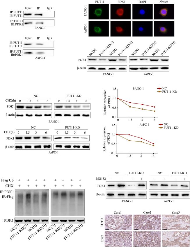
Cao et al.                                                                                                                           FUT11 Promoted Pancreatic Cancer

                                  A                                   B

 FIGURE 1 | Identification of key hypoxia-related genes in PC. (A) Intersection analysis of differentially expressed genes in PC samples under hypoxia based on the gene
 expression profiles of GSE67549 and GSE9350. (B) The mRNA expression of ANGPTL4, ANKRD37, ANKZF1, ADM, C4ORF3, ERO1L, FAM162A, FUT11, BNIP3L,
 KDM3A, MXI1, NDRG1, KCTD11, PDX1, PFKFB1, PPF1A4, SLC2A1 and P4HA1 in PC tissues compared with non-tumor tissues analyzed by GEPIA online tool.

metastasis as shown in Supplementary Table 2. We split the                              and PANC-1 cells under normoxia, as well as decreasing the
patients into high and low FUT11 expression groups based on                             stimulative impact of hypoxia (Figures 3A, B). Simultaneously,
the expression of FUT11 with the median value of 5.6. The                               suppression of FUT11 reduced the colony formation of AsPC-1
expression of FUT11>5.6 was defined as high expression, while                            and PANC-1 cells in normoxia, as well as reducing the
the expression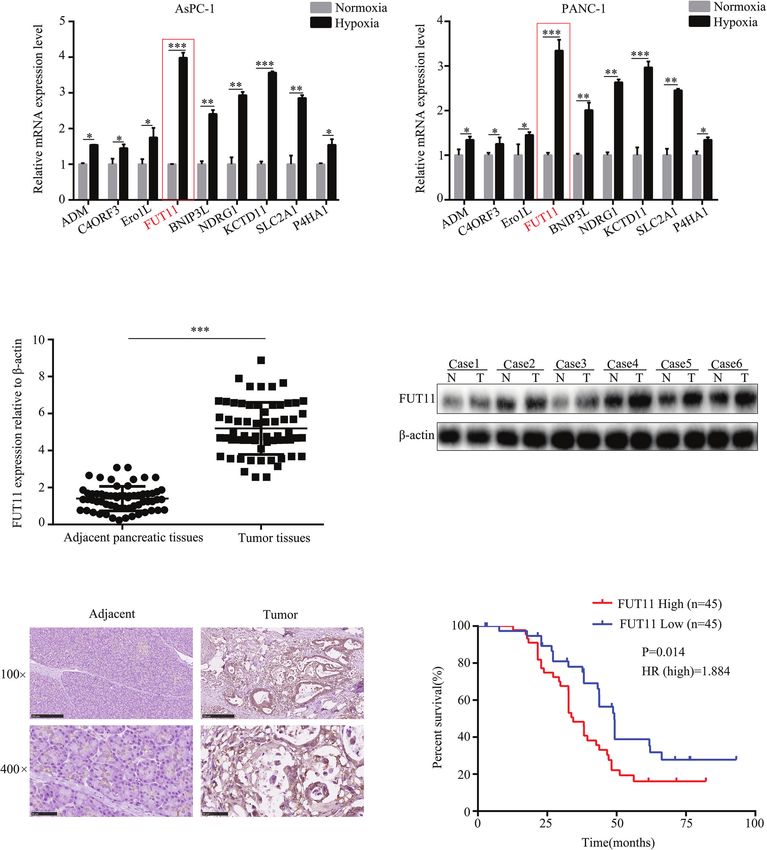
Cao et al. FUT11 Promoted Pancreatic Cancer
A
B C
D E
FIGURE 2 | FUT11 was up-regulated in PC and predicted poor outcome. (A) qRT-PCR analysis on the mRNA expression of ADM, C4ORF3, ERO1L, FUT11 BNIP3L,
KDM3A, MXI1, NDRG1 and KCTD11 in AsPC-1 and PANC-1 cells under normoxia and hypoxia. (B) qRT-PCR analysis of mRNA level of FUT11 in 62 pairs of matched
PC tissue and adjacent normal tissues. (C) Western blot analysis of protein level of FUT11 in PC tissue and adjacent normal tissues. (D) IHC analysis of the protein level
of FUT11 in pancreatic cancer and normal pancreatic tissue. (E) 90 patients with PC were divided into high- and low-expression groups based on the expression of
FUT11. Kaplan survival curve showed the overall survival of high FUT11(red) and low FUT11(blue) expression group. *P < 0.05; **P < 0.01; ***P < 0.001.
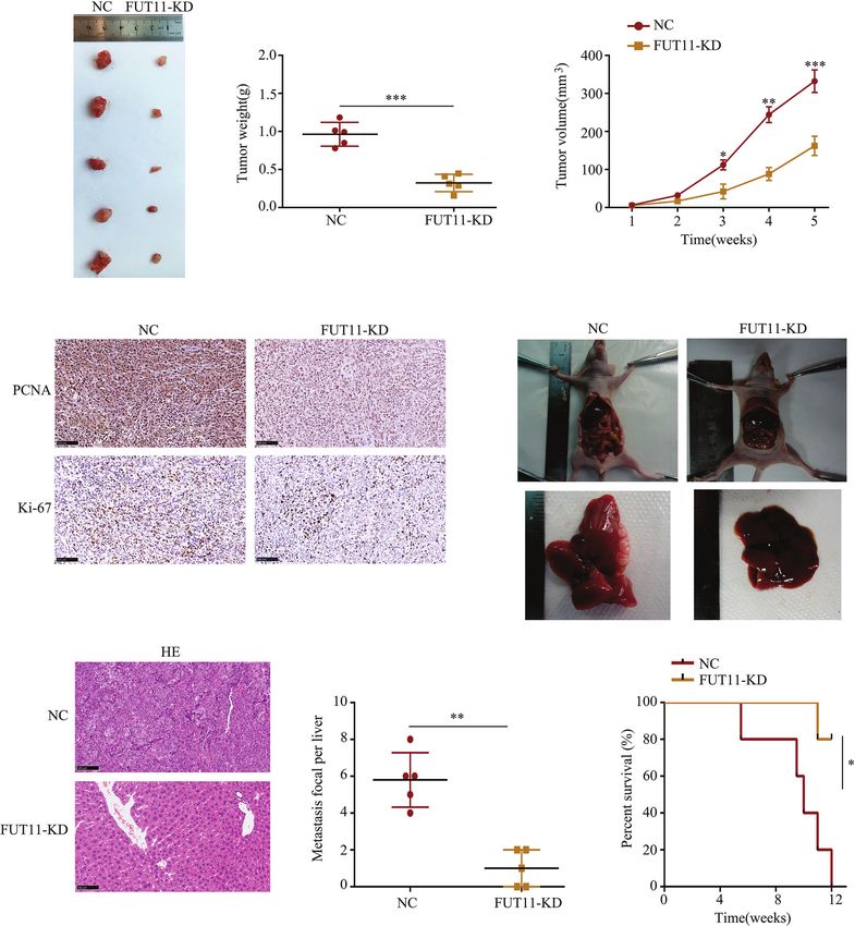
Knockdown of FUT11 Inhibits the PC that KI67 and PCNA was decreased in the tumor tissues with low
Cells Proliferation and Metastasis In Vivo FUT11 expression (Figure 4D). The effects of FUT11 on the
hepatic metastasis of PANC-1 cells was evaluated in by injecting
The effects of FUT11 knockdown in vivo was also determined. the FUT11 knockdown and negative control PANC-1 cells into
We found that tumor tissues derived from FUT11 knockdown the spleen capsule. Results showed that FUT11 knockdown
cells showed slower growth rate and lower tumor weight than significantly reduced the metastatic foci in the liver
that derived negative control cells (all P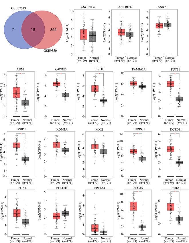
Cao et al.                                                                                                                          FUT11 Promoted Pancreatic Cancer

                 A                                                               B

                 C

                 D

                 E                                                                             F

 FIGURE 3 | FUT11 regulated the proliferation and migration of PC cells under normoxia and hypoxia in vitro. Targeted FUT11 lentivirus and negative control
 lentivirus were used to construct FUT11 knockdown cells and negative control cells, that were cultured either in normoxia or hypoxia. Cells were divided into four
 groups: NC(N), cells transfected with negative lentiviral and cultured in normoxia; FUT11-KD(N), cells with FUT11 knockdown and cultured in normoxia; NC(H), cells
 transfected with negative lentiviral and cultured in hypoxia; FUT11-KD(H), cells with FUT11 knockdown and cultured in hypoxia. (A) The effect of FUT11 on PC cell
 proliferation detected by CCK-8 assay. (B) The effect of FUT11 on PC cell proliferation detected by EDU assay. (C)The effect of FUT11 on PC cell colony forming
 ability detected by colony formation assay. (D) The effect of FUT11 on PC cell migratory ability detected by Transwell assays. (E) Western blotting used to detect the
 protein level of N-cadherin and E-cadherin of each group. (F) Immunofluorescent staining on the expression of N-cadherin and E-cadherin of each group. *P < 0.05;
 **P < 0.01.

FUT11 Co-Localized With PDK1 in PC                                                       spectrometry analysis to determine proteins interacting with
Cells and Regulated the Expression of                                                    FUT11. A total of 700 proteins were found to interact with
PDK1 via Maintaining Its Stability Under                                                 FUT11 as shown in Supplementary Table 3. Among the 700
                                                                                         interacted proteins with FUT11, PDK1 has been revealed as an
Hypoxia
                                                                                         oncogene in pancreatic cancer, indicating that the effect of FUT11
In order to explore the molecular mechanism of FUT11 in PC                               on PC cells might be associated with PDK1. Immunoprecipitation
development, we used immunoprecipitation with mass                                       (IP) and immunofluorescence (IF) analysis demonstrated that

Frontiers in Oncology | www.frontiersin.org                                          7                                         June 2021 | Volume 11 | Article 675991
Cao et al.                                                                                                                            FUT11 Promoted Pancreatic Cancer

                        A                               B                                              C

                   D                                                                         E

                   F                                               G                                              H

 FIGURE 4 | Inhibition of FUT11 suppressed the proliferation and metastasis of PANC-1 cells in vivo. (A) Representative image of tumor tissues in negative control
 group and FUT11 knockdown group. (B) The mean tumor weight of FUT11 knockdown and negative control groups. (C) The proliferative rate of tumor tissues with
 FUT11 knockdown and negative control. (D) IHC staining of Ki-67 and PCNA protein expression in transplanted tumors under different experimental conditions.
 (E) Live metastatic tumor model (n=5). (F) IHC staining and HE images showing the metastatic foci in liver in indicated groups. (G) Statistical analysis of the average
 numbers of visible liver metastatic foci. (H) Kaplan–Meier survival curves for each experimental group, (n=5). *P < 0.05; **P < 0.01; ***P < 0.001.

FUT11 directly bound to and co-localized with PDK1 (Figures 5A,                         of PDK1. The results showed that the degradation of PDK1 was
B). The suppression of FUT11 prominently decreased the protein                          increased in FUT11 knockdown cells (Figure 5D). To investigate
level of FUT11 under normoxia and hypoxia in AsPC-1 and                                 whether FUT11 protect PDK1 via inhibiting ubiquitination, we
PANC-1 cell (Figure 5C). A previous study showed that the                               performed the ubiquitination assay. The results showed that the
members of FUT family can regulate the expression of related                            suppression of FUT11 increased PDK1 ubiquitination under
proteins by stabilizing their binding proteins and decreasing their                     normoxia and hypoxia in AsPC-1 and PANC-1 cell (Figure 5E).
ubiquitination (13). Therefore, we considered that FUT11 may bind                       Moreover, it is interesting that treatment with MG132 (10mM)
to PDK1 and protect it from degradation. We used cycloheximide                          restored the reduction of PDK1 induced by FUT11 suppression
(CHX) to inhibit the synthesis of protein and detect the degradation                    under hypoxia (Figure 5F). Furthermore, the expression of FUT11

Frontiers in Oncology | www.frontiersin.org                                         8                                           June 2021 | Volume 11 | Article 675991
Cao et al. FUT11 Promoted Pancreatic Cancer
A B
C
D
E F
G
FIGURE 5 | FUT11 bound to PDK1 and regulated its expression. (A) Immunoprecipitation on the binding between FUT11 and PDK1. (B) Immunofluorescence
showed FUT11 co-localized with PDK1 in PC cells.(C) Western blot on the expression of FUT11 in sh-scramble and FUT11 knockdown PC cells under normoxia
and hypoxia. (D) The ubiquitination assay showed the ubiquitination level of PDK1 in sh-scramble and FUT11 knockdown PC cells under normoxia and hypoxia.
(E) CHX was used to inhibit the protein synthesis, and the degradation of PDK1 in sh-scramble and FUT11 knockdown cells under hypoxia detected using Western
blot. (F) Western blot on the expression of PDK1 in sh-scramble and FUT11 cells treated with MG132 under hypoxia. (G) IHC images showed the co-expression of
FUT11 and PDK1.
Frontiers in Oncology | www.frontiersin.org 9 June 2021 | Volume 11 | Article 675991Cao et al. FUT11 Promoted Pancreatic Cancer
and PDK1 was investigated in our PC clinical samples, results HIF1a inhibited the proliferation of PC cells under hypoxia,
indicated that both FUT11 and PDK1 co-expressed in the PC while overexpression of FUT11 in HIF1a knockdown cells
tissues (Figure 5G). relieved the suppressive effects of HIF1a knockdown on cell
growth (Figures 8B, C). Similarly, the colony number of cells
PDK1 Overexpression Under Hypoxia with HIF1a inhibition was obviously decreased. Overexpression
Decreased the Inhibitory Effect of FUT11 of FUT11 in HIF1a knockdown cells relieved the inhibitory
Knockdown effects of HIF1a knockdown on colony forming ability under
To verify whether FUT11 promoted the proliferation of hypoxia (Figure 8D). Furthermore, transwell assays
pancreatic cancer via increasing the expression of PDK1, we demonstrated that HIF1a suppression remarkable decreased
overexpressed PDK1 in FUT11 knockdown PC cells. Then, we the migratory ability of PC cells in hypoxia, while
used CCK-8 and EDU assays to monitor the cell viability. The overexpression of FUT11 in HIF1a knockdown cells reversed
results indicated that increased the expression of PDK1 the inhibitory effects of HIF1a knockdown on cell
significantly increased the proliferation of FUT11 low- migration (Figure 8E).
expressed PC cells (Figures 6A, B). In addition, the colony
formation ability of the cells co-transfected with targeting FUT11
lentivirus and PDK1 overexpression lentivirus under hypoxia DISCUSSION
was higher than cells transfected with targeting FUT11 lentivirus
Although the therapy for PC had been improved significantly,
alone (Figure 6C). Similarly, PDK1 overexpressed in FUT11
the prognosis of patients with PC was still poor (15). Moreover,
knockdown PC cells remarkably increased the migratory ability
due to early metastasis, most PC patients lost the best time for
of PC cells (Figure 6D). Results of Western blot showed that
treatment. Recently, increasing evidences showed that the distant
overexpressed PDK1 in FUT11 knockdown cells significantly
metastasis in early stage of PC cells were driven by signals from
increased the protein level of N-cadherin and decreased the
tumor environment, including hypoxia (16). Therefore,
protein level E-cadherin (Figure 6E). Furthermore, western blot
uncovering the mechanism of hypoxia-regulated response in
demonstrated the PDK1 significantly activated the AKT/mTOR
PC cells is critical for the treatment of PC.
pathway, while overexpressed PDK1 in FUT11 knockdown cells
Previous studies had revealed that bioinformatics is a
significantly reversed the inhibitory effects of FUT11 knockdown
powerful tool to identify genes associated with the
on the activation of AKT/mTOR pathway (Figure 6F).
development of tumors, including PC (17). Furthermore,
online database GEO and TCGA storing thousands of gene
FUT11 Was a Target Gene of HIF1a database of tumor tissues that provide enough analytical data.
Hypoxia-inducible factors including HIF1a were the most direct In the current study, we used the bioinformatics tool to identify
hypoxia response elements. To explore the regulatory network of novel hypoxia-related genes. Through analyzing the gene
FUT11, we further determined whether FUT11 was directly expression profile, we found 18 genes were differentially
regulated by HIF1a. After obtaining the motif of HIF1a in expressed between hypoxic and normoxic PC samples. Among
JASPAR database (Figure 7A). we found that there is a hypoxia- these 18 genes, 9 of them including ADM, C4orf3, ERO1L,
responsive element (HRE) in the promoter of FUT11 (Figure 7B). FUT11, BNIP3L, NDRG1, KCTD11, SLC2A1 and P4HA1 were
The results indicated that compared with the control group, hypoxia highly expressed in PC tissues. Furthermore, FUT11 was
significantly increased the luciferase activity in the cells transfected increased the most significant in PC cells under hypoxia
with the vector contained full-length FUT11 promoter, while the condition, up-regulated in PC tissues and predicted poor
lack of HRE reduced the luciferase activity. Furthermore, inhibition prognosis of PC patients. These findings suggested that FUT11
of HIF1a reversed hypoxia-induced luciferase activity (Figure 7C). may be a novel hypoxia-related gene.
Anti-HIF1a antibody was enrolled to perform ChIP assays in The fucosyltransferase (FUT) family are the key enzymes in
PANC-1 cells. Results indicated that the HRE in the FUT11 cell-surface antigen synthesis during various biological processes
promoter was the major region mediating HIF1a-induced such as tumor proliferation, metastasis and drug resistance (18,
transcription (Figure 7D). In addition, it is interesting that 19). At present, a total of 13 members consisted FUT1 to FUT11,
FUT11 was co-expressed with HIF1a in TCGA PC samples protein O-fucosyltransferase 1 (POFUT1) and POFUT2 were
(Figure 7E) and our clinical PC samples (Figure 7F). identified. A number of studies had demonstrated that some
members of the FUTs play roles as oncogenes in various types of
Restoration of FUT11 Reversed the cancers. FUT8 was up-regulated in non-small cell lung cancer
Inhibitory Effects of HIF1a Knockdown and promoted the process of epithelial–mesenchymal transition
on PC Cells (20). POFUT1 increased the activity of Notch1 signaling
To determine whether FUT11 was involved in the biological pathway and promoted the progression of colorectal cancer
function of PC cells induced by HIF1a under hypoxia, we (21). As shown in the previous studies, inhibition of FUTs
constructed negative control cells, FUT11 overexpressed cells, including FUT11 significantly decreased the expression and
HIF1a knockdown cells and FUT11 overexpressed plus HIF1a activity of ERK1/2 and p38 MAPK pathways, as well as the
knockdown cells, and cultured them under hypoxia (Figure 8A). progression of human invasive ductal carcinoma (22). FUT11
CCK-8 and EDU assays results showed that suppression of was highly expressed in gynecological cancers, and
Frontiers in Oncology | www.frontiersin.org 10 June 2021 | Volume 11 | Article 675991Cao et al. FUT11 Promoted Pancreatic Cancer
A B
C
D
E F
FIGURE 6 | Overexpression of PDK1 reversed the inhibitory effect of FUT11 inhibition on PC cell proliferation and migration. Cells were divided into four groups:
negative control group (NC), PDK1 overexpressed group (PDK1-UP), FUT11 knockdown group (FUT11-KD) and PDK1 overexpressed plus FUT11 knockdown
group (PDK1-UP + FUT11-KD). All cells were cultured in hypoxia. (A) CCK-8 assay on cell viability of each group. (B) EDU assay on cell proliferation of each group.
(C) Colony formation on the colony forming ability of cells in each group. (D) Transwell assay on the cell migratory ability of each group. (E) Western blot on the
protein level of FUT11, PDK1, N-cadherin and E-cadherin in each group. (F) Western blot on the protein level of phosphorylated AKT, AKT, phosphorylated mTOR
and mTOR in each group. *P < 0.05; **P < 0.01.
overexpression of FUT11 in patients predicted poor outcome found that FUT11 inhibition significantly decreased proliferation
(23). However, the effect of FUT11 on proliferation and and migration of PC cells in both hypoxic and normoxic
metastasis of human PC cells have not yet been defined. Using environment. These results were in consistent with previous
CCK-8 assay, colony formation assay and transwell assay, we studies. Via performing xenograft tumor model and in vivo
Frontiers in Oncology | www.frontiersin.org 11 June 2021 | Volume 11 | Article 675991Cao et al. FUT11 Promoted Pancreatic Cancer
A B
C D
E F
FIGURE 7 | HIF1a regulates the expression of FUT11 by binding to the HRE of the FUT11 promoter. (A) The motif of HIF1a. (B) The hypoxia response element
(HRE) in the promoter of FUT11 (Vector 1: vector with full-length FUT11 promoter; Vector 2: vector with truncated FUT11 promoter which lack of HRE). (C) PANC‐1
and AsPC-1 cells transfected with either a full‐length or truncated FUT11 promoter‐pGL3 reporter vector and cultured under hypoxia, with or without si‐HIF1a. After
48 hours, luciferase activity was measured using the dual‐luciferase reporter assay system. (D) ChIP assays with anti-HIF1a antibody verifying the binding between
HIF1a and hypoxia response element of the FUT11 promoter under normoxia and hypoxia. (E) Co-expression of FUT11 and HIF1a based on the data from PC
tissues via online database GEPIA. (F) Co-expression of FUT11 and HIF1a based on the data from our clinical PC tissues (n=62). **P < 0.01.
metastatic tumor model, we found that FUT11 inhibition immunoprecipitation with mass spectrometry analysis, PDK1
decreased the PC cells proliferation and metastasis in vivo. was identified as one of the potential downstream genes of
These results suggested that FUT11 was linked to hypoxia, FUT11, which co-expressed with FUT11. Furthermore, using
because it had the potential to regulate PC cells proliferation immunoprecipitation and Western blot, we found that FUT11
and migration under normoxia and hypoxia. directly bound to PDK1 and regulated its expression in normoxia
PDK1 has emerged as an important oncogene in many types and hypoxia. Based on previous studies, FUTs can bind to a
of cancers including PC (24). Lucero-Acuna A et al. has been series of proteins and maintain their stability via blocking the
reported that the expression of PDK1 is up-regulated in human binding site of protease (13). Therefore, we determined whether
PC and promotes cancer cell growth and mobility (25). Xia S FUT11 regulated PDK1 via maintaining its stability. Consistent
et al. showed that knockdown of PDK1 forces cells containing with our speculation, knockdown of FUT11 under hypoxia
activated p21(Ras) to undergo apoptosis in PC cells (26). i n c r e a s e d th e d e g r a d a ti o n of P D K 1 . F u r t h e r m o r e ,
Previous studies have shown that, one of the targets of PDK-1 overexpression of PDK1 in PC under hypoxia relieved the
was AKT, which can be activated by phosphorylation on two inhibitory impacts of FUT11 knockdown on cell proliferation
residues (T308 and S473) for full oncogenic activity (27). and migration.
However, the mechanisms of PDK1 in regulating tumor The relationship among hypoxia-inducible factors, hypoxia
progression is not clear. In the current study, through microenvironment, hypoxia related genes and the development
Frontiers in Oncology | www.frontiersin.org 12 June 2021 | Volume 11 | Article 675991Cao et al. FUT11 Promoted Pancreatic Cancer
A B
C
D
E
FIGURE 8 | Overexpression of FUT11 reversed the inhibitory effects of HIF1a knockdown on PC cell proliferation and migration under hypoxia. PANC-1 and AsPC-1
cells were divided into four groups: negative control (NC); FUT11 overexpression (FUT11-UP); HIF1a inhibition (si-HIF1a); HIF1a inhibition plus FUT11 overexpression (si-
HIF1a+FUT11-UP). All groups of cells were cultured in hypoxia. (A) Western blot on the expression of HIF1a and FUT11 in each group of cells. (B) CCK-8 assay on the
cell viability in each group of cells. (C) EDU assay on the proliferation in each group of cells. (D) Colony formation assay on the colony forming ability in each group of
cells. (E) Transwell assays on the migratory ability in each group of cells. The data were shown as means ± S.D. of three independent assays. *P < 0.05, **P < 0.01.
of PC were widely reported in previous studies. For example, HIF1a which promotes PC cell proliferation and mobility (31).
PAFAH1B2 regulated by HIF1a under hypoxia promoted the In the present study, we provided the first evidence that FUT11
growth and mobility of PC cells (28). MTA1 was regulated by was a novel target gene of HIF1a, which involved in the
HIF-a/VEGF axis and promoted the development of PC (29). biological function mediating by HIF1a under hypoxia.
Similarly, overexpression of hydroxyproline via EGLN/HIF1A is In conclusion, our present study demonstrated that FUT11 is
associated with distant metastasis in PC (30). Similarly, our a new hypoxia related gene, and is overexpressed in pancreatic
previous study also demonstrated that YEATS2 directly targets cancer tissues and related to poor prognosis of pancreatic cancer
Frontiers in Oncology | www.frontiersin.org 13 June 2021 | Volume 11 | Article 675991Cao et al. FUT11 Promoted Pancreatic Cancer
patients. FUT11 is regulated by HIF1a and promotes PC cells AUTHOR CONTRIBUTIONS
proliferation and migration via maintaining the stability of
PDK1 mediates AKT/mTOR signaling pathway. FUT11 could WC, ZZ, ZY, and SL contributed to the experiment design, and
be an effective target for overcoming the hypoxia response of PC. data analysis. RP, HW, WC, and SL contributed to the
experiment implementation, YN, XZ, HC, and SL contributed
to manuscript draft and data analysis. All authors contributed to
the article and approved the submitted version.
DATA AVAILABILITY STATEMENT
The original contributions presented in the study are included in
the article/Supplementary Material. Further inquiries can be
directed to the corresponding authors. SUPPLEMENTARY MATERIAL
The Supplementary Material for this article can be found online at:
https://www.frontiersin.org/articles/10.3389/fonc.2021.675991/
full#supplementary-material
ETHICS STATEMENT
Supplementary Table 1 | The primers used for quantitative real-time PCR (qRT-
The studies involving human participants were reviewed and PCR) and plasmid sequence.
approved by The Ethics Committee of GuiZhou Medical
University Ethics. The patients/participants provided their written Supplementary Table 2 | The Association between FUT11 expression and
clinicopathological features of PC patients.
informed consent to participate in this study. The animal study was
reviewed and approved by Animal Experimental Ethical Inspection Supplementary Table 3 | Mass spectrometry of immunoprecipitation proteins
Form of Guizhou Medical University. interacting with FUT11.
REFERENCES 12. Liang L, Gao C, Li Y, Sun M, Xu J, Li H, et al. miR-125a-3p/FUT5-FUT6 Axis
Mediates Colorectal Cancer Cell Proliferation, Migration, Invasion and
1. Lee KG, Roy V, Laszlo M, Atkins KM, Lin KJ, Tomassian S, et al. Symptom Pathological Angiogenesis Via PI3K-Akt Pathway. Cell Death Dis (2017) 8
Management in Pancreatic Cancer. Curr Treat Options Oncol (2021) 22(1):8. (8):e2968. doi: 10.1038/cddis.2017.352
doi: 10.1007/s11864-020-00801-4 13. Kumar S, Das S, Rachagani S, Kaur S, Joshi S, Johansson SL, et al. NCOA3-
2. Chin V, Nagrial A, Sjoquist K, O’Connor CA, Chantrill L, Biankin AV, et al. Mediated Upregulation of Mucin Expression Via Transcriptional and Post-
Chemotherapy and Radiotherapy for Advanced Pancreatic Cancer. Cochrane Translational Changes During the Development of Pancreatic Cancer.
Database Syst Rev (2018) 3:CD011044. doi: 10.1002/14651858.CD011044.pub2 Oncogene (2015) 34(37):4879–89. doi: 10.1038/onc.2014.409
3. Hirata E, Sahai E. Tumor Microenvironment and Differential Responses to 14. Lin G, Zhao R, Wang Y, Han J, Gu Y, Pan Y, et al. Dynamic Analysis of N-
Therapy. Cold Spring Harb Perspect Med (2017) 7(7):a026781. doi: 10.1101/ glycomic and Transcriptomic Changes in the Development of Ovarian Cancer
cshperspect.a026781 Cell Line A2780 to its Three Cisplatin-Resistant Variants. Ann Transl Med
4. Wozniak M, Pastuch Gawolek G, Makuch S, Wisniewski J, Ziolkowski P, (2015) 8(6):289. doi: 10.21037/atm.2020.03.12
Szeja W, et al. Overcoming Hypoxia-Induced Chemoresistance in Cancer 15. Kurihara K, Hanada K, Shimizu A. Endoscopic Ultrasonography Diagnosis of
Using a Novel Glycoconjugate of Methotrexate. Pharm (Basel) (2020) 14 Early Pancreatic Cancer. Diagn (Basel) (2020) 10(12):1086. doi: 10.3390/
(1):13. doi: 10.3390/ph14010013 diagnostics10121086
5. Zhang Q, Han Z, Zhu Y, Chen J, Li W. Role of Hypoxia Inducible Factor-1 in 16. Bhandari V, Hoey C, Liu LY, Lalonde E, Ray J, Livingstone J, et al. Molecular
Cancer Stem Cells (Review). Mol Med Rep (2021) 24(1):17. doi: 10.3892/ Landmarks of Tumor Hypoxia Across Cancer Types. Nat Genet (2019) 51
mmr.2020.11655 (2):308–18. doi: 10.1038/s41588-018-0318-2
6. Guo FJ, Shao YP, Wang YP, Jin YM, Liu SS, Wang QY. Mir-92 Stimulates 17. Lv K, Yang J, Sun J, Guan J. Identification of Key Candidate Genes for
VEGF by Inhibiting Von Hippel-Lindau Gene Product in Epithelial Ovarian Pancreatic Cancer by Bioinformatics Analysis. Exp Ther Med (2019) 18
Cancer. J Biol Regul Homeost Agents (2017) 31(3):615–24. (1):451–8. doi: 10.3892/etm.2019.7619
7. Ebright RY, Zachariah MA, Micalizzi DS, Wittner BS, Niederhoffer KL, 18. Gan CZ, Li G, Luo QS, Li HM. miR-339-5p Downregulation Contributes to
Nieman LT, et al. HIF1A Signaling Selectively Supports Proliferation of Taxol Resistance in Small-Cell Lung Cancer by Targeting Alpha1,2-
Breast Cancer in the Brain. Nat Commun (2020) 11(1):6311. doi: 10.1038/ fucosyltransferase 1. IUBMB Life (2017) 69(11):841–9. doi: 10.1002/iub.1679
s41467-020-20144-w 19. Wang S, Zhang X, Yang C, Xu S. MicroRNA-198-5p Inhibits the Migration and
8. Kutkowska J, Strzadala L, Rapak A. Hypoxia Increases the Apoptotic Invasion of Non-Small Lung Cancer Cells by Targeting Fucosyltransferase 8. Clin
Response to Betulinic Acid and Betulin in Human Non-Small Cell Lung Exp Pharmacol Physiol (2019) 46(10):955–67. doi: 10.1111/1440-1681
Cancer Cells. Chem Biol Interact (2021) 333:109320. doi: 10.1016/ 20. Li F, Zhao S, Cui Y, Guo T, Qiang J, Xie Q, et al. Alpha1,6-Fucosyltransferase
j.cbi.2020.109320 (FUT8) Regulates the Cancer-Promoting Capacity of Cancer-Associated Fibroblasts
9. Cui XG, Han ZT, He SH, Wu XD, Chen TR, Shao CH, et al. HIF1/2alpha (Cafs) by Modifying EGFR Core Fucosylation (CF) in Non-Small Cell Lung Cancer
Mediates Hypoxia-Induced LDHA Expression in Human Pancreatic Cancer (NSCLC). Am J Cancer Res (2020) 10(3):816–37. doi: 10.21203/rs.2.18620/v1
Cells. Oncotarget (2017) 8(15):24840–52. doi: 10.18632/oncotarget.15266 21. Du Y, Li D, Li N, Su C, Yang C, Lin C, et al. POFUT1 Promotes Colorectal
10. Fan LF, Diao LM, Chen DJ, Liu MQ, Zhu LQ, Li HG, et al. Expression of Cancer Development Through the Activation of Notch1 Signaling. Cell Death
HIF-1 Alpha and its Relationship to Apoptosis and Proliferation in Lung Dis (2018) 9(10):995. doi: 10.1038/s41419-018-1055-2
Cancer. Ai Zheng (2002) 21(3):254–8. 22. Carrascal MA, Silva M, Ramalho JS, Pen C, Martins M, Pascoal C, et al.
11. Hwang H, Jeong HK, Lee HK, Park GW, Lee JY, Lee SY, et al. Machine Learning Inhibition of Fucosylation in Human Invasive Ductal Carcinoma Reduces
Classifies Core and Outer Fucosylation of N-Glycoproteins Using Mass E-selectin Ligand Expression, Cell Proliferation, and ERK1/2 and P38
Spectrometry. Sci Rep (2020) 10(1):318. doi: 10.1038/s41598-019-57274-1 MAPK Activation. Mol Oncol (2018) 12(5):579–93. doi: 10.1002/1878-0261
Frontiers in Oncology | www.frontiersin.org 14 June 2021 | Volume 11 | Article 675991Cao et al. FUT11 Promoted Pancreatic Cancer
23. Zhang X, Wang Y. Identification of Hub Genes and Key Pathways Associated J Recept Signal Transduct Res (2018) 38(4):352–8. doi: 10.1080/10799893.
With the Progression of Gynecological Cancer. Oncol Lett (2019) 18(6):6516– 2018.1531887
24. doi: 10.3892/ol.2019.11004 30. Chiba N, Sunamura M, Nakagawa M, Koganezawa I, Yokozuka K, Kobayashi T,
24. Wang C, Liu H, Qiu Q, Zhang Z, Gu Y, He Z. TCRP1 Promotes NIH/3T3 Cell et al. Overexpression of Hydroxyproline Via EGLN/HIF1A Is Associated With
Transformation by Over-Activating PDK1 and AKT1. Oncogenesis (2017) 6 Distant Metastasis in Pancreatic Cancer. Am J Cancer Res (2020) 10(8):2570–81.
(4):e323. doi: 10.1038/oncsis.2017.18 31. Zeng Z, Lei S, He Z, Chen T, Jiang J. YEATS2 Is a Target of HIF1alpha and
25. Lucero-Acuna A, Jeffery JJ, Abril ER, Nagle RB, Guzman R, Pagel MD, et al. Promotes Pancreatic Cancer Cell Proliferation and Migration. J Cell Physiol
Nanoparticle Delivery of an AKT/PDK1 Inhibitor Improves the Therapeutic Effect (2021) 236(3):2087–98. doi: 10.1002/jcp.29995
in Pancreatic Cancer. Int J Nanomed (2014) 9:5653–65. doi: 10.2147/IJN.S68511
26. Xia S, Chen Z, Forman LW, Faller DV. Pkcdelta Survival Signaling in Cells Conflict of Interest: The authors declare that the research was conducted in the
Containing an Activated p21Ras Protein Requires PDK1. Cell Signal (2009) 21 absence of any commercial or financial relationships that could be construed as a
(4):502–8. doi: 10.1016/j.cellsig.2008 potential conflict of interest.
27. Lin HJ, Hsieh FC, Song H, Lin J. Elevated Phosphorylation and Activation of
PDK-1/AKT Pathway in Human Breast Cancer. Br J Cancer (2005) 93 Copyright © 2021 Cao, Zeng, Pan, Wu, Zhang, Chen, Nie, Yu and Lei. This is an
(12):1372–81. doi: 10.1038/sj.bjc.6602862 open-access article distributed under the terms of the Creative Commons Attribution
28. Ma C, Guo Y, Zhang Y, Duo A, Jia Y, Liu C, et al. PAFAH1B2 is a HIF1a License (CC BY). The use, distribution or reproduction in other forums is permitted,
Target Gene and promSilvaotes Metastasis in Pancreatic Cancer. Biochem provided the original author(s) and the copyright owner(s) are credited and that the
Biophys Res Commun (2018) 501(3):654–60. doi: 10.1016/j.bbrc.2018.05.039 original publication in this journal is cited, in accordance with accepted academic
29. Sun X, Zhang Y, Li B, Yang H. MTA1 Promotes the Invasion and Migration of practice. No use, distribution or reproduction is permitted which does not comply with
Pancreatic Cancer Cells Potentially Through the HIF-alpha/VEGF Pathway. these terms.
Frontiers in Oncology | www.frontiersin.org 15 June 2021 | Volume 11 | Article 675991You can also read