Ubiquitin specific peptidase 17 promotes cisplatin resistance via PI3K/AKT activation in non small cell lung cancer
←
→
Page content transcription
If your browser does not render page correctly, please read the page content below
ONCOLOGY LETTERS 20: 67-74, 2020

      Ubiquitin‑specific peptidase 17 promotes cisplatin resistance
        via PI3K/AKT activation in non‑small cell lung cancer
                         SHENGCHAO ZHANG, ZHENGLANG XU, JUN YUAN and HAO CHEN

    Department of Thoracic Surgery, Qingpu Branch Zhongshan Hospital, Fudan University, Shanghai 201700, P.R. China

                                    Received November 8, 2018; Accepted November 22, 2019

                                                     DOI: 10.3892/ol.2020.11568

Abstract. The suppression of ubiquitin‑specific peptidase             show an initial positive response to cisplatin treatment, but
17 (USP17) has previously been found to result in reduced             in many cases this response is not sustained and cancer cells
tumorigenesis and invasion of non‑small cell lung cancer              become resistant to the treatment, this has become a major
(NSCLC) cells. However, the functions and underlying mech-            clinical challenge in the treatment of NSCLC (4). Therefore,
anisms of USP17 in NSCLC progression remain unclear. In               the identification of novel therapeutic targets in NSCLC treat-
the present study, cisplatin treatment was found to upregulate        ment that can complement current therapy is urgently required.
USP17 expression in a dose‑dependent manner. Furthermore,                 The ubiquitin‑proteasome system regulates cellular protein
USP17‑overexpressing (USP17‑OE) NSCLC A549 and H1299                  levels with specificity and precision to optimize cellular
cells were generated for mechanistic studies. The results from        functions (5,6). Moreover, certain studies have demonstrated
the Cell Counting Kit‑8 assay revealed increased cell prolif-         that ubiquitination can modify protein functions, including
eration in USP17‑OE cells compared with that of control cells.        the regulation of subcellular localization and protein‑protein
Moreover, the viability of USP17‑OE cells was significantly           interactions (5‑7). The association between ubiquitin and
higher than that of the control cells, when treated with cisplatin.   tumor biology has long been recognized, and the suppression
The results of the biochemical studies demonstrated enhanced          of the proteasome has proven to be effective in the treatment of
PI3K and AKT phosphorylation in USP17‑OE NSCLC cells,                 various types of cancer, such as myeloma and gastrointestinal
whereas USP17‑knockdown decreased these levels of phos-               cancer (6). Deubiquitinating enzymes (DUBs) can reverse
phorylation. By contrast, an AKT inhibitor abolished the              protein ubiquitination by digesting ubiquitin chains (8).
USP17‑mediated enhancement of proliferation. Moreover,                    Ubiquitin‑specific protease 17 (USP17), a DUB, is involved
suppression of USP17 or the combination of the AKT inhibitor          in the regulation of inflammation (9) and cell motility (10),
and cisplatin significantly reduced cell viability. Overall, the      the development of Th17 cells (11) and oncogenesis (12‑17).
results of the present study suggest that PI3K/AKT activation         It has been found that USP17 is highly expressed in several
is the underlying mechanism of USP17‑mediated cisplatin               types of tumor, including colon, esophageal and cervical
resistance in NSCLC.                                                  tumors (14). In addition, USP17 regulates the Ras pathway by
                                                                      altering the intracellular localization of Ras and other small
Introduction                                                          GTPases, which are crucial regulators of cellular prolifera-
                                                                      tion and migration (10). USP17 is required for the trafficking
Lung cancer is the one of the leading causes of cancer‑related        and oncogenic signaling of mutant epidermal growth factor
death worldwide (1), and non‑small cell lung cancer (NSCLC)           receptor (EGFR) in NSCLC cells (15). These studies verified
accounts for almost 80% of these deaths (2). Cisplatin‑based          the role of USP17 in promoting tumor growth and metastasis.
chemotherapy is the main treatment used for patients with             McFarlane et al (16) reported upregulation of USP17 in
major NSCLC (2). However, the long‑term prognosis for                 patients with NSCLC. Moreover, patients with USP17‑positive
patients with NSCLC still remains poor, with the 5‑year               tumors had significantly shorter recurrence‑free survival
survival rate being only ~11% (3). Patients with NSCLC often          times compared with those with USP17‑negative tumors,
                                                                      and USP17 expression was associated with the recurrence of
                                                                      disease at distant sites. In addition, an in vivo study, in which
                                                                      human NSCLC cells were inoculated into nude mice, found
                                                                      that the suppression of USP17 in NSCLC cells inhibited tumor
Correspondence to: Dr Hao Chen, Department of Thoracic
Surgery, Qingpu Branch Zhongshan Hospital, Fudan University,          growth and invasion (17). However, the biological function
1158 East Gongyuan Road, Qingpu, Shanghai 201700, P.R. China          of USP17 that directly regulates NSCLC progression has not
E‑mail: h.chen@fudan.edu.cn                                           been studied fully.
                                                                          The re‑emergence of cancer cells is often due to the activa-
Key words: ubiquitin‑specific peptidase 17, non‑small cell lung       tion of survival signals, including increased activation of the
cancer, AKT inhibitor, cisplatin                                      PI3K/AKT pathway (18‑20), which has been associated with
                                                                      NSCLC progression. The PI3K/AKT pathway is an important
                                                                      pathway downstream of EGFR. Deregulation of this pathway,
68 ZHANG et al: USP17 PROMOTES CISPLATIN RESISTANCE VIA PI3K/AKT ACTIVATION IN NSCLC
due to gene amplifications, activating oncogene mutations or Cell Counting Kit‑8 assay (CCK‑8). Cells (5x10³/well) were
the loss of PTEN, has been observed in several types of human treated with cisplatin (1 µM) or MK2206 (1 µM; both purchased
cancer, such as colorectal, gastric, lung, ovarian and thyroid from Selleck Chemicals) for 0, 24, 48, 72, 96 and 120 h. The
cancer (20‑23). CCK‑8 assay was conducted according to the kit instructions
In the present study, the aim was to explore the functions (cat. no. CK04; Dojindo Molecular Technologies, Inc.). Cells with
and underlying molecular mechanisms of USP17 in NSCLC or without USP17 overexpression (OE) and cells treated with
cells. Moreover, the effects of inhibiting of USP17 downstream MK2206 were examined. Briefly, cells in logarithmic growth
PI3K/AKT pathway in cisplatin sensitivity of NSCLC cells phase were trypsin‑digested and resuspended in RPMI‑1640
were also investigated. medium. Cells were plated at equal densities (2,000 cells/100 µl
per well) in 96‑well plates and incubated at 37˚C and 5% CO2
Materials and methods for continuous detection over a 5‑day period. At the beginning
of the second day, cell growth was terminated by the addition of
Cell lines. The human NSCLC A549 and H1299 cell lines were 10 µl of CCK‑8 solution (5 mg/ml) to the culture medium. After
purchased from The Cell Bank of Type Culture Collection of 2 h, the OD490 values were determined using a microplate
the Chinese Academy of Sciences. A549 cells were main- reader (BioTek Instruments, Inc.).
tained in Dulbecco's modified Eagle's medium, and H1299
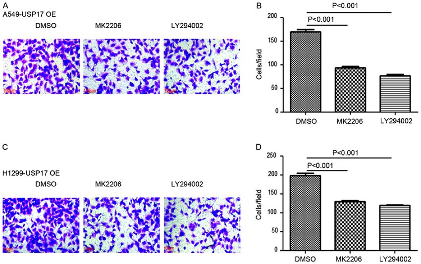
cells were maintained in RPMI‑1640 medium. All media were Transwell assay. USP17‑OE cells and those (2x10 4/well)
supplemented with 10% heat‑inactivated fetal bovine serum treated with MK2206 (1 µM) or LY294002 (5 µM; both
(FBS; Invitrogen; Thermo Fisher Scientific, Inc.), penicillin purchased from Selleck Chemicals) for 48 h were examined.
(100 U/ml), and streptomycin (100 µg/ml) in a humidified Cells (2x10⁴) were detached and resuspended in serum‑free
atmosphere of 5% CO2 at 37˚C. All cells were confirmed to be medium and seeded in the upper chamber of Matrigel‑coated
free from mycoplasma contamination. Transwell (precoating with Matrigel at 4˚C for 60 min) inserts
with a pore size of 8 µm. Culture medium containing 10%
Plasmids and reagents. USP17 short hairpin (sh)RNA FBS as a chemoattractant was added to the lower chamber.
and USP17 overexpression lentiviruses were purchased After 24 h of incubation, cells on the upper surface of the insert
from Hanyin Biotech Co. Polybrene (cat. no. 107689; were gently removed with a cotton swab. Invasive cells (on the
Sigma‑Aldrich; Merck KGaA) was used as an infection lower surface of the insert) were fixed with 4% paraformal-
reagent. The target sequence for USP17 shRNA‑knockdown dehyde (Sigma‑Aldrich; Merck KGaA) at room temperature
(KD) was 5'‑CTCT TGAGAATGTGCCGAT‑3' (the shRNA for 15 min, stained with crystal violet (1%) at room tempera-
was packaged into the lentivirus). The negative control (NC) ture for 60 min, and counted under a microscope (Olympus,
comprised an empty vector without target sequences. To CKX31); five random microscopic fields were examined for
generate stable cell lines, supernatant containing lentivirus each insert using magnification, x200.
(1x10⁶ TU) was added to A549 and H1299 cells (1x10⁵/well),
which were subsequently screened with 1 µg/ml puromycin Western blot analysis. Total protein was extracted from cells
for 2 weeks. Experiments were performed 72 h after using RIPA buffer (Beyotime Institute of Biotechnology).
infection. Protein was quantified using a BCA assay. Protein lysates
(50 µg/lane) were separated via 8‑10% SDS‑PAGE and
Reverse transcription‑quantitative (RT‑q)PCR. Total RNA transferred onto nitrocellulose membranes. After blocking
was extracted from cells using TRIzol® (Invitrogen; Thermo with 5% fat‑free milk at room temperature for 30 min,
Fisher Scientific, Inc.). Total RNA (100 ng) was used for cDNA the membranes were incubated with primary antibodies
synthesis using the Stratagene AffinityScript QPCR cDNA (1:500) at 4˚C overnight. The primary antibodies used were:
Synthesis kit (Agilent Technologies, Inc.). The temperature Goat anti‑human USP17 (cat. no. AP5491b; Abgent, Inc.),
protocol for the RT step was 5 min at 65˚C, 60 min at 42˚C and mouse anti‑actin (cat. no. 3700P), anti‑human AKT (cat.
15 min at 70˚C. The cDNA samples were diluted 10‑fold with no. 4685), anti‑human phosphorylated (p)‑AKT (cat. no. 4060),
nuclease‑free H2O, of which 2 µl was combined with Brilliant anti‑human p‑PI3K (cat. no. 4228) and anti‑human PI3K (cat.
III Ultra‑Fast SYBR® Green qPCR Master mix (Applied no. 4257; all purchased from Cell Signaling Technology, Inc.).
Biosystems; Thermo Fisher Scientific, Inc.). Human RPL13A Following this, membranes were incubated with horseradish
was used as an internal reference control. The primer sequences peroxidase‑conjugated secondary antibodies (1:3,000; cat.
were as follows: Human USP17 forward, 5'‑GAGATTCTC nos. 705‑035‑147, 705‑035‑150 and 705‑035‑152; Jackson
CGATGTCACAGG C‑3' and reverse, 5'‑TCCGTCGTGACA ImmunoResearch Laboratories, Inc.) at room temperature
ACTCCACCCA‑3'; human RPL13A forward, 5'‑CTCA AG for 60 min. Immunoreactive proteins were visualized using
GTGT TTGACG GCATCC‑3' and reverse, 5'‑TACT TCCAG an enhanced chemiluminescence reagent (EMD Millipore).
CCAACCTCGTGAG ‑3'. The relative expression of target Relative expression of p‑PI3K and p‑Akt was normalized to
genes was determined using the 2‑∆∆Cq method (24). The qPCR that of actin, and then relative to the normalized total PI3K
cycling conditions comprised an initial denaturation step of or Akt values. Quantity One software version 4.6.9 (Bio‑Rad
3 min at 95˚C, followed by 45 cycles at 95˚C (10 sec) and 58˚C Laboratories, Inc.) was used to quantify the relative band
(45 sec); data were acquired at the end of the annealing/exten- intensities.
sion phase. Melt curve analysis was performed at the end of
each run between 58‑95˚C and the data were analyzed using Statistical analysis. Quantitative variables were compared
Microsoft Excel 2013 (Microsoft Corporation). using one‑way ANOVA to compare differences between twoONCOLOGY LETTERS 20: 67-74, 2020 69 Figure 1. Increased expression of USP17 is observed in non‑small cell lung cancer cells treated with cisplatin. Increasing concentrations of cisplatin increased the protein expression level of USP17 in a dose‑dependent manner, as shown in (A) western blot images and the (B) corresponding histogram, in A549 cells; and (C) western blot images and the (D) corresponding histogram, in H1299 cells. USP, ubiquitin‑specific peptidase. or more groups, followed by Tukey's test for post‑hoc analysis. results demonstrate that the overexpression of USP17 increases Statistical analysis was performed using SPSS version 19.0 proliferation and viability in NSCLC cells, independent of (IBM Corp.) and GraphPad Prism version 5.0 (GraphPad cisplatin treatment. Software, Inc.). P
70                      ZHANG et al: USP17 PROMOTES CISPLATIN RESISTANCE VIA PI3K/AKT ACTIVATION IN NSCLC

Figure 2. USP17‑OE in non‑small cell lung cancer cells. Reverse transcription‑quantitative PCR analysis of USP17‑OE efficiency in (A) A549 and (B) H1299
cells. Western blot analysis of USP17‑OE efficiency in (C) A549 and (D) H1299 cells. USP, ubiquitin‑specific peptidase; NC, negative control; OE, overexpression.

Figure 3. Overexpression of ubiquitin‑specific peptidase 17 increases the proliferation and viability of non‑small cell lung cancer cells. Cell Counting Kit‑8
analysis comparing USP17‑OE with the NC in (A) A549 and (B) H1299 cells treated with DMSO; and (C) A549 and (D) H1299 cells treated with cisplatin
(1 µM). ***P
ONCOLOGY LETTERS 20: 67-74, 2020                                                               71

Figure 4. USP17‑OE enhances activation of the PI3K/AKT pathway in non‑small cell lung cancer cells. Western blot analysis of p‑PI3K, PI3K, p‑AKT and
AKT levels in (A‑C) A549 and (D‑F) H1299 cells with or without USP17‑OE. USP, ubiquitin‑specific peptidase; OE, overexpression; NC, negative control;
p, phosphorylated.

Figure 5. Inhibition of AKT activation abolishes USP17‑mediated enhancement of proliferation and viability in USP17‑OE cells. Cell Counting Kit‑8 assay in
USP17‑OE A549 (A and C) and H1299 cells (B and D) treated with DMSO control or MK2206, alone or in combination with cisplatin. ***P
72                     ZHANG et al: USP17 PROMOTES CISPLATIN RESISTANCE VIA PI3K/AKT ACTIVATION IN NSCLC

Figure 6. USP17 suppression decreases PI3K/AKT pathway activation and the viability of non‑small cell lung cancer cells. Western blot analysis of p‑PI3K,
PI3K, p‑AKT and AKT protein levels in (A‑C) A549 and (D‑F) H1299 cells with or without USP17 KD. (G) Cell Counting Kit‑8 assay in USP17 KD and
NC (G) A549 and (H) H1299 cells treated with cisplatin. ***P
ONCOLOGY LETTERS 20: 67-74, 2020                                                      73

   Overall, the findings of the present study demonstrate              demonstrate the therapeutic potential of targeting PI3K/AKT
the promotion of NSCLC cell proliferation and viability by             pathways downstream of USP17 to prevent NSCLC progression.
USP17, via the activation of the PI3K/AKT pathway.
                                                                       Acknowledgements
Discussion
                                                                       Not applicable.
The USP family is one of the five subfamilies of DUB enzymes,
which cleave polyubiquitin chains from proteins, a number of           Funding
studies have reported that USP17 has oncogenic characteristics.
Firstly, it has been shown that high levels of USP17 in lung,          The present study was supported by the Shanghai Qingpu
colon, esophageal and cervical tumor samples promoted G1/S             District Health and Planning Commission Research Fund
transition and cellular proliferation (14‑17). Additionally, it was    (grant no. W2017‑21) and the Qingpu District Science and
demonstrated that USP17 was expressed highly in NSCLC                  Technology Commission (grant no. QKY2017‑03).
tissues, and patients with high levels of USP17 exhibited lower
survival rates (16,17). Moreover, USP17 can be induced by cyto-        Availability of data and materials
kines such as interleukin (IL)‑4 and IL‑6 (28). In the present
study, it was found that cisplatin treatment upregulated USP17         The datasets used and/or analyzed during the current study are
expression in a dose‑dependent manner. Moreover, increased             available from the corresponding author on reasonable request.
cell proliferation was found in USP17‑OE cells compared
with that of control cells, which is consistent with previous          Authors' contributions
studies (14‑17). Furthermore, it was demonstrated that the
viability of USP17‑OE cells was significantly higher than that of      SZ and HC contributed to designing research studies,
the control cells, when treated with cisplatin. In the present study   conducting experiments, acquiring data, analyzing data,
USP17 regulated cisplatin sensitivity in NSCLC. USP17 was              providing reagents and writing the manuscript. ZX and JY
initially identified as a regulator of cell viability via signaling    contributed to conducting experiments and acquiring data.All
pathways associated with cell death in cervical cancer (29).           authors read and approved the final manuscript.
    It was previously reported that USP17 expression could
regulate Ras cellular localization and activation, via the deubiq-     Ethics approval and consent to participate
uitination of Ras‑converting enzyme 1, thereby inhibiting
phosphorylation of the downstream kinases, dual specificity            Not applicable.
mitogen‑activated protein kinase kinase and ERK (30). In
osteosarcoma, USP17 facilitated cell migration and invasion            Patient consent for publication
by deubiquitinating and stabilizing SMAD4 (31). USP17‑ and
Skp1‑cullin‑1‑F‑box protein β F‑box/WD repeat‑containing               Not applicable.
protein 1A‑regulated degradation of differentially expressed
in chondrocytes protein 1 has been shown to control the                Competing interests
DNA‑damage response (32,33), thus indicating that the
expression of USP17 may be associated with sensitivity to              The authors declare that they have no competing interests.
chemotherapeutics, including cisplatin.
    In the present study, USP17 was found to promote the               References
growth of NSCLC cells via the activation of the PI3K/AKT
pathway. Aberrant activation of the PI3K/AKT pathway is                 1. Mao Y, Yang D and Krasna MJ: Epidemiology of lung cancer.
often detected in numerous types of human cancer; hence,                   Surg Oncol Clin N Am 25: 439‑445, 2016.
                                                                        2. Azar FE, Azami‑Aghdash S, Pournaghi‑Azar F, Mazdaki A,
targeting this pathway may have therapeutic potential for the              Rezapour A, Ebrahimi P and Yousefzadeh N: Cost‑effectiveness
management of these tumors (34‑37). Emerging evidence                      of lung cancer screening and treatment methods: A systematic
                                                                           review of systematic reviews. BMC Health Serv Res 17: 413,
indicates that the activation of PI3K/AKT signaling by                     2017.
hypoxia may be a contributing factor to drug resistance in              3. Jacobsen MM, Silverstein SC, Quinn M, Waterston LB,
certain types of human cancer, including NSCLC and prostate                Thomas CA, Benneyan JC and Han PKJ: Timeliness of access to
                                                                           lung cancer diagnosis and treatment: A scoping literature review.
cancer (38‑40). PI3K activation is regulated by various mole-              Lung Cancer 112: 156‑164, 2017.
cules in NSCLC, such as DIX domain‑containing protein 1,                4. Giaccone G: Clinical perspectives on platinum resistance.
GRB2‑associated‑binding protein 2 and microRNAs (41‑43).                   Drugs 59: 9‑37, 2000.
    There are limitations of the present study. Firstly, the func-      5. D'Andrea A and Pellman D: Deubiquitinating enzymes: A new
                                                                           class of biological regulators. Crit Rev Biochem Mol Biol 33:
tions of USP17 in the response of NSCLC cells to cisplatin                 337‑352, 1998.
treatment require further investigation, specifically using             6. Weathington NM and Mallampalli RK: Emerging therapies
in vivo animal models (44,45). Moreover, as the underlying                 targeting the ubiquitin proteasome system in cancer. J Clin
                                                                           Invest 124: 6‑12, 2014.
mechanisms of USP17 in the cisplatin response have not been             7. Welchman RL, Gordon C and Mayer RJ: Ubiquitin and ubiq-
investigated in this study, it should be examined in the future.           uitin‑like proteins as multifunctional signals. Nat Rev Mol Cell
    Considerable effort has been made to identify agents that              Biol 6: 599‑609, 2005.
                                                                        8. Nijman SM, Luna‑Vargas MP, Velds A, Brummelkamp TR,
target the activity of DUBs. However, no effective drugs have yet          Dirac AM, Sixma TK and Bernards R: A genomic and functional
entered into clinical trials. The findings from the present study          inventory of deubiquitinating enzymes. Cell 123: 773‑786, 2005.
74 ZHANG et al: USP17 PROMOTES CISPLATIN RESISTANCE VIA PI3K/AKT ACTIVATION IN NSCLC
9. Song H, Tao L, Chen C, Pan L, Hao J, Ni Y, Li D, Li B and Shi G: 28. Baek KH: Cytokine‑regulated protein degradation by the ubiqui-
USP17‑mediated deubiquitination and stabilization of HDAC2 tination system. Curr Protein Pept Sci 7: 171‑177, 2006.
in cigarette smoke extract‑induced inflammation. Int J Clin Exp 29. Shin JM, Yoo KJ, Kim MS, Kim D and Baek KH: Hyaluronan‑
Pathol 8: 10707‑10715, 2015. and RNA‑binding deubiquitinating enzymes of USP17 family
10. de la Vega M, Kelvin AA, Dunican DJ, McFarlane C, Burrows JF, members associated with cell viability. BMC Genomics 7: 292,
Jaworski J, Stevenson NJ, Dib K, Rappoport JZ, Scott CJ, et al: The 2006.
deubiquitinating enzyme USP17 is essential for GTPase subcel- 30. Burrows JF, Kelvin AA, McFarlane C, Burden RE, McGrattan MJ,
lular localization and cell motility. Nat Commun 2: 259, 2011. De la Vega M, Govender U, Quinn DJ, Dib K, Gadina M, et al:
11. Han L, Yang J, Wang X, Wu Q, Yin S, Li Z, Zhang J, Xing Y, USP17 regulates Ras activation and cell proliferation by blocking
Chen Z, Tsun A, et al: The E3 deubiquitinase USP17 is a posi- RCE1 activity. J Biol Chem 284: 9587‑9595, 2009.
tive regulator of retinoic acid‑related orphan nuclear receptor γt 31. Song C, Liu W and Li J: USP17 is upregulated in osteosarcoma
(RORγt) in Th17 cells. J Biol Chem 289: 25546‑25555, 2014. and promotes cell proliferation, metastasis, and epithelial‑mesen-
12. Borbely G, Haldosen LA, Dahlman‑Wright K and Zhao C: chymal transition through stabilizing SMAD4. Tumour Biol 39:
Induction of USP17 by combining BET and HDAC inhibitors in 1010428317717138, 2017.
breast cancer cells. Oncotarget 6: 33623‑33635, 2015. 32. Wang M, He SF, Liu LL, Sun XX, Yang F, Ge Q, Wong WK and
13. Hu M, Chen H, Han C, Lan J, Xu Y, Li C, Xue Y and Lou M: Meng JY: Potential role of ZEB1 as a DNA repair regulator in
Expression and functional implications of USP17 in glioma. colorectal cancer cells revealed by cancer‑associated promoter
Neurosci Lett 616: 125‑131, 2016. profiling. Oncol Rep 38: 1941‑1948, 2017.
14. McFarlane C, Kelvin AA, de la Vega M, Govender U, Scott CJ, 33. Kim J, D'Annibale S, Magliozzi R, Low TY, Jansen P,
Burrows JF and Johnston JA: The deubiquitinating enzyme Shaltiel IA, Mohammed S, Heck AJ, Medema RH and
USP17 is highly expressed in tumor biopsies, is cell cycle Guardavaccaro D: USP17‑ and SCFβTrCP‑regulated degrada-
regulated, and is required for G1‑S progression. Cancer Res 70: tion of DEC1 controls the DNA damage response. Mol Cell
3329‑3339, 2010. Biol 34: 4177‑4185, 2014.
15. McCann AP, Smyth P, Cogo F, McDaid WJ, Jiang L, Lin J, 34. Zhu J, Sun Y, Lu Y, Jiang X, Ma B, Yu L, Zhang J, Dong X and
Evergren E, Burden RE, Van Schaeybroeck S, Scott CJ and Zhang Q: Glaucocalyxin A exerts anticancer effect on osteosar-
Burrows JF: USP17 is required for trafficking and oncogenic coma by inhibiting GLI1 nuclear translocation via regulating
signaling of mutant EGFR in NSCLC cells. Cell Commun PI3K/Akt pathway. Cell Death Dis 9: 708, 2018.
Signal 16: 77, 2018. 35. Koundouros N and Poulogiannis G: Phosphoinositide
16. McFarlane C, McFarlane S, Paul I, Arthur K, Scheaff M, Kerr K, 3‑Kinase/Akt Signaling and Redox Metabolism in Cancer. Front
Stevenson M, Fennell DA and Johnston JA: The deubiquitinating Oncol 8: 160, 2018.
enzyme USP17 is associated with non‑small cell lung cancer 36. Zheng J, Zhang M, Zhang L, Ding X, Li W and Lu S:
(NSCLC) recurrence and metastasis. Oncotarget 4: 1836‑1843, HSPC159 promotes proliferation and metastasis by inducing
2013. epithelial‑mesenchymal transition and activating the PI3K/Akt
17. Zhang S, Yuan J and Zheng R: Suppression of Ubiquitin‑Specific pathway in breast cancer. Cancer Sci 109: 2153‑2163, 2018.
Peptidase 17 (USP17) inhibits tumorigenesis and invasion in 37. Zhong C, Chen Y, Tao B, Peng L, Peng T, Yang X, Xia X and
non‑small cell lung cancer cells. Oncol Res 24: 263‑269, 2016. Chen L: LIM and SH3 protein 1 regulates cell growth and
18. Graff JR, Konicek BW, McNulty AM, Wang Z, Houck K, Allen S, chemosensitivity of human glioblastoma via the PI3K/AKT
Paul JD, Hbaiu A, Goode RG, Sandusky GE, et al: Increased AKT pathway. BMC Cancer 18: 722, 2018.
activity contributes to prostate cancer progression by dramatically 38. Gong T, Cui L, Wang H, Wang H and Han N: Knockdown of
accelerating prostate tumor growth and diminishing p27Kip1 KLF5 suppresses hypoxia‑induced resistance to cisplatin in
expression. J Biol Chem 275: 24500‑24505, 2000. NSCLC cells by regulating HIF‑1α‑dependent glycolysis through
19. Roy HK, Olusola BF, Clemens DL, Karolski WJ, Ratashak A, inactivation of the PI3K/Akt/mTOR pathway. J Transl Med 16:
Lynch HT and Smyrk TC: AKT proto‑oncogene overexpres- 164, 2018.
sion is an early event during sporadic colon carcinogenesis. 39. O'Reilly D, Johnson P and Buchanan PJ: Hypoxia induced cancer
Carcinogenesis 23: 201‑205, 2002. stem cell enrichment promotes resistance to androgen depriva-
20. Saini MK and Sanyal SN: PTEN regulates apoptotic cell death tion therapy in prostate cancer. Steroids 152: 108497, 2019.
through PI3‑K/Akt/GSK3β signaling pathway in DMH induced 40. Liu W, Yu WM, Zhang J, Chan RJ, Loh ML, Zhang Z,
early colon carcinogenesis in rat. Exp Mol Pathol 93: 135‑146, 2012. Bunting KD and Qu CK: Inhibition of the Gab2/PI3K/mTOR
21. Xue G, Restuccia DF, Lan Q, Hynx D, Dirnhofer S, Hess D, signaling ameliorates myeloid malignancy caused by Ptpn11
Ruegg C and Hemmings BA: Akt/PKB‑mediated phosphoryla- (Shp2) gain‑of‑function mutations. Leukemia 31: 1415‑1422,
tion of Twist1 promotes tumor metastasis via mediating cross‑talk 2017.
between PI3K/Akt and TGF‑β signaling axes. Cancer Discov 2: 41. Wang L, Cao XX, Chen Q, Zhu TF, Zhu HG and Zheng L:
248‑259, 2012. DIXDC1 targets p21 and cyclin D1 via PI3K pathway activa-
22. Li Y, Yang Q, Guan H, Shi B, Ji M and Hou P: ZNF677 suppresses tion to promote colon cancer cell proliferation. Cancer Sci 100:
Akt phosphorylation and tumorigenesis in thyroid cancer. Cancer 1801‑1808, 2009.
Res 78: 5216‑5228, 2018. 42. Zhao W, Sun Q, Yu Z, Mao S, Jin Y, Li J, Jiang Z, Zhang Y,
23. Tang Y, Xiao G, Chen Y and Deng Y: LncRNA MALAT1 Chen M, Chen P, et al: MiR‑320a‑3p/ELF3 axis regulates
promotes migration and invasion of non‑small‑cell lung cancer cell metastasis and invasion in non‑small cell lung cancer via
by targeting miR‑206 and activating Akt/mTOR signaling. PI3K/Akt pathway. Gene 670: 31‑37, 2018.
Anticancer Drugs 29: 725‑735, 2018. 43. Zhao J, Xu J and Zhang R: SRPX2 regulates colon cancer cell
24. Livak KJ and Schmittgen TD. Analysis of relative gene expres- metabolism by miR‑192/215 via PI3K‑Akt. Am J Transl Res 10:
sion data using real‑time quantitative PCR and the 2(‑Delta Delta 483‑490, 2018.
C(T)) method. Methods 25: 402‑408, 2001. 44. Zhang B, Liu L, Guan H, Wang H, Zhang Z and Zhou P: HepG2
25. Malkomes P, Lunger I, Luetticke A, Oppermann E, Haetscher N, cell cycle related gene transcriptional profiles are altered by a
Serve H, Holzer K, Bechstein WO and Rieger MA: Selective novel vanillin derivative BVAN08. J Med Discov 2: 17036, 2017.
AKT Inhibition by MK‑2206 represses colorectal cancer‑initi- 45. Deng N and Chen Y: Application of CRISPR‑Cas9 gene editing
ating stem cells. Ann Surg Oncol 23: 2849‑2857, 2016. system: Non‑viral delivery strategies and improvements. J Med
26. Wisinski KB, Tevaarwerk AJ, Burkard ME, Rampurwala M, Discov 3: 17057, 2018.
Eickhoff J, Bell MC, Kolesar JM, Flynn C and Liu G: Phase I
Study of an AKT Inhibitor (MK‑2206) combined with lapatinib
in adult solid tumors followed by dose expansion in advanced This work is licensed under a Creative Commons
HER2+ breast cancer. Clin Cancer Res 22: 2659‑2667, 2016. Attribution-NonCommercial-NoDerivatives 4.0
27. Agarwal E, Chaudhuri A, Leiphrakpam PD, Haferbier KL, International (CC BY-NC-ND 4.0) License.
Brattain MG and Chowdhury S: Akt inhibitor MK‑2206
promotes anti‑tumor activity and cell death by modulation of
AIF and Ezrin in colorectal cancer. BMC Cancer 14: 145, 2014.You can also read