Notch intracellular domain regulates glioblastoma proliferation through the Notch1 signaling pathway
←
→
Page content transcription
If your browser does not render page correctly, please read the page content below
ONCOLOGY LETTERS 21: 303, 2021

                   Notch intracellular domain regulates glioblastoma
                  proliferation through the Notch1 signaling pathway
          YIXUAN WANG1,2*, QIAN SUN1,2*, RONGXIN GENG1,2, HAO LIU1,2, FAN'EN YUAN1,2, YANG XU1,2,
                 YANGZHI QI1,2, HONGXIANG JIANG1,2, QIANXUE CHEN1,2 and BAOHUI LIU1,2

1
    Department of Neurosurgery; 2Central Laboratory, Renmin Hospital of Wuhan University, Wuhan, Hubei 430060, P.R. China

                                      Received April 9, 2020; Accepted November 24, 2020

                                                   DOI: 10.3892/ol.2021.12564

Abstract. Notch intracellular domain (NICD), also known             Introduction
as the activated form of Notch1 is closely associated with
cell differentiation and tumor invasion. However, the role of       Glioma is a common malignant tumor of the central nervous
NICD in glioblastoma (GBM) proliferation and the underlying         system and prognosis is extremely poor even after years of
regulatory mechanism remains unclear. The present study             treatment (1). Glioblastoma (GBM) as the most malignant
aimed to investigate the expression of NICD and Notch1              type of glioma is classified as grade IV according to the 2016
downstream gene HES5 in human GBM and normal brain                  WHO classification criteria (2). Numerous patients undergo
samples and to further detect the effect of NICD on human           total resection followed by adjuvant chemoradiotherapy,
GBM cell proliferation. For this purpose, western blotting and      which is the current standard treatment for GBM, but the
immunohistochemical staining were performed to analyze the          overall survival and quality of life have been disappointing (3).
expression of NICD in human GBM tissues, while western blot‑        Currently, GBM has ~12,000 newly diagnosed cases annually
ting and reverse‑transcription quantitative PCR experiments         in the United States (4). The Central Brain Tumor Registry
were used to analyze the expression of Hes5 in human GBM            of the United States recently described 1‑ and 5‑year overall
tissues. A Flag‑NICD vector was used to overexpress NICD in         survival rates of 40.2 and 5.6%, respectively for GBM from
U87 cells and compound E and small interfering (si) Notch1          2000‑2015 (5). Abnormal proliferation of cells is an important
were used to downregulate NICD. Cellular proliferation              feature of GBM and inhibiting the proliferation of GBM cells
curves were generated and BrdU assays performed to evaluate         could suppress tumor growth and improve the survival of
the proliferation of U87 cells. The results demonstrated that       patients (6). GBM proliferation is associated with a number
compared with normal brain tissues, the level of NICD protein       of molecular pathways, including the neurogenic locus notch
in human GBM tissues was upregulated and the protein and            homolog protein 1 (Notch1) signaling pathway (7).
mRNA levels of Hes5 were also upregulated in GBM tissues                The Notch1 signaling pathway participates in cell prolifera‑
indicating that the Notch1 signaling pathway is activated in        tion, metastasis, embryo development and tissue generation (8).
GBM. Overexpression of NICD promoted the proliferation of           Previous studies have found that the Notch signaling pathway
U87 cells in vitro while downregulation of NICD by treatment        is abnormally activated in gliomas (9,10) and that hyperactiva‑
with compound E or siNotch1 suppressed the proliferation of         tion of Notch1 can promote differentiation and vascularization
U87 cells in vitro. In conclusion, NICD was upregulated in          of malignant glioma stem cells, thereby accelerating tumor
human GBM and NICD promoted GBM proliferation via the               invasion and metastasis (11). The most common target genes
Notch1 signaling pathway. NICD may be a potential diagnostic        in the classic notch signaling pathway are members of the
marker and therapeutic target for GBM treatment.                    HES family of transcription repressors (12). Hairy enhancer
                                                                    of split 1 (Hes1) and hairy enhancer of split 5 (Hes5) are
                                                                    important target genes downstream of the Notch1 signaling
                                                                    pathway, and they have been reported to be associated with
                                                                    GBM proliferation (13,14). Recently, more research revealed
Correspondence       to:    Professor     Baohui     Liu    or
                                                                    that the Notch signaling pathway was closely associated
Professor Qianxue Chen, Department of Neurosurgery, Renmin
                                                                    with abnormal proliferation of GBM cells, but the regulatory
Hospital of Wuhan University, 238 Jiefang Street, Wuhan, Hubei
430060, P.R. China                                                  mechanism behind this remains unclear (15,16). Hence, an
E‑mail: bliu666@whu.edu.cn                                          improved understanding of Notch pathway components, such
E‑mail: chenqx666@whu.edu.cn                                        as the Notch intracellular domain (NICD) is an important step
                                                                    towards understanding the exact function of Notch and the
*
    Contributed equally                                             development of GBM targeted therapy.
                                                                        In further studies investigating the Notch1 signaling
Key words: glioblastoma, proliferation, Hes5, Notch intracellular   pathway, a molecule called the Notch intracellular domain
domain, Notch1                                                      (NICD) was identified that has attracted the attention of
                                                                    researchers. The intracellular fragment of Notch1 can be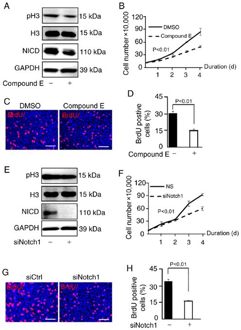
2 WANG et al: NICD, A NEW ACTIVATOR OF GBM CELL PROLIFERATION
cleaved by γ‑secretase to release NICD, the activated form of histological diagnosis of GBM; and ii) performed surgical
Notch1 (17). NICD then is transferred into the nucleus where it resection. The exclusion criteria for patients were: i) History of
can act as a transcriptional activation factor (18). The expres‑ multiple tumors or severe cerebral vascular or cardiovascular
sion of NICD in glioma can be influenced by hypoxia (19). diseases (heart stent surgery or admission to the acute ward
Chronic hypoxia (48 h) can continuously enhance the expres‑ due to cardiovascular disease within the last 6 months), severe
sion of hypoxia inducible factor‑2 α which can activate the vascular lesions, tuberculosis and extensive acute inflammation
Notch signaling pathway (20,21). The upregulation of NICD (including, but not limited to acute meningitis and vasculitis)
induces the activation of Notch1, which promotes GBM within the last 3 months; and ii) participation in a clinical
invasion and migration by promoting β ‑catenin and NF‑κ B trial within the last 3 months. The histological diagnosis of
signaling (22). The role of NICD in the abnormal proliferation glioma was confirmed by the pathologists of the Department
of GBM remains unclear. of Pathology, Renmin Hospital of Wuhan University (Wuhan,
The aim of the present study was to explore whether the China). All tumor samples were subjected to pathological
expression of NICD in GBM is different from that in normal examination and related molecular testing [Methylation of
brain tissue, and to better understand the role and regulatory O6‑methylguanine‑DNA methyltransferase (MGMT)], 1p19q,
mechanism of NICD in GBM. The present study revealed the and isotrate dehydrogenase (IDH)1/IDH2], which were all
function of NICD in GBM proliferation and its role in the defined according to the 2016 WHO classification (1). All
Notch1 signaling pathway which may provide a new molecular clinical information for the patients is presented in Table SI.
tool for the diagnosis and treatment of GBM. The procurement and use of tissue in the present study were
approved by the Institutional Ethics Committee of the Faculty
Materials and methods of Medicine, Renmin Hospital of Wuhan University [approval
number, 2012LKSZ (010) H]. Written informed consent for use
Antibodies and reagents. Primary antibodies used were of tissue was obtained from all patients in the present study.
as follows: i) Anti‑NICD [1:1,000 for western blotting and
1:200 for immunohistochemistry (IHC); cat. no. 2421; Cell culture. U87 (glioblastoma cell line of unknown origin;
Cell Signaling Technology, Inc.]; ii) anti‑Hes5 (1:1,000 for CL‑0238) was purchased from the Cell Bank of Type Culture
western blotting; cat. no. sc‑13859; Santa Cruz Biotechnology, Collection of the Chinese Academy of Sciences. The U87 cell
Inc.); iii) anti‑phosphorylated (p)‑Histone H3 (1:1,000 for line was STR authenticated. Cells were cultured in DMEM
western blotting; cat. no. sc‑8656; Santa Cruz Biotechnology, high glucose medium (cat. no. GNM12800; Jinuo Biomedical
Inc.); iv) anti‑Histone H3 (1:1,000 for western blotting; Technology Co., Ltd.) supplemented with 10% fetal bovine
cat. no. GB13102‑1; Wuhan Servicebio Technology, Co., Ltd); serum (FBS) (cat. no. 04‑001‑1A; Biological Industries) and
v) anti‑Notch1 (1:2,000 for WB; cat. no. ab27526; Abcam); and 100 U/ml penicillin and 100 µg/ml streptomycin. Cells were
vi) anti‑GAPDH (1:2,000 for western blotting; cat. no. 5174; incubated in humidified air with 5% CO2 at 37˚C.
Cell Signaling Technology, Inc.). Secondary antibodies used
were as follows: i) Alex Fluor 680/790 (1:10,000 for western Plasmid construction and transfection. A Flag‑NICD and
blotting; cat. no. ANT091; Antgene); ii) HRP‑labeled (1:100 Flag‑control plasmid was provided by Dr Chundong Yu
for IHC; cat. no. GB23303; Wuhan Servicebio Technology, (Xiamen University, Xiamen, China) and its construction has
Co., Ltd). The antibodies used in the BrdU assay were been described in a previous study by Lin et al (23). An siRNA
anti‑BrdU (1:200; cat. no. 552598; Becton and Dickinson (siNotch1) and a non‑targeting control siRNA (siCtrl) were
and Company) and Alexa Fluor 594 (1:100; cat. no. A11032; purchase from Thermo Scientific Inc. The sequences used were
Invitrogen; Thermo Fisher Scientific, Inc.). Compound E as follows: siRNA‑Notch1, sense 5'‑GCAACCUGCAGUGUA
(cat. no. AG‑CR1‑0081; Adipogen Life Sciences, Inc.) was AUAATT‑3' and antisense 5'‑UUAU UACACUGCAGGU UG
used as an inhibitor of NICD at an intervention concentration CTT‑3'; siRNA‑non‑targeting control, sense 5'‑UUCUCCGAA
of 1 µM which was in accordance with the manufacturer's CGUGUCACGU TT‑3' and antisense 5'‑ACGUGACACGUU
instructions. CGGAGAATT‑3'. U87 cells were seeded in 6‑well plates at a
density of about 5x10⁵ cells/well and cultured overnight at 37˚C.
Tissue samples. Human control brain tissues and GBM tissues Plasmids 2 µg per well were transfected by Lipofectamine
were acquired from the Department of Neurosurgery, Renmin 3000® transfection reagent (Thermo Fisher Scientific, Inc.),
Hospital of Wuhan University (Wuhan, China). GBM tissues and siRNA 200 pM per well was transfected by Lipofectamine
were sampled during resection surgeries and stored at ‑80˚C. RNAiMax reagent (Thermo Fisher Scientific, Inc.) according
A total of 19 pathologically diagnosed GBM samples were to the manufacturer's instructions. Plasmids and siRNA were
enrolled in this study. All the clinical samples were collected incubated with Lipofectamine 3000 ® transfection reagent
between December 2012 and September 2014. The mean ages for 15 min at room temperature before transfection and U87
were 47 and 35 years for GBM and controls, respectively. cells were transfected for 48 h prior to performing subsequent
The average weight of the sample was generally ~3 g of experiments.
which 0.5 g was used for western blotting and the rest was
used for immunohistochemistry and other related experi‑ Western blotting. U87 cell samples and brain tissues were lysed
ments. Control brain tissues used in the present study were on ice in RIPA buffer (50 mm Tris, 150 mm NaCl, 0.5% EDTA
normal tissues collected from 11 non‑tumor patients during and 0.5% NP-40) with PMSF protease inhibitors (Beyotime
emergency surgeries of traumatic brain injury. Patients in the Institute of Biotechnology) and an inhibitor cocktail (Roche
present study had the following inclusion criteria: i) confirmed Diagnostics). The concentration of total proteins was detected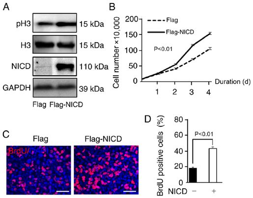
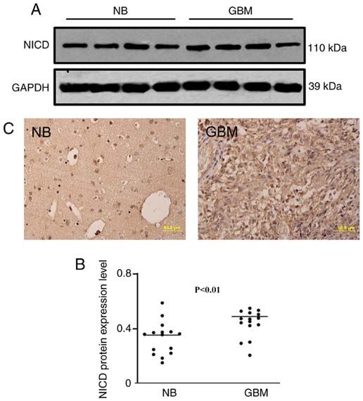
ONCOLOGY LETTERS 21: 303, 2021                                                             3

using a bicinchoninic acid (BCA) kit (Biosharp Life Sciences).
Protein was loaded and separated by 10% SDS‑PAGE and then
transferred onto PVDF membranes (cat. no. 88520; Thermo
Fisher Scientific, Inc.). In total ~20 µg/well of protein was
added. After blocking with 5% skimmed milk for 30 min at
room temperature, membranes were incubated with primary
antibodies overnight and with secondary antibodies for 1 h.
The samples were visualized with a LI‑COR Odyssey Infrared
Imaging System (LI‑COR Biosciences). GAPDH was used as
a loading control.

Immunohistochemical staining. Human brain tissues were
fixed in 4% paraformaldehyde at room temperature for 48 h
and embedded in paraffin and cut into sections. The thickness
of sections used for immunohistochemical staining was 6‑µm
and these sections were cut using a paraffin section machine
(cat. no. HM340E; Thermo Fisher Scientific, Inc.). After
paraffin sections were deparaffinized at 65˚C for 2 h, hydrated
with 100/95/75% ethanol for 10 min each, antigen recovery
was performed by exposing sections to 0.01 mol/l citrate buffer
(pH 6.0) at 100˚C for 20 min. Endogenous peroxidase was
removed by treatment with 3% H2O2. Samples were blocked
with 1% BSA (Amresco, LLC) at room temperature for 1 h and
were then incubated with primary antibodies at 37˚C overnight.    Figure 1. NICD is upregulated in human GBM tissues. (A and B) Western
Following 3 washes with PBS, the sections were incubated          blotting demonstrated that the NICD protein level was significantly
with secondary antibody for 1 h at room temperature. DAB          increased in human GBM tissue compared with normal brain tissue (P
4                                       WANG et al: NICD, A NEW ACTIVATOR OF GBM CELL PROLIFERATION

Figure 2. Notch1 signaling pathway is activated in GBM tissues.
(A and B) Western blotting demonstrated that the Hes5 protein level was
significantly increased in human GBM tissue compared with normal brain
tissue (P
ONCOLOGY LETTERS 21: 303, 2021                                                   5

data demonstrated that NICD induced the proliferation of U87           NICD is the activated form of Notch1 and it serves an
cells in vitro.                                                   important role in the Notch1 signaling pathway (36). Notably,
                                                                  most studies of NICD explored is effect on cell differen‑
Downregulation of NICD suppresses the proliferation of U87        tiation (19), invasion (22) and apoptosis in glioma (37). It is
cells. To further confirm the relationship between NICD and       generally accepted that the growth, metastasis, and invasion
U87 cell proliferation, U87 cells were observed when NICD         of tumors are inseparable from the proliferation of tumor
was downregulated. Compound E is an inhibitor of γ‑secretase      cells (38), however, it was still unclear whether NICD could
and thus has an inhibitory effect on NICD (27). The expres‑       regulate cell proliferation in GBM. Hence, the present study
sion of NICD and the level of H3 phosphorylation was clearly      explored the connection between NICD and proliferation to
downregulated after cells were treated with compound E            fill in the gaps in the knowledge regarding GBM.
(Fig. 4A). Consequently, the number of U87 cells was signifi‑          In the present study, the expression of NICD in human
cantly decreased compared with the control group (DMSO)           GBM tissues which has not been thoroughly explored in
as shown in the proliferation curve (Fig. 4B), and the propor‑    previous research was explored and the findings demon‑
tion of BrdU‑positive U87 cells was significantly reduced         strated an increase in NICD protein level in GBM tissues
compared with the control group (DMSO) in the BrdU assay          compared with normal brain tissues. To determine whether
(Fig. 4C and D). In addition, Brdu postive cells in the control   NICD could regulate cell proliferation in GBM, NICD
group (NICD‑) (Fig. 3D) were less compared with the control       was overexpressed in vitro using the plasmid Flag‑NICD.
group (compound E‑) (Fig. 4D) as vector transfection in the       Phosphorylated histone H3 levels, a key marker of cell
NICD‑ group had a certain inhibiting effect on cells while        proliferation have been demonstrated to be upregulated by
3 µm DMSO treatment in the compound E‑ group had almost           induced cell growth (39). The findings of the present study
no toxic effect. In addition, w U87 cells were transfected with   showed demonstrated that phosphorylated histone H3 levels
siNotch1 to suppress the activity of the Notch1 signaling         were increased and cell growth was promoted following the
pathway; the results demonstrated that the NICD level and         overexpression of NICD. In conclusion, in the present study
the level of H3 phosphorylation were reduced compared with        NICD was activated in GBM tissues and promoted GBM
the control group (siNotch1‑) (Fig. 4E) and the proliferation     proliferation in vitro.
of U87 cells was inhibited as well compared with the control           In addition, to explore whether NICD regulates cell prolif‑
group (siCtrl) (Fig. 4F‑H). In conclusion, NICD was inhibited     eration through the Notch1 signaling pathway, compound E
by compound E and knockdown of Notch1 also reduced the            and siNotch1 were used to suppress the activation of NICD
expression of NICD and suppressed the proliferation of U87        and Notch1, respectively. Compound E, a selective inhibitor
cells.                                                            of γ‑secretase that has the ability to suppress the expression
                                                                  of NICD (40) was observed to decrease NICD protein levels
Discussion                                                        and phosphorylated histone H3 levels as well as to repress
                                                                  the growth of U87 cells in the present study. In addition,
The Notch1 signaling pathway has been reported to associate       it has been reported that overexpression of NICD in PC12
with various cancers, such as lung cancer and gastric cancer      cells can promote cell apoptosis and inhibit cell prolifera‑
and participate in several neoplastic biological behaviors        tion by regulating S phase arrest (41). Similar results were
including drug resistance and apoptosis (28,29). Although         obtained in the present study where NICD was inhibited
some studies have demonstrated that Notch1 is related to the      and cell proliferation suppressed when Notch1 was knocked
abnormal proliferation of GBM cells (30‑33), the specific         down by siNotch1. Hence, in the present study a reduced
regulatory mechanism of this remains unclear. As significant      level of NICD suppressed the proliferation of GBM cells and
effectors downstream of the Notch1 signaling pathway, Hes1        NICD regulated proliferation through the Notch1 signaling
and Hes5 have recently received increasing attention (25).        pathway.
Hes1 is expressed at low levels in the human brain, and several        GBM is still the most malignant type of glioma, and it
studies have detected uninduced Hes1 expression in GBM            exhibits abnormal proliferation and a high mortality rate
tissues (34,35). In comparison, the Hes5 expression level was     according to the statistical analysis of glioma cases in US
relatively high (34), however, the expression of Hes5 in human    between 2010 and 2014 (4). Early diagnosis and precision
GBM is still controversial and remains to be solved. Hence,       treatments have great significance for providing an improved
the present study investigated the level of Hes5 fixed GBM        prognosis for patients with GBM (42). The findings of the
samples. Interestingly, the expression of Hes5 was higher         present study emphasized the significance of NICD in the
at both the protein and mRNA levels in the GBM samples            Notch1 signaling pathway regulation of the proliferation of
compared with normal control brain samples, indicating            GBM. Hence, NICD may be a potential diagnostic marker and
an activated state of the Notch1 signaling pathway in GBM         therapeutic target for GBM.
tissues which has not been previously reported to the best of
our knowledge. According to the existing literature, HES5         Acknowledgements
serves an important role in the regulation of mammalian
neuronal differentiation and the maintenance of neural stem       Not applicable.
cells and can actively regulate the self‑renewal of neuronal
stem cells (13). Based on the findings of the present study, it   Funding
may be speculated that HES5 in GBM may have a significant
impact on the occurrence and development of GBM.                  No funding was received.
6 WANG et al: NICD, A NEW ACTIVATOR OF GBM CELL PROLIFERATION
Availability of data and materials 11. Guichet PO, Guelfi S, Teigell M, Hoppe L, Bakalara N, Bauchet L,
Duffau H, Lamszus K, Rothhut B and Hugnot JP: Notch1 stimu‑
lation induces a vascularization switch with pericyte‑like cell
The datasets used and/or analyzed during the current study are differentiation of glioblastoma stem cells. Stem Cells 33: 21‑34,
available from the corresponding author on reasonable request. 2015.
12. Aster JC, Pear WS and Blacklow SC: The varied roles of Notch
in cancer. Annu Rev Pathol 12: 245‑275, 2017.
Authors' contributions 13. Ohtsuka T, Ishibashi M, Gradwohl G, Nakanishi S, Guillemot F
and Kageyama R: Hes1 and Hes5 as notch effectors in mamma‑
lian neuronal differentiation. EMBO J 18: 2196‑2207, 1999.
BL and QC designed the research. YW and QS performed 14. Zhang YM, Chen SX, Dai QF, Jiang ST, Chen AL, Tang CZ
the experiments. RG, HL, FY and YX analyzed the data and and Zhang YQ: Effect of acupuncture on the notch signaling
wrote the manuscript. YQ and HJ participated in data analysis pathway in rats with brain injury. Chin J Integr Med 24:
537‑544, 2018.
and revised the article for important intellectual content. All 15. Purow BW, Haque RM, Noel MW, Su Q, Burdick MJ, Lee J,
authors have read and approved the final manuscript. Sundaresan T, Pastorino S, Park JK, Mikolaenko I, et al:
Expression of Notch‑1 and its ligands, Delta‑like‑1 and Jagged‑1,
is critical for glioma cell survival and proliferation. Cancer
Ethics approval and consent to participate Res 65: 2353‑2363, 2005.
16. Hu GW, Wu L, Kuang W, Chen Y, Zhu XG, Guo H and Lang HL:
All tissue specimens used in this study were approved by the Knockdown of linc‑OIP5 inhibits proliferation and migration of
glioma cells through down‑regulation of YAP‑NOTCH signaling
Institutional Ethics Committee of the Faculty of Medicine, pathway. Gene 610: 24‑31, 2017.
Renmin Hospital of Wuhan University (approval no. 2012LKSZ 17. Hu S, Chen Q, Lin T, Hong W, Wu W, Wu M, Du X and Jin R:
(010) H) (Wuhan, China). Written informed consent was The function of Notch1 intracellular domain in the differentia‑
tion of gastric cancer. Oncol Lett 15: 6171‑6178, 2018.
obtained from all the patients. 18. Tamura K, Taniguchi Y, Minoguchi S, Sakai T, Tun T, Furukawa T
and Honjo T: Physical interaction between a novel domain of the
Patient consent for publication receptor Notch and the transcription factor RBP‑J kappa/Su(H).
Curr Biol 5: 1416‑1423, 1995.
19. Hu YY, Fu LA, Li SZ, Chen Y, Li JC, Han J, Liang L, Li L,
Not applicable. Ji CC, Zheng MH and Han H: Hif‑1α and Hif‑2α differentially
regulate Notch signaling through competitive interaction with
the intracellular domain of Notch receptors in glioma stem cells.
Competing interests Cancer Lett 349: 67‑76, 2014.
20. Renfrow JJ, Soike MH, Debinski W, Ramkissoon SH,
The authors declare that they have no competing interests. Mott RT, Frenkel MB, Sarkaria JN, Lesser GJ and Strowd RE:
Hypoxia‑inducible factor 2α: A novel target in gliomas. Future
Med Chem 10: 2227‑2236, 2018.
References 21. Patel SA and Simon MC: Biology of hypoxia‑inducible
factor‑2alpha in development and disease. Cell Death Differ 15:
628‑634, 2008.
1. Reni M, Mazza E, Zanon S, Gatta G and Vecht CJ: Central 22. Zhang X, Chen T, Zhang J, Mao Q, Li S, Xiong W, Qiu Y, Xie Q
nervous system gliomas. Crit Rev Oncol Hematol 113: 213‑234, and Ge J: Notch1 promotes glioma cell migration and invasion by
2017. stimulating β‑catenin and NF‑κ B signaling via AKT activation.
2. Louis DN, Perry A, Reifenberger G, von Deimling A, Cancer Sci 103: 181‑190, 2012.
Figarella‑Branger D, Cavenee WK, Ohgaki H, Wiestler OD, 23. Lin X, Liu B, Yang X, Yue X, Diao L, Wang J and Chang J:
Kleihues P and Ellison DW: The 2016 World Health Organization Genetic deletion of Rnd3 results in aqueductal stenosis leading
classification of tumors of the central nervous system: A to hydrocephalus through up‑regulation of Notch signaling. Proc
summary. Acta Neuropathol 131: 803‑820, 2016. Natl Acad Sci USA 110: 8236‑8241, 2013.
3. Le Rhun E, Rhun EL, Taillibert S and Chamberlain MC: The 24. Livak KJ and Schmittgen TD: Analysis of relative gene expres‑
future of high‑grade glioma: Where we are and where are we sion data using real‑time quantitative PCR and the 2(‑Delta Delta
going. Surg Neurol Int 6 (Suppl 1): S9‑S44, 2015. C(T)) method. Methods 25: 402‑408, 2001.
4. Ostrom QT, Gittleman H, Liao P, Vecchione‑Koval T, Wolinsky Y, 25. Ronchi CL, Sbiera S, Altieri B, Steinhauer S, Wild V, Bekteshi M,
Kruchko C and Barnholtz‑Sloan JS: CBTRUS Statistical Kroiss M, Fassnacht M and Allolio B: Notch1 pathway in adreno‑
Report: Primary brain and other central nervous system tumors cortical carcinomas: Correlations with clinical outcome. Endocr
diagnosed in the United States in 2010‑2014. Neuro Oncol 19 Relat Cancer 22: 531‑543, 2015.
(suppl_5): v1‑v88, 2017. 26. Elmaci İ, Altinoz MA, Sari R and Bolukbasi FH: Phosphorylated
5. Ostrom QT, Gittleman H, Truitt G, Boscia A, Kruchko C and histone H3 (PHH3) as a novel cell proliferation marker and
Barnholtz‑Sloan JS: CBTRUS statistical report: Primary brain prognosticator for meningeal tumors: A short review. Appl
and other central nervous system tumors diagnosed in the United Immunohistochem Mol Morphol 26: 627‑631, 2018.
States in 2011‑2015. Neuro Oncol 20 (suppl_4): iv1‑iv86, 2018. 27. Yang T, Arslanova D, Gu Y, Augelli‑Szafran C and Xia W:
6. Han D, Yu T, Dong N, Wang B, Sun F and Jiang D: Napabucasin, Quantification of gamma‑secretase modulation differentiates
a novel STAT3 inhibitor suppresses proliferation, invasion and inhibitor compound selectivity between two substrates Notch
stemness of glioblastoma cells. J Exp Clin Cancer Res 38: 289, and amyloid precursor protein. Mol Brain 1: 15, 2008.
2019. 28. Jun HT, Stevens J and Kaplan‑Lefko P: Top NOTCH targets:
7. Ji Y, Sun Q, Zhang J and Hu H: MiR‑615 inhibits cell prolif‑ Notch signaling in cancer. Drug Development Research 69:
319‑328, 2010.
eration, migration and invasion by targeting EGFR in human 29. Pancewicz‑Wojtkiewicz J: Epidermal growth factor receptor and
glioblastoma. Biochem Biophys Res Commun 499: 719‑726, notch signaling in non‑small‑cell lung cancer. Cancer Med 5:
2018. 3572‑3578, 2016.
8. Gil‑García B and Baladrón V: The complex role of NOTCH 30. Feng HB, Wang J, Jiang HR, Mei X, Zhao YY, Chen FR, Qu Y,
receptors and their ligands in the development of hepatoblas‑ Sai K, Guo CC, Yang QY, et al: β‑elemene selectively inhibits the
toma, cholangiocarcinoma and hepatocellular carcinoma. Biol proliferation of glioma stem‑like cells through the downregula‑
Cell 108: 29‑40, 2016. tion of Notch1. Stem Cells Transl Med 6: 830‑839, 2017.
9. Stockhausen MT, Kristoffersen K and Poulsen HS: The func‑ 31. Wang BQ, Yang B, Yang HC, Wang JY, Hu S, Gao YS and
tional role of Notch signaling in human gliomas. Neuro Oncol 12: Bu XY: MicroRNA‑499a decelerates glioma cell proliferation
199‑211, 2010. while accelerating apoptosis through the suppression of Notch1
10. Lino MM, Merlo A and Boulay JL: Notch signaling in glioblas‑ and the MAPK signaling pathway. Brain Res Bull 142: 96‑106,
toma: A developmental drug target? BMC Med 8: 72, 2010. 2018.ONCOLOGY LETTERS 21: 303, 2021 7
32. Hai L, Zhang C, Li T, Zhou X, Liu B, Li S, Zhu M, Lin Y, Yu S, 38. Xie Q, Mittal S and Berens ME: Targeting adaptive glioblastoma:
Zhang K, et al: Notch1 is a prognostic factor that is distinctly An overview of proliferation and invasion. Neuro Oncol 16:
activated in the classical and proneural subtype of glioblastoma 1575‑1584, 2014.
and that promotes glioma cell survival via the NF‑κ B(p65) 39. Villani V, Mahadevan KK, Ligorio M, Fernández‑Del Castillo C,
pathway. Cell Death Dis 9: 158, 2018. Ting DT, Sabbatino F, Zhang I, Vangel M, Ferrone S,
33. Liu Q, Zheng JM, Chen JK, Yan XL, Chen HM, Nong WX and Warshaw AL, et al: Phosphorylated histone H3 (PHH3) is a
Huang HQ: Histone deacetylase 5 promotes the proliferation superior proliferation marker for prognosis of pancreatic neuro‑
of glioma cells by upregulation of Notch 1. Mol Med Rep 10: endocrine tumors. Ann Surg Oncol 23: 609‑617, 2016.
2045‑2050, 2014. 40. Arumugam TV, Cheng YL, Choi Y, Choi YH, Yang S, Yun YK,
34. Cheung HC, Corley LJ, Fuller GN, McCutcheon IE and Cote GJ: Park JS, Yang DK, Thundyil J, Gelderblom M, et al: Evidence that
Polypyrimidine tract binding protein and Notch1 are indepen‑ gamma‑secretase‑mediated Notch signaling induces neuronal
dently re‑expressed in glioma. Mod Pathol 19: 1034‑1041, 2006. cell death via the nuclear factor‑kappaB‑Bcl‑2‑interacting medi‑
35. Saito N, Fu J, Zheng S, Yao J, Wang S, Liu DD, Yuan Y, ator of cell death pathway in ischemic stroke. Mol Pharmacol 80:
Sulman EP, Lang FF, Colman H, et al: A high Notch pathway 23‑31, 2011.
activation predicts response to γ secretase inhibitors in proneural 41. Li B, Duan P, Han X, Yan W and Xing Y: NICD inhibits cell
subtype of glioma tumor‑initiating cells. Stem Cells 32: 301‑312, proliferation and promotes apoptosis and autophagy in PC12
2014. cells. Mol Med Rep 16: 2755‑2760, 2017.
36. Liu B, Lin X, Yang X, Dong H, Yue X, Andrade KC, Guo Z, 42. Alexander BM and Cloughesy TF: Adult glioblastoma. J Clin
Yang J, Wu L, Zhu X, et al: Downregulation of RND3/RhoE in Oncol 35: 2402‑2409, 2017.
glioblastoma patients promotes tumorigenesis through augmen‑
tation of notch transcriptional complex activity. Cancer Med 4:
1404‑1416, 2015. This work is licensed under a Creative Commons
37. Wang Y, Wang H, Ge H and Yang Z: AG‑1031 induced autoph‑ Attribution-NonCommercial-NoDerivatives 4.0
agic cell death and apoptosis in C6 glioma cells associated with International (CC BY-NC-ND 4.0) License.
Notch‑1 signaling pathway. J Cell Biochem 119: 5893‑5903, 2018.You can also read