Porcine Parvovirus 7: Evolutionary Dynamics and Identification of Epitopes toward Vaccine Design - MDPI
←
→
Page content transcription
If your browser does not render page correctly, please read the page content below
Article
Porcine Parvovirus 7: Evolutionary Dynamics and
Identification of Epitopes toward Vaccine Design
Dongliang Wang, Jinhui Mai, Yi Yang                  and Naidong Wang *
 Hunan Provincial Key Laboratory of Protein Engineering in Animal Vaccines, Laboratory of Functional
 Proteomics (LFP), Research Center of Reverse Vaccinology (RCRV), College of Veterinary Medicine,
 Hunan Agricultural University, Changsha 410128, China; dongliangwang@stu.hunau.edu.cn (D.W.);
 jinhuimai@stu.hunau.edu.cn (J.M.); yiyang@hunau.edu.cn (Y.Y.)
 * Correspondence: naidongwang@hunau.edu.cn

 Received: 5 June 2020; Accepted: 1 July 2020; Published: 5 July 2020

 Abstract: Porcine parvovirus 7 (PPV7) belonging to the genus Chapparvovirus in the family Parvoviridae,
 has been identified in the USA, Sweden, Poland, China, South Korea and Brazil. Our objective was to
 determine the phylogeny, estimate the time of origin and evolutionary dynamics of PPV7, and use
 computer-based immune-informatics to assess potential epitopes of its Cap, the main antigenic viral
 protein, for vaccines or serology. Regarding evolutionary dynamics, PPV7 had 2 major clades, both of
 which possibly had a common ancestor in 2004. Furthermore, PPV7 strains from China were the
 most likely ancestral strains. The nucleotide substitution rates of NS1 and Cap genes were 8.01 × 10⁻⁴
 and 2.19 × 10⁻³ per site per year, respectively, which were higher than those reported for PPV1-4.
 The antigenic profiles of PPV7 Cap were revealed and there were indications that PPV7 used antigenic
 shift to escape from the host’s immune surveillance. Linear B cell epitopes and CD8 T cell epitopes of
 Cap with good antigenic potential were identified in silico; these conserved B cell epitopes may be
 candidates for the PPV7 vaccine or for the development of serological diagnostic methods.

 Keywords: PPV7; Cap; B cell and T cell epitopes; vaccine; evolution

1. Introduction
      The family Parvoviridae contains two subfamilies: Parvovirinae and Densovirinae, whose hosts
are vertebrates and arthropods, respectively [1]. The subfamily Parvovirinae is further divided
into nine genera: Tetraparvovirus, Copiparvovirus, Erythroparvovirus, Dependoparvovirus, Aveparvovirus,
Bocaparvovirus, Amdoparvovirus, Protoparvovirus and Chapparvovirus [1,2]. To date, seven genotypes of
porcine parvoviruses (PPV) have been discovered in pigs, and belong to four genera: Protoparvovirus
(PPV1), Tetraparvovirus (PPV2–3), Copiparvovirus (PPV4-6) and Chapparvovirus (PPV7), based on the
similarity of non-structural protein 1 (NS1) [2,3]. PPV1, first identified in 1965 in Germany [4], is a major
cause of reproductive failure in pigs, characterized by mummified fetuses, infertility, early embryonic
death, stillbirths and delayed returns to the estrus [5,6]. Subsequently, six other PPV genotypes
(PPV2–PPV7) were identified using novel techniques, including next-generation sequencing [2,7–11].
      In 2016, PPV7 was first identified by the metagenomic sequencing of rectal swab samples from
pigs in the USA [2]. Subsequently, PPV7 infections were reported in Sweden, Poland, China, South
Korea and Brazil [12–19]. PPV7 is a small, single-stranded linear and non-enveloped DNA virus with
a genome of ~4 kb that contains two major open reading frames (ORFs): ORF1 encodes non-structural
protein 1 (NS1) responsible for viral replication, and ORF2 encodes the major structural capsid protein
(Cap) or VP2 protein [2]. PPV Cap is also the major antigenic component, with an important role in
eliciting neutralizing antibodies against viral infection [20]. In addition, it is a prospective antigen for
subunit vaccine design and serological diagnosis. A vaccine based on the Cap could elicit antibodies to

Vaccines 2020, 8, 359; doi:10.3390/vaccines8030359                              www.mdpi.com/journal/vaccines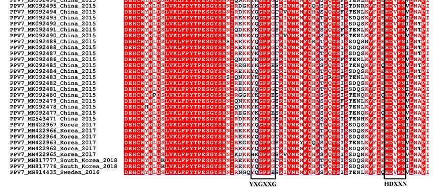
Vaccines 2020, 8, 359                                                                             2 of 13

neutralize virus infection by blocking virus entry [21]. Thus, the Cap could be regarded as an effective
antigenic component for PPV7 vaccine design and development. Currently, there is no, or very limited,
information about its antigenic structure and immunogenic profiles, thus, the immune profiles of PPV7
Cap are required to develop PPV7 Cap-based vaccines against PPV7 infection.
     Despite limited knowledge regarding PPV7 pathogenicity, its presence in aborted pig fetuses
suggests that it causes reproductive failure [17]. Co-infections are more frequent than single infection
in swine herd; multiple infectious pathogens such as PCV2 and PPV can impact the outcome of
respiratory infections and deserve [22]. In addition, co-infections with PPV7 and PCV2 were recently
reported [23], although whether this co-infection enhanced the severity of porcine circovirus associated
diseases (PCVADs) has yet to be determined. To better understand the molecular evolution and genetic
diversity of this newly emerging PPV7, we analyzed the phylogeny and estimated the time of origin
and evolutionary dynamics of PPV7. Importantly, computer-based immune-informatics were used to
assess the potential B cell and CD8 T cell epitopes of the PPV7 Cap, which will greatly facilitate the
development of new generation vaccines against PPV7 infection and contribute to the development of
effective serological diagnostic methods.

2. Materials and Methods

2.1. Sequence Datasets
     A total of 45 complete (or partial) genomes, and 59 NS1 and Cap complete coding sequences,
were collected from GenBank to perform phylogenetic analysis. Detailed information of PPV7 and
reference Parvoviridae sequences are summarized in Tables S1 and S2, respectively.

2.2. Multiple Sequence Alignment
     NS1 and Cap amino acid sequences were derived from 59 PPV7 genomes. Sequence identity was
analyzed and aligned with the Clustal W method of the MegAlign program of DNASTAR, version 7.10
(Lasergene) (DNASTAR, Inc., Wisconsin, USA). Amino acid sequences were aligned with the ESPript
3.0 online tool (ESPript 3.0, http://espript.ibcp.fr/ESPript/ESPript/, 4 July 2020).

2.3. Phylogenetic and Evolution Dynamic Analysis
     Sequences were aligned with Clustal W, implemented in MEGA 7 software [24].
A maximum-likelihood (ML) tree was constructed with NS1 and Cap amino acid sequences, using
MEGA 7 with the Jones–Taylor–Thornton (JTT) model and 1000 bootstrap replicates. The p-distance
method was used to reconstruct a neighbor joining (NJ) tree with 1000 bootstrap replicates.
     The most recent common ancestor (tMRCA) and rates of nucleotide substitutions per site per
year were estimated using the Bayesian Markov chain Monte Carlo (MCMC) method within the
BEAST package (Version 1.10.4) [25]. A general time reversible (GTR) substitution model with a
proportion of gamma and invariant distributed rate heterogeneity (GTR + G + I) with an uncorrelated
relaxed lognormal molecular clock was selected. Chain length for the run was 1 × 10⁸ generations,
with sampling at every 10,000 generations. After removing the initial 10% of samples as burn-in,
only runs with an estimate sample size (ESS) > 200 were accepted, based on the software Tracer
(v1.7.1) (Tracer, http://tree.bio.ed.ac.uk/software/tracer/, 4 July 2020). A maximum clade credibility
(MCC) tree was reconstructed with TreeAnnotator (v1.10.4) and displayed in Figtree (v1.4.4) (Figtree,
http://tree.bio.ed.ac.uk/software/figtree/, 4 July 2020).

2.4. Selection Pressures Analysis
    The detection of selected NS1 and Cap coding sequences of PPV7 was performed using
DATAMONKEY (Datamonkey, http://www.datamonkey.org/, 4 July 2020). Positive selected sites
were detected using 4 algorithms, including fixed effects likelihood (FEL), single-likelihood ancestor
counting (SLAC), fast unconstrained Bayesian approximation (FUBAR) and mixed effects model
Vaccines 2020, 8, 359                                                                             3 of 13

of evolution (MEME) [26–28]. A site was considered as a positive selection position only if it was
identified by at least 2 algorithms, and with p < 0.1 in SLAC, p < 0.05 in FEL and MEME and posterior
probability > 0.9 in FUBAR were considered significant. The selection pressure analysis of genomes
was determined with MEGA7 software by calculating the differences between non-synonymous (dN)
and synonymous substitution (dS) rates, for the aligned genes. The calculated value of dN-dS was
used to evaluate selection pressure (dN-dS > 0: positive selection; dN-dS < 0: purifying selection;
and dN-dS = 0: neutral selection) [29].

2.5. Structural Analysis
     The physicochemical properties of PPV7 Cap were assessed with the ProtParam webserver
(ProtParam, https://web.expasy.org/protparam/, 4 July 2020). The secondary structure of PPV7 Cap
was predicted with PSIPRED [30].

2.6. B Cell Epitope Prediction
     Linear B cell epitopes of the PPV7 Cap were predicted using the BepiPred 2.0 online tool in
IEDB (Immune Epitope Database) (BepiPred 2.0, http://www.iedb.org/, 4 July 2020), with a threshold
of 0.55 (corresponding specificity > 0.817 and sensitivity < 0.292); only epitopes with > 7 residues
were considered for subsequent antigenicity analysis. Antigenicity testing was done with the VaxiJen
v2.0 server online tool [31] (VaxiJen v2.0, http://www.ddg-pharmfac.net/vaxijen/VaxiJen/VaxiJen.html,
4 July 2020).

2.7. CD8 T Cell Epitope Prediction
     For CD8 T cell epitopes prediction, 3 porcine MHC-I molecules SLA-1*04:01, SLA-2*04:01 and
SLA-3*04:01 [32–34] were selected, based on the IEDB recommended 2.22 algorithm in IEDB, with a
peptide size of 9 residues. Higher scoring peptides (rank ≤ 2%) (threshold 0.5% and 2.0% rank for strong
and weak binders, respectively) based on the prediction were chosen for subsequent immunogenicity
evaluation by the VaxiJen v2.0 server.

2.8. Peptide Modelling and Molecular Docking
     The 3D structures of all peptides were modelled with the PEP-FOLD3 online server [35].
All peptides were docked to correspond to SLA-2*04:02 (PDB ID: 6A6H) and SLA-3*04:01 (PDB
ID: 5H94), using the PatchDock rigid-body docking server, using the defined threshold [36]. Based on
geometry docking algorithm in PatchDock, the docking transformation with good molecular shape
complementarity was selected, and docked complexes were refined with the FireDock server [37,38].
Complexes with high global energy, attractive Vander Waal (vdW) energy and hydrogen bonding
energy were used for subsequent analyses. Protein peptide connections were examined with the
LigPlus tool and analyzed with Pymol.

3. Results

3.1. Construction of Phylogenetic Tree and Evolution Dynamic Analysis
     To better understand genetic relationships between PPV7 and other strains of Parvoviridae,
a phylogenetic tree was constructed, using the maximum likelihood (ML) and neighbor joining (NJ)
methods, based on the NS1 and Cap amino acid sequences, respectively. According to phylogenetic
analyses, all PPV7 strains were located in a branch belonging to the Chapparvovirus genus (Figure 1
and Figure S1). Additionally, PPV7 was more genetically close to PPV1, compared to the PPV strains
among the other genotypes (Figure 1). Furthermore, the maximum clade credibility (MCC) tree was
reconstructed with 45 complete PPV7 genomes. It appeared that PPV7 strains may have had a common
ancestor in 2004 (95% highest posterior density (HPD): 1986–2014); furthermore, the PPV7 strains from
China were the most likely ancestral strains, based on currently available sequences (Figure 2).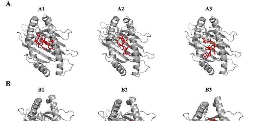
Vaccines 2020, 8, x                                                                                               4 of 13
Vaccines 2020, 8, x                                                                                              4 of 13
strains from China were the most likely ancestral strains, based on currently available sequences
strains
(Figurefrom
        2). China were the most likely ancestral strains, based on currently available sequences
(Figure 2).
Vaccines 2020, 8, 359                                                                                             4 of 13

      Figure 1. Phylogenetic analysis of viruses in the Parvoviridae family. Trees were constructed based on
      Figure
      Figure
      the NS11.  Phylogenetic
             1. (A)
                 Phylogenetic  analysis
                               analysis
                     and Cap (B)        ofacid
                                  aminoof  viruses
                                           viruses  in the
                                                   in
                                               sequences    Parvoviridae
                                                        the Parvoviridae  family.
                                                                         family.
                                                             by the neighbor       Trees were
                                                                                  Trees
                                                                                joining  werephylogenetic
                                                                                         (NJ)  constructed based
                                                                                               constructed based on
                                                                                                                 on
                                                                                                            method,
      the
      the NS1
          NS1
      using     (A)
            the(A)  and  Cap  (B)
                    and Capmodel
                  p-distance      amino
                              (B) amino  acid
                                    with acid  sequences
                                         1000 sequences      by the  neighbor
                                                             by the neighbor
                                               bootstrap replicates            joining   (NJ)
                                                                               joining >(NJ)
                                                                       and bootstrap          phylogenetic  method,
                                                                                          50%.phylogenetic method,
      using the
      using the p-distance
                 p-distance model
                             model with
                                   with 1000
                                         1000 bootstrap
                                               bootstrap replicates
                                                            replicates and  bootstrap >
                                                                       and bootstrap      50%.
                                                                                       > 50%.

      Figure 2. Maximum clade credibility (MCC) tree for the complete genome sequences of PPV7. The tree
      contained 2 major clades: Clade 1 (red) and Clade 2 (blue).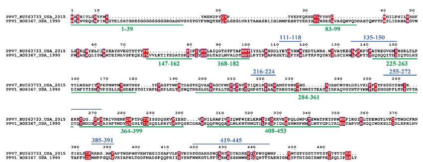
Vaccines 2020, 8, 359 5 of 13
The evolutionary rates of the NS1 and Cap genes of PPV7 were estimated. The mean evolutionary
rates of the NS1 and Cap genes were 8.01 × 10⁻⁴ per site per year (95% HPD: 3.67 × 10⁻⁶ –1.9 × 10⁻³ )
and 2.19 × 10⁻³ per site per year (95% HPD: 1.28 × 10⁻⁴ –5.06 × 10⁻³ ), respectively.
3.2. Selection Pressures Analysis
Five sites (24, 106, 158, 270 and 446) among Cap were confirmed by all 4 methods to be under
positive selection. Furthermore, 2 sites (195 and 441) were also confirmed to be under positive selection
in Cap by at least 2 methods with p < 0.05 by FEL and MEME, p < 0.1 by SLAC, and a posterior
probability > 0.9 by FUBAR (Table 1). In addition, 8 NS1 sites (69, 342, 426, 465, 474, 498, 525 and 603)
were detected as positive selection positions by all 4 methods, whereas the other 9 sites (27, 83, 376, 500,
547, 549, 550, 579 and 618) were detected as positive selection positions by at least 2 methods (Table 2).
The overall mean differences of dN-dS were −4.122 for Cap gene and −15.371 for NS1 gene, indicating
that both Cap and NS1 were under purifying selection.
Table 1. Selection analysis of PPV7 Cap coding sequences.
FEL SLAC FUBAR MEME
Site dN-dS p-Value Site dN-dS p-Value Site dN-dS Post.Pro Site β+ p-Value
24 2.492 0.004 24 5.741 0.018 24 6.900 0.997 24 2.5 0.01
106 2.157 0.023 106 4.943 0.028 106 5.706 0.979 106 24.21 0
158 1.552 0.033 158 3.652 0.098 158 3.244 0.972 158 1.55 0.05
195 1.442 0.909 195 166.2 0
270 1.777 0.026 270 5.107 0.048 270 4.363 0.974 270 14.87 0
441 2.926 0.944 441 18.72 0
446 1.649 0.015 446 3.970 0.055 446 3.901 0.988 446 3.43 0.02
Table 2. Selection analysis of PPV7 NS1 coding sequences.
FEL SLAC FUBAR MEME
Site dN-dS p-Value Site dN-dS p-Value Site dN-dS Post.Pro Site β+ p-Value
27 1.519 0.016 27 2.24 0.977 27 1.52 0.03
69 2.815 0.002 69 3.466 0.069 69 4.953 0.997 69 2.82 0
83 2.627 0.96 83 148.66 0
342 5.477 0.01 342 8.331 0.005 342 12.563 0.993 342 173.79 0
376 4.566 0.066 376 5.16 0.965
426 8.215 0.005 426 10.973 0.002 426 17.289 0.999 426 40.1 0
465 1.905 0.034 465 3.658 0.088 465 2.882 0.979 465 1.9 0.05
474 4.118 0.019 474 5.486 0.026 474 7.406 0.983 474 4.12 0.03
498 4.261 0 498 5.326 0.031 498 8.12 1 498 4.25 0
500 5.817 0.028 500 2.963 0.956
525 7.599 0.003 525 10.047 0.002 525 15.702 0.999 525 7.6 0
547 5.486 0.026 547 4.412 0.97
549 4.267 0.059 549 2.533 0.956
550 3.658 0.088 550 2.944 0.992 550 1.9 0.01
579 2.081 0.01 579 3.455 0.994 579 2.08 0.02
603 1.271 0.025 603 3.778 0.081 603 1.998 0.974 603 1.27 0.04
618 4.105 0.077 618 2.663 0.95
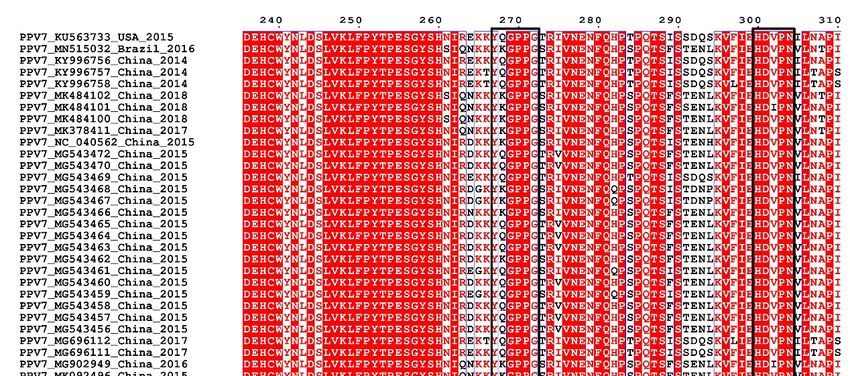
3.3. Sequence and Structural Characteristics
Sequence alignments revealed that PPV7 genomes (total 45 of isolates deposited in GenBank)
exhibited 91.9–100% nucleotide homology. Furthermore, PPV7 NS1 and Cap genes (total 59 of each
deposited in GenBank) had 92.8–100% and 85.7–100% homology, respectively, and the derived amino
acid sequence of both proteins shared 89.9–100% and 82.4–100% identity, respectively. The Ca2+ binding
loop (YXGXG) was present in the Caps of PPV1, PPV2, PPV3 and PPV5 [1,10], and “YXGXR” was in
PPV6 [11], but absent in PPV4. However, the potential Ca2+ binding loop was the 267 YXGXXG272 motif
in PPV7, and “269 GXX271 ” (“269 GPP271 ”) were strictly conserved in PPV7 (Figure 3). Furthermore,
compared to the catalytic motif (HDXXY) of the putative secretory phospholipase A2 (PLA2) in
PPV5 [10], a similar motif 300 HDXXN304 was present in PPV7, whereas a point mutation occurred at
position 304 (N 304 Y) in the 59 of PPV7 Cap proteins (Figure 3).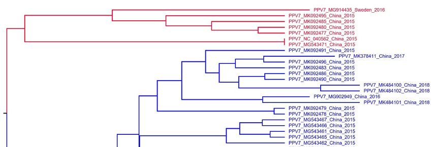
Vaccines 2020, 8, x                                                                                                  6 of 13

compared to the catalytic motif (HDXXY) of the putative secretory phospholipase A2 (PLA2) in PPV5
[10], a similar motif 300HDXXN304 was present in PPV7, whereas a point mutation occurred at position
Vaccines 2020, 8, 359                                                                          6 of 13
304 (N 304 Y) in the 59 of PPV7 Cap proteins (Figure 3).

       Figure 3.
      Figure       Sequencealignment
              3. Sequence    alignmentof     Ca2+2+binding
                                          of Ca     bindingloop
                                                             loopand
                                                                   andputative
                                                                        putativesecretory
                                                                                 secretoryphospholipase
                                                                                            phospholipaseA2 A2(PLA2)
                                                                                                               (PLA2)
       motif of PPV7.    The conserved    amino   acids of the Ca 2+  binding loop ( 267 YXGXXG 272 ) and the catalytic
      motif of PPV7. The conserved amino acids of the Ca binding loop ( YXGXXG ) and the catalytic
                                                                   2+                267        272
                   300 HDXXN
       residues ((300         304 ) are indicated at the bottom of the alignment.
      residues        HDXXN304) are   indicated at the bottom of the alignment.

      A representative strain (KU563733) was used for a structural analysis of PPV7 Cap.
      A representative strain (KU563733) was used for a structural analysis of PPV7 Cap. The
 The physicochemical properties of the PPV7 Cap, computed using the ProtParam tool, concluded that
physicochemical properties of the PPV7 Cap, computed using the ProtParam tool, concluded that it
 it contained 469 aa, with a molecular weight of 54,257.8 Da. The isoelectric point (PI) of this protein
contained 469 aa, with a molecular weight of 54,257.8 Da. The isoelectric point (PI) of this protein was
 was 7.23, indicating that it is positive in nature. Out of 469 residues, 42 (Asp + Glu) were negatively
7.23, indicating that it is positive in nature. Out of 469 residues, 42 (Asp + Glu) were negatively
 charged and 42 (Arg + Lys) were positively charged. The instability index of (II) was computed to
charged and 42 (Arg + Lys) were positively charged. The instability index of (II) was computed to be
 be 38.58, indicating that it was a stable protein. The predicted aliphatic index was 56.78 and the grand
38.58, indicating that it was a stable protein. The predicted aliphatic index was 56.78 and the grand
 average of hydropathicity (GRAVY) for the protein sequence was −0.695. This protein contained
average of hydropathicity (GRAVY) for the protein sequence was −0.695. This protein contained 7483
 7483 atoms, and was described by the formula C2469H3639N647O714S14. The estimated half-life was
atoms, and was described by the formula C2469H3639N647O714S14. The estimated half-life was 30
 30 h (mammalian reticulocytes, in vitro), > 20 h (yeast, in vivo) and > 10 h (Escherichia coli, in vivo).
h (mammalian reticulocytes, in vitro), > 20 h (yeast, in vivo) and > 10 h (Escherichia coli, in vivo).
      The secondary structure of the PPV7 Cap was analyzed by PSIPRED with (24%) beta sheets, (12%)
      The secondary structure of the PPV7 Cap was analyzed by PSIPRED with (24%) beta sheets,
 helixes and (64%) loops present in the structure (Figure S2). VaxiJen v2.0 was used to evaluate the
(12%) helixes and (64%) loops present in the structure (Figure S2). VaxiJen v2.0 was used to evaluate
 antigenicity of the Cap. By setting the threshold at 0.4 for higher specificity, antigenicity score was
the antigenicity of the Cap. By setting the threshold at 0.4 for higher specificity, antigenicity score
 0.4426 for the Cap, implying an excellent antigenic potential.
was 0.4426 for the Cap, implying an excellent antigenic potential.
3.4. Linear B Cell Epitopes Prediction and Analysis of Cap
3.4. Linear B Cell Epitopes Prediction and Analysis of Cap
       In total, 10 potential linear B-cell epitopes were predicted with BepiPred 2.0, 6 of which were
      In total, 10 potential linear B-cell epitopes were predicted with BepiPred 2.0, 6 of which were
chosen for subsequent analyses based on antigenicity scores evaluated by VaxiJen v2.0 (Table 3).
chosen for subsequent analyses based on antigenicity scores evaluated by VaxiJen v2.0 (Table 3). The
The prediction of the secondary structure and sequence alignment of PPV7 Caps suggested that amino
acid mutations occurred predominantly in loops, and the six potential epitopes were all located in
these loops (Figure S3). Among these 6 epitopes, epitopes C and E were highly conserved (with 98.3
Vaccines 2020, 8, x 7 of 13
prediction of the secondary structure and sequence alignment of PPV7 Caps suggested that amino
acid mutations occurred predominantly in loops, and the six potential epitopes were all located
Vaccines 2020, 8, 359
in
7 of 13
these loops (Figure S3). Among these 6 epitopes, epitopes C and E were highly conserved (with 98.3
and 100% identity, respectively) throughout all 59 isolates (Table 3 and Figure S3). The epitope C
and 100% (IQELRPGKN)
sequences identity, respectively) throughout
in 58 isolates all 59 isolates
were identical, except(Table 3 and Figure
that 2 mutations S3). The
occurred epitope
in one isolate C
sequences (IQELRPGKN) in 58 isolates were identical, except that 2 mutations
of PPV7-77 (IQELMPRKN). However, these 2 mutations in this isolate significantly decreased the occurred in one isolate
of PPV7-77
antigenic (IQELMPRKN).
score from 0.7908 toHowever,
0.2766. Thethese
other2 mutations
4 epitopes in this isolate
exhibited highsignificantly
variations amongdecreased
thesethe59
antigenic
PPV7 scorewith
isolates, from1 0.7908 to selection
positive 0.2766. Thesiteother
(270K)4 epitopes
and 2 sitesexhibited
(441K andhigh446P)
variations
present among these 59
in epitopes D
PPV7 isolates, with 1 positive selection site (270K) and 2 sites (441K and 446P)
and F, respectively. Generally, variations of the capsid surface, caused by residue mutations, maypresent in epitopes D
and F,antigenic
alter respectively. Generally,
profiles, whichvariations of the
results in the capsid surface,
differences in caused by residue activities.
cross-protective mutations,Thus,
may alter
the
antigenic profiles, which results in the differences in cross-protective activities.
alteration of the PPV7 capsid antigenic profile is one of the strategies of PPV7 to adapt to the hostThus, the alteration
of the PPV7
during capsid
the virus antigenic profile is one of the strategies of PPV7 to adapt to the host during the
evolution.
virusFurther,
evolution. we reviewed 8 linear B cell epitope regions of PPV1 Cap deposited in IEDB database,
according to the corresponding peptide overlap (Table S3). Except for regions A and D, 6 of the 8
Table 3. Linear B cell epitopes prediction of PPV7 Cap using BepiPred 2.0.
linear B cell epitope regions were mapped to the most exposed surface regions of the PPV1 capsid
(Figure S4A). Comparison
Epitope Start End
of the epitope sequences revealed Length
Sequence
that thereIdentity
were noVaxijen
homologous
v2.0
(59 Isolates) Score
sequences between the PPV7 and PPV1 (Figure 4) due to the low identity (~11.6%) of the Cap,
A 111 118 YETGYHNW 8 72.88% 0.5888
suggesting B that 135there may150 be a lack ofLVPKPTTATKEGVGNS
cross-reactivity between the two 16 viruses. 18.64% 0.4355
C 216 224 IQELRPGKN 9 98.31% 0.7908
D 255 272 ESGYSHNIREKKYQGPPG 18 25.42% 0.6235
E
Table
385
3.391
Linear B cell epitopes prediction of PPV7 Cap using
KRRSRMF 7
BepiPred 2.0.
100.00% 1.6044
F 419 445 TGGARRSWQARTRDTRDKEPQQPWYQW 27 10.17% 0.4966
Identity (59
Epitope Start End Sequence Length Vaxijen v2.0 Score
Isolates)
A
Further, we111 118 8 linearYETGYHNW
reviewed B cell epitope regions8of PPV172.88%
Cap deposited in 0.5888
IEDB database,
B 135 150 LVPKPTTATKEGVGNS 16 18.64% 0.4355
accordingC to the216
corresponding
224
peptide overlap (Table S3).
IQELRPGKN 9
Except for regions A 0.7908
98.31%
and D, 6 of the 8
linear B cell
D epitope
255 regions
272 were mapped to
ESGYSHNIREKKYQGPPG the most exposed
18 surface
25.42% regions of the PPV1 capsid
0.6235
(Figure S4A).
E Comparison
385 391 of the epitope sequences revealed
KRRSRMF 7 that there were no homologous
100.00% 1.6044 sequences
between Fthe PPV7 TGGARRSWQARTRDTRD
419and445
PPV1 (Figure 4) due to the low identity
27 (~11.6%)
10.17% of the Cap, suggesting that
0.4966
KEPQQPWYQW
there may be a lack of cross-reactivity between the two viruses.
Figure 4.
Figure Sequencealignment
4. Sequence alignmentof
ofpredicted
predictedPPV7
PPV7andandidentified
identifiedPPV1
PPV1linear
linearBBcell
cellepitopes
epitopesfrom
fromCap.
Cap.
Blue lines represent predicted PPV7 B cell epitopes and green lines represent identified PPV1 B cellB
Blue lines represent predicted PPV7 B cell epitopes and green lines represent identified PPV1
cell epitopes.
epitopes.
3.5. CD8 T Cell Epitopes Prediction and Interaction Study of Predicted Peptides with SLA Alleles
3.5. CD8 T Cell Epitopes Prediction and Interaction Study of Predicted Peptides with SLA Alleles
A total of 12 peptides from the Cap were predicted as CD8 T cell epitopes, whose antigenicities
A total of 12 peptides from the Cap were predicted as CD8 T cell epitopes, whose antigenicities
were evaluated with a VaxiJen v2.0 sever (Table S4). The 3D structures of all MHC class-I peptides
were evaluated with a VaxiJen v2.0 sever (Table S4). The 3D structures of all MHC class-I peptides
were modelled via PEP-FOLD3, and a best model for each peptide was used for the subsequent
were modelled via PEP-FOLD3, and a best model for each peptide was used for the subsequent
molecular docking with SLA proteins. Among all 12 peptides, 3 peptides were docked to MHC
molecular docking with SLA proteins. Among all 12 peptides, 3 peptides were docked to MHC class-
class-I SLA-2*04:02, whereas 6 peptides were docked to MHC class-I SLA-3*04:01. All 9 peptides
I SLA-2*04:02, whereas 6 peptides were docked to MHC class-I SLA-3*04:01. All 9 peptides had high
had high binding affinities. For the 3 peptide-SLA-2*04:02 molecular docking, the binding efficiency
binding affinities. For the 3 peptide-SLA-2*04:02 molecular docking, the binding efficiency of each
of each epitope was evaluated by the global and vdW energies, computed to range from −23.20 to
−54.08 kcal/mol and −22.21 to −30.77 kcal/mol, respectively (Table 4). All 3 peptides were predicted
to be able to be docked into the groove of the SLA-2*04:02 molecule and form stable hydrogenVaccines 2020, 8, 359 8 of 13 bonds with the residues (within 3.1Å) in the groove of the SLA. Furthermore, Asn66 and Asn70 residues from the SLA groove were most abundantly involved in bonding with various peptides (Figure 5A and Figure S5A). In addition, global and vdW energies of the 6 peptide-SLA-3*04:01 dockings ranged from −17.93 to −44.82 kcal/mol and −18.96 to −26.27 kcal/mol, respectively (Table 4). Of these 6 peptides, hydrogen bonds
Vaccines 2020, 8, 359                                                                                                          9 of 13

             Table 4. Molecular docking results of SLA-2*0402 and SLA-3*0401 with MHC I peptides.
                                                      Global        vdW        H-Bond
                                                                                           Interacting   Vaxijen     Identity
      No.      Allele     Start   End     Peptide     Energy       Energy       Energy
                                                                                            Residues      Score    (59 Isolates)
                                                    (kcal/mol)   (kcal/mol)   (kcal/mol)
                                                                                             Arg65,
       A1    SLA-2*0402   111     119   YETGYHNWY    −26.32       −22.21        −1.43        Gly69,      0.5659      72.88%
                                                                                             Asn70
                                                                                             Asn66,
       A2    SLA-2*0402   409     417   AFVLPTVRY    −23.20       −24.38        −1.49                    0.7382      91.53%
                                                                                             Tyr159
                                                                                             Asn66,
       A3    SLA-2*0402   445     453   WNPYMTGTY    −54.08       −30.77        −3.19        Asn70,      1.2177      32.20%
                                                                                             Gln155
       B1    SLA-3*0401   166     174   TAPETNWTW    −25.49       −21.17        −0.82                    1.4351      96.61%
                                                                                            Asn78,
                                                                                            Asn81,
       B2    SLA-3*0401   170     178   TNWTWTWNT    −37.42       −22.35        −4.1        Tyr85,       1.2242      33.90%
                                                                                            Lys147,
                                                                                            Trp148
                                                                                            Asp70,
       B3    SLA-3*0401   220     228   RPGKNAMSF    −44.82       −24.06        −1.49       Ser151,      0.5339      91.53%
                                                                                            Trp153
                                                                                            Asp70,
       B4    SLA-3*0401   385     393   KRRSRMFAP    −33.28       −26.27        −3.37       Ser151,      1.2953      100.00%
                                                                                            Gly152
       B5    SLA-3*0401   386     394   RRSRMFAPT    −32.49       −18.96        −0.51       Asp70        0.9148      100.00%
                                                                                            Arg63,
       B6    SLA-3*0401   423     431   RRSWQARTR    −17.93       −20.24        −2.17       Gln156,      0.6504      76.27%
                                                                                            Glu167

4. Discussion
      Since the discovery of PPV7 in 2016, most studies have focused on the genetic characterization
of individual isolates and epidemiological investigations [13–17]. However, the origin and evolution
of this newly emerging PPV are also of interest. To better understand the evolution and genetic
relationships of various PPV7 strains, we constructed an MCC tree based on the complete genome
sequences of PPV7. We have determined that PPV7 strains have two major clades and may have a
common ancestor in approximatively 2004. Furthermore, the PPV7 strains isolated from China are
the most likely ancestral strains, based on the collected sequences (Figure 2). To better define PPV7
genotypes, we constructed the NJ and ML trees using NS1 and Cap, respectively, but they did not
display similar clusters (data not shown). Thus, a phylogenetic tree analysis did not provide strong
evidence for PPV7 genotyping based on current sequences.
      The mean evolutionary rates of the PPV7 NS1 gene rate (8.01 × 10⁻⁴ per site per year) were higher
than that of the PPV1 NS1 gene (3.03 × 10⁻⁵ per site per year) from the previous report [39]. In the
PPV7 genotype, the Cap gene had a more rapid evolutionary rate (2.19 × 10⁻³ per site per year) than the
NS1 gene (8.01 × 10⁻⁴ per site per year), which was comparable to the rates of most RNA viruses [40].
In addition, the evolutionary rate of the PPV7 Cap gene was also higher than the rates of the PPV1,
PPV2, PPV3 and PPV4 Cap genes (10−4 per site per year) [39,41]. We also analyzed selective pressures
of the PPV7 Cap and NS1 genes. The overall mean difference of dN-dS was −4.122 for the Cap gene,
which was higher than that of the NS1 gene (−15.371). Therefore, we inferred that both the Cap and
NS1 genes are under purifying selection, whereas the Cap gene undergoes more stringent purifying
selection than the NS1 gene. This was supported by the higher evolutionary rate of Cap compared to
NS1, and further supported the notion of the correlation between selective pressure and evolutionary
rate. PPV7 employs the antigenic variations/shift to resist selective pressures from the host’s immune
system in the absence of vaccine-induced immune pressure.
      The PPV Cap is considered a primary target for eliciting neutralizing antibodies, and has been
used for the key antigen of the subunit vaccine against PPV [20,21]. With virtually no information
regarding the immune responses against PPV7, computer-based immune-informatics can be used for
analysis of antigenic profiles and assist in vaccine development against this newly emerging virus,
thereby decreasing cost and time. By selecting the effective antigenic components (epitopes) exposed
on the surface, epitope-based vaccines have great potential, as they are capable of inducing strong
immune responses in hosts. In this study, we explored potential B cell and CD8 T cell epitopes of PPV7
Cap that may elicit immune responses in the host. After filtering, a total of six linear B cell epitopes
Vaccines 2020, 8, 359 10 of 13
were predicted in PPV7 Cap located in loops (Figure S3). However, four of the six epitopes were highly
variable in residue compositions (Figure S3), although it was not determined whether these epitopes
were located on the capsid surface. We tried to predict the 3D structure of PPV7 Cap, but failed due to
low identity (~11.6%) with the template of PPV1 Cap (data not shown). The loops of the PPV1 Cap
are generally located on the capsid surface, and these loops were the dominant B cell epitope regions
and were considered to be important for viral infection and immunogenicity (Figure S4). Amino acid
mutations occurred predominantly in these loops [42], and as PPV1 Cap loops are generally located on
the capsid surface, these amino acid mutations may influence receptor binding or antigenicity. Thus,
high variations of the epitopes located in PPV7 Cap loops may influence receptor binding or antigenicity,
if these epitopes are mostly located on the capsid surface; therefore, perhaps PPV7 uses antigenic shift to
escape host immune responses. Interestingly, epitope C (IQELMPRKN to IQELRPGKN) had two point
mutations (see underlined residues) in isolate PPV7-77, which dramatically decreased the antigenicity
score from 0.7908 to 0.2766. Thereafter, the antigenicity of the peptide containing the double-point
mutation in isolate PPV7-77 greatly decreased, which may be one of the strategies of this virus to evade
the host’s immune responses. Of note, epitope E (KRRSRMF) was highly antigenic (antigenicity score
= 1.6044) and strictly conserved among all the isolates of PPV7. Therefore, this potential candidate can
be used as a dominant antigen for PPV7 serodiagnosis. Additionally, we predicted several potential
CD8 T cell epitopes derived from the PPV7 Cap, and identified nine peptides able be docked onto the
SLA with high binding affinities. Based on molecular docking, peptides with potential to SLA and
high immunogenicity scores may prove highly immunogenic. Importantly, two conserved overlapped
peptides (KRRSRMFAP and RRSRMFAPT) with high immunogenicity scores (1.2953 and 0.9148)
should be experimentally tested for the PPV7 vaccine in future studies.
Many parvoviruses, including porcine parvovirus (PPV), human parvovirus B19 (B19V) and
human bocavirus 1 (HBoV1), cause infections of their hosts. Among them, B19V is an important
human pathogen responsible for a variety of diseases and causes various pathological symptoms,
including nonimmune hydrops fetalis and fetal death in pregnant women [43]. Vaccination is the
most effective strategy against B19V pathogenesis and infection. Currently, there are two generations
of B19V based virus like particles (VLPs) vaccines. The first generation of B19 VLPs consists of two
viral structural proteins (VP1 and VP2) produced in the baculovirus expression system, and this
induces neutralizing antibodies [44]. These vaccines, produced in insect cells, induced side effects, e.g.,
reactive symptoms in the host; therefore, baculovirus-based vaccines were abandoned during clinical
trials. The second generation of B19 VLPs vaccines, composed of only VP2 protein, are expressed in
Saccharomyces cerevisiae [44]. Until now, research for vaccines against B19V has been a huge challenge,
due to the unavailability of viral antigens, a good cell line model and a virus-infected animal model.
Moreover, there is almost no viremia in most B19V-infected patients when symptoms start to appear.
Neutralizing antibodies against B19V are insufficient, emphasizing the importance of developing a
vaccine that induces innate and cellular immunity against B19V. The research work of the B19V vaccine
provides a further design reference for the PPV7 vaccine.
Based on accumulated evidence, this newly emerging PPV7 has epidemic potential in the global
swine population. There is limited information about its epidemiology, transmission, pathogenesis
and molecular biology, or indeed, how PPV7 emerges in swine. Regardless, its presence in aborted pig
fetuses and its co-infection with PCV2 imply that PPV7 threatens swine herd health security. Notably,
the substitution rate of PPV7 was higher than PPV1-4, which may enable PPV7 to adapt to various
environmental conditions and cause a substantial threat to the swine herd. Thus, a vaccine against
PPV7 is needed to control this emerging virus infection. Inactivated vaccines against PPV1 have been
used for 30 years because they hindered or reduced virus transmission. Low homology between PPV7
Cap and PPV1 suggested the PPV1 vaccine strains were not closely matched with PPV7 strains. Novel
vaccine formulations containing PPV1 and the newly circulating strains PPV7 may overcome some
potential weaknesses of current vaccines, perhaps increasing vaccine efficacy.Vaccines 2020, 8, 359 11 of 13
5. Conclusions
In conclusion, this study has provided evidence on the evolutionary dynamics of PPV7.
We concluded that PPV7 has a more rapid evolutionary rate than other PPV genotypes. In addition,
antigenic profiles of the PPV7 Cap were revealed by immunoinformatics, and there were indications
that PPV7 evades the host’s immune responses via antigenic shifts during virus evolution, in the
absence of vaccine-induced immune pressure. In addition, these potential B cell epitopes identified
in this study may serve as antigens for PPV7 vaccine or for serological diagnosis, with further
experiments warranted.
Supplementary Materials: The following are available online at http://www.mdpi.com/2076-393X/8/3/359/s1,
Figure S1: Phylogenetic analysis of viruses in the Parvoviridae family, Figure S2: Secondary structure analysis of
PPV7 Cap via PSIPRED, Figure S3: Sequence alignment of PPV7 Cap, Figure S4: Mapping the location of identified
PPV1 B cell epitope regions on 3D structure (PDB ID: 1K3V) of capsid (A) and Cap monomer (B,C), Figure S5: 2D
graphical representation of molecular interaction analysis of MHC class-I alleles binding peptides to SLA-2*0402
(A) and SLA-3*0401 (B) protein, Table S1: Detailed information of PPV7 strain sequences used in this study,
Table S2: Detailed information of reference Parvoviridae sequences used in this study, Table S3: Identified linear B
cell epitopes in PPV1 Cap deposited in IEDB database, Table S4: CD8 T cell epitopes prediction of PPV7 Cap.
Author Contributions: Conceptualization, D.W., N.W.; methodology, D.W., J.M.; software, D.W., J.M.; validation,
D.W., J.M.; formal analysis, D.W., J.M.; investigation, D.W.; resources, D.W.; data curation, D.W.; writing—original
draft preparation, D.W., N.W.; writing—review and editing, N.W., Y.Y.; visualization, D.W.; supervision, N.W.,
Y.Y.; project administration, N.W., Y.Y.; funding acquisition, D.W., N.W. All authors have read and agreed to the
published version of the manuscript.
Funding: This research was funded by Hunan Provincial Natural Science Foundation of China, grant number
2018JJ2177; Double first-class construction project of Hunan Agricultural University, grant number SYL2019048
and Postgraduate Research and Innovation Project of Hunan Province, grant number CX2018B394.
Conflicts of Interest: The authors declare no conflict of interest.
References
1. Cotmore, S.F.; Agbandje-Mckenna, M.; Chiorini, J.A.; Mukha, D.V.; Davison, A. The family Parvoviridae.
Arch. Virol. 2013, 159, 1239–1247. [CrossRef] [PubMed]
2. Palinski, R.M.; Mitra, N.; Hause, B.M. Discovery of a novel Parvovirinae virus, porcine parvovirus 7,
by metagenomic sequencing of porcine rectal swabs. Virus Genes 2016, 52, 564–567. [CrossRef] [PubMed]
3. Streck, A.F.; Canal, C.W.; Truyen, U. Molecular epidemiology and evolution of porcine parvoviruses.
Infect. Genet. Evol. 2015, 36, 300–306. [CrossRef] [PubMed]
4. Mayr, A.; Mahnel, H. Cultivation of hog cholera virus in pig kidney cultures with cytopathogenic effect.
Zent. Bakteriol Orig 1965, 195, 157–166.
5. Mengeling, W.L.; Lager, K.M.; Vorwald, A.C. The effect of porcine parvovirus and porcine reproductive and
respiratory syndrome virus on porcine reproductive performance. Anim. Reprod. Sci. 2000, 60, 199–210.
[CrossRef]
6. Hueffer, K.; Parrish, C. Parvovirus host range, cell tropism and evolution. Curr. Opin. Microbiol. 2003, 6,
392–398. [CrossRef]
7. Hijikata, M.; Abe, K.; Win, K.M.; Shimizu, Y.K.; Yoshikura, H. Identification of new parvovirus DNA sequence
in swine sera from Myanmar. Jpn. J. Infect. Dis. 2002, 54, 244–245.
8. Lau, S.K.P.; Woo, P.C.Y.; Tse, H.; Fu, C.T.Y.; Au, W.-K.; Chen, X.-C.; Tsoi, H.-W.; Tsang, T.H.F.; Chan, J.S.Y.;
Tsang, D.N.C.; et al. Identification of novel porcine and bovine parvoviruses closely related to human
parvovirus. J. Gen. Virol. 2008, 89, 1840–1848. [CrossRef]
9. Cheung, A.K.; Wu, G.; Wang, D.; Bayles, D.O.; Lager, K.M.; Vincent, A.L. Identification and molecular cloning
of a novel porcine parvovirus. Arch. Virol. 2010, 155, 801–806. [CrossRef]
10. Xiao, C.T.; Giménez-Lirola, L.G.; Jiang, Y.-H.; Halbur, P.G.; Opriessnig, T.; Elankumaran, S. Characterization
of a novel porcine parvovirus tentatively designated PPV5. PLoS ONE 2013, 8, e65312. [CrossRef]
11. Ni, J.; Qiao, C.; Han, X.; Han, T.; Kang, W.; Zi, Z.; Cao, Z.; Zhai, X.; Cai, X. Identification and genomic
characterization of a novel porcine parvovirus (PPV6) in China. Virol. J. 2014, 11, 203. [CrossRef]
12. Anne-Lie, B.M.; Ye, X.; Caroline, F.; Per, W.; Mikael, B. Characterisation of the virome of tonsils from
conventional pigs and from specific pathogen-free pigs. Viruses 2018, 10, 382.Vaccines 2020, 8, 359 12 of 13
13. Miłek, D.; Woźniak, A.; Stadejek, T. The detection and genetic diversity of novel porcine parvovirus 7 (PPV7)
on Polish pig farms. Res. Vet. Sci. 2018, 120, 28–32. [CrossRef] [PubMed]
14. Xing, X.; Zhou, H.; Tong, L.; Chen, Y.; Sun, Y.; Wang, H.; Zhang, G. First identification of porcine parvovirus
7 in China. Arch. Virol. 2018, 163, 209–213. [CrossRef] [PubMed]
15. Wang, Y.; Yang, K.-K.; Wang, J.; Wang, X.-P.; Zhao, L.; Sun, P.; Li, Y.-D. Detection and molecular characterization
of novel porcine parvovirus 7 in Anhui province from Central-Eastern China. Infect. Genet. Evol. 2019, 71,
31–35. [CrossRef] [PubMed]
16. Wang, W.; Cao, L.; Sun, W.; Xin, J.; Zheng, M.; Tian, M.; Lu, H.; Jin, N. Sequence and phylogenetic analysis of
novel porcine parvovirus 7 isolates from pigs in Guangxi, China. PLoS ONE 2019, 14, e0219560. [CrossRef]
17. Ouh, I.-O.; Park, S.; Lee, J.-Y.; Song, J.Y.; Cho, I.-S.; Kim, H.-R.; Park, C.-K. First detection and genetic
characterization of porcine parvovirus 7 from Korean domestic pig farms. J. Vet. Sci. 2018, 19, 855–857.
[CrossRef]
18. Da Silva, M.S.; Budaszewski, R.F.; Weber, M.N.; Cibulski, S.P.; Paim, W.P.; Mósena, A.C.S.; Canova, R.;
Varela, A.P.M.; Mayer, F.Q.; Pereira, C.W.; et al. Liver virome of healthy pigs reveals diverse small ssDNA
viral genomes. Infect. Genet. Evol. 2020, 81, 104203. [CrossRef]
19. Chung, H.-C.; Nguyen, V.-G.; Huynh, T.-M.-L.; Park, Y.-H.; Park, K.-T.; Park, B.-K. PCR-based detection
and genetic characterization of porcine parvoviruses in South Korea in 2018. BMC Vet. Res. 2020, 16, 113.
[CrossRef]
20. Antonis, A.F.G.; Bruschke, C.J.M.; Rueda, P.; Maranga, L.; Casal, J.I.; Vela, C.; Hilgers, L.A.T.; Belt, P.B.G.M.;
Weerdmeester, K.; Carrondo, M.J.T. A novel recombinant virus-like particle vaccine for prevention of porcine
parvovirus-induced reproductive failure. Vaccine 2006, 24, 5481–5490. [CrossRef]
21. Ji, P.; Liu, Y.; Chen, Y.; Wang, A.; Jiang, D.; Zhao, B.; Wang, J.; Chai, S.; Zhou, E.; Zhang, G. Porcine parvovirus
capsid protein expressed in Escherichia coli self-assembles into virus-like particles with high immunogenicity
in mice and guinea pigs. Antivir. Res. 2017, 139, 146–152. [CrossRef]
22. Saade, G.; Deblanc, C.; Bougon, J.; Marois-Créhan, C.; Fablet, C.; Auray, G.; Belloc, C.; Leblanc-Maridor, M.;
Gagnon, C.A.; Zhu, J.; et al. Coinfections and their molecular consequences in the porcine respiratory tract.
Vet. Res. 2020, 51, 80. [CrossRef] [PubMed]
23. Miłek, D.; Woźniak, A.; Podgórska, K.; Stadejek, T. Do porcine parvoviruses 1 through 7 (PPV1-PPV7) have
an impact on porcine circovirus type 2 (PCV2) viremia in pigs? Vet. Microbiol. 2020, 242, 108613. [CrossRef]
24. Kumar, S.; Stecher, G.; Tamura, K. MEGA7: Molecular evolutionary genetics analysis version 7.0 for bigger
datasets. Mol. Biol. Evol. 2016, 33, 1870–1874. [CrossRef] [PubMed]
25. Drummond, A.J.; Rambaut, A. BEAST: Bayesian evolutionary analysis by sampling trees. BMC Evol. Biol.
2007, 7, 214. [CrossRef] [PubMed]
26. Kosakovsky, P.S.L.; Frost, S.D.W. Not so different after all: A comparison of methods for detecting amino
acid sites under selection. Mol. Biol. Evol. 2005, 22, 1208–1222. [CrossRef]
27. Ben, M.; Sasha, M.; Amandla, M.; Thomas, W.; Daniel, S.; Pond, S.L.K.; Konrad, S. FUBAR: A fast,
unconstrained bayesian AppRoximation for inferring selection. Mol. Biol. Evol. 2013, 30, 1196–1205.
28. Murrell, B.; Wertheim, J.O.; Moola, S.; Weighill, T.; Scheffler, K.; Pond, S.L.K. Detecting individual sites
subject to episodic diversifying selection. PLoS Genet. 2012, 8, e1002764. [CrossRef]
29. Hughes, A.L.; Nei, M. Nucleotide substitution at major histocompatibility complex class II loci: Evidence for
overdominant selection. Proc. Natl. Acad. Sci. USA 1989, 86, 958–962. [CrossRef]
30. Buchan, D.W.A.; Minneci, F.; Nugent, T.; Bryson, K.; Jones, D.T. Scalable web services for the PSIPRED
Protein Analysis Workbench. Nucleic Acids Res. 2013, 41, W349–W357. [CrossRef]
31. Doytchinova, I.A.; Flower, D.R. VaxiJen: A server for prediction of protective antigens, tumour antigens and
subunit vaccines. BMC Bioinform. 2007, 8. [CrossRef] [PubMed]
32. Pedersen, L.E.; Harndahl, M.; Nielsen, M.; Patch, J.R.; Jungersen, G.; Buus, S.; Golde, W.T. Identification of
peptides from foot-and-mouth disease virus structural proteins bound by class I swine leukocyte antigen
(SLA) alleles, SLA-1*0401 and SLA-2*0401. Anim. Genet. 2013, 44, 251–258. [CrossRef] [PubMed]
33. Pedersen, L.E.; Rasmussen, M.; Harndahl, M.; Nielsen, M.; Buus, S.; Jungersen, G. A combined prediction
strategy increases identification of peptides bound with high affinity and stability to porcine MHC class I
molecules SLA-1*04:01, SLA-2*04:01, and SLA-3*04:01. Immunogenetics 2016, 68, 157–165. [CrossRef]Vaccines 2020, 8, 359 13 of 13
34. Gutiérrez, A.H.; Martin, W.D.; Bailey-Kellogg, C.; Terry, F.; Moise, L.; Groot, A.S.D. Development and
validation of an epitope prediction tool for swine (PigMatrix) based on the pocket profile method.
BMC Bioinform. 2015, 16, 290. [CrossRef] [PubMed]
35. Alexis, L.; Pierre, T.; Julien, R.; Marek, V.; Philippe, D.; Pierre, T.J.N.A.R. PEP-FOLD3: Faster de novo structure
prediction for linear peptides in solution and in complex. Nucleic Acids Res. 2016, 44, W449–W454.
36. Schneidman-Duhovny, D.; Inbar, Y.; Nussinov, R.; Wolfson, H.J. PatchDock and SymmDock: Servers for
rigid and symmetric docking. Nucleic acids Res. 2005, 33, W363–W367. [CrossRef] [PubMed]
37. Andrusier, N.; Nussinov, R.; Wolfson, H.J. FireDock: Fast interaction refinement in molecular docking.
Proteins Struct. Funct. Bioinform. 2007, 69, 139–159. [CrossRef]
38. Efrat, M.; Dina, S.D.; Nelly, A.; Ruth, N.; Wolfson, H.J. FireDock: A web server for fast interaction refinement
in molecular docking. Nucleic Acids Res. 2008, 36, W229–W232.
39. Ren, X.; Tao, Y.; Cui, J.; Suo, S.; Cong, Y.; Tijssen, P. Phylogeny and evolution of porcine parvovirus. Virus Res.
2013, 178, 392–397. [CrossRef]
40. Jenkins, G.M.; Rambaut, A.; Pybus, O.G.; Holmes, E.C. Rates of molecular evolution in RNA viruses:
A quantitative phylogenetic analysis. J. Mol. Evol. 2002, 54, 156–165. [CrossRef]
41. Cadar, D.; Lo´´rincz, M.; Kiss, T.; Novosel, D.; Podgorska, K.; Becskei, Z.; Tuboly, T.s.; Csa´gola, A. Emerging
novel porcine parvoviruses in Europe: Origin, evolution, phylodynamics and phylogeography. J. Gen. Virol.
2013, 94, 2330–2337. [CrossRef] [PubMed]
42. Streck, A.F.; Bonatto, S.L.; Homeier, T.; Souza, C.K.; Truyen, U. High rate of viral evolution in the capsid
protein of porcine parvovirus. J. Gen. Virol. 2011, 92, 2628–2636. [CrossRef] [PubMed]
43. Qiu, J.; Söderlund-Venermo, M.; Young, N.S. Human parvoviruses. Clin. Microbiol. Rev. 2017, 30, 43–113.
[CrossRef] [PubMed]
44. Das, P.; Chatterjee, K.; Chattopadhyay, N.R.; Choudhuri, T. Evolutionary aspects of Parvovirus B-19V
associated diseases and their pathogenesis patterns with an emphasis on vaccine development. Virusdisease
2019, 30, 32–42. [CrossRef]
© 2020 by the authors. Licensee MDPI, Basel, Switzerland. This article is an open access
article distributed under the terms and conditions of the Creative Commons Attribution
(CC BY) license (http://creativecommons.org/licenses/by/4.0/).You can also read