Systemic mastocytosis with flushing and hypotension: A case report and literature review
←
→
Page content transcription
If your browser does not render page correctly, please read the page content below
EXPERIMENTAL AND THERAPEUTIC MEDICINE 21: 404, 2021

                Systemic mastocytosis with flushing and hypotension:
                         A case report and literature review
                                     AN‑TIAN CHEN1*, XIN‑YU REN2* and WEI CHEN1

                       Departments of 1Cardiology and 2Pathology, Peking Union Medical College Hospital,
               Chinese Academy of Medical Sciences and Peking Union Medical College, Beijing 100730, P.R. China

                                       Received April 13, 2020; Accepted August 19, 2020

                                                   DOI: 10.3892/etm.2021.9835

Abstract. Systemic mastocytosis (SM) is a heterogeneous             infiltration of mast cells in various organs and systems (1). SM
disease of the bone marrow, which is characterized by the           is not common and has been reported with an incidence of 13
abnormal proliferation and infiltration of mast cells in one        in 100,000 people; however, the real prevalence may be higher
or more organs, such as the skin, bone marrow, digestive            because of underdiagnosis (2). The clinical manifestations are
tract, liver and spleen. Urticaria pigmentosa is a typical but      associated with the acute release of diverse mast cell media‑
infrequent manifestation of SM. Other clinical presentations        tors, such as histamine, heparin, eosinophil chemotactic factor,
are non‑specific, varying from pruritus and hypotension to          slow reaction substance and 5‑hydroxytryptamine (5‑HT). The
multiple organ dysfunction, which may be lethal when hemo‑          symptoms include pruritus, episodic flushing, syncope, gastro‑
dynamic changes occur, such as the sharp decline in blood           intestinal symptoms, musculoskeletal pain and neuropsychiatric
pressure observed in the present case. In patients who lack skin    disorders. However, mast cell activation syndrome (MCAS)
lesions, the diagnosis of SM is frequently challenging. The         refers to a range of disorders characterized by accumulated mast
present study reported on a 58‑year‑old male who presented          cells and/or the abnormal release of mediators inside these cells,
with episodic flushing and syncope. The patient demonstrated        which seldom causes abnormalities in routine laboratory param‑
marked neutrophilia and reduced blood potassium concentra‑          eters or imaging examinations (3,4). Bone marrow biopsy and
tions soon after the onset of each episode, which was able to       immunohistochemical analysis of the specimen may contribute
last several hours, ranging from once to four times a year. SM      to the classification of SM or MCAS. Skin lesions, such as
without skin lesions was suspected and confirmed after multi‑       urticaria pigmentosa, are among the classical presentations of
focal bone marrow aspiration, which revealed dense infiltrates      mastocytosis (1). In patients without skin lesions, mastocytosis
of mast cells (≥15 mast cells), with positive toluidine blue and    is frequently misdiagnosed as another disease, such as anaphy‑
CD117 staining. The present case illustrates the significance of    laxis, or may even remain unidentified. In individuals presenting
taking SM or mast cell activation syndrome into consideration       with mast cell mediator release symptoms, mastocytosis should
when unexplained recurrent hypotension or even syncope are          be suspected, and serum tryptase levels, bone marrow smear
observed, care should be taken to exclude differential diag‑        and biopsy may help to diagnose the condition (1). The present
noses, as some of them may have much poorer prognoses and           study reported on a case of SM, which may increase the aware‑
require alternative treatments.                                     ness of clinicians of this disease.

Introduction                                                        Case report

Systemic mastocytosis (SM) is a heterogeneous disease of the        Acute chief complaints and history of SM. A 58‑year‑old
bone marrow characterized by the abnormal proliferation and         male presented with recurrent flushing, hypotension and loss
                                                                    of consciousness, the severity of which varied, was admitted
                                                                    to the Department of Cardiology in September 2013 to
                                                                    obtain a diagnosis. The patient's symptoms started when he
                                                                    was 46 years old and notably included recurrent episodes of
Correspondence to: Dr Wei Chen, Department of Cardiology,           flushing, hypotension and unconsciousness.
Peking Union Medical College Hospital, Chinese Academy of
Medical Sciences and Peking Union Medical College, 1 Shuaifuyuan,
                                                                    Medical history. The patient had a 19‑year history of
Beijing 100730, P.R. China
E‑mail: chenwei6@medmail.com.cn
                                                                    hypertension, with a blood pressure that rose to as high as
                                                                    160/110 mmHg but had been controlled at ~130/90 mmHg for
*
    Contributed equally                                             years.

Key words: systemic mastocytosis, mast cell activation syndrome,    Medication history. The pre‑admission medications included
hypotension, flushing, bone marrow biopsy, pathology                indapamide tablets (2.5 mg once a day) and metoprolol tartrate
                                                                    tablets (25 mg every 12 h).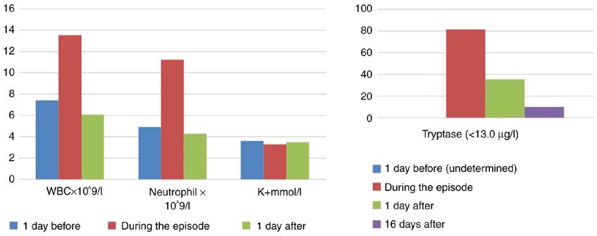
2 CHEN et al: SYSTEMIC MASTOCYTOSIS WITH FLUSHING AND HYPOTENSION
Physical examination. There was no significant change in 0‑60 KU/l). Inhalant/food/mold allergen, anti‑C1Q antibody
blood pressure when the patient moved from recumbent to and C1 inhibitor tests were negative. Serum tryptase was
orthostatic positions. Cardiorespiratory and abdominal phys‑ 81.6 ng/ml during onset, decreased to 36.3 ng/ml 9 days later
ical examinations were negative. There was also no swelling and remained at 9.85 ng/ml (normal, 0‑13.0 ng/ml) on the
in either lower extremity. 16th day (Fig. 1).
Episode prior to admission. The patient had a 12‑year history Imaging examinations. Electrocardiogram (ECG), neck
of recurrent facial, anterior chest and skin flush with bulbar vascular ultrasound (CVUS), transcranial doppler ultrasound
conjunctival congestion, and experienced stuffy nose, as (TCD), cerebral MRI angiography, thyroid ultrasound,
well as discomfort in the chest and upper abdomen, but did echocardiography, octreotide imaging, gastroenteroscopy,
not present with associated chest pain, dyspnea or fever. The contrast‑enhanced CT scans of the chest, abdomen and pelvis,
patient's blood pressure declined 1‑2 h after the onset of each and positron emission tomography/CT of the chest, abdomen
episode to 70‑90 mmHg systolic with skin flush, which was and pelvis were all unremarkable.
able to recover to 120/80 mmHg without treatment after
several hours. A temporary loss of consciousness was observed Pathological examination. The peripheral blood smears both
during several severe onsets, with a minimum systolic blood during the onsets and remission were all negative. Multifocal
pressure of 20‑30 mmHg. Vasoactive medications, such as bone marrow smear (posterior ilium/anterior iliac/sternum)
dopamine and noradrenaline, were administered to maintain exhibited active proliferation and phagocytic cells were
blood pressure. Following treatment, the skin flush and bulbar observed. The percentage of basophils in certain tissues was up
conjunctival hyperemia were gradually be relieved. Vasoactive to 0.5% and clusters of basophils were observed. The pathology
medications were discontinued after several hours to two days of the bone marrow biopsy (posterior ilium) was as follows:
depending on the severity of each attack. The frequency of Megakaryocytes were identified and mast cell infiltration
the aforementioned symptoms varied from once to four times existed, which supported the diagnosis of SM. Mast cell‑specific
yearly, with similar symptoms but to different extents. There staining was then performed; methylaniline blue staining was
was no apparent association between the attacks and the positive but scattered. Immunohistochemistry revealed posi‑
season, time of day or night, region of stay or diet. The onset tive staining for CD117 (mast cells ++), CD15 (granulocytes +),
was also not associated with the patient being in an active, myeloperoxidase (granulocytes +) and CD20 (diffuse +), and
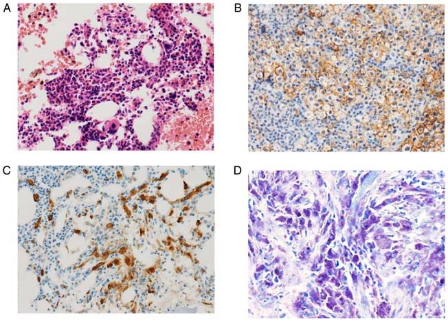
resting, sleeping, standing or recumbent state. negative staining for CD3 (Fig. 2). Tests for the cKIT/D816V
and factor interacting with poly(A) polymerase α and cleavage
An episode during hospitalization. On the day after admission, and polyadenylation‑specific factor 1/platelet‑derived growth
there was a new onset with no stimulus. The patient presented factor receptor α (FIP1L1/PDGFRα) gene mutations in bone
with bulbar conjunctival congestion, flush of the face and marrow were negative.
anterior chest skin, as well as nasal congestion. After ~1 h, the Mast cells infiltrated in the bone marrow pathology
patient's blood pressure began to decline. Intensive care and (CD117; Fig. 2) were further counted and the result suggested
fluid infusion therapy were started, but the blood pressure still a ≥15 mast cell aggregation in the field of view and multifo‑
dropped to a minimum of 72/43 mmHg ~2 h after the begin‑ cality, consistent with the major diagnostic criteria for SM (1).
ning of the attack and then gradually increased. The redness
of the skin was also relieved as the blood pressure increased. Final diagnosis. The final diagnosis of the present case was
There was no unconsciousness, diarrhea, itching or dyspnea inert SM, MCAS and increased excitability of the vasovagal
during the onset; however, the patient had no complete recol‑ nerve, all of which contributed to the flushing and hypotension
lection of the time of the episode. status in the present case.
Laboratory examinations. The leukocyte (13.53x10⁹/l) and Treatment. The patient refused to use glucocorticosteroids and
neutrophil (11.22x10 9/l) concentrations exhibited marked medications to control the mast cell mediator release symp‑
increases during the attack and blood potassium was decreased toms. As an alternative, an adrenaline pen was carried as a
(3.3 mmol/l; normal range 3.5‑5.5 mmol/l), while the hemo‑ backup. Metoprolol was prescribed prior to admission due to
globin and platelet counts were normal. Following the attack, hypertension and was not recommended following discharge,
the complete blood count and blood potassium spontaneously since it may make the adrenergic response less effective.
returned to the normal range (Fig. 1). Laboratory tests of the liver
and renal function and arterial blood gas were also within normal Outcome and follow‑up. The patient was followed up for
limits. No abnormalities were found in the erythrocyte sedimen‑ 4 years by telephone and no further onset was observed. The
tation rate, C‑reactive protein and complement C3 and C4. patient did not undergo any tests of serum tryptase or bone
Antinuclear antibodies, anti‑double‑stranded DNA, marrow puncture after discharge from our hospital.
anti‑soluble nuclear antigen antibodies and tumor markers
were all negative. Plasma protein gel electrophoresis, blood Discussion
immuno‑fixation electrophoresis, test of thyroid function,
three repeated tests of levels of calcitonin and 24‑h urinary The present case was that of a middle‑aged male with a
catecholamine examinations were also negative. 12‑year history of episodes of recurrent flushing, hypotension
The head‑up tilt test was positive with nitroglycerin. and loss of consciousness. The characteristics of the symp‑
Serum total immunoglobulin E (IgE) was 90 KU/l (normal, toms were as follows: i) No obvious stimulus of each onset,EXPERIMENTAL AND THERAPEUTIC MEDICINE 21: 404, 2021 3 Figure 1. Changes in the WBC count, potassium and tryptase prior to, during and after the episodic period of the patient. Normal ranges: WBC, 3.50‑9.50x10⁹/l; Neutrophil, 2.00‑7.50x10⁹/l; K+, 3.5‑5.5 mmol/l; and Tryptase,
4                           CHEN et al: SYSTEMIC MASTOCYTOSIS WITH FLUSHING AND HYPOTENSION

render cardiogenic syncope highly unlikely. While in patients       of the attack, and multiple focal mast cell infiltration (≥15 mast
with reflex syncope episodes, prodromal symptoms, such as           cell aggregation in the field of view) was observed in the bone
dizziness, panic and weakness are common, which did not             marrow biopsy, which supports the diagnosis of SM.
appear in this patient. Although the orthostatic tilt test was           SM belongs to a group of mast cell monoclonal prolifera‑
positive after nitroglycerin induction, the syncope induced         tive diseases under the umbrella term of mast cell hyperplasia,
by the test was not accompanied by redness of the skin and          which was first reported by Ellis (11) in 1949. In 2016, the
hyperemia of the bulbar conjunctiva. It remains challenging         World Health Organization divided mast cell hyperplasia
to explain the full clinical picture of the patient by increased    into three categories, including skin type, SM and mast cell
excitability of the vasovagal nerve, which could only explain       sarcoma (12). The diagnostic criteria for SM include one
hypotension but would not be responsible for elevated serum         major criterion and four minor criteria. The major criterion
tryptase levels.                                                    is multifocal dense mast‑cell infiltration (≥15 mast cell aggre‑
     One notable symptom of the patient of the present study        gation in the field of view) in the bone marrow and/or other
was that the skin flush prior to each onset, except when            nonskin tissue sections (12). The minor criteria are as follows:
syncope was accompanied by a marked drop in blood pressure          i) >25% spindle or atypical mast cell infiltration in non‑skin
in parallel during the onset. Therefore, flush and hypoten‑         tissue sections or bone marrow smears or >25% mast cell
sion were the principal clinical characteristics of the patient.    are immature or atypical; ii) mutations in codon 816 of the
Murali et al (7) provided a comprehensive list summarizing          KIT gene detected in bone marrow, blood or other non‑skin
disorders for differential diagnosis of patients with flushing      tissues; iii) mast cells in bone marrow, blood or other non‑skin
and hypotension, including exogenous and endogenous factors.        tissues express CD25 with/without CD2 in addition to normal
Exogenous factors, such as diet and medication, may give rise       mast cell markers; and iv) serum tryptase levels >20 ng/ml
to severe allergic reactions that frequently lead to recurrent      (unless an associated myeloid neoplasm exists that invalidates
skin flushing, hypotension and shock. Slightly elevated IgE         this parameter) (12). SM may be diagnosed when the primary
and clinical symptoms suggest a possibility of allergy (8).         criterion and one minor criterion, or three minor criteria, are
However, allergens and the association between the onset of         fulfilled (1).
symptoms and diet, medication and contactors were all absent,            SM is further divided into several types according to the
and no seasonal or geographical patterns were identified for        B and C findings (1). B findings include: i) Bone marrow
the attacks. Therefore, exogenous factors are less likely to        biopsy reveals >30% infiltration of mast cells in the field of
cause this situation.                                               view and serum tryptase level >200 ng/ml; ii) non‑mast cell
     Possible endogenous factors include sepsis, carcinoid          lineage dysplasia or myeloproliferation, but cannot meet the
syndrome, endocrine tumor, idiopathic capillary leakage             criteria to diagnose an associated hematological neoplasm
syndrome and mastocytosis (5). First, the present patient           (AHN), the blood counts are normal or slightly abnormal; and
maintained a normal body temperature during the attack              iii) Hepatomegaly and palpable splenomegaly without functional
and the hypotension was transient and at times completely           abnormality, and/or palpation or imaging lymphadenopathy.
self‑limited, which bears no support for sepsis. Furthermore,       C findings include: i) Bone marrow dysfunction revealed by
carcinoid syndrome refers to a group of argyrophilic cell           ≥1 cytopenia(s) (absolute neutrophil count 20 ng/ml at the time       (with monoclonal hyperplasia), secondary (secondary to
EXPERIMENTAL AND THERAPEUTIC MEDICINE 21: 404, 2021 5
Table I. Subtypes of SM.
Subtype of SM Definition
Cutaneous mastocytosis Urticaria pigmentosa/maculopapular cutaneous mastocytosis; diffuse cutaneous mastocytosis;
solitary mastocytoma of skin
Indolent SMa No ‘C’ findings. No evidence of associated hematological neoplasm
Smoldering SMa No ‘C’ findings but with 2 or more ‘B’ findings. No evidence of associated clonal hematological
nonmast cell lineage disease
SM with associated Evidence of associated clonal hematological nonmast cell lineage disease
hematological neoplasm
Aggressive SMa 1 or more ‘C’ findings. No evidence of MCL
MCL Bone marrow biopsy indicates a diffuse infiltration by atypical or immature mast cells. Bone
marrow aspirate smears contain ≥20% mast cells. In typical MCL, mast cells account for ≥10% of
the peripheral blood white cells. There is also an aleukemic MCL variant (200 ng/ml (20). Certain studies have suggested that serum
mediators exist (4). The diagnosis of the present case also falls tryptase levels are parallel to the infiltration of bone marrow
into the category of primary MCAS. mast cells in patients with ISM, but there is a poor correlation
The etiology of SM remains to be fully established, and between the level of serum tryptase and the clinical symptoms
according to the literature, it is frequently associated with a of mast cell mediator release (21). Of note, in the present
functional acquired mutation of the KIT gene. KIT (CD117) case, white blood cells were significantly elevated during
is a type III tyrosine kinase receptor expressed in mast cells, the onset, mainly neutrophilic granulocytes, and decreased
and a mutation in this gene may induce the clonal prolifera‑ plasma potassium levels were detected (Fig. 1), all of which
tion of mast cells, without the control of hematopoietic stem recovered spontaneously after remission, which is consistent
cell factor (14,15). The most common mutation site is D816V with the literature (7). The pro‑inflammatory and chemotactic
(>80%) (14,15). Other causes, such as a tet methylcytosine factors released by mast cells may affect leukocyte migration
dioxygenase 2 mutation, increased levels of hematopoietic stem and recruitment, but the decrease in serum potassium may be
cell factors and the persistent expression of heat shock protein related to the redistribution of intracellular and extracellular
32, may also be associated with the pathogenesis of SM (16,17). potassium ions (4,22).
In addition to mast cell mediator release syndrome, the As there is currently no cure for SM, current treatments focus
infiltration of viscera by mast cells may occur in SM. Skin on methods to control the symptoms, improve the quality of life
involvement may present urticaria and result in the forma‑ and delay disease progression. The present treatments exhibit a
tion of skin mastocytoma (1). Anemia, thrombocytopenia, marked variation depending on the subtype of SM. For patients
enlargement of the liver and spleen or lymph nodes, portal with ISM, the major treatments are avoidance of the induction
hypertension, osteoporosis and pathological fracture may of mast cell release, control of the release symptoms of mast
occur when there are endocrine manifestations, osteoporosis cells and local surgery (15). Drugs that are able to control the
and osteosclerosis (18,19). According to the presence or symptoms include antihistamines, including H1 or H2 receptor
absence of type B or C features, SM may be further divided blockers, such as cetirizine and ranitidine, mast cell membrane
into seven subtypes (1) (Table I): Cutaneous mastocytosis, stabilizers, such as sodium tryptophan, leukotriene inhibi‑
indolent SM (ISM), smoldering SM, SM with an associated tors, such as montelukast, and nonsteroidal anti‑inflammatory
hematological neoplasm (SM‑AHN), aggressive SM (ASM), drugs (23). If the symptoms cause hypotension or shock, the first
mast cell leukemia (MCL) and mast cell sarcoma (12,15). In choice is adrenaline treatment, and glucocorticoid treatment is
the present case, there was no evidence of type B or C features optional (24). In certain cases, shock was reported as a symptom
and no hematological malignancy, and mast cell lineages were but more frequently defined as anaphylactic shock (25‑27). Other
identified; therefore, the present case may be classified as ISM. drugs, such as midostaurin and masitinib, have undergone phase
Elevated levels of serum tryptase, which is one of the mediators 2 or 3 trials and still require further evaluation (28,29). ASM6 CHEN et al: SYSTEMIC MASTOCYTOSIS WITH FLUSHING AND HYPOTENSION
should be treated with chemotherapy. Commonly used regimens Availability of data and materials
include interferon‑α alone or in combination with prednisone,
or kratobine alone (15). In addition, tyrosine kinase inhibi‑ The datasets used and/or analyzed during the current study are
tors (TKIs), including imatinib, dashatinib and midostaurin, available from the corresponding author on reasonable request.
hydroxyurea and mitoxaline, may also be used, among which
imatinib is the only drug approved by the US Food and Drug Authors' contributions
Administration for SM and is suitable for patients with SM with
KIT mutations that are sensitive to imatinib or those without ATC performed the literature search/selection/review and
KIT D816V mutations (15). Over 80% of adults with mastocy‑ collected clinical information and drafted the manuscript.
tosis carry this mutation (30‑32). Midostaurin is the only TKI XYR provided critical pathological reports and diagnostic
approved as a monotherapy for ASM (33). Hematopoietic stem consulting services. ATC and XYR contributed equally to this
cell transplantation has been reported to improve the survival study. WC conceived the study and revised the manuscript. All
rate among patients with SM (34,35), but this requires to be authors read and approved the final manuscript.
confirmed by larger studies.
The prognosis of different subtypes of SM is significantly Ethics approval and consent to participate
different from that of indolent life‑threatening diseases (36).
The prognosis of ISM is relatively good, with a median Not applicable.
survival time of 198 months, which exhibits no significant
difference between patients with ISM and healthy controls. Patient consent for publication
The progression and prognosis of ISM are not associated with
the ultrasonography findings (37). ASM and SM‑AHN are Consent for publication of the patient's data/images in this case
associated with poor prognosis, with median survival rates of report was obtained.
41 and 24 months, respectively. By contrast, MCL has a dismal
prognosis, with a median survival time of only 2 months (15). Competing interests
The patient in the present study belonged to the ISM group,
and hematology specialists in the Peking Union Medical The authors declare that they have no competing interests.
College Hospital recommended two treatment options for the
patient. The first was using drugs to control the release of mast References
cell mediators together with epinephrine and glucocorticoid
treatment when the symptoms are severe. Another consider‑ 1. Pardanani A: Systemic mastocytosis in adults: 2019 update on
ation is that this patient suffered a significant decline in blood diagnosis, risk stratification and management. Am J Hematol 94:
363‑377, 2019.
pressure during the onset of symptoms, which may lead to 2. Brockow K: Epidemiology, prognosis, and risk factors in
a lack of blood supply to vital organs and potentially fatal mastocytosis. Immunol Allergy Clin North Am 34: 283‑295,
arrhythmia. Therefore, it was recommended that the patient 2014.
3. Valent P: Mast cell activation syndromes: Definition and clas‑
begins treatment with interferon and/or glucocorticoid imme‑ sification. Allergy 68: 417‑424, 2013.
diately. However, the patient did not accept the aforementioned 4. Valent P, Akin C, Arock M, Brockow K, Butterfield JH, Carter MC,
scheme due to the potential side effects of the medications, and Castells M, Escribano L, Hartmann K, Lieberman P, et al:
Definitions, criteria and global classification of mast cell disor‑
he purchased and carried an adrenalin pen as a precaution. ders with special reference to mast cell activation syndromes: A
In conclusion, the diagnosis and treatment of the present consensus proposal. Int Arch Allergy Immunol 157: 215‑225,
case were in accordance with SM/MCAS. To date, this disease 2012.
5. Runser LA, Gauer RL and Houser A: Syncope: Evaluation and
has been rarely reported, with high rates of missed diagnosis differential diagnosis. Am Fam Physician 95: 303‑312, 2017.
and misdiagnosis. Therefore, the understanding of this disease 6. Brignole M, Moya A, de Lange FJ, Deharo JC, Elliott PM,
should be expanded. In addition to detection of postural hypo‑ Fanciulli A, Fedorowski A, Furlan R, Kenny RA, Martín A, et al:
2018 ESC Guidelines for the diagnosis and management of
tension, and cardiac and vascular factors in syncope patients, syncope. Eur Heart J 39: 1883‑1948, 2018.
the possibility of SM/MCAS should be taken into consider‑ 7. Murali MR, Castells MC, Song JY, Dudzinski DM and
ation when encountering symptoms such as redness of the skin Hasserjian RP: Case records of the Massachusetts General
Hospital. Case 9‑2011. A 37‑year‑old man with flushing and
and severe hypotension. Bone marrow or histopathological hypotension. N Engl J Med 364: 1155‑1165, 2011.
examination, mast cell‑specific and immunohistochemical 8. Campbell DE and Mehr S: Fifty years of allergy: 1965‑2015.
staining, KIT gene detection and serum tryptase determina‑ J Paediatr Child Health 51: 91‑93, 2015.
9. Rubin de Celis Ferrari AC, Glasberg J and Riechelmann RP:
tion are of great significance in the diagnosis of SM/MCAS. Carcinoid syndrome: Update on the pathophysiology and treat‑
ment. Clinics (Sao Paulo) 73 (Suppl 1): e490s, 2018.
Acknowledgements 10. Siddall E, Khatri M and Radhakrishnan J: Capillary leak
syndrome: Etiologies, pathophysiology, and management.
Kidney Int 92: 37‑46, 2017.
The authors wish to thank Dr Haiyan He (Department of 11. Ellis JM: Urticaria pigmentosa; a report of a case with autopsy.
Cardiology, Peking Union Medical College Hospital) for her Arch Pathol (Chic) 48: 426‑435, 1949.
12. Arber DA, Orazi A, Hasserjian R, Thiele J, Borowitz MJ,
help in preparing and editing this manuscript. Le Beau MM, Bloomfield CD, Cazzola M and Vardiman JW: The
2016 revision to the World Health Organization classification of
Funding myeloid neoplasms and acute leukemia. Blood 127: 2391‑2405,
2016.
13. Metcalfe DD: Mast cells and mastocytosis. Blood 112: 946‑956,
No funding was received. 2008.EXPERIMENTAL AND THERAPEUTIC MEDICINE 21: 404, 2021 7
14. Arock M, Sotlar K, Akin C, Broesby‑Olsen S, Hoermann G, 28. van Anrooij B, Oude Elberink JNG, Span LFR, de Monchy JGR,
Escribano L, Kristensen TK, Kluin‑Nelemans HC, Hermine O, Rosati S, Mulder AB and Kluin‑Nelemans JC: Midostaurin in
Dubreuil P, et al: KIT mutation analysis in mast cell neoplasms: patients with indolent systemic mastocytosis: An open‑label
Recommendations of the European Competence Network on phase 2 trial. J Allergy Clin Immunol 142: 1006‑1008.e7, 2018.
Mastocytosis. Leukemia 29: 1223‑1232, 2015. 29. Lortholary O, Chandesris MO, Bulai Livideanu C, Paul C,
15. Pardanani A: Systemic mastocytosis in adults: 2017 update on Guillet G, Jassem E, Niedoszytko M, Barete S, Verstovsek S,
diagnosis, risk stratification and management. Am J Hematol 91: Grattan C, et al: Masitinib for treatment of severely symp‑
1146‑1159, 2016. tomatic indolent systemic mastocytosis: A randomised,
16. Kondo R, Gleixner KV, Mayerhofer M, Vales A, Gruze A, placebo‑controlled, phase 3 study. Lancet 389: 612‑620, 2017.
Samorapoompichit P, Greish K, Krauth MT, Aichberger KJ, 30. Hartmann K, Escribano L, Grattan C, Brockow K, Carter MC,
Pickl WF, et al: Identification of heat shock protein 32 (Hsp32) as Alvarez‑Twose I, Matito A, Broesby‑Olsen S, Siebenhaar F,
a novel survival factor and therapeutic target in neoplastic mast Lange M, et al: Cutaneous manifestations in patients with masto‑
cells. Blood 110: 661‑669, 2007. cytosis: Consensus report of the European Competence Network
17. Tefferi A, Lim KH, Abdel‑Wahab O, Lasho TL, Patel J, on Mastocytosis; the American Academy of Allergy, Asthma &
Patnaik MM, Hanson CA, Pardanani A, Gilliland DG and Immunology; and the European Academy of Allergology and
Levine RL: Detection of mutant TET2 in myeloid malignan‑ Clinical Immunology. J Allergy Clin Immunol 137: 35‑45, 2016.
cies other than myeloproliferative neoplasms: CMML, MDS, 31. Valent P, Akin C, Hartmann K, Nilsson G, Reiter A, Hermine O,
MDS/MPN and AML. Leukemia 23: 1343‑1345, 2009. Sotlar K, Sperr WR, Escribano L, George TI, et al: Advances in
18. Greene LW, Asadipooya K, Corradi PF and Akin C: Endocrine the classification and treatment of mastocytosis: Current status
manifestations of systemic mastocytosis in bone. Rev Endocr and outlook toward the future. Cancer Res 77: 1261‑1270, 2017.
Metab Disord 17: 419‑431, 2016. 32. Kristensen T, Vestergaard H and Møller MB: Improved detection
19. Johansson C, Roupe G, Lindstedt G and Mellstrom D: Bone of the KIT D816V mutation in patients with systemic mastocy‑
density, bone markers and bone radiological features in mastocy‑ tosis using a quantitative and highly sensitive real‑time qPCR
tosis. Age and ageing 25: 1‑7, 1996. assay. J Mol Diagn 13: 180‑188, 2011.
20. Lim KH, Tefferi A, Lasho TL, Finke C, Patnaik M, Butterfield JH, 33. Kayser S, Levis MJ and Schlenk RF: Midostaurin treatment in
McClure RF, Li CY and Pardanani A: Systemic mastocytosis in FLT3‑mutated acute myeloid leukemia and systemic mastocy‑
342 consecutive adults: Survival studies and prognostic factors. tosis. Expert Rev Clin Pharmacol 10: 1177‑1189, 2017.
Blood 113: 5727‑5736, 2009. 34. Nakamura R, Chakrabarti S, Akin C, Robyn J, Bahceci E,
21. Pardanani A, Lim KH, Lasho TL, Finke CM, McClure RF, Greene A, Childs R, Dunbar CE, Metcalfe DD and Barrett AJ:
Li CY and Tefferi A: WHO subvariants of indolent mastocytosis: A pilot study of nonmyeloablative allogeneic hematopoietic
Clinical details and prognostic evaluation in 159 consecutive stem cell transplant for advanced systemic mastocytosis. Bone
adults. Blood 115: 150‑151, 2010. Marrow Transplant 37: 353‑358, 2006.
22. Valent P, Sillaber C, Baghestanian M, Bankl HC, Kiener HP, 35. Ustun C, Reiter A, Scott BL, Nakamura R, Damaj G, Kreil S,
Lechner K and Binder BR: What have mast cells to do with Shanley R, Hogan WJ, Perales MA, Shore T, et al: Hematopoietic
edema formation, the consecutive repair and fibrinolysis? Int stem‑cell transplantation for advanced systemic mastocytosis.
Arch Allergy Immunol 115: 2‑8, 1998. J Clin Oncol 32: 3264‑3274, 2014.
23. Scherber RM and Borate U: How we diagnose and treat systemic 36. Theoharides TC, Valent P and Akin C: Mast cells, mastocytosis,
mastocytosis in adults. Br J Haematol 180: 11‑23, 2018. and related disorders. N Engl J Med 373: 163‑172, 2015.
24. van der Weide HY, van Westerloo DJ and van den Bergh WM: 37. de Mol CL, Hermans MA, Gerth van Wijk R, van Hagen PM and
Critical care management of systemic mastocytosis: When every van Daele PL: Routine abdominal ultrasonography has limited
wasp is a killer bee. Crit Care 19: 238, 2015. value in the care for patients with indolent systemic mastocy‑
25. Chen G, Chen L, Qin X, Xie X, Li G and Xu B: Systemic masto‑ tosis. Hematology 22: 544‑547, 2017.
cytosis with recurrent anaphylactic shock and multiple organ
dysfunction failure. Clin Lab 61: 179‑182, 2015. This work is licensed under a Creative Commons
26. Chatterjee M, Sengupta S, Chakravartyr C, Ramasubban S, Attribution-NonCommercial-NoDerivatives 4.0
Bhartia S, Khan S and Agarwal VK: Indolent systemic mastocy‑ International (CC BY-NC-ND 4.0) License.
tosis manifesting as protracted anaphylactic shock. Indian J Crit
Care Med 22: 311‑313, 2018.
27. Kors JW, van Doormaal JJ and de Monchy JG: Anaphylactoid
shock following Hymenoptera sting as a presenting symptom of
systemic mastocytosis. J Intern Med 233: 255‑258, 1993.You can also read