AN ANATOMICAL STUDY OF THE BRONCHIAL VASCULAR SYSTEM AND ITS VARIATIONS IN DISEASE
←
→
Page content transcription
If your browser does not render page correctly, please read the page content below
Thorax: first published as 10.1136/thx.5.3.207 on 1 September 1950. Downloaded from http://thorax.bmj.com/ on February 29, 2020 by guest. Protected by
Thorax (1950), 5, 207.

AN ANATOMICAL STUDY OF THE BRONCHIAL
   VASCULAR SYSTEM AND ITS VARIATIONS
               IN DISEASE
                                          BY
         PAUL MARCHAND, J. C. GILROY, AND V. H. WILSON
From the Thoracic Surgery Unit and Department of Medicine, Baragwanath Hospital,
                                    Johannesburg
     During the course of an anatomical study of specimens of emphysematous lungs
 evidence was found to suggest that the bronchial circulation might play a more
 important part in producing the disease picture than has been appreciated hitherto.
 Consequently we undertook a detailed investigation of the bronchial arteries and
 veins in both normal and diseased lungs.
     In some respects our findings differ from presently accepted views of the
 anatomy of these vessels.
                     MATERIAL AND METHODS OF INVESTIGATION
     The material us.d in our experiments is set out in Tables I and II.

                                                                                                                                                               copyright.
     The specimens have varied from very fresh lungs obtained at operation to
 necropsy material injected at least 24 hours after death and after rigor mortis had
 passed off. In all, a total of 63 lung specimens were injected.
     The following injection techniques were employed:
     The Vinylite Corrosion Technique (Puckett and Newman, 1940).-In this pro-
cedure coloured plastic was injected into the vessels under a positive pressure which
 varied from 2 to 5 lb. per sq. in. The organic material was digested off the cast
by immersion in either concentrated hydrochloric acid or a 40% solution of caustic
potash. After digestion a careful dissection was necessary to remove the finer
radicles of the pulmonary vessels in order to visualize for study the bronchial circu-
lation. The lungs, in the majority of instances, were inflated with air before the
injection of the blood vessels; in other cases vinylite was formed into the bronchi
as an alternative to inflation with air. In preparing demonstration specimens, casts
of the bronchi were made by pouring Wood's metal into the trachea before opening
the chest.
    When injecting complete lungs obtained at necropsy the pulmonary artery was
filled through a cannula tied into the main stem; the pulmonary veins were filled
through a cannula in the left auricular appendix, the aorta being ligated at its
origin to prevent the escape of vinylite into the systemic vessels. The bronchial
arteries were injected through a cannula tied into the divided descending aorta,
the arch of the aorta being clamped at the origin of the left subclavian artery and
the intercostal vessels being tied or clamped before or during the injection. The
veins which enter the azygos and hemiazygos veins were injected through these
veins in a retrograde manner.
       Q
Thorax: first published as 10.1136/thx.5.3.207 on 1 September 1950. Downloaded from http://thorax.bmj.com/ on February 29, 2020 by guest. Protected by
208            PAUL MARCHAND, J. C. GILROY, and V. H. WILSON
    When dealing with specimens obtained at operation, injection was accomplished
through the cut ends of the hilar vessels, and the bronchial artery was dissected
out and cannulated directly. Foetal lungs were injected in the same way. The
ductus arteriosus was ligated and the left auricle was clamped in such a way as to
prevent the loss of injection mass through the foramen ovale and the atrio-ventricular
valve. In these specimens the bronchial arteries were injected with a 7% solution
of vinylite in acetone to ensure penetration of the finer vessels. In all our other
injections in foetal and in adult lungs a 12j% solution was used. Both lungs were
always injected in the necropsy specimens.
    Perfusion of the arteries with saline before injecting the vinylite was not done
because we found that this procedure caused waterlogging of the tissues with con-
sequent occlusion of small vessels. Satisfactory injection casts of the pulmonary
vessels can be obtained with vinylite after manual expression of clots from the
larger vessels before injecting them. The specimens injected by this technique are
tabulated (Table I).
                                         TABLE I
             TABLE OF SPECIMENS INJECTED BY THE VINYLITE-CORROSION TECHNIQUE

                          Type of Specimen                                No. of Lungs Injected
NORMAL LUNGS
  Complete injection        ..    ..     ..    ..     ..     ..      ..      5 Necropsy
  Bronchial arteries alone              ..    ..      ..     ..      ..      2 material

                                                                                                                                                                        copyright.
  Bronchial arteries plus azygos and hemiazygos veins        ..      ..      2 J(both lungs)
                                                                  Total      9
ABNORMAL LUNGS
  Lung Abscess:
    (a) Complete injection (3 pneumonectomy, 1 lobectomy)           .               4
    (b) Pulmonary artery and vein only (1 lobectomy, 2 pneumonectomy)               3
  Bronchiectasis:
    (a) Complete injection (lobectomy) ..       ..     ..    ..    ..               2
    (b) Pulmonary artery and vein (lobectomy)          ..    ..    ..               3
  Pulmonary Tuberculosis:
    (a) Complete injection (pneumonectomy)             ..    ..    ..               2
  Emphysema: Complete injection (necropsy)             ..    ..    ..               2
  Partial atelectasis due to compression of main bronchus by tracheo-
       bronchial tubercular glands (necropsy) ..
    (a) Complete injection               ..     ..           ..    ..
  Congenital dysgenesis of the lung whereby the left main bronchus
       opened into an infected cystic space bounded by fibrous tissue
    (a) Pulmonary artery, pulmonary vein and bronchial artery
       (pneumonectomy) ..          ..     ..    ..     ..    ..    ..
                                                                  Total            18
FOETAL LUNGS
 Before respiration (full term)
   (a) Bronchial and pulmonary arteries only..        ..     ..      ..             2
   (b) Bronchial arteries and pulmonary arteries and veins           ..             2

   Injection of Radio-Opaque Media.-This method has been used to demonstrate
the course and connexions of the bronchial arteries and of those veins which drain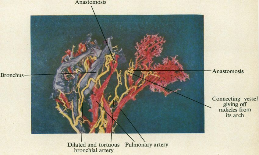
Thorax: first published as 10.1136/thx.5.3.207 on 1 September 1950. Downloaded from http://thorax.bmj.com/ on February 29, 2020 by guest. Protected by
    ANATOMICAL STUDY OF BRONCHIAL VASCULAR SYSTEM                                                            209

into the systemic circulation. The routes of injection have been the same as those
employed for the vinylite injections.
    We have used lipiodol (Lafay 40%) 75% with 25% acetone but this solution,
although it gives excellent radio-opacity, has the disadvantage of penetrating
capillary vessels. Recently we have modified the formula of Hill (1929) who used
a bismuth oxychloride suspension in gum acacia by substituting a saturated solu-
tion of sodium iodide for the water component of the original formula. This gives
the solution a bright yellow colour which is easily recognizable and adds materially
to its radio-opacity. The advantage of Hill's formula is that the solution will not
penetrate the capillaries. The use of this method does not preclude subsequent
injection with vinylite and both techniques have been used in the same specimen on
three occasions (two normal lung specimens and one of emphysema). We felt that
the penetration of the bronchial arterial injection with vinylite might not be com-
plete if it were preceded by an injection of bismuth oxychloride. We have therefore
made it our usual practice to use one or other method alone.
    After completion of the injection, stereoscopic radiographs of these specimens
were taken at four feet. These lungs were finally dissected, a special effort being
made to follow the bronchial artery and its branches as far into the substance of
the lung as possible.
    The specimens treated by this method are tabulated in Table II.
                                                TABLE II

                                                                                                                                                                                         copyright.
                           Type of Specimen                                              No. of Lungs Injected
NORMAL LUNGS
  (1) Bronchial arteries (necropsy)      ..    ..     ..   ..     ..                              10
  (2) Veins draining into
      (a) Azygos vein or superior vena cava (necropsy)     ..     ..                               8
      (b) Hemiazygos vein or left innominate vein or superior inter-
            costal vein    ..    ..   ..         ..        ..        ..        ..                  6
ABNORMAL LUNGS
  Bronchial arteries
  (1) Lung abscess (pneumonectomy)         ..         ..        ..        ..        ..             2
  (2) Emphysema (necropsy)     ..          ..         ..        ..        ..        ..             2
FOETAL LuNGs
  (1) Bronchial arteries   ..    ..        ..         ..        ..        ..        ..             2

   Injections with Gelatine.-Two specimens of emphysema and two normal lungs
have been injected through the main pulmonary artery with a carmine gelatine
preparation after the formula of Moore (1929) to which we added 20% yellow
oxide of lead in order to obtain radio-opacity. Serial sections of the bronchi and
bronchioles were cut and examined.
    RESULTS OF INJECTION EXPERIMENTS IN SPECIMENS
                  OF NORMAL LUNGS
    The bronchial arteries show some inconstancy in their origin, distribution, and
course, though this has been less than we expected. The commonest site of origin
is from the antero-lateral aspect of the aorta about i in. to 1 in. distal to the origin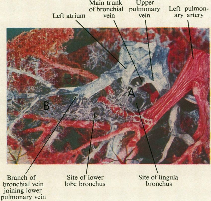
Thorax: first published as 10.1136/thx.5.3.207 on 1 September 1950. Downloaded from http://thorax.bmj.com/ on February 29, 2020 by guest. Protected by
210              PAUL MARCHAND, J. C. GILROY, and V. H. WILSON

of the left subclavian artery. Usually the arteries to the right and left lungs arise
separately although they may have a common origin from the aorta. Either artery
may arise from an intercostal artery, and Miller (1947) states that the bronchial
artery may originate from the internal mammary artery.
    There are usually two main arteries, one to each lung. In addition, one or more
smaller arteries arise directly from the aorta about another half inch distal to the
main bronchial arteries, but they may arise from any part of the aorta above the
diaphragm (Fig. 1).
    Where the lung is supplied by more than one bronchial artery the upper and
                                                                 normally placed
                                                                 vessel supplies the
                                                                 greater portion of
                                                                 the lung; the addi-
                                                                 tional artery seldom
                                                                 supplies more than
                                                                 the posterior and
                                                                 medial aspects of
                                                                 the lower iobe,
                                                                 although in one of
                                                                ~~our specimens      it
                                                            ~ ~ ~ ~ ~ ~ ~ ~ ~ ~ ~ ~ ~ ~ ~ ~ ~ ~. . . .

                                                                 supplied most of
                                                                 the right lower lobe.

                                                                                                                                                                                                      copyright.
                                                                 The additional
                                                                 bronchial a r t e r y
                                                                 traverses the pul-
                                                                 monary ligament,
                                                                 and the lower its
                                                                 origin fo the
                                                                 aorta the more
                                                                                                         limited seems to
                                                                                                         be its distribution.
                                                                                                         This artery may be
                                                                                                         the source of annoy-
                                                                                                         ing, and sometimes
                                                                                                         of troublesome
                                                                                                         bleeding, when the
                                                                                                         pulmonary ligament
                                                                                                         is divided during
                                                                                                         pneumonectomy or
                                                                                                         lower lobectomy.
  FIG. 1.-Injection through the aorta of the bronchial arteries of a
      normal lung with radio-opaque bismuth suspension. This is one         Several    small
      of the two lungs that showed no pulmnonary arterial filling. Both arteries which arise
      lungs are supplied by a single main bronchial artery, the point
      where the right bronchial artery divides being obscured by the either directly from
      smudged area where the medium has escaped into interstitial the aorta or from
      tissue. Note how the arteries branch and re-anastomose after the the extra-pulmonary
      main artery has entered the hilum and given off branches which core
      run with the maiin sejmental bronchicus.o fth
      I L&tA W11644 %,la%, ARA"lat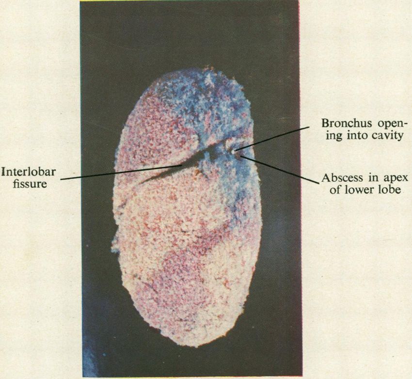
Thorax: first published as 10.1136/thx.5.3.207 on 1 September 1950. Downloaded from http://thorax.bmj.com/ on February 29, 2020 by guest. Protected by
         Nor-.2M       in     X;                             V u
FiG. 2.-Wood's metal cast of bronchial tree. Red vinylite was injected into the
    aorta, and shows the manner of origin of the bronchial arteries as they arise
    from the aorta. In this specimen both lower lobes are partly supplied by acces-
    sory bronchial arteries arising from the aorta a few centimetres distal to the
    main bronchial arteries. This specimen is photographed from behind and
    shows the aorta crossing the left main bronchus.

                                                                                                                                                                                                                   copyright.
                                                        Anastomosis

                                                                                                                          -Anastomosis
                    Bronchus -

                                                                                                                                 Connecting vessel
                                                                                                                          .1,,       giving off
                                                                                                                                   radicles from
                                                                                                                                      its arch

                                                           I              I       \\
                                                    Dilated  and tortuous Pulmonary artery
                                                        bronchial artery

                                  FIG. 4.-Vinylite cast of the subapical segment of the right upper lobe of an emphy-
                                       sematous lung. The bronchi show up blue, the bronchial arteries yellow, and
                                       the pulmonary artery red. This specimen has also been flattened by placing in
                                       warm water. There are several anastomoses to be seen demonstrating the
                                       dilatation and tortuosity of the parent bronchial arteries, the radicles arising
                                       from the arch of the connecting vessel, and the manner in which the connecting
                                       vessel arises from the parent pulmonary artery. The bronchi have been removed
                                       from the right-hand portion of the specimen.                   [Facing page 210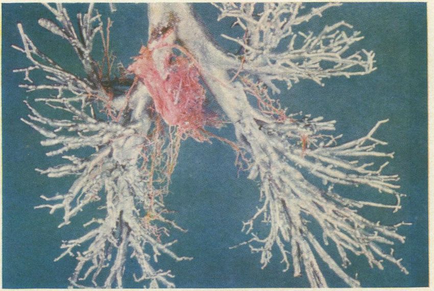
Thorax: first published as 10.1136/thx.5.3.207 on 1 September 1950. Downloaded from http://thorax.bmj.com/ on February 29, 2020 by guest. Protected by
    ANATOMICAL STUDY OF BRONCHIAL VASCULAR SYSTEM                                   211

bronchial artery, pass to the mediastinal aspect of the lung and then spread out
subpleurally. Neither the bronchial nor the intercostal arteries leave the aorta at
right angles to its axis, but they pass upwards initially forming an acute angle with
the aorta (Fig. 2). During systole the effect of the elongation and dilatation of
the aorta is to accentuate this angulation.
    The main bronchial arteries usually arise from the aorta just after it has crossed
behind the left main bronchus, the artery to. the right lung passing upwards to cross
the mid-line at a point level with or just below the tracheal bifurcation. Both
arteries enter the lung closely applied to the membranous portion of the main stem
bronchus. The main bronchial artery continues along the posterior aspect of the
main stem bronchus. Branches of this vessel are then given off to supply each main
segmental bronchus, the anteriorly placed ones being reached by winding round the
lateral aspect of the main bronchus. These branches are intimate relations of the
outer wall of the bronchus.
     The mediastinal pleura is prolonged into the lung with the bronchi, and by
 careful dissection the bronchial arteries can be followed well into the substance of
 the lung without coming in contact with the lung parenchyma; they lie between
 the invaginated pleura and the fibrous layer covering the bronchi. The invaginated
 pleura fuses with the outer bronchial coat at the origin of the segmental bronchi.
 If the bronchial arteries are followed further than this point the interstitial tissue
 of the lung itself must be exposed. The bronchial arteries are now truly intra-
pulmonary and from here onwards branch repeatedly and by re-anastomosis form

                                                                                                                                                                 copyright.
an arterial mesh around the bronchi (Fig. 1). Small branches constantly pierce
the bronchial wall to form an intramural network as described by Miller ((1947).
In normal lung specimens the bronchial arteries cannot be traced by our methods
further along the surface of a bronchus than the third dividing point of a segmental
bronchus.
     The arteries which arise from the aorta distal to the origin of the left subclavian
artery include bronchial, pleural, and mediastinal arteries, and these supply the
tracheo-bronchial tree from the thoracic inlet as far as the respiratory bronchiole.
They also supply the tracheo-bronchial and interbronchial lymph glands and vasa
vasorum to the pulmonary vessels; these vasa vasorum never communicate with
the lumen of the pulmonary artery. Branches from these vessels also pass along
the interlobular septa to supply the visceral pleura of the interlobar fissures. We
have been unable to demonstrate a bronchial arterial supply to the greater part of
the costal portion of the visceral pleura and this agrees with the findings of von Hayek
(1940) and Verloop (1948). This area of the pleura is supplied by the pulmonary
artery.
     The existence of macroscopic anastomoses between bronchial and pulmonary
arteries has been denied by Miller (1947), but Braus (1931) and Liebow, Hates,
and Lindskog (1949) have demonstrated such anastomoses in normal and diseased
lungs. We have demonstrated them in three of the four normal lung specimens in
which both the bronchial and pulmonary arteries were injected with vinylite, one
of these specimens being the lungs of a child aged two, who had died from menin-
gitis. We have also demonstrated our radio-opaque medium (which will not pass
through capillaries) in the pulmonary arteries of eight of ten normal lung specimens
in which the bronchial arteries alone were injected.
(!Iloni ;
Thorax: first published as 10.1136/thx.5.3.207 on 1 September 1950. Downloaded from http://thorax.bmj.com/ on February 29, 2020 by guest. Protected by
212 PAUL MARCHAND, J. C. GILROY, and V. H. WILSON
These anastomoses have a constant anatomical design. As the bronchial artery
approaches the pulmonary artery with which it is to anastomose it becomes widened
and tortuous and gives off a branch which arches over to enter the pulmonary artery
FIG. 3.-Isolated seg-
ment of a vinylite cast
of pulmonary artery
and bronchial artery
from a lobe which was
the site of a lung
abscess. The specimen
has been placed in
ILI -:._.
x . . }PLI ImuI i ir1-l
1 -\ warm water and flat-
| ai dlcr\ tened so as to photo-
graph more readily.
The bronchial artery,
Bianmch ~1giving rise to the con-
vessel, showed
-forn l~~i
o
l!i necting
onch
much greater cork-
I30n I -1 5I. AItc
i 1ll'I
screw tortuosity before
.
un
on i x\
monar\X 41 tr.;c
lm1cr\
e \ ~ .i
~' ,_ '111 11 shows This
this procedure.
~~~~~~~~~~~~~~~~~~specimen
in .i > tmanner
@t . inP 2which thethe
9t -)
si n .lI 11 i> s 81 ( } [] L' 1Thorax: first published as 10.1136/thx.5.3.207 on 1 September 1950. Downloaded from http://thorax.bmj.com/ on February 29, 2020 by guest. Protected by
Main Bronchial artery dividing into branches
Left atrium bronchus which follow the segmental bronchi
/ /
t. V." .t .I
j.=.
Bronchial vein X1 ..
passing to left
atrium
Bronchial artery
showing admixture
' with red from the
pulmonary artery
Bronchial vein
and artery
copyright.
FIG. 5.-Complete cast of a normal left lung of an infant. The bronchi are filled
with Wood's metal. The pulmonary artery has been injected red, the bronchial
artery yellow, and the left atrium, pulmonary veins, and bronchial veins blue.
(The atrium and pulmonary veins appear whitish in parts because of the bleaching
action of the hot photographic lamps.) This specimen shows the manner of
division of the bronchial arteries and a bronchial artery giving rise to a con-
necting vessel. (The actual anastomosis is hidden, but the admixture of red
and yellow vinylite is clearly shown.) The termination of the bronchial veins
in the atrium is obscured by the atrium itself.
Pulmonary
artery
Main )
bronchus Network of
_ bronchial
Bronchial venules
artery i
Bronchial
vein enter-
ing atrium
Left
atrium
Lower
pulmonary
vein
Atrio-ventricular Left ventricle
valve
FIG. 6.-Vinylite and Wood's metal cast of section of the lung of a male aged 50 in
whom the heart and lungs were normal. The pulmonary artery was injected
with blue vinylite. The left atrium, and through this the left auricle, pulmonary
veins, and bronchial veins were injected with red vinylite. The bronchial artery
was injected yellow but has been removed, except for a small segment, so as to
demonstrate the bronchial vein around the segmental bronchi and extending
over the lowet lobe bronchus. The main bronchial vein can be seen entering
the left atrium and its size can be compared with that of the adjacent bronchial
artery. [Facing page 212Thorax: first published as 10.1136/thx.5.3.207 on 1 September 1950. Downloaded from http://thorax.bmj.com/ on February 29, 2020 by guest. Protected by
ANATOMICAL STUDY OF BRONCHIAL VASCULAR SYSTEM 213
vessels less than 20 microns in diameter yet the distinctive yellow colour of the
suspension, which appeared in the main pulmonary artery during the injection of
the bronchial arteries was unmistakable, and indicates the presence of pre-capillary
anastomoses between bronchial and pulmonary arteries.
THE BRONCHIAL VEINS
Miller (1947) stated that true bronchial veins are found only at the hilum of the
lung and that they arise "from the first or the first two dividing points of the
bronchial tree," and that they may also receive branches from that part of th-
pleura in close proximity to the hilum. He describes these veins as draining into
the azygos, hemiazygos, or one of the intercostal veins. Within the lung he mentions
venous plexuses in the walls of the bronchi and bronchioles which unite at the
bifurcation of a bronchus or bronchiole to form a radicle which empties into the
pulmonary vein.
We have found that two distinct groups of veins exist, namely, the deep or true
bronchial veins which are intrapulmonary vessels related to the bronchi and which
drain into the pulmonary veins or into the left auricle direct, and a second group
of veins which drain the subpleural and hilar structures into the azygos and
hemiazygos veins as described by Miller (1947). Miller makes no mention of the
extensive communication between this second group of veins and the pulmonary
veins.
On the basis of our findings we would suggest the following nomenclature.
copyright.
The Deep or True Bronchial Veins.-Whenever vinylite was injected through
the left auricular appendix in order to fill the pulmonary veins we were able to
demonstrate these bronchial veins. They first appear as macroscopic structures
in the region of the terminal bronchiole. At this level they appear as a very fine
lacy network surrounding the bronchiole, and they become more obvious by the
accession of further radicles from the intrabronchial venous plexuses. Eventually
they are seen as several vessels running longitudinally along the outer wall of the
larger bronchi (Fig. 5). Near the hilum the several vessels unite to form a single
trunk which is slightly larger in diameter than the bronchial artery, and this vein
terminates either in the atrium direct (Fig. 6) or into a main pulmonary vein close
to the atrium. If the injection to demonstrate these veins is not made via the left
atrium, their mode of termination may easily be missed.
We have found one main deep bronchial vein in each lung. On the left side
it forms an anterior relation to the main stem bronchus just below the origin of the
left upper lobe bronchus. If it enters the atrium it does so anywhere between the
two pulmonary veins but usually close to the lower one.
The mode of termination of the bronchial vein on the right side is essentially
the same, but because of the high situation of the right upper lobe bronchus, it
crosses the lower part of the right main bronchus in relation to the middle lobe
bronchus.
Throughout their intrapulmonary course these bronchial veins communicate
freely with the pulmonary veins.
The Pleuro-Hilar Veins.-There is a rich subpleural network of veins, most of
which pass t9wards the mediastinum along the anterior and posterior surfaces ofThorax: first published as 10.1136/thx.5.3.207 on 1 September 1950. Downloaded from http://thorax.bmj.com/ on February 29, 2020 by guest. Protected by
214 PAUL MARCHAND, J. C. GILROY, and V. H. WILSON
the lung, and drain into one or more veins which run beneath the pleura at the
junction of lung and mediastinum. These latter veins are present on both the
anterior and posterior surfaces of the hilum. A limited area of the visceral pleura
however drains into the pulmonary vessels. Apart from the pleural vessels the
veins at the junction of lung and mediastinum receive tributaries from the extra-
pulmonary bronchi, the hilar lymph glands and the vasa vasorum of the pulmonary
x essels at the hilum. " Pleuro-hilar veins " seems an appropriate name as it
describes the regions whose venous drainage passes into these veins. The veins
on the right side usually empty into the azygos vein just before that vessel enters
the superior vena cava. In two specimens the termination has actually been into
the latter vessel.
The veins on the left enter the hemiazygos, the superior intercostal or the left
innominate vein. As the veins on the left side have this somewhat inconstant
termination, they are often difficult to demonstrate.
In every lung injected through these pleuro-hilar veins, we were able to show
free filling of the pulmonary veins. Communications between the pleuro-hilar and
pulmonary veins occur at the hilum and in the subpleural areas. The communica-
tion at the hilum is via veins in the region of the hilar bronchi. These are essentially
extrapulmonary in character as they lie in the potential space between invaginated
pleura and the dense outer coat of the bronchi. These are the bronchial veins
described by Miller (1947). They are bronchial veins in the sense that they drain
the large bronchi at the hilum, but their only connexion with the intrapulmonary
copyright.
bronchial venous system is an indirect one by way of communicating vessels to
the pulmonary veins, and probably by a limited connexion with the intramural
venous plexuses.
The venous arrangement is therefore one whereby the intrapulmonary bronchi
and bronchioles drain via the deep or true bronchial veins and empty into the
pulmonary venous system (we include the left atrium in this system), whereas the
subpleural areas and the hilar structures drain into systemic veins by a separate
system of vessels which communicate freely with the pulmonary veins.
As previously stated, the arterial supply to the hilar and subpleural structures
is usually quite separate from the bronchial artery, and therefore we prefer to term
both the arteries and the veins " pleuro-hilar vessels." If an accurate description of
function is to be achieved, the term "bronchial " should only be applied to those
vessels which supply and drain the intrapulmonary portions of the bronchial tree.
THE BRONCHIAL VASCULAR SYSTEM IN DISEASED LUNGS
LUNG ABSCESS AND BRONCHIECTASIS
Sections cut through the wall of an acute lung abscess or from the surrounding
area of pneumonitis show that the radicles of the pulmonary artery are obliterated
by infected thrombi. In chronic lung abscess organized thrombi with evidence of
recanalization can be shown. In vinylite casts the pulmonary artery and veins
demarcate the abscess area distinctly (Fig. 7). It will be noted that the vascular
pattern in the abscess areas has been altered. Very little pulmonary artery remainsThorax: first published as 10.1136/thx.5.3.207 on 1 September 1950. Downloaded from http://thorax.bmj.com/ on February 29, 2020 by guest. Protected by
FIG. 7.-Vinylite cast of the left lung
_ Bronchus open- removed at operation for two
chronic lung abscesses, one in
ing into cavity the subapical area of the upper
lobe and the other in the apex
of the lower lobe. The bronchi
were filled with yellow vinylite,
Interlobar -.-Abscess in apex the pulmonary arteries red, and
fissure of lower lobe the pulmonary veins blue. The
two abscess areas, separated by
the interlobar fissure, are clearly
demarcated by the blue of the
pulmonary vein. Very little
pulmonary artery is to be seen
in the abscess areas. The cavity
in the apex of the lower lobe can
be seen. The yellow mass of
vinylite which occupied it has
been removed. The area of
pulmonary arterial destruction
extends for some distance around
the actual cavity. The bronchial
artery was not injected.
copyright.
Mtain trunk Upper
of bronchial pulmonary Left pulmon-
Left atrium vein vein ary arterv
N. I I
/I\
Branch of Site of lower Site of lingula
bronchial vein lobe bronchus bronchus
joining lower
pulmonary vein
FIG. 8.-Vinylite cast of the left lung from a case of emphysema associated with cor
pulmonale. (Pulmonary artery red, pulmonary veins, bronchial veins, and left
atrium blue.) The bronchi were not injected, but their course is outlined by the
enlarged bronchial veins, which extend as a dilated plexus between the points
marked A and B. Not adequately visualized is the extension of the bronchial
veins along the course of the upper lobe bronchi and along the branches of the
main bronchus. [Facing page 214Thorax: first published as 10.1136/thx.5.3.207 on 1 September 1950. Downloaded from http://thorax.bmj.com/ on February 29, 2020 by guest. Protected by
ANATOMICAL STUDY OF BRONCHIAL VASCULAR SYSTEM 215
and consequently the abscess areas are demarcated by the blue dye in the pulmonary
veins. Injection of the bronchial artery demonstrates that the arterial supply to the
abscess area is provided by this vessel.
Macroscopic anastomoses between the bronchial and pulmonary arteries are
plentifully demonstrated; these are seen not only immediately adjacent to the
abscess, but are widespread throughout the affected lobe. They are also increased
in the remaining, apparently normal lobes of a diseased lung. Anatomically the
anastomoses are identical with those found in normal lungs, being situated mainly
in the region of the smaller bronchi.
In cases of early bronchiectasis in which the infection is limited to the wall of
the bronchus, the number of anastomoses found is not noticeably increased, but
once infection spreads to the parenchyma of the lung and consequent fibrosis has
occurred, the anastomoses become conspicuous and widespread throughout the
affected lobes. Wood and Miller (1937-8) and Liebow et al. (1949) have also
described arterial anastomoses in cases of lung abscess and bronchiectasis.
Two specimens of pulmonary tuberculosis, affecting both lungs and in which
fibrosis was widespread, also displayed a similar increase in the number of arterial
anastomoses.
GENERALIZED PULMONARY DISEASE ASSOCIATED WITH COR PULMONALE
Three autopsy specimens of emphysematous lungs, one of which was associated
copyright.
with long-standing atelectasis of the left lung, the right lung being markedly emphy-
sematous, were injected with vinylite. In a further two cases of emphysema the
pulmonary arteries were injected with gelatine. The lung pathology has been
confirmed microscopically in all cases. All the patients from whom these specimens
were obtained exhibited the cardiac signs of cor pulmonale during their illness.
The Bronchial Artery.-The vinylite casts show broncho-pulmonary arterial
anastomoses to be widespread throughout both lungs, including the partially
atelectatic lung. These anastomoses were structurally identical with those found in
normal lungs. A striking feature of these anastomoses was the calibre of the
connecting vessel which often approximated that of the pulmonary radicle which it
entered. This is not peculiar to the lungs of cor pulmonale because we have seen
it in specimens of lung abscess and even occasionally in specimens of normal
lungs (Fig. 4).
The two lungs injected with gelatine must also have had widespread arterial
anastomoses because the gelatine which was injected into the pulmonary artery
appeared in considerable amounts in the aorta by way of the bronchial arteries.
The Bronchial Veins.-Whereas normally the true bronchial veins are delicate
vessels, our vinylite casts of the vessels in generalized lung disease showed a very
conspicuous venous network outlining the course of the bronchi. The longitudinal
venous trunks united to form main vessels two or three times as large as normal
ones. The connections with the pulmonary veins were abnormally dilated and easily
recognizable (Fig. 8). Serial section of the smaller bronchi in the gelatine-injected
lungs revealed dilatation of the intra-bronchial venous plexuses within the walls of
the bronchi.Thorax: first published as 10.1136/thx.5.3.207 on 1 September 1950. Downloaded from http://thorax.bmj.com/ on February 29, 2020 by guest. Protected by
216 PAUL MARCHAND, I. C. GILROY, and V. H. WILSON
DISCUSSION
The question as to whether or not the bronchial artery anastomoses with the
pulmonary artery, has been the subject of much controversy. Miller (1934), who
worked with dogs, stated that pre-capillary anastomoses did not exist, and he
expressed the opinion that vasa vasorum to the pulmonary vessels were being
mistaken for true anastomoses. We cannot agree with this, and our findings together
with those of Braus (1934), von Hayek (1940), and Verloop (1948) prove that
broncho-pulmonary arterial anastomoses exist in normal human lungs. The vinylite
technique will only demonstrate the anastomoses between the comparatively large
arteries of the bronchial and pulmonary systems. Verloop (1948) made a detailed
histological study of the arteries forming these anastomoses in normal human lungs
and described pre-capillary anastomoses along the surface, and within the walls, of
all the smaller bronchi. In addition there are superficial anastomoses between
bronchial and pulmonary radicles under the visceral pleura, though these are far
less numerous than the anastomoses related to the bronchi. He concluded that the
total number of broncho-pulmonary anastomoses must be very great.
Verloop (1948) also found anastomoses in foetal lungs but remarks on the fact
that they were few in number. From our experiments with radio-opaque bismuth
oxychloride injection we are in agreement that anastomoses between bronchial and
pulmonary arteries are already present in foetal lungs. In addition to pre-capillary
anastomoses there is probably an extensive capillary anastomosis between pulmonary
and bronchial arteries which subsequently become arterialized to form the very
copyright.
extensive anastomatic system found in the adult.
The increase in the number of macroscopic arterial anastomoses found in dis-
eased lungs, as compared with normal, must be due to dilatation of existing anas-
tomoses. They cannot be a direct result of revascularization following infection
or necrosis, because in specimens of lung abscess they occur remote from the affected
lung segments. They are also increased in emphysematous lungs, a generalized
disease in which local infection and necrosis are absent. Apart from the numerical
increase, the macroscopic anastomoses found in diseased lungs are structurally
identical with those in the normal. The function of these anastomoses is still
under investigation, but there is little doubt that the dilatation is due to the passage
of an increased volume of blood through the anastomoses which therefore appear
to be concerned with the redistribution of blood flow within the lungs.
With the exception of von Hayek (1940) all recent workers agree that blood
must flow from bronchial to pulmonary artery when the anastomoses become func-
tionally active. This conclusion is based upon supposed pressure differences between
the two systems. Fig. 9 represents diagrammatically our views upon the possible
effects the broncho-pulmonary anastomoses have upon the distribution of blood
flow within the lungs.
Normally there is probably little interchange of blood between the two systems
due to constriction of the wall of the muscular connecting vessel (Fig. 9-0().
Bloomer, Harrison, Lindskog, and Liebon (1949) showed that after ligation of
the pulmonary artery in dogs, oxygenated blood circulated through the pulmonary
arteries distal to the point of ligature. If the bronchial artery and the connectingThorax: first published as 10.1136/thx.5.3.207 on 1 September 1950. Downloaded from http://thorax.bmj.com/ on February 29, 2020 by guest. Protected by
ANATOMICAL STUDY OF BRONCHIAL VASCULAR SYSTEM 217
I
con necil$ing
Yeas (
13ronch io I Pulmonanry
Artery Artery
'i
fo Bronchial
CrcI ;o+on
copyright.
RevvolnrI of flow
I in~~~Pulmonary Art.
FIG. 9.-Diagrammatic representation of possible interchange of blood between pulmonary and
bronchial circulations.
vessel are patent and there is obstruction to the pulmonary artery, such as was
produced in Bloomer's experiment, the pressure within the pulmonary artery distal
to the obstruction will drop and bronchial blood will be directed into the pulmonary
circulation. Virchow (1851) demonstrated that ligation of the pulmonary artery did
not result in gangrene of the lung and the arterial anastomoses functioning in this
direction (Fig. 9-0) may be an important factor in preventing necrosis following a
pulmonary embolus.
The main source of oxygenated blood to the foetal lung is probably via the
bronchial arteries reaching the pulmonary alveolar capillaries through the arterial
anastomoses.
East and Barnard (1938) published a description of four cases of Fallot's tetra-
logy which at necropsy showed complete stenosis of the pulmonary artery, yet the
affected individuals survived until the ages of 33, 30, 20, and six years respectively.
The bronchial arteries were the sple source of pulmonary blood and in two were
grossly dilated. Blood could only have reached the alveolar capillaries throughThorax: first published as 10.1136/thx.5.3.207 on 1 September 1950. Downloaded from http://thorax.bmj.com/ on February 29, 2020 by guest. Protected by
218 PAUL MARCHAND, J. C. GILROY, and V. H. WILSON
these anastomoses passing from bronchial to pulmonary artery. We have been
unable to study the bronchial arteries in a case of Fallot's tetralogy at necropsy but
they are probably frequently dilated. The exposure of the pulmonary artery in the
course of Blalock's operation for the relief of Fallot's tetralogy is often difficult,
because of the overlying plexus of dilated vessels. These vessels have been men-
tioned by Keith (1909) and by Fatti and Gilroy (1949) and are probably derived
from the pleuro-hilar arteries which arise direct from the aorta or from the extra-
pulmonary portion of the bronchial artery. Verloop (1948) has demonstrated that
arterial anastomoses between the pleural and pulmonary arteries occur in the sub-
pleural areas, and it is along this pathway that systemic blood will reach the
pulmonary arteries. This is precisely what Blalock's operation accomplishes on a
larger scale.
Fig. 9-(G illustrates the direction of blood flow in conditions associated with
obstruction of the small pulmonary arterial radicles such as occur in atelectasis.
This has been demonstrated by blood oxygen estimations on samples of blood
taken at operation and the results will be reported fully in a further paper. If the
atelectasis is lobular or lobar, blood is diverted into the pulmonary arterial branches
of the remaining segments of lung. With total collapse of a lung the oxygenated
arterial blood derived from the bronchial arteries, may actually be diverted to the
other lung.
In all the above-mentioned cases blood has been diverted from bronchial to
copyright.
pulmonary artery and this has been accompanied by an increased delivery of blood
from the aorta to the bronchial arteries. Under such circumstances one would
expect to find the extrapulmonary bronchial artery to be dilated. We have demon-
strated numerous and widespread broncho-pulmonary arterial anastomoses in lungs
with tuberculosis, lung abscess, and emphysema, especially when associated with
cor pulmonale. This has been taken as a probable indication that in these condi-
tions an appreciable volume of blood crosses from one system of arteries to the
other. In none of these latter specimens however was the extrapulmonary portion
of the bronchial artery dilated. We have considered the possibility of the passage
of blood from pulmonary to bronchial artery, a possibility which only von Hayek
(1940) has entertained. All other workers consider that hydrostatic pressure dif-
ferences between the two systems would preclude pulmonary arterial blood from
entering the bronchial artery. We are not convinced that such pressure differences
do, in fact, exist. We have been unable to cannulate the bronchial artery at opera-
tion, but on occasions when it has been cut during the course of lobectomy, we
have noticed that it does not spurt with the vigour of an artery such as the digital.
Also the anastomoses occur in the substance of the lung arising from small branches
of a bronchial artery which has divided and re-anastomosed repeatedly. It is pos-
sible that at the site of the anastomoses the pressures in the pulmonary and bronchial
arteries are approximately equal. Apart from these considerations, the construc-
tion of the bronchial artery in the anastomatic area is such as to make pressure
differences of little consequence in regulating the direction of blood flow. Von
Hayek (1940), Mauer (1941), van der Zwaag (1940), and Verloop (1948) have
described thick-walled muscular bronchial arteries within the lung, different from
the extrapulmonary bronchial artery which histologically is typically systemic inThorax: first published as 10.1136/thx.5.3.207 on 1 September 1950. Downloaded from http://thorax.bmj.com/ on February 29, 2020 by guest. Protected by
ANATOMICAL STUDY OF BRONCHIAL VASCULAR SYSTEM 219
structure. Verloop (1948) made a painstaking study of the histology of the con-
necting artery and its parent vessels, and he found that the segment of bronchial
artery which gives rise to the anastomatic vessel always has an hypertrophied muscu-
lar coat. This hypertrophy mainly involves the longitudinal muscle layer within
the intima, but the circular muscle in the media is also frequently increased. Von
Hayek (1940) also comments upon the very muscular wall of the bronchial arteries
which give off connecting branches to form anastomoses with the pulmonary arteries.
Both workers have found that these bronchial arteries are capable of active occlu-
sion of their lumina. The connecting vessel has a thinner wall than its parent
bronchial vessel, but does possess substantial muscular fibres which run longitu-
dinally. The pulmonary artery has only a few circular muscle fibres in its media
and these are separated by thick elastic laminae. As Brenner (1935) remarks, the
smaller pulmonary arteries seem best adapted to follow passively changes in the
pulmonary blood pressure. They can have very little active control by virtue of
their vaso-motor powers. The bronchial arteries and connecting vessels on the
other hand, seem admirably designed to respond to vaso-motor control. If there-
fore, the bronchial artery proximal to the anastomoses is occluded by muscular
constriction of its wall, blood could be diverted from the pulmonary to the bronchial
artery (Fig. 9- ). Under these circumstances possible differences in hydrostatic
pressure would not be operative. The function of the anastomosis in such an event
would appear to be in the nature of an attempt to prevent the development, or
minimize the effects, of pulmonary hypertension, and a consequence would be to
copyright.
divert unoxygenated pulmonary arterial blood into the bronchial circulation. We
believe that blood does flow in this direction across the anastomoses between pul-
monary and bronchial arteries in conditions of raised pulmonary arterial pressure.
In localized disease the anastomoses are sufficient to counteract the effect of the
disease process on the pulmonary arterial pressure and so prevent strain upon the
right ventricle. In generalized disease, even though the anastomoses are widespread
they may fail eventually in the function of obviating pulmonary hypertension. Wood
and Miller (1937) remark that broncho-pulmonary anastomoses are most marked
in heart disease with right-sided hypertrophy. We have found in vinylite casts
that the density of the anastomoses in a particular segment of lung from a case of
cor pulmonale is no greater than that in a segment affected with lung abscess. In
the former, however, these macroscopic arterial anastomoses are widespread
throughout both lungs.
Whatever the direction of the blood flow, the arterial anastomoses appear to
be functionally significant in many varied forms of diseased lungs. The components
of the anastomosis seem to have dilated in response to the continued increase in
the blood flow traversing them from one system of arteries to the other.
The anastomoses between the pulmonary vein and the veins which we term the
"pleuro-hilar " veins provide a ready decompressive mechanism in cases of raised
pulmonary venous pressure. Ferguson, Kobilak, and Deitrick (1944) suggested that
in cases of mitral stenosis the blood flQw might be reversed from the pulmonary
vein to the bronchial vein and thence to the systemic circulation. The bronchial
vein to which they refer is presumably the vein described by us as the pleuro-hilar
vein. Bland and Sweet (1949) described an operation for the relief of tight mitral
stenosis based in principle upon these decompressive anastomoses. Hughes (1944)Thorax: first published as 10.1136/thx.5.3.207 on 1 September 1950. Downloaded from http://thorax.bmj.com/ on February 29, 2020 by guest. Protected by
220 PAUL MARCHAND, J. C. GILROY, and V. H. WILSON
described two cases of anomalous pulmonary veins seen in mitral stenosis; it is
possible that he was describing dilated pleuro-hilar veins serving a decompressive
role.
The true bronchial veins within the lung drain into the pulmonary circulation
and anastomose freely with the pulmonary vein. Therefore pressure changes within
the pulmonary vein will readily be transmitted to the true bronchial veins. Micro-
scopically the bronchial venous radicles within the bronchial wall are arranged in
two groups of very thin-walled vessels lying in the submucosa and on the outer
aspect of the muscular layer. In the larger bronchi they are well separated from
the bronchial lumen but in the bronchioles where the submucosa is very thin, these
veins are situated very close to the lumen. In conditions associated with a raised
pulmonary venous pressure congestion of these venous plexuses may partially
occlude the bronchiole.
The hypertrophy and dilatation of the true bronchial vein which we found in
emphysematous lungs associated with cor pulmonale may have been due to a sus-
tained increase in pulmonary venous pressure. It is however possible that this
hypertrophy followed a long-continued increase in blood flow through the bronchial
vascular system.
No longer can the bronchial and pulmonary circulations be regarded as closed
circuits. They communicate freely with each- other on both the arterial and venous
sides. The bronchial circulation is unique in that the artery arises from the
systemic circulation, whilst its venous component drains into the pulmonary system.
copyright.
SUMMARY
1. Anatomical studies of the vascular pattern encountered in 39 normal lungs
and 24 diseased lungs are described.
2. True bronchial vessels are differentiated from vessels which supply and drain
the sub-pleural and hilar structures.
3. Anastomoses between bronchial and pulmonary arteries in normal and
diseased lungs have been described.
4. The anastomoses are most numerous in diseased lungs and are considered
to be of functional importance.
5. The possible direction of blood flow through these anastomoses has been
discussed. Their effect is considered to be of very great importance upon the
haemodynamics of the pulmonary circulation in many diseased states.
6. The function of the pleuro-hilar veins in cases of pulmonary venous hyper-
tension has been discussed and it is suggested that these veins act as an important
decompressive mechanism.
We wish to express our gratitude to Professor Raymond A. Dart for granting us facilities
in the Department of Anatomy, University of the Witwatersrand, where most of this work
was done. Professor Joseph Gillman, Physiology Department, and Dr. T. Gillman, Anatomy
Department, have rendered invaluable assistance in the choice of techniques, and in the prepara-
tion of this paper. Mr. L. Fatti, Thoracic Surgical Unit, Baragwanath Hospital, supplied the
operation material for injection and has constantly helped us with this investigation. The
photography is the work of Mr. A. Hughes, Anatomy Department, University of the
Witwatersrand.Thorax: first published as 10.1136/thx.5.3.207 on 1 September 1950. Downloaded from http://thorax.bmj.com/ on February 29, 2020 by guest. Protected by
ANATOMICAL STUDY OF BRONCHIAL VASCULAR SYSTEM 221
REFERENCES
Bland, E. F., and Sweet, R. H. (1949). J. Amer. mod. Ass., 140, 1259.
Bloomer, W. E., Harrison, W., Lindskog, G. E., and Liebow, A. A. (1949). Amer. J. Physiol., 157,
317.
Braus, H. (1934). Anatomie des Menschen, 2nd ed., vol. 2, p. 203. Berlin.
Brenner, 0. (1935). Arch. intern. Med., 56, 211.
East, T., and Barnard, W. G. (1938). Lancet, 1, 834.
Fatti, L., and Gilroy, J. C. (1949). Brit. Heart J., 11, 398.
Ferguson, F. C., Kobilak, R. E., and Deitrick, J. E. (i944). Amer. Heart J., 28, 445.
Hill, E. C. (1929). Bull. Johns Hopk. Hosp., 44, 248.
Hayek, H. von (1940). Anat. Anz., 89, 216.
(1940). Z. Anat. EntwGesch., 110, 412.
Hughes, C. W., and Rumore, P. C. (1944). Arch. Path., 37, 364.
Keith, A. (1909). Lancet, 1, 745.
Liebow, A. A., Hales, M. R., and Lindskog, G. E. (1949). Amer. J. Path., 25, 211.
Mauer, G. (1941). Frankfurt. Z. Path., 55, 208.
Merkel, H. (1941). Virchows Arch., 308, 303.
Miller, W. S. (1947). The Lung, 2nd ed. Springfield.
Moore, R. A. (1929). J. tech. Meth., 12, 55.
Puckett, W. O., and Neumann, C. P. (1940). Anat. Rec., 78, 105.
Rivkind, A. V. (1949). Abstr. World Med., 5, 718. Abstr. No. 2486.
Verloop, M. C. (1948). Acta anat., Basel, 5, 171.
Virchow, R. (1851). Virchows Arch., 3, 427.
Wood, D. A., and Miller, M. (1938). J. thorac. surg., 7, 649.
Zuckerkandl, E. (1883). S.B. Akad. Wiss. Wien., 87, Part 3, p. 171.
Zwaag, G. L. van der (1940). Sclerosis arteriae pulmonalis primaria. Thesis, Groningen.
copyright.You can also read