UC Riverside UC Riverside Previously Published Works - eScholarship
←
→
Page content transcription
If your browser does not render page correctly, please read the page content below
UC Riverside
UC Riverside Previously Published Works

Title
Contralateral Hemispheric Cerebral Blood Flow Measured With Arterial Spin Labeling Can
Predict Outcome in Acute Stroke.

Permalink
https://escholarship.org/uc/item/6692f2xp

Authors
Thamm, Thoralf
Guo, Jia
Rosenberg, Jarrett
et al.

Publication Date
2019-10-17

DOI
10.1161/strokeaha.119.026499

Peer reviewed

 eScholarship.org                                Powered by the California Digital Library
                                                                 University of California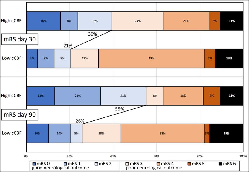
CONTRALATERAL CEREBRAL BLOOD FLOW PREDICTS OUTCOME

Contralateral Hemispheric Cerebral Blood Flow Measured with Arterial
             Spin Labeling Can Predict Outcome in Acute Stroke

Thoralf Thamm1,2, Jia Guo, PhD1,3, Jarrett Rosenberg, PhD1, Tie Liang, EdD1, Michael P Marks,
 MD1, Soren Christensen, PhD1, Huy M Do, MD1, Stephanie M Kemp, BS1, Emma Adair, BS1,
Irina Eyngorn, MD1, Michael Mlynash, MD MS1, Tudor G Jovin, MD4, Bart P Keogh, MD5, Hui
 J Chen, MD6, Maarten G Lansberg, MD PhD1, Gregory W Albers, MD1, and Greg Zaharchuk,
                      MD PhD1*, on behalf of the iCAS Study Investigators.

1 - Stanford University, Stanford, CA, United States
2 - Center for Stroke Research Berlin, Charité - Universitätsmedizin Berlin, Berlin, Germany
3 - University of California Riverside, Riverside, CA, United States
4 - Cooper University Hospital, Camden, NJ, United States
5 - Swedish Medical Center, Seattle, WA, United States
6 - Eden Medical Center, Castro Valley, CA, United States

* Correspondence to:
Stanford University
Department of Radiology
300 Pasteur Drive, Grant Building, Room S047
Stanford, CA 94305-5105
Phone: 650-736-6172
E-mail: gregz@stanford.edu
https://orcid.org/0000-0001-5781-8848

Tables and Figures: Tables 2; Figures 3; Supplemental Tables 4; Supplemental Figures 2.

Subject Terms: Ischemic Stroke; Magnetic Resonance Imaging (MRI); Prognosis

Keywords: Acute stroke; Cerebral blood flow; Outcome; Magnetic resonance imaging;

Perfusion imaging; Arterial spin labeling

Total Manuscript Word Count: 5983/5500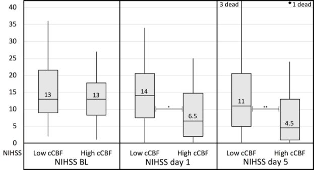
Abstract

Background and Purpose: Imaging is frequently used to select acute stroke patients for intra-

arterial treatment (IAT). Quantitative cerebral blood flow (CBF) can be measured non-invasively

with arterial spin labeling (ASL) magnetic resonance imaging (MRI). CBF levels in the

contralateral (unaffected) hemisphere may affect capacity for collateral flow and patient outcome.

The goal of this study was to determine whether higher contralateral CBF (cCBF) in acute stroke

identifies patients with better 90-day functional outcome.

Methods: Patients were part of the prospective, multicenter ‘Imaging Collaterals in Acute Stroke‘

(iCAS) study between 2013 and 2017. Consecutive patients were enrolled after being diagnosed

with anterior circulation acute ischemic stroke. Inclusion criteria were ischemic anterior circulation

stroke, baseline National Institutes of Health Stroke Scale (NIHSS)>=1, pre-stroke modified

Rankin Score (mRS)
Conclusion: Higher quantitative contralateral CBF at baseline is a significant predictor of good

neurological outcome at day 90. cCBF levels may inform decisions regarding stroke triage,

treatment of acute stroke, and general outcome prognosis.

Clinical Trial Registration:

Imaging Collaterals in Acute Stroke (iCAS), unique identifier: NCT02225730, clinical trial

registration URL: https://clinicaltrials.gov/ct2/show/NCT02225730

Abstract word count: 291/300

                                                                                              3
Introduction
Following an acute vessel occlusion, perfusion of the ischemic penumbra is sustained by collateral
flow. Evaluation of cerebral hemodynamics in the stroke-affected, ipsilateral hemisphere to
identify appropriate patients for intra-arterial thrombectomy (IAT) is a common practice.1-4 To the
best of our knowledge, no studies have investigated the predictive role of quantitative perfusion
biomarkers in the contralateral, unaffected hemisphere. Higher cerebral blood flow (CBF) in the
contralateral hemisphere might be associated with a greater capacity to mobilize collateral flow to
affected regions in the ipsilateral hemisphere or be a marker for better cardiac output, and thereby
be associated with better neurological outcomes.
Arterial spin labeling (ASL) MRI enables measurement of quantitative CBF without the need for
contrast agents and can be acquired in the acute stroke setting. 5 ASL differs from the more
commonly-applied contrast-enhanced methods with MRI or CT perfusion-weighted imaging
(PWI), which do not routinely provide quantitative CBF information, instead providing relative
measurements. Meanwhile, ASL is prone to underestimate CBF in ischemic regions due to
prolonged arterial arrival times.6-8 However, these issues are less prevalent in the contralateral
hemisphere, where there is usually no acute ischemic event or large vessel occlusion. In this study,
we hypothesized that the high contralateral CBF (cCBF) at the time of ischemic stroke is associated
with better outcome.
4Materials and Methods Study Design and Patient Selection The cohort is part of the ongoing observational, prospective, multi-center ‘Imaging Collaterals in Acute Stroke‘ (iCAS) study (unique identifier: NCT02225730, clinical trial registration URL: https://clinicaltrials.gov/ct2/show/NCT02225730). The iCAS study has been approved by the Institutional Review Board. Informed consent was obtained from all subjects. The data that support the findings of this study are available from the corresponding author upon reasonable request. Patients were enrolled between November 2013 and August 2017, after being diagnosed with an acute ischemic stroke at four participating sites: Stanford University (Stanford, CA, United States), University of Pittsburgh Medical Center (UPMC, Pittsburgh, PA, United States), Swedish Medical Center (Seattle, WA, United States), and Eden Medical Center (Castro Valley, CA, United States). Patients imaged =1, (c)
with labeling duration (LD) = 1.45s, post-labeling delay (PLD) = 2.025s (at 1.5T); and a multi-

delay (5-delay) ASL with LD = 2s, PLD = 0.7, 1.275, 1.85, 2.425, and 3s (at 3.0T). Proton density

(PD)-weighted images were collected for quantification. The ASL images were reconstructed with

an interpolated resolution of 1.9x1.9x6mm³ (1.5T) or 1.9x1.9x4mm3 (3.0T). Isotropic DWI images

were acquired with TR/TE 4000/77.5ms, b=1000 s/mm2 and spatial resolution of 0.9x0.9x5mm3.

Infarct volume was calculated using RAPID software (version 4.5.1, iSchemaView, Menlo Park,

CA, USA).

For single-delay ASL, the CBF maps were quantified using the simplified equation from Alsop et

al.5 For multi-delay ASL, arterial transit time (ATT) corrected CBF maps were generated from a

kinetic signal model10 with assumed arterial and tissue T1 of 1.65s and 1.5s, respectively. This was

performed using an automated standardized script in MATLAB R2013b (version 8.2.0.701, The

MathWorks, Inc., Natick, MA, USA) using SPM8 (Statistical Parametric Mapping, The Wellcome

Trust Centre for Neuroimaging, University College London, UK).

All perfusion and diffusion images were first registered to the T1-weighted structural scans and

then registered and normalized to the Montreal Neurological Institute (MNI) template. The atlas’

standard gray matter mask was then applied. CBF values were extracted automatically in gray

matter regions-of-interest at four supratentorial levels corresponding to those assessed as part of

the Alberta Stroke Program Early CT Score (ASPECTS) system (Supplemental Figure I, please

see http://stroke.ahajournals.org). Mean contralateral CBF (cCBF) was calculated across all ROIs

in the unaffected brain hemisphere. Additional analyses based on follow-up imaging and

corresponding imaging outcome measures are reported in the Supplemental Material

(Supplemental Methods, please see http://stroke.ahajournals.org).
                                                                                                  6
Clinical Assessment Neurological outcome was assessed by using NIHSS and the modified Rankin Score (mRS) at several timepoints. NIHSS was measured at baseline before any intervention (NIHSS baseline), at 24 hrs (NIHSS day 1), and at day 5 or at discharge (NIHSS day 5). mRS was assessed at day 30 (mRS day 30) and day 90 (mRS day 90) either by telephone or clinical visit. The primary endpoint was good neurological outcome at day 90, defined as mRS day 90
level of significance was set to α
Results

Patient Population

109 patients underwent MRI. Of these, 32 were excluded for the following reasons: poor image

quality and/or motion artifacts (n=19), no ASL performed at baseline (n=7), pre-stroke mRS >=3

(n=3), OIT >24hrs (n=1), baseline NIHSS=0 (n=1), and bilateral stroke (n=1). Seventy-seven (77)

patients were included in the analysis (Table 1): 41 (53%) females, age 66 (55-76) yrs, OIT 4.8

(3.6-7.7) hrs, baseline NIHSS 13 (9-20). Sixty-nine (90%) were scanned at 3T and the rest at 1.5T.

A total of 46 patients (60%) received tissue plasminogen activator (tPA) before the baseline MRI

scan. Following the baseline MRI, 41 patients underwent IAT (53%); in 33/41 (80%) of those, the

final Thrombolysis in Cerebral Infarction (TICI) score was 2b or 3. When comparing cCBF values

for single-delay (37.6 ml/100g/min, IQR 30.2-43.6) vs. multi-delay ASL (39.0 ml/100g/min, IQR

31.8-44.5), there was no significant difference found (p=0.87). There was also no difference in the

fraction of patients in the high and low cCBF groups regarding ASL type (Supplemental Table II,

please see http://stroke.ahajournals.org).

Dichotomized cCBF

There were no significant differences in the clinical baseline characteristics between high and low

cCBF groups (Table 1). In particular, there was no significant linear correlation between DWI

lesion size and cCBF (Supplemental Figure II, please see http://stroke.ahajournals.org). Median

NIHSS at baseline/day 1/day 5 for low and high cCBF groups was 13/14/11 and 13/6.5/4.5,

respectively (Figure 1). While there was no significant difference at baseline, NIHSS was

significantly different between groups on both day 1 (p=0.016) and day 5 (p=0.003). High cCBF

                                                                                                 9
significantly predicted good clinical outcomes as assessed by mRS at day 90 (p=0.011) (Figure 2),
with 55% of high cCBF patients in the mRS 0-2 group compared with only 26% for the low cCBF
group. There was also a trend towards better outcome at day 30, but this was not statistically
significant.
Multivariable analysis
The univariate analysis identified the variables high cCBF, baseline NIHSS, DWI lesion volume,
and intra-arterial treatment as significant factors for outcome prediction. In the multivariable
analysis, patients were roughly 5 times more likely to be in the good 90-day clinical outcome group
rather than poor outcome group if they presented with high cCBF at baseline imaging (OR 4.6,
95% CI 1.4-14.7, p=0.011), while controlling for other significant contributing factors (p=66 ml (n=18, 23%) had poor 90-day outcome.
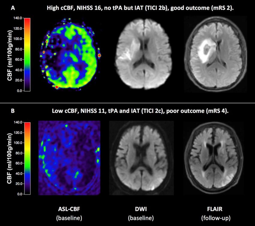
Representative cases are shown in Figure 3.
10Discussion
In this study, we demonstrated that higher CBF in the contralateral hemisphere is a strong
determinant of good 90-day clinical outcome. This quantitative parameter, cCBF, varied strongly
among patients, showing that there are marked inter-individual differences in CBF in the unaffected
brain during acute ischemic stroke.
To quantify perfusion in acute stroke patients, MR- or CT-based bolus perfusion imaging is most
commonly used, with a ratio between the affected and unaffected regions being a commonly
reported metric.1, 12-15 For example, the recent DEFUSE-3 trial used a cutoff of 30% of CBF based
on the unaffected regions as a measure of irreversibly infarcted tissue using CTP. 1 However, this
semi-quantitative approach neglects the fact that the CBF levels in non-affected regions may differ
substantially between patients. Fully quantitative approaches may provide more information;5 for
example, Harston et al. have reported absolute CBF values in the acute stroke setting when
evaluating serial perfusion imaging and tissue fate prediction.16 Our results show, first, that there
is a wide range of CBF in the contralateral hemisphere of stroke patients, which is in line with
recent reports on patient-level mean cCBF.16 This is not entirely unexpected as it is well-known
that baseline CBF varies widely in patients of similar age, even though the explicit reasons for
these variations are poorly understood.17 When assessing patients using relative measures, this
important information is lost, which could give insight into the patient’s cardiac output or ability
of the brain to deliver collateral flow. Other alternatives for quantitative perfusion measurements
are H215O water PET and xenon CT;18, 19 however, these approaches are difficult to implement in
the acute settings, require radiation, and have high cost and personnel needs. ASL MRI is
becoming increasingly available and has already been applied in the acute stroke setting in several
11prior studies.7, 14, 16, 20 While acquiring MRI in the acute stroke setting is often challenging, the
WAKE-UP study showed that patients with unknown stroke onset profit from MRI and suggested
general feasibility of MRI in an acute setting.21, 22 There are a few studies that propose CT and MR
bolus perfusion can be used in a quantitative manner to measure CBF;23-25 however, this is still
uncommon in clinical routine and has not been validated to the degree that ASL has. A comparison
of ASL-cCBF with other imaging techniques related to blood flow, such as multiphase CT, would
be valuable.
Recently, Raza and Rangaraju reviewed different existing prognostic scores to evaluate acute
ischemic stroke clinically before endovascular treatment.26 Looking at 10 different prognostic
scores in acute ischemic stroke, the authors found a ceiling effect for the scores’ area under the
curve (AUC) with a prognostic accuracy of 0.8.26 Only three scores included radiographic
parameters additionally to traditional clinical information, either based on CT ASPECTS regional
infarct lesion core (PRE score27, HIAT2 score28) or MRI DWI lesion core volume (SAD score29).
After the introduction of the mismatch concept for MRI30 and its translation to CT13, 31, there are
several reports on other promising imaging parameters12, 32, 33 and collaterals with good outcome.33-
35 While, however, predominately age, baseline NIHSS, CT ASPECTS, and MRI DWI lesion
volume have been evaluated so far,27, 29, 36 no such model includes quantitative CBF, largely due
to the historical challenges of implementing such methods in the acute setting.
cCBF measurements could affect patient triage, therapy, and care at various different timepoints.
High cCBF might represent a biomarker for underlying good overall health,37-39 sufficient cardiac
output,40, 41 and/or capacity to provide good collateral flow.42, 43 While we do not intend to alter
any treatment recommendations with this observational study, our results may point at a future role
12of cCBF as an objective and quantitative biomarker for therapy stratification: Dichotomized cCBF
could be utilized as treatment decision tool supporting clinicians in deciding which patients might
be suitable for IAT. One possibility is that patients with high cCBF on baseline MRI might be
appropriate for more aggressive treatment.
Based on this study, we cannot assess the precise mechanism of why higher cCBF is associated
with better outcomes; the size of the strokes and the baseline NIHSS scores were similar between
the high and low cCBF groups. Potential causes of inter-individual baseline CBF levels are age,
sex, end-tidal CO2 level, cerebral diaschisis, and white matter (WM) lesion burden due to small
vessel disease.44-49 Older patients generally tend to show lower brain perfusion.37, 50-52 Younger
women tend to have higher brain perfusion compared to postmenopausal women and men, possibly
due to differences in hematocrit.44, 53 Another potential cause of higher cCBF could be higher
arterial CO2 levels, as could occur in patients with concomitant lung disease. 46 Unfortunately, we
do not have information on CO2 levels for our patients. However, if CO2 levels were increased in
sicker patients due to lung disease, this would be unexpected to improve their prognosis, as was
seen with higher cCBF.
cCBF could also be affected by the presence of transhemispheric diaschisis, in which reduced
afferent input to the contralateral hemisphere leads to depressed cCBF.54-56 A xenon CT study in
acute stroke patients found that cCBF was about 35% lower than in age-matched normal subjects.47
One argument against diaschisis as a cause of reduced cCBF in our cohort is the lack of relationship
of DWI lesion size with cCBF, although it is still possible that it could reflect a broader network-
based effect.
13Lower cCBF could also be related to small vessel ischemic disease (leukoaraiosis). 48, 49, 57 Prior
stroke studies have shown that a high burden of white matter lesions is an indicator for bad
neurological outcome.58, 59 Small vessel disease leads to vascular dysfunction which then could
result in CBF decrease and neuronal dysfunction. Our acute MR imaging protocol did not include
fluid-attenuated inversion recovery (FLAIR) imaging, so we could not evaluate directly the role of
leukoaraiosis on cCBF levels.
There are several limitations to this study. First, the sample size is limited and 17% of otherwise
eligible patients were excluded from the study, primarily due to patient motion which is common
in the acute stroke setting. Second, because of the prospective enrollment of patients in the iCAS
study, studies were performed at different field strengths with slightly different ASL sequences
(single vs. multi-delay), though this likely increases generalizability of the results. There were no
differences in the percentage of patients in each group with single- and multi-delay ASL, nor was
there any difference in the mean values of CBF based on the ASL type. Third, patients received
different types of treatment (none/tPA/IAT/both). The study’s regression model included
information on reperfusion for the 53% of patients who received an IAT after the initial MRI exam.
While cCBF is available for all patients, this study cannot assess the extent of reperfusion and
collateral circulation of patients who received no or only tPA treatment. Fourth, as expected, the
acute DWI lesion volume at baseline had a large impact on outcome, though we found a strong
effect of cCBF even when this was controlled for in our analysis. Fifth, we note that use of cCBF
is not applicable in bilateral stroke because there is no unaffected hemisphere. Even the presence
of large non-acute contralateral infarcts could confound the interpretation of cCBF, though
excluding them from the analysis would be straightforward. Sixth, there was no systematic
information recorded on the contralateral arterial vessel status at the cervical level; intracranial
14MRA was available in 75/77 subjects and only 1 patient in the low cCBF group (cCBF 36.7 ml/100
g/min) had a significant stenosis or occlusion (ICA occlusion). But since the cervical region was
not imaged, we cannot determine whether stenosis or occlusion at the carotid bifurcation was
associated with low cCBF.
15Conclusion
Quantitative contralateral CBF is a significant predictor of clinical outcome at 90 days. High cCBF
predicts good outcome, even when controlled for baseline NIHSS, DWI lesion size, and intra-
arterial treatment. Quantitative CBF is a predictive measurement that may be valuable for acute
stroke triage, treatment, and general outcome prognosis.
16Sources of Funding
This research was supported by the National Institutes of Health (grant R01-NS066506).
Disclosures
Thoralf Thamm received an academic scholarship from the German Academic Scholarship
Foundation (Studienstiftung des deutschen Volkes) during the conduct of the study. Dr Marks owns
stock in ThrombX Medical Inc. Dr Christensen has an equity interest in iSchemaView. Dr Do is a
consultant for Microvention. Dr Jovin received a research grant from Stryker Neurovascular as PI
of the AURORA trial; is an investor/advisor for Anaconda, FreeOx Biotech, Corindus, and
Route92; owns stock in VizAi and Blockade Medical; and is a consultant for Cerenovus. Dr
Lansberg is a consultant for Novo Nordisk, Genentech, Biogen and Moleac. Dr Albers is a
consultant for and has an equity interest in iSchemaView; he also reports a patent for an automated
AIF. Dr Zaharchuk received funding support from GE Healthcare and Bayer Healthcare; and has
an equity interest in Subtle Medical. The other authors report no conflicts.
17References
1. Albers GW, Marks MP, Kemp S, Christensen S, Tsai JP, Ortega-Gutierrez S, et al.
Thrombectomy for stroke at 6 to 16 hours with selection by perfusion imaging. N Engl J
Med. 2018;378:708-718
2. Nogueira RG, Jadhav AP, Haussen DC, Bonafe A, Budzik RF, Bhuva P, et al.
Thrombectomy 6 to 24 hours after stroke with a mismatch between deficit and infarct. N
Engl J Med. 2018;378:11-21
3. Borst J, Berkhemer OA, Roos YB, van Bavel E, van Zwam WH, van Oostenbrugge RJ, et
al. Value of computed tomographic perfusion-based patient selection for intra-arterial acute
ischemic stroke treatment. Stroke. 2015;46:3375-3382
4. Asadi H, Dowling R, Yan B, Wong S, Mitchell P. Advances in endovascular treatment of
acute ischaemic stroke. Internal medicine journal. 2015;45:798-805
5. Alsop DC, Detre JA, Golay X, Gunther M, Hendrikse J, Hernandez-Garcia L, et al.
Recommended implementation of arterial spin-labeled perfusion mri for clinical
applications: A consensus of the ismrm perfusion study group and the european consortium
for asl in dementia. Magn Reson Med. 2015;73:102-116
6. Haga S, Morioka T, Shimogawa T, Akiyama T, Murao K, Kanazawa Y, et al. Arterial spin
labeling perfusion magnetic resonance image with dual postlabeling delay: A correlative
study with acetazolamide loading (123)i-iodoamphetamine single-photon emission
computed tomography. J Stroke Cerebrovasc Dis. 2016;25:1-6
7. Wang DJ, Alger JR, Qiao JX, Gunther M, Pope WB, Saver JL, et al. Multi-delay multi-
parametric arterial spin-labeled perfusion mri in acute ischemic stroke - comparison with
dynamic susceptibility contrast enhanced perfusion imaging. NeuroImage. Clinical.
2013;3:1-7
8. Fan AP, Guo J, Khalighi MM, Gulaka PK, Shen B, Park JH, et al. Long-delay arterial spin
labeling provides more accurate cerebral blood flow measurements in moyamoya patients:
A simultaneous positron emission tomography/mri study. Stroke. 2017;48:2441-2449
9. Dai W, Garcia D, de Bazelaire C, Alsop DC. Continuous flow driven inversion for arterial
spin labeling using pulsed radiofrequency and gradient fields. Magn Reson Med.
2008;60:1488-1497
1810. Buxton RB, Frank LR, Wong EC, Siewert B, Warach S, Edelman RR. A general kinetic
model for quantitative perfusion imaging with arterial spin labeling. Magn. Reson. Med.
1998;40:383-396
11. Adams HP, Jr., Bendixen BH, Kappelle LJ, Biller J, Love BB, Gordon DL, et al.
Classification of subtype of acute ischemic stroke. Definitions for use in a multicenter
clinical trial. Toast. Trial of org 10172 in acute stroke treatment. Stroke. 1993;24:35-41
12. Haussen DC, Dehkharghani S, Rangaraju S, Rebello LC, Bouslama M, Grossberg JA, et al.
Automated ct perfusion ischemic core volume and noncontrast ct aspects (alberta stroke
program early ct score): Correlation and clinical outcome prediction in large vessel stroke.
Stroke. 2016;47:2318-2322
13. Campbell BC, Christensen S, Levi CR, Desmond PM, Donnan GA, Davis SM, et al.
Comparison of computed tomography perfusion and magnetic resonance imaging
perfusion-diffusion mismatch in ischemic stroke. Stroke. 2012;43:2648-2653
14. Hernandez DA, Bokkers RP, Mirasol RV, Luby M, Henning EC, Merino JG, et al. Pseudo-
continuous arterial spin labeling quantifies relative cerebral blood flow in acute stroke.
Stroke. 2012;43:753-758
15. Campbell BC, Christensen S, Levi CR, Desmond PM, Donnan GA, Davis SM, et al.
Cerebral blood flow is the optimal ct perfusion parameter for assessing infarct core. Stroke.
2011;42:3435-3440
16. Harston GW, Okell TW, Sheerin F, Schulz U, Mathieson P, Reckless I, et al. Quantification
of serial cerebral blood flow in acute stroke using arterial spin labeling. Stroke.
2017;48:123-130
17. Lee C, Lopez OL, Becker JT, Raji C, Dai W, Kuller LH, et al. Imaging cerebral blood flow
in the cognitively normal aging brain with arterial spin labeling: Implications for imaging
of neurodegenerative disease. J Neuroimaging. 2009;19:344-352
18. Zaharchuk G, Bammer R, Straka M, Newbould RD, Rosenberg J, Olivot JM, et al.
Improving dynamic susceptibility contrast mri measurement of quantitative cerebral blood
flow using corrections for partial volume and nonlinear contrast relaxivity: A xenon
computed tomographic comparative study. J Magn Reson Imaging. 2009;30:743-752
19. Rao MR, Stewart NJ, Griffiths PD, Norquay G, Wild JM. Imaging human brain perfusion
with inhaled hyperpolarized (129)xe mr imaging. Radiology. 2018;286:659-665
1920. Yu S, Ma SJ, Liebeskind DS, Yu D, Li N, Qiao XJ, et al. Aspects-based reperfusion status
on arterial spin labeling is associated with clinical outcome in acute ischemic stroke
patients. J Cereb Blood Flow Metab. 2018;38:382-392
21. Thomalla G, Boutitie F, Fiebach JB, Simonsen CZ, Nighoghossian N, Pedraza S, et al.
Stroke with unknown time of symptom onset: Baseline clinical and magnetic resonance
imaging data of the first thousand patients in wake-up (efficacy and safety of mri-based
thrombolysis in wake-up stroke: A randomized, doubleblind, placebo-controlled trial).
Stroke. 2017;48:770-773
22. Thomalla G, Simonsen CZ, Boutitie F, Andersen G, Berthezene Y, Cheng B, et al. Mri-
guided thrombolysis for stroke with unknown time of onset. N Engl J Med. 2018;379:611-
622
23. Calamante F, Gadian DG, Connelly A. Quantification of perfusion using bolus tracking
magnetic resonance imaging in stroke: Assumptions, limitations, and potential implications
for clinical use. Stroke. 2002;33:1146-1151
24. Kudo K, Terae S, Katoh C, Oka M, Shiga T, Tamaki N, et al. Quantitative cerebral blood
flow measurement with dynamic perfusion ct using the vascular-pixel elimination method:
Comparison with h2(15)o positron emission tomography. AJNR Am J Neuroradiol.
2003;24:419-426
25. Wintermark M, Flanders AE, Velthuis B, Meuli R, van Leeuwen M, Goldsher D, et al.
Perfusion-ct assessment of infarct core and penumbra: Receiver operating characteristic
curve analysis in 130 patients suspected of acute hemispheric stroke. Stroke. 2006;37:979-
985
26. Raza SA, Rangaraju S. A review of pre-intervention prognostic scores for early
prognostication and patient selection in endovascular management of large vessel occlusion
stroke. Interventional neurology. 2018;7:171-181
27. Rangaraju S, Aghaebrahim A, Streib C, Sun CH, Ribo M, Muchada M, et al. Pittsburgh
response to endovascular therapy (pre) score: Optimizing patient selection for endovascular
therapy for large vessel occlusion strokes. J Neurointerv Surg. 2015;7:783-788
28. Sarraj A, Albright K, Barreto AD, Boehme AK, Sitton CW, Choi J, et al. Optimizing
prediction scores for poor outcome after intra-arterial therapy in anterior circulation acute
ischemic stroke. Stroke. 2013;44:3324-3330
2029. Liggins JTP, Yoo AJ, Mishra NK, Wheeler HM, Straka M, Leslie-Mazwi TM, et al. A score
based on age and dwi volume predicts poor outcome following endovascular treatment for
acute ischemic stroke. Int J Stroke. 2015;10:705-709
30. Warach S, Dashe JF, Edelman RR. Clinical outcome in ischemic stroke predicted by early
diffusion-weighted and perfusion magnetic resonance imaging: A preliminary analysis. J.
Cereb. Blood Flow Metab. 1996;16:53-59
31. Bivard A, McElduff P, Spratt N, Levi C, Parsons M. Defining the extent of irreversible
brain ischemia using perfusion computed tomography. Cerebrovasc Dis. 2011;31:238-245
32. Wannamaker R, Guinand T, Menon BK, Demchuk A, Goyal M, Frei D, et al. Computed
tomographic perfusion predicts poor outcomes in a randomized trial of endovascular
therapy. Stroke. 2018;49:1426-1433
33. Bivard A, Levi C, Lin L, Cheng X, Aviv R, Spratt NJ, et al. Validating a predictive model
of acute advanced imaging biomarkers in ischemic stroke. Stroke. 2017;48:645-650
34. de Havenon A, Haynor DR, Tirschwell DL, Majersik JJ, Smith G, Cohen W, et al.
Association of collateral blood vessels detected by arterial spin labeling magnetic resonance
imaging with neurological outcome after ischemic stroke. JAMA neurology. 2017;74:453-
458
35. Marks MP, Lansberg MG, Mlynash M, Olivot JM, Straka M, Kemp S, et al. Effect of
collateral blood flow on patients undergoing endovascular therapy for acute ischemic
stroke. Stroke. 2014;45:1035-1039
36. Flint AC, Faigeles BS, Cullen SP, Kamel H, Rao VA, Gupta R, et al. Thrive score predicts
ischemic stroke outcomes and thrombolytic hemorrhage risk in vista. Stroke.
2013;44:3365-3369
37. Zhang N, Gordon ML, Goldberg TE. Cerebral blood flow measured by arterial spin labeling
mri at resting state in normal aging and alzheimer's disease. Neuroscience and
biobehavioral reviews. 2017;72:168-175
38. Joris PJ, Mensink RP, Adam TC, Liu TT. Cerebral blood flow measurements in adults: A
review on the effects of dietary factors and exercise. Nutrients. 2018;10
39. Beason-Held LL, Moghekar A, Zonderman AB, Kraut MA, Resnick SM. Longitudinal
changes in cerebral blood flow in the older hypertensive brain. Stroke. 2007;38:1766-1773
2140. Jefferson AL, Liu D, Gupta DK, Pechman KR, Watchmaker JM, Gordon EA, et al. Lower
cardiac index levels relate to lower cerebral blood flow in older adults. Neurology.
2017;89:2327-2334
41. Jefferson AL, Himali JJ, Beiser AS, Au R, Massaro JM, Seshadri S, et al. Cardiac index is
associated with brain aging: The framingham heart study. Circulation. 2010;122:690-697
42. Zaharchuk G, Do HM, Marks MP, Rosenberg J, Moseley ME, Steinberg GK. Arterial spin-
labeling mri can identify the presence and intensity of collateral perfusion in patients with
moyamoya disease. Stroke. 2011;42:2485-2491
43. Bang OY, Saver JL, Buck BH, Alger JR, Starkman S, Ovbiagele B, et al. Impact of
collateral flow on tissue fate in acute ischaemic stroke. J Neurol Neurosurg Psychiatry.
2008;79:625-629
44. Liu W, Lou X, Ma L. Use of 3d pseudo-continuous arterial spin labeling to characterize sex
and age differences in cerebral blood flow. Neuroradiology. 2016;58:943-948
45. Bahrani AA, Powell DK, Yu G, Johnson ES, Jicha GA, Smith CD. White matter
hyperintensity associations with cerebral blood flow in elderly subjects stratified by
cerebrovascular risk. J Stroke Cerebrovasc Dis. 2017;26:779-786
46. Pavilla A, Arrigo A, Mejdoubi M, Duvauferrier R, Gambarota G, Saint-Jalmes H.
Measuring cerebral hypoperfusion induced by hyperventilation challenge with intravoxel
incoherent motion magnetic resonance imaging in healthy volunteers. J Comput Assist
Tomogr. 2018;42:85-91
47. Rubin G, Levy EI, Scarrow AM, Firlik AD, Karakus A, Wechsler L, et al. Remote effects
of acute ischemic stroke: A xenon ct cerebral blood flow study. Cerebrovasc Dis.
2000;10:221-228
48. Brown R, Benveniste H, Black SE, Charpak S, Dichgans M, Joutel A, et al. Understanding
the role of the perivascular space in cerebral small vessel disease. Cardiovascular research.
2018;114:1462-1473
49. Shi Y, Thrippleton MJ, Makin SD, Marshall I, Geerlings MI, de Craen AJ, et al. Cerebral
blood flow in small vessel disease: A systematic review and meta-analysis. J Cereb Blood
Flow Metab. 2016;36:1653-1667
2250. Parkes LM, Rashid W, Chard DT, Tofts PS. Normal cerebral perfusion measurements using
arterial spin labeling: Reproducibility, stability, and age and gender effects. Magn Reson
Med. 2004;51:736-743
51. Bertsch K, Hagemann D, Hermes M, Walter C, Khan R, Naumann E. Resting cerebral
blood flow, attention, and aging. Brain Res. 2009;1267:77-88
52. Restom K, Bangen KJ, Bondi MW, Perthen JE, Liu TT. Cerebral blood flow and bold
responses to a memory encoding task: A comparison between healthy young and elderly
adults. Neuroimage. 2007;37:430-439
53. Pagani M, Salmaso D, Jonsson C, Hatherly R, Jacobsson H, Larsson SA, et al. Regional
cerebral blood flow as assessed by principal component analysis and (99m)tc-hmpao spet
in healthy subjects at rest: Normal distribution and effect of age and gender. European
journal of nuclear medicine and molecular imaging. 2002;29:67-75
54. Slater R, Reivich M, Goldberg H, Banka R, Greenberg J. Diaschisis with cerebral
infarction. Stroke. 1977;8:684-690
55. Dobkin JA, Levine RL, Lagreze HL, Dulli DA, Nickles RJ, Rowe BR. Evidence for
transhemispheric diaschisis in unilateral stroke. Arch Neurol. 1989;46:1333-1336
56. Lagreze HL, Levine RL, Pedula KL, Nickles RJ, Sunderland JS, Rowe BR. Contralateral
flow reduction in unilateral stroke: Evidence for transhemispheric diaschisis. Stroke.
1987;18:882-886
57. Promjunyakul NO, Dodge HH, Lahna D, Boespflug EL, Kaye JA, Rooney WD, et al.
Baseline nawm structural integrity and cbf predict periventricular wmh expansion over
time. Neurology. 2018;90:e2119-e2126
58. Helenius J, Henninger N. Leukoaraiosis burden significantly modulates the association
between infarct volume and national institutes of health stroke scale in ischemic stroke.
Stroke. 2015;46:1857-1863
59. Kim GM, Park KY, Avery R, Helenius J, Rost N, Rosand J, et al. Extensive leukoaraiosis
is associated with high early risk of recurrence after ischemic stroke. Stroke. 2014;45:479-
485
23Figures and Figure Legends
Figure 1. Boxplot diagram depicting NIHSS at baseline (BL), day 1, and day 5 for low and high
cCBF groups.
Legend:
While there were no differences for NIHSS at baseline (p=0.28), the differences were significant
between groups at day 1 (*p=0.016) and day 5 (**p=0.003). cCBF indicates contralateral cerebral
blood flow; and NIHSS, National Institutes of Health Stroke Scale.
24Figure 2. mRS outcome at day 30 and 90 stratified by dichotomized cCBF (low vs. high cCBF
group).
Legend:
Patients with high cCBF were significantly more likely to have a good outcome at day 90 (Fisher’s
exact test, p=0.011). There was a trend towards similar good outcomes at day 30, but this did not
reach statistical significance (p=0.09). cCBF indicates contralateral cerebral blood flow; and mRS,
modified Rankin Score.
25Figure 3. Representative patient cases.
Legend:
A: 46 year-old female. Left-sided hemiplegia, partial facial drop with total gaze paralysis, moderate
sensory loss, complete hemianopia, and profound hemi-neglect. Baseline NIHSS of 16, DWI lesion
37 mL, cCBF 58.1 ml/100g/min. No tPA but IAT with successful reperfusion (TICI 2b). Day 90
mRS = 2. B: 66 year-old male. Severe aphasia, partial facial paralysis, and bilateral hemianopia.
Baseline NIHSS of 11, DWI lesion 0 ml, cCBF 28.3 ml/100g/min. Both, tPA and IAT with
successful reperfusion (TICI 2c). Day 90 mRS = 4. CBF indicates cerebral blood flow; cCBF,
26contralateral CBF; NIHSS, National Institutes of Health Stroke Scale; tPA, tissue plasminogen
activator; IAT, intra-arterial therapy; TICI, thrombolysis in cerebral infarction; mRS, modified
Rankin Score; ASL, arterial spin labeling; DWI, diffusion-weighted imaging; and FLAIR, fluid-
attenuated inversion recovery.
27Table 1. Demographics and baseline imaging information.
Variable Full cohort High cCBF Low cCBF p; High vs.
(n=77) group (n=38) group (n=39) low cCBF
Age, median (IQR), years 66 (55-76) 63 (56-78) 66 (54-76) 0.98
Sex, female, No. (%) 41 (53) 23 (61) 18 (46) 0.26
Onset-to-imaging time (OIT), 4.8 (3.6-7.7) 4.7 (3.2-7.7) 5.0 (4.3-7.0) 0.62
median (IQR), hrs
DWI lesion volume, median 16.7 (6.1-51.2) 17.8 (6.4-49.7) 15.2 (5.4-67.9) 0.84
(IQR), ml
NIHSS baseline, median (IQR) 13 (9-20) 13 (8-18) 13 (9-22) 0.28
cCBF, median (IQR), 38.9 (31.2-44.5) 44.6 (41.6-50.8) 31.2 (28.0-35.0)Table 2. Univariate and multivariable analyses (logistic regression) for good outcome (mRS 0-2)
using dichotomized cCBF groups while controlling for potential confounders. In the adjusted
multivariable model, DWI lesion volume and baseline NIHSS were identified as significant
contributing factors.
Univariate analysis (unadjusted) Multivariable analysis (adjusted)†
Variable Odds ratio 95% CI p Odds ratio 95% CI p
Age 0.98 0.95 - 1.02 0.31 --- --- ---
Sex 0.72 0.30 - 1.85 0.52 --- --- ---
Previous stroke 0.38 0.07 – 2.01 0.26 --- --- ---
Onset-to-imaging-time (OIT) 0.96 0.82 - 1.12 0.61 --- --- ---
NIHSS at baseline 0.86 0.80 - 0.92mRS indicates modified Rankin Score; NIHSS, National Institutes of Health Stroke Scale; DWI,
diffusion-weighted imaging; tPA, tissue plasminogen activator; TICI, thrombolysis in cerebral
infarction; and cCBF, contralateral cerebral blood flow.
30You can also read