Prognostic role of ALK-1 and h-TERT expression in glioblastoma multiforme: correlation with ALK gene alterations
←
→
Page content transcription
If your browser does not render page correctly, please read the page content below
Journal of Pathology and Translational Medicine 2021; 55: 212-224
https://doi.org/10.4132/jptm.2021.03.15 ORIGINAL ARTICLE
Prognostic role of ALK-1 and h-TERT expression in glioblastoma
multiforme: correlation with ALK gene alterations
Dalia Elsers1, Doaa F. Temerik2, Alia M. Attia3, A. Hadia4, Marwa T. Hussien5
1
Department of Pathology, Faculty of Medicine, Assiut University, Assiut; Departments of 2Clinical Pathology, 3Radiation Oncology, 4Medical Oncology, and
5
Oncologic Pathology, South Egypt Cancer Institute, Assiut University, Assiut, Egypt
Background: Anaplastic lymphoma kinase (ALK) is a receptor tyrosine kinase that is expressed in the developing central and peripheral
nervous systems during embryogenesis. Human telomerase reverse transcriptase (h-TERT) protein resumption is the main process of
preservation of telomeres that maintains DNA integrity. The present study aims to evaluate the prognostic role of ALK-1 and h-TERT
protein expression and their correlation with ALK gene alterations in glioblastoma multiforme (GBM). Methods: The current study is a
retrospective study on a cohort of patients with GBM (n = 53) that attempted to detect ALK gene alterations using fluorescence in situ
hybridization. ALK-1 and h-TERT proteins were evaluated using immunohistochemistry. Results: Score 3 ALK-1 expression was signifi-
cantly associated with male sex, tumor multiplicity, Ki labeling index (Ki LI), and type of therapeutic modality. Score 3 h-TERT expression
exhibited a significant association with Ki LI. ALK gene amplifications (ALK-A) were significantly associated with increased Ki LI and
therapeutic modalities. Score 3 ALK-1 protein expression, score 3 h-TERT protein expression, and ALK-A were associated with poor
overall survival (OS) and progression-free survival (PFS). Multivariate analysis for OS revealed that ALK gene alterations were an inde-
pendent prognostic factor for OS and PFS. Conclusions: High protein expression of both ALK-1 and h-TERT, as well as ALK-A had a
poor impact on the prognosis of GBM. Further studies are needed to establish the underlying mechanisms.
Key Words: ALK-1; h-TERT; ALK gene; Glioblastoma multiforme; Prognosis
Received: February 1, 2021 Revised: March 14, 2021 Accepted: March 15, 2021
Corresponding Author: Marwa T. Hussien, MD, PhD, Department of Oncologic Pathology, South Egypt Cancer Institute, Assiut University, Assiut 171516, Egypt
Tel: +20-88-208671, Fax: +20-088-2087709, E-mail: marwat.hussien@aun.edu.eg
Glioblastoma multiforme (GBM) is a grade IV glioma and is mosome 2p23. The ALK protein has restricted expression in some
considered the most common and lethal malignant central ner- tissues such as pericytes, endothelial, and neural cells in the brain,
vous system (CNS) neoplasm in adults. The incidence rate of and is also found in some types of lymphoma, lung carcinoma,
GBM is three cases per 100,000 persons in the adult popula- neuroblastomas, and GBMs [3]. Some studies showed that neg-
tion, with a median survival of 15 months. CNS tumor diagnoses ative ALK-1 expression in GBM predicted better overall survival
should consist of a histopathological name based on genetic fea- (OS) [4]. Other studies revealed no significant impact on OS with
tures [1]. According to the recent World Health Organization different ALK expression levels in GBM [5].
classification of CNS tumors, GBM that lacks the isocitrate de- ALK gene rearrangements are mostly linked to subcellular
hydrogenase (IDH) mutation is termed GBM, IDH-wild type, changes in the ALK protein, which can assessed by immunohis-
while tumors that harbor an IDH mutation are termed GBM, tochemistry (IHC) and can likewise be evaluated with the fluo-
IDH-mutant type. Tumors that lack any diagnostic mutation are rescence in situ hybridization (FISH) technique with higher sen-
categorized as GBM, not otherwise specified [2]. GBM necessi- sitivity and specificity [5]. Both tests use an in vitro diagnostic
tates robust diagnostic and management strategies [1]. assay that the Food and Drug Administration (FDA) approved
The anaplastic lymphoma kinase (ALK) is a member of the for crizotinib response prediction. FISH is a qualitative test that
insulin receptor superfamily of receptor tyrosine kinases. The detects ALK gene rearrangements with all potential fusion part-
genomic locus that codes the ALK gene is located at human chro- ners. Other methods of molecular testing such as real-time poly-
© 2021 The Korean Society of Pathologists/The Korean Society for Cytopathology pISSN 2383-7837
This is an Open Access article distributed under the terms of the Creative Commons Attribution Non-Commercial License (https://creativecommons.org/licenses/
212 by-nc/4.0) which permits unrestricted non-commercial use, distribution, and reproduction in any medium, provided the original work is properly cited. eISSN 2383-7845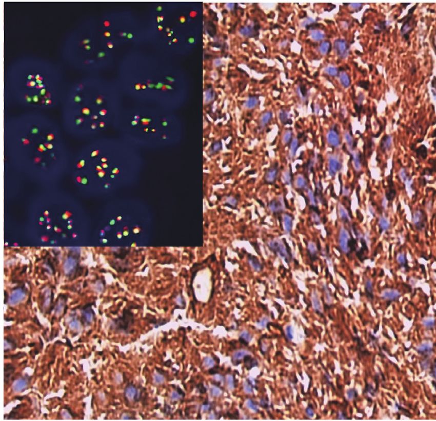
ALK-1, h-TERT in glioblastoma mutiforme • 213

merase chain reaction targeted only the fusion site. Therefore,      their relationship to conventional therapy for GBM.
other probable clinically important fusion sites will not be de-
tected. ALK gene analysis using FISH helps to detect a specific                   MATERIALS AND METHODS
type of gene aberration, such as gene rearrangement and ampli-
fications [6]. ALK gene amplification (ALK-A) has been recog-           This was a retrospective study that included 53 patients pri-
nized in several types of cancer as anaplastic large cell lymphoma   marily diagnosed with GBM. All were recruited and diagnosed
(ALCL), hepatocellular carcinoma (HCC), esophageal squamous          in Pathology Department, Assiut University Hospital and South
cell carcinoma, and GBM [7].                                         Egypt Cancer Institute, between April 2014 and April 2018.
   Telomerase is composed of a reverse transcriptase catalytic       Patients were followed until May 2020. All patients were eligible
subunit human telomerase reverse transcriptase (h-TERT) and          at age 20–80 years, if they had no previous diagnosis of cancer
an RNA template, the human telomerase RNA component                  and no previous CNS surgery for any cause. The exclusion criteria
(hTERC). The h-TERT coded by the TERT gene is an active cat-         were patients with primary malignant brain tumors other than
alytic protein subset located at chromosome 5p15.33. The oth-        GBM or with brain metastasis, and patients with no follow-up
er subset of telomere is hTERC or hTR, coded by the human            records.
telomerase RNA component (TERC gene), which positioned at               With regards to the treatment modalities used in the current
chromosome 3q26. In cancer cells, the resumption of h-TERT           research, all patients underwent surgical intervention, which in-
protein is the chief process of telomere preservation [8]. The ex-   cluded gross total resection, subtotal resection, or biopsy. Follow-
pression of h-TERT is an important determinant of telomere           ing surgical intervention, fractionated conformal radiation ther-
activity. Generally, h-TERT is not expressed in normal tissues,      apy was delivered to Gross tumor volume 1, which included the
which has been attributed to strict h-TERT regulation. However,      T2/FLAIR abnormality and the surgical cavity if present for a total
h-TERT was highly expressed in malignant tumors including            dose of 46 Gy in 23 fractions at 2 Gy per fraction, once daily, for
breast cancer, HCC, thyroid cancer and gliomas [9]. Some studies     five days per week. Gross tumor volume 2 included T1 contrast
on GBM patients documented that patients with absent h-TERT          enhanced abnormality and the surgical cavity if present for a
expression had significantly longer OS than patients with strong     boost dose of 14 Gy in seven fractions with 2 Gy per fraction,
h-TERT expression [10]. However, another study revealed no           once daily, for five days per week. All patients were treated using
significant effect of h-TERT expression on survival [11].            a megavoltage linear accelerator and photon energies of 6 MV
   The standard treatment for newly diagnosed GBM is maxi-           or more. Adjuvant radiotherapy only was given in 12 patients
mum safe resection followed by concurrent temozolomide (TMZ)         (22.6%) while the remaining patients received chemotherapy as
and radiotherapy with adjuvant TMZ for six cycles. This study        a part of a treatment protocol. Chemotherapy treatment consist-
reported a median OS of 14.6 months after median follow-up           ed of TMZ, which was given concomitantly with radiotherapy
duration of 28 months [12]. Despite multimodal treatment of          (75 mg/m2/day, started from the first day of radiotherapy until
GBM, patient outcomes are still non-satisfying for neuro-oncol-      the end of radiation) in 27 patients (50.9%) or given as concurrent
ogists. Molecular characterization of GBM with clinical concern      chemoradiotherapy (CCRT) and adjuvant (150–200 mg/m2/for
is still under research for emerging targeted therapy. A novel       5 days/every 28 days for 6 or 12 cycles) in 14 patients (26.4%).
targeted ALK inhibitor therapy as crizotinib, which is an FDA-          Follow-up evaluation included history and neurological exami-
approved treatment for lung cancer, is currently used in clinical    nation, laboratory investigations, assessment of treatment related
trials for ALK-positive ALCL and in a few GBM patients [13].         toxicity, and magnetic resonance imaging (MRI) or magnetic res-
However, emerging resistance to treatment with ALK inhibitors        onance spectroscopy imaging that were available for review. Pa-
in patients remains a major concern. A recent study revealed that    tients were evaluated for response using MRI and or magnetic
a subset of patients with ALK-positive lung adenocarcinomas          resonance spectroscopy, which were performed within 48 hours
harbor additional TERT amplification, which leads to unstable        of surgery, before the first cycle, after every 3 cycles of adjuvant
genomes with obvious fast relapse and therapeutic failure [14].      TMZ, and every three months after termination of treatment.
   To date, there is no data detailing the relationship between         The GBM specimens used for evaluation of ALK-1, h-TERT
ALK-1 and h-TERT expression in GBM. The present study                immunohistochemical protein expression, and ALK gene altera-
aimed to assess ALK-1 and h-TERT protein expression, their           tions using the FISH technique. Clinicopathological parameters
correlation with ALK gene alterations in GBM prognosis, and          collected from the patient’s archives sheets included patient age,

https://doi.org/10.4132/jptm.2021.03.15                                                                              https://jpatholtm.org/
214 • Elsers D et al.

sex, tumor site, presence of tumor calcification, tumor multiplicity,   positive controls.
tumor size, and Ki-67 labeling index (Ki LI) with cutoff point            Ki-67 positivity was identified as a brown nuclear expression.
of 14% [15].                                                            KI LI was defined as the percentage of positive tumor nuclei in
                                                                        1,000 tumor cells with cutoff value of 14% [15].
Immunohistochemistry
   Formalin-fixed paraffin-embedded (FFPE) slides from the GBM          Fluorescence in situ hybridization
tissue blocks, were retrieved from pathology lab and included              An ALK break-apart probe set (XT ALK BA Dual Color, Break
for IHC study. The slides stained with hematoxylin and eosin            Apart Rearrangement Probe [reference number: D-6001-100-
were reviewed histologically before staining by the two patho-          OG], Metasystems, Altlussheim, Germany) was used for FISH
logic consultants in this research. The FFPE blocks were cut into       to detect gene rearrangements and copy number changes. In
3–4-μm thickness, and then put on positively charged glass slides       each case, we examined around 200 nuclei from at least 5–8 ar-
(PCS). Sections were de-paraffinized and rehydrated, followed by        eas. We excluded nuclei with apparent overlapping or truncation.
antigen retrieval, which was done with Tris-EDTA in a water                Four-micrometer-thick tissue sections were cut from FFPE
bath at 90°C for 45 minutes. The primary monoclonal mouse               GBM tissue and were put on PCS. The unstained slides were
anti-Human ALK/CD246 antibody (clone ALK-1), ready-to-use               placed overnight at 60°C on a hotplate. Then, the slides were im-
(code IR641, 117498-002, CVR No. 33211317, Dako, Glostrup,              mersed 3 times in xylene for 5 minutes and dehydrated twice in
Denmark), Ki-67antibody (clone MIB-1), ready-to-use (code               100% ethanol for 5 minutes at room temperature. In sequence,
IR626, primary mononclonal mouse, Dako), and a primary rab-             the slides were immersed for 20 minutes in 0.2 N HCl, in puri-
bit polyclonal anti–Human TERT antibody (catalog #213737,               fied water for 3 minutes, and in 1 M sodium thiocyanate at 80°C
United State Biological 4 Technology, Salem, MA, USA) were              for 30 minutes. The slides were incubated for 30 minutes in
applied. Anti-TERT antibody was used at a dilution of 1/75              Protease Solution previously warmed to 37°C after removal of
(optimum dilution according to datasheet). Both incubated for           excess water and washed in purified water for 3 minutes. Then,
one hour at room temperature in an airtight humid chamber. A            dehydration of slides in 70%, 80%, and 100% ethanol for one
universal staining kit “Ultra Vision Detection System Anti-Poly-        minute each was done and slides were allowed to dry. Then, they
valent, HRP/DAB (ready-to-use)” (catalog #TP-015-HD, LAB                were placed in a dark room. Ten microliters of probe mixture
VISION Corp., Fremont, CA, USA) was applied following the               were applied to a slide, immediately covered by a coverslip, and
manufacturer’s instructions.                                            sealed with rubber cement. They were placed in a hybridizer in-
                                                                        strument (DakoCytomation, Glostrup, Denmark) at 73°C for 3
Evaluation of ALK-1 and h-TERT IHC expression                           minutes followed by an overnight hybridization at 37°C. At the
   ALK-1 positivity was identified as a brown cytoplasmic ex-           end of the hybridization period, we removed the rubber cement
pression. A four-tier scoring system was used for evaluation of         from the slides and placed them in 2 × saline sodium citrate (SSC;
ALK-1 positivity [5]; score 1 was corresponded to weak cytoplas-        post hybridization wash) at 73°C temperature for 2 minutes,
mic expression of ALK-1, moderate cytoplasmic expression was            then immersed them for 1 minute in 2 × SSC at room tempera-
considered as score 2, and strong cytoplasmic expression was con-       ture and allowed the slides to dry. Ten microliters of DAPI counter-
sidered as score 3 (Fig. 1). Negative staining was scored as 0.         stain was applied to the target area and covered by a coverslip [6].
   Positive brown nuclear staining of h-TERT was deemed posi-
tive. A three-tier evaluation system was used for scoring of h-         Analysis of ALK gene alterations using FISH technique
TERT IHC protein expression [11]. Score 1 corresponded to nu-              We analyzed the prepared slides under an oil immersion objec-
clear expression in < 5% of tumor cells. h-TERT expression in           tive (100 ×) with a fluorescence microscope (M1, Carl Zeiss Mi-
between 5 and 50% tumor cells was scored as 2, and expression           croscopy GmbH, Gottingen, Germany) equipped with appro-
in more than 50% of tumor cells was deemed to score 3 (Fig. 2).         priate filters and a charge-coupled device camera using FISH
Positive cytoplasmic staining of ALK-1 protein in anaplastic            imaging with the capturing software Metafer 5 (a Metafer slide
lymphoma cells was used as a positive control. Positive nuclear         scanning system [Metasystems]). Non-rearranged ALK showed
staining of h-TERT in melanoma cells were used as positive con-         fusion (yellow signals) or very close abutment of the probes adja-
trol. Negative control done using the same protocol of IHC unless       cent to the 3' (red) and the 5' (green) ends of the gene. Rearranged
the addition of the primary antibody on tissue section of specific      ALK appeared as splitting of 3' and 5' signals. Tumor tissues

https://jpatholtm.org/                                                                                https://doi.org/10.4132/jptm.2021.03.15
ALK-1, h-TERT in glioblastoma mutiforme • 215
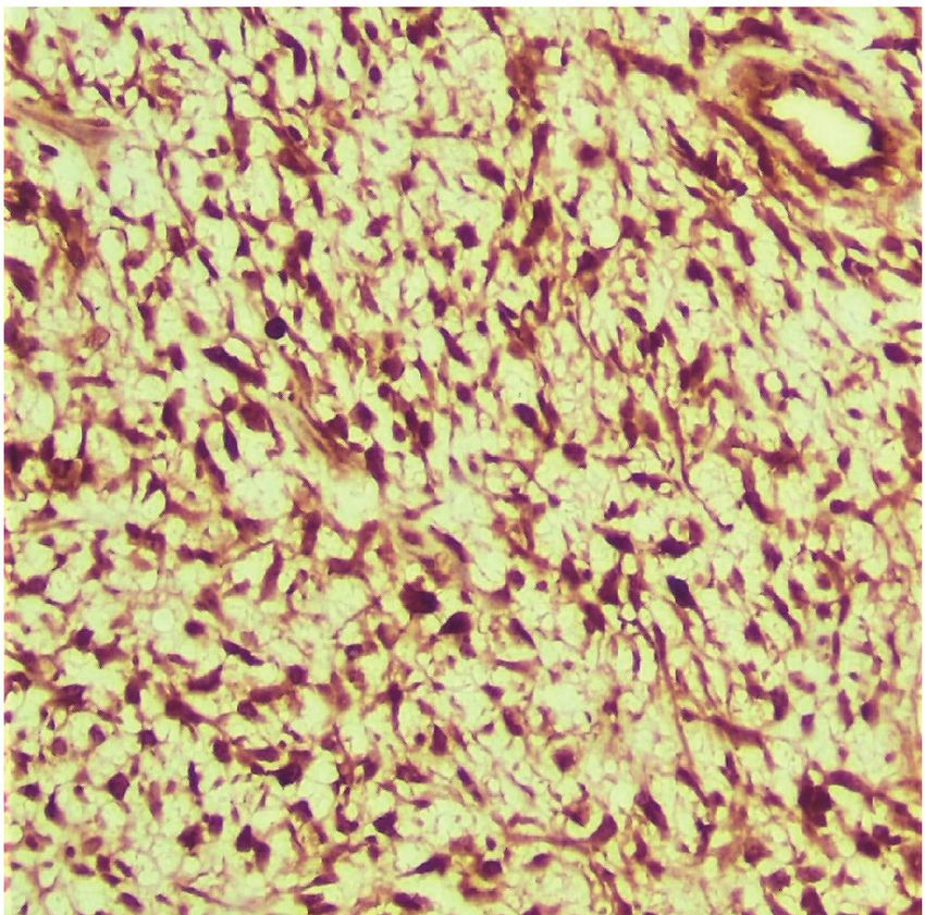
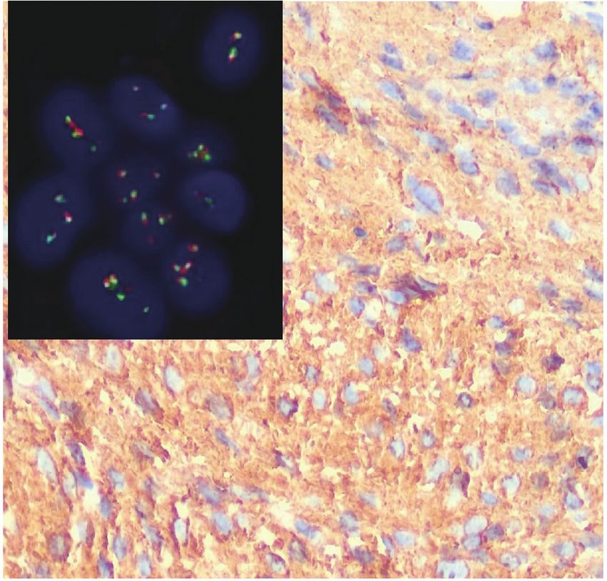
A B
C D
Fig. 1. Expression of anaplastic lymphoma kinase 1 (ALK-1) immunohistochemistry in tumor cells and ALK gene alterations in glioblastoma
multiforme (GBM). (A) A case of GBM shows strong cytoplasmic expression of ALK-1 in tumor cells (score 3). The inset illustrates ALK gene
amplification for the same case. (B) A case of GBM shows moderate cytoplasmic staining of ALK-1 in tumor cells. The inset illustrates ALK
gene gain for the same case. (C) A case of GBM shows weak cytoplasmic expression of ALK-1 in tumor cells (score 1). The inset illustrates
ALK gene rearrangement for the same case. (D) A case of GBM showed negative expression of ALK-1 (score 0). The inset illustrates that the
ALK gene was negative for rearrangement with a normal copy number for the same case.
were considered ALK-FISH positive (ALK-rearranged) if > 15% logical data. Chi-square test was used only when ≤ 20% of the
tumor cells showed splitting of red and green signals [16]. The cells had an expected count less than 5. Correlation between ALK
mean cutoff copy number of 3 to 5 fusion signals in ≥ 10% of gene alterations and ALK and TERT IHC protein expression were
cells represented ALK–copy number gain (ALK-CNG), while done via Spearman correlation coefficient test. OS was calculated
the presence of ≥ 6 copies of ALK per cell in ≥ 10% of analyzed from the date of surgical resection to the date of death from any
cells represented ALK-A (Fig. 1) [17]. cause or last follow-up. Progression-free survival (PFS) was cal-
culated from the date of surgical resection to the date of progres-
Statistical analysis sion or date of last follow-up or death.
The analysis for this study was done through using the SPSS Kaplan-Meier curves were used to analyze OS and PFS. Com-
ver. 21 (IBM Corp., Armonk, NY, USA). Fisher exact test was parison of survival was determined by log-rank test. Multivari-
used to detect the association between ALK protein, TERT pro- ate analysis using Cox proportional hazard model of predictors
tein expression, ALK gene alterations, and various clinicopatho- of outcome variables were applied. The value of significance was
https://doi.org/10.4132/jptm.2021.03.15 https://jpatholtm.org/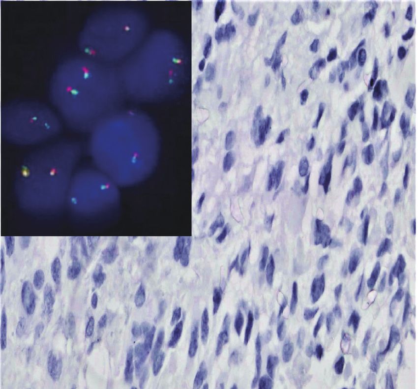
216 • Elsers D et al.

                                          A                                             B                                               C

Fig. 2. Expression of human telomerase reverse transcriptase (TERT) immunohistochemistry in tumor cells of glioblastoma multiforme. (A)
Strong nuclear expression of TERT in > 50% of tumor cells (score 3). (B) Moderate nuclear staining of TERT in 5%–50% of tumor cells (score
2). (C) Weak nuclear expression of TERT in < 5% of tumor cells (score 1).

determined as p < .05.                                                  Association between h-TERT IHC protein expression and
                                                                        clinicopathological parameters
                          RESULTS                                          h-TERT expression was detected in the studied cases with vari-
                                                                        able percentages and staining intensities. Strong h-TERT nucle-
   The current study included 53 GBM patients. The study in-            ar expression in > 50% of tumor cells (score 3) was noted in 29
cluded 17 patients (32.1%) who were < 50 years of age, while 36         (54.7%) cases, while moderate expression in 5%–50% of tumor
patients (67.9%) were ≥ 50 years old. Thirty-six cases (67.9%)          cells (score 2) was present in 16 cases (30.2%) and eight cases
were males. The most prominent tumor location was at the pa-            (15.1%) showed weak expression in < 5% of tumor cells (score 1).
rieto-occipital region, constituting 28.4% of patients. The me-            h-TERT expression showed significant association with the
dian tumor size was 5 cm. Calcification was present in 32 cases         presence of calcifications (p = .016). Score 3 h-TERT expression
(60.4%). There were multiple tumors in six cases (11.3%).               exhibited a significant association with Ki LI (p = .005). There
   After a median follow-up duration of 12 months (range, 3 to 19       was no significant correlation between h-TERT expression and
months), 21 out of 53 patients (39.6%) were still alive (Table 1).      other clinicopathological variables (Table 2).

Association between ALK-1 IHC protein expression and                    Association between ALK gene alterations and
clinicopathological parameters                                          clinicopathological parameters
   ALK-1 expression was detected in 45 cases out of 53 (84.9%).            Thirty-four cases out of 53 showed ALK gene alterations (64.2%).
Strong ALK cytoplasmic expression (score 3) was noted in 17             ALK-A was detected in four cases (7.5%), and all of these cases
GBM tumors (32.1%). Thirteen cases (24.5%) showed moderate              had strong ALK immunohistochemical expression. ALK-CNG
cytoplasmic expression (score 2), while 15 cases (28.3%) showed         was noted in 12 cases (22.6%), while 18 cases (34.0%) showed
weak cytoplasmic expression (score 1). Eight cases (15.1%) were         ALK gene rearrangement. Nineteen cases (35.9%) were negative.
negative (score 0).                                                     ALK-A was significantly associated with increased Ki LI (p =
   Score 3 ALK-1 expression showed significant association with         .044). The type of therapeutic modality was positively associat-
male sex (p = .038), tumor multiplicity (p = .046), and Ki LI (p ≤      ed with ALK gene alterations (p = .027). There was no significant
.001). The type of therapeutic modality was positively associated       association between ALK gene alteration and clinicopathological
with ALK-1 protein expression (p = .030). The clinicopatholog-          variables (Table 3).
ical associations with ALK-1 protein IHC expression are sum-
marized in Table 2.                                                     Correlation between ALK gene alterations, ALK-1 and
                                                                        h-TERT IHC protein expression
                                                                          A strong positive correlation was noted between ALK protein
                                                                        IHC expression and ALK gene alterations (p = .001, r = 0.616).

https://jpatholtm.org/                                                                               https://doi.org/10.4132/jptm.2021.03.15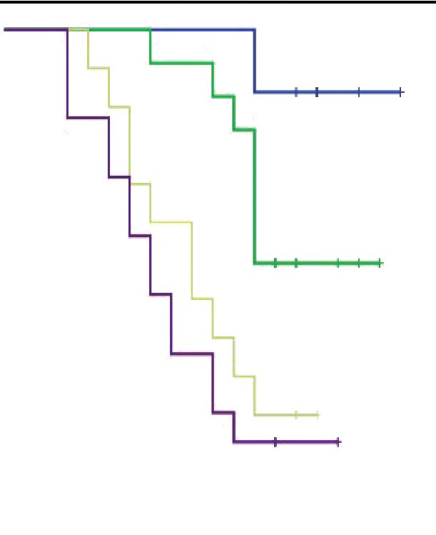
ALK-1, h-TERT in glioblastoma mutiforme • 217

Table 1. Clinicopathological characteristics of the patients                    Outcome analysis
Variable                                           No. (%)                         The median follow-up duration of the 53 GBM patients was
Age (yr)                                                                        12 months (range, 3 to 19 months). During follow-up, 32/53
  < 50                                            17 (32.1)
                                                                                patients (60.4%) died as a result of tumor progression. According
  ≥ 50                                            36 (67.9)
Sex
                                                                                to Kaplan-Meier analysis, the median OS was 12 months (95%
  Male                                            36 (67.9)                     confidence interval [CI], 10.449 to 13.551), while the 19-month
  Female                                          17 (32.1)                     OS rate was 39.6%. A total of 36/53 patients (67.9%) developed
Site                                                                            disease progression. The median time to progression was 10
  CC                                               4 (7.5)
  FP                                              12 (22.6)
                                                                                months (range, 2 to 19 months). According to Kaplan-Meier
  PO                                              15 (28.4)                     analysis, the median PFS was 10 months (95% CI, 7.860 to
  TO                                               7 (13.2)                     12.140). The PFS rate at 19 months was 32.1%.
  PS                                               6 (11.3)                        ALK-1 protein expression (score 3), h-TERT protein expres-
  TP                                               9 (17.0)
                                                                                sion (score 3), and ALK-A were associated with poor OS and
Calcification
  Absent                                          21 (39.6)                     PFS (p < .001, p = .031, and p < .001) and (p < .001, p = .040, and
  Present                                         32 (60.4)                     p < .001), respectively. Cases that exhibited high Ki LI had poor
Multiplicity                                                                    OS and short PFS (p = .016) and (p = .022), respectively (Figs. 3, 4).
  Single                                          47 (88.7)
                                                                                   Regarding the type of therapeutic modalities, patients treated
  Multiple                                         6 (11.3)
Tumor size                                                                      with adjuvant radiotherapy only had poor OS and PFS compared
  Median (interquartile range)                     5 (4–7)                      to those who were treated with CCRT or CCRT and adjuvant
Type of surgical resection                                                      TMZ (p = .002) and (p = .004), respectively (Table 5, Figs. 3E, 4E).
  GTR                                             19 (35.0)
                                                                                   Multivariate analysis for OS and disease-free survival was ap-
  STR                                             25 (47.0)
  Biopsy                                           9 (17.0)
                                                                                plied to clinicopathological features that were significant in uni-
Ki LI (%)                                                                       variate analysis to adjust for confounders. Our results revealed
  < 14                                            37 (69.8)                     that ALK gene alteration was the only independent prognostic
  ≥ 14                                            16 (30.2)                     factor for OS and PFS (p < .001; hazard ratio [HR], 7.514; 95%
Status
  Living                                          21 (39.6)
                                                                                CI, 3.292 to 17.155) and (p ≤ .001; HR, 4.711; 95% CI, 2.429
  Dead                                            32 (60.4)                     to 9.136), respectively (Table 6).
Therapeutic modalities
  RTH only                                        12 (22.6)                                              DISCUSSION
  CCRT                                            14 (26.4)
  CCRT and adjuvant TMZ                           27 (50.9)
                                                                                   GBM exhibits a vast group of modifications, both genetic and
CC, corpus callosum; FP, fronto-parietal; PO, parieto-occipital; TO, tempe-
ro-occipital; PS, parasagittal; TP, tempero-parietal; GTR, gross total resec-   epigenetic, which create a great number of mutation subsets, some
tion; STR, subtotal resection; Ki LI, Ki labeling index; RTH, radiotherapy;     of which have a proven effect in survival and therapy response [1].
CCRT, concurrent chemoradiotherapy; TMZ, temozolomide.
                                                                                   In the present study, high ALK-1 expression showed a signif-
                                                                                icant association with male sex, however, no significant relation
Moderate correlation was noted between ALK gene alterations                     between ALK-1 expression and patient’s age. These findings are
and TERT IHC (p = .007, r = 0.476), as (score 2) TERT expres-                   not matched with the study done by Karagkounis et al. [5], which
sion was associated with ALK gene rearrangement (56.2%), while                  reported that ALK overexpression is more common in older in-
(score 3) TERT expression was associated with ALK-CNG and                       dividuals (> 59 years) and that was no association between ALK
ALK-A (51.8%). There was a strong positive correlation between                  expression and patient’s sex. This discrepancy was due to division
ALK-1 and TERT IHC protein expression (p = .002, r = 0.602).                    of their cases into subgroups according to IDH1 protein expres-
The relationships and correlations between ALK gene alterations,                sion with cutoff median age of 59 years, which was not imple-
ALK, and TERT IHC protein expression are presented in Ta-                       mented in the current study.
bles 3 and 4, respectively.                                                        One persistent debate is whether ALK-1 immunohistochem-
                                                                                ical overexpression was associated with ALK gene mutation or
                                                                                amplification in GBM cases. In the current study, we reported that

https://doi.org/10.4132/jptm.2021.03.15                                                                                         https://jpatholtm.org/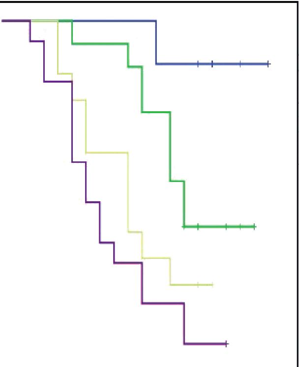
218 • Elsers D et al.

Table 2. Association between ALK-1, h-TERT IHC protein expression, and clinicopathological parameters
                                                            ALK-1 IHC                                                          h-TERT IHC
Parameter
                                 Score 0       Score 1        Score 2       Score 3         p-value         Score 1       Score 2        Score 3       p-value
Age (yr)                                                                                     .236                                                       .123a
  < 50                           2 (11.8)      6 (35.3)       6 (35.3)      3 (17.6)                        3 (17.6)      8 (47.1)       6 (35.3)
  ≥ 50                           6 (16.7)      9 (25.0)       7 (19.4)     14 (38.9)                        5 (13.9)      8 (22.2)      23 (63.9)
Sex                                                                                          .038                                                       .267a
  Male                           7 (19.4)      6 (16.7)     10 (27.8)      13 (36.1)                        6 (16.7)     13 (36.1)      17 (47.2)
  Female                         1 (5.9)       9 (52.9)      3 (17.6)       4 (23.5)                        2 (11.8)      3 (17.6)      12 (70.6)
Site                                                                                         .425                                                       .119
  CC                             2 (50.0)      1 (25.0)       0             1 (25.0)                        0             3 (75.0)       1 (25.0)
  FP                             1 (8.4)       4 (33.3)       4 (33.3)      3 (25.0)                        2 (16.7)      2 (16.7)       8 (66.6)
  PO                             2 (13.3)      7 (46.7)       2 (13.3)      4 (26.7)                        5 (33.3)      2 (13.3)       8 (53.4)
  TO                             2 (28.5)      1 (14.3)       3 (42.9)      1 (14.3)                        1 (14.3)      4 (57.1)       2 (28.6)
  PS                             0             0              2 (33.3)      4 (66.7)                        0             1 (16.6)       5 (83.4)
  TP                             1 (11.2)      2 (22.2)       2 (22.2)      4 (44.4)                        0             4 (44.4)       5 (55.6)
Calcification                                                                                .315                                                       .016*
  Absent                         1 (4.8)       6 (28.6)       5 (23.8)      9 (42.8)                        3 (14.3)      2 (9.5)       16 (76.2)
  Present                        7 (21.9)      9 (28.1)       8 (25.0)      8 (25.0)                        5 (15.6)     14 (43.8)      13 (40.6)
Multiplicity                                                                                 .046                                                       .501
  Single                         8 (17.1)     14 (29.8)      13 (27.7)     12 (25.5)                        8 (17.0)     15 (31.9)      24 (51.1)
  Multiple                       0             1 (16.7)       0             5 (83.3)                        0             1 (16.7)       5 (83.3)
Tumor size                                                                                   .670                                                       .834
  < Median                       4 (14.2)      6 (21.4)       9 (32.2)      9 (32.2)                        5 (17.8)      7 (25.0)      16 (57.2)
  ≥ Median                       4 (14.2)      9 (32.2)       7 (25.0)      8 (28.6)                        3 (12.0)      9 (36.0)      13 (52.0)
Type of surgical resection                                                                   .266                                                       .133
  GTR                            3 (15.8)      4 (21.1)       6 (31.6)      6 (31.6)                        5 (26.3)      3 (15.8)      11 (57.9)
  STR                            5 (20.0)      7 (28.0)       7 (28.0)      6 (24.0)                        3 (12.0)     11 (44.0)      11 (44.0)
  Biopsy                         0             4 (44.4)       0             5 (55.6)                        0             2 (22.0)      29 (54.7)
Ki LI (%)                                                                                   < .001                                                      .005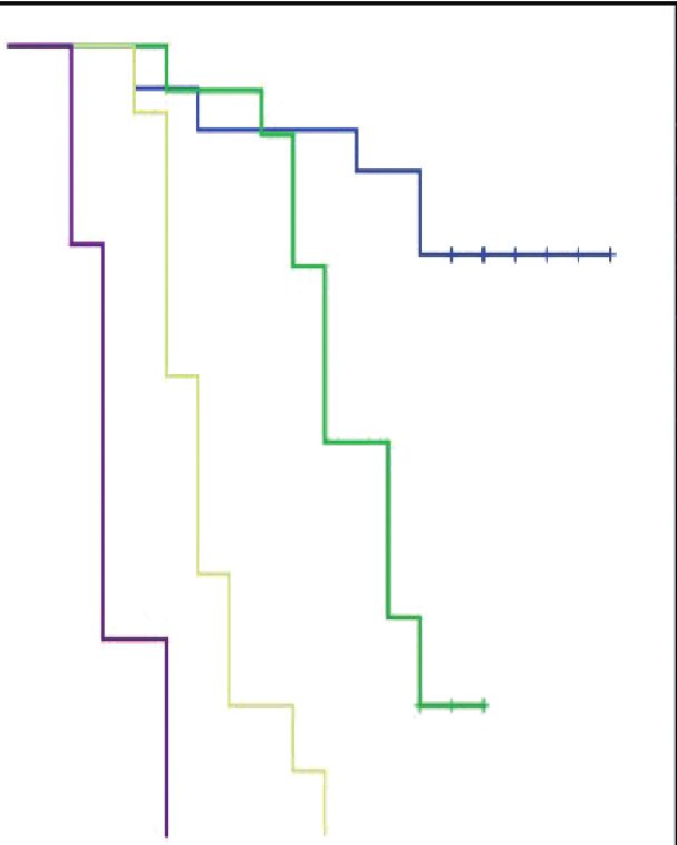
ALK-1, h-TERT in glioblastoma mutiforme • 219

Table 3. Association between ALK gene alteration, clinicopatho-                   Table 4. Correlation between ALK gene alterations, ALK-1, and h-
logical parameters, and ALK-1 and h-TERT expression                               TERT IHC protein expression
                              ALK gene alterations                                                                         Spearman’s rho
                                                                        p-value
               Negative Rearrangement         Gain      Amplification                                          h-TERT          ALK-1          ALK gene
Age (yr)                                                                 .430                                 expression     expression       alterations
   < 50          7 (41.2)      7 (41.2)      3 (17.6)     0                       h-TERT expression
   ≥ 50        12 (33.3)      11 (30.6)      9 (25.0)     4 (11.1)                  Correlation coefficient     1.000           0.602            0.476
Sex                                                                      .181       Sig. (2-tailed)                             0.002a           0.007a
   Male        10 (27.8)      12 (33.3)     10 (27.8)     4 (11.1)                  No.                          53              53               53
   Female        9 (52.9)      6 (35.3)      2 (11.8)     0                       ALK-1 expression
Site                                                                     .955       Correlation coefficient     0.602           1.000            0.616
   CC            2 (50.0)      1 (20.0)      1 (20.0)     0                         Sig. (2-tailed)             0.002a                           0.001a
   FP            4 (33.3)      3 (25.0)      3 (25.0)     2 (16.7)                  No.                          53              53               53
   PO            6 (40.0)      5 (33.3)      3 (20.0)     1 (6.7)                 ALK gene alterations
   TO            2 (28.6)      3 (42.8)      1 (14.3)     1 (14.3)                  Correlation coefficient     0.476           0.616            1.000
   PS            2 (33.3)      1 (16.7)      3 (50.0)     0                         Sig. (2-tailed)             0.007a          0.001a
   TP            3 (33.3)      5 (55.6)      1 (11.1)     0                         No.                          53              53               53
Calcification                                                            .710     ALK-1, anaplastic lymphoma kinase 1; h-TERT, human telomerase reverse
  Absent         6 (28.6)      7 (33.3)      6 (28.6)     2 (9.5)                 transcriptase; IHC, immunohistochemistry.
  Present      13 (40.6)      11 (34.4)      6 (18.8)     2 (6.2)
                                                                                  a
                                                                                   Significant; correlation is significant at the 0.01 level (2-tailed).
Multiplicity                                                             .162
                                                                                  revealed that ALK gene aberrations detected by FISH and ALK
  Single       19 (40.4)      15 (31.9)     10 (21.3)     3 (6.4)
  Multiple       0             3 (50.0)      2 (33.3)     1 (16.7)                overexpression by IHC in rhabdomyosarcoma were significantly
Size                                                                     .836     correlated [21]. On the other hand, other studies found no asso-
  < Median 12 (38.7)           9 (29.0)      7 (22.6)     3 (9.7)                 ciation between ALK gene aberrations detected by FISH and
   > Median      7 (31.8)      9 (40.9)      5 (22.7)     1 (4.6)
                                                                                  ALK expression by IHC in various cancer types such as esopha-
Type of surgical resection                                               .125
   GTR           9 (47.4)      3 (15.8)      5 (26.3)     2 (10.5)                geal squamous cell carcinoma and colorectal carcinoma [22]. Fur-
   STR           8 (32.0)      9 (36.0)      7 (28.0)     1 (4.0)                 thermore, a study of a neuroblastoma cell line harboring ALK-A
   Biopsy        2 (22.2)      6 (66.7)      0            1 (11.1)                was accompanied by reduction of ALK levels as a result of N-
Ki- LI                                                                   .044
                                                                                  linked glycosylation inhibition with subsequent inhibition of its
   < 14%       15 (40.5)      15 (40.5)      6 (16.2)     1 (2.8)
   ≥ 14%         4 (25.0)      3 (18.8)      6 (37.4)     3 (18.8)
                                                                                  phosphorylated downstream molecules such as AKT and STAT3
ALK-1 IHC                                                                .001     [23]. This controversy between the relationship between ALK
   Score 0       8 (100)       0             0            0                       gene extra-copies on FISH and ALK protein expression may be
   Score 1       6 (40.0)      9 (60.0)      0            0                       attributed to either transcriptional or post-transcriptional mod-
   Score 2       2 (15.4)      5 (38.4)      6 (46.2)     0
   Score 3       3 (17.7)      4 (23.5)      6 (35.3)     4 (23.5)
                                                                                  ifications, or degradation, such as N-linked glycosylation acti-
h-TERT IHC                                                               .008     vation or inhibition, which lead to ALK activation and reduction,
   Score 1       6 (75.0)      2 (25.0)      0            0                       respectively.
   Score 2       6 (37.5)      9 (56.2)      1 (6.3)      0                          Regarding h-TERT immunohistochemical expression, the cur-
   Score 3       7 (24.1)      7 (24.1)     11 (38.0)     4 (13.8)
                                                                         .027
                                                                                  rent work revealed strong h-TERT nuclear expression in about
Therapeutic modalities
   RTH           2 (16.7)      3 (25.0)      3 (25.0)     4 (33.3)                half of the studied cases; this is compatible with the study done
  CCRT &      11 (40.7)      10 (37.0)      6 (22.3)     0                       by Potharaju et al. [10], which showed that 60% of GBM patients
    adjuvant
     TMZ
                                                                                  expressed strong h-TERT. In spite of the use of h-TERT IHC as
   CCRT          6 (42.9)      5 (35.7)      3 (21.4)     0                       a mirror to detect tumors harboring TERT-mutation, h-TERT
Significant at p < .05.                                                           IHC was not able to identify the differences between TERT-
ALK-1, anaplastic lymphoma kinase 1; h-TERT, human telomerase reverse             mutated GBMs and TERT-nonmutant GBMs. h-TERT protein
transcriptase; CC, corpus callosum; FP, fronto-parietal; PO, parieto-occipi-
tal; TO, tempero-occipital; PS, parasagittal; TP, tempero-parietal; GTR,          overexpression was noted even among gliomas with wildtype
gross total resection; STR, subtotal resection; Ki LI, Ki labeling index; IHC,    TERT. Moreover, h-TERT IHC widely varied through the TERT-
immunohistochemistry; RTH, radiotherapy; CCRT, concurrent chemoradio-
therapy; TMZ, temozolomide.                                                       mutant gliomas such as oligodendrogliomas and GBMs. This
                                                                                  indicates that h-TERT IHC expression may be regulated by var-
                                                                                  ious mechanisms along with TERT promoter mutations [24].

https://doi.org/10.4132/jptm.2021.03.15                                                                                               https://jpatholtm.org/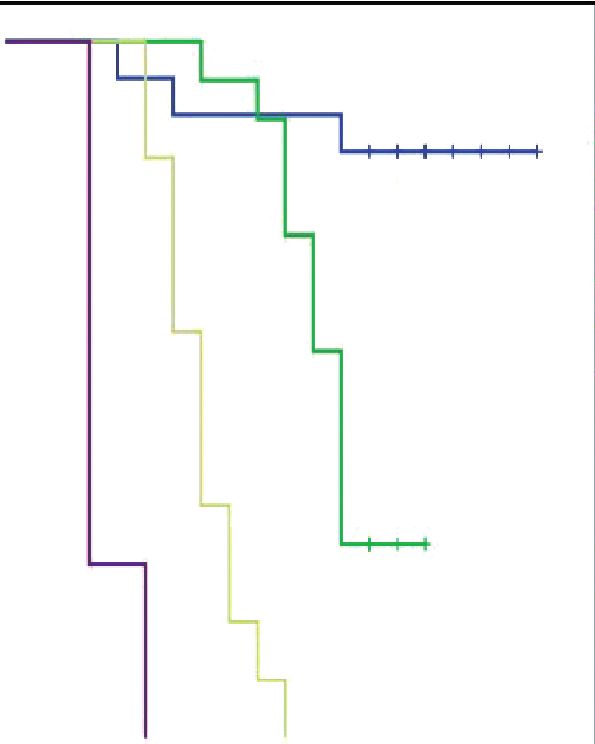
220 • Elsers D et al.
Survival functions Survival functions
1.0 Ki LI 1.0 ALK-1 IHC expression
< 14% Score 0
≥ 14% Score 1
0.8 0.8
< 14%-censored Score 2
Cumulative survival
Cumulative survival
≥ 14%-censored Score 3
0.6 0.6 Score 0-censored
Score 1-censored
Score 2-censored
0.4 0.4 p < .001
p = .016 Score 3-censored
Log rank = 19.639
Log rank = 5.758
0.2 0.2
0.0 0.0
0 5 10 15 20 0 5 10 15 20
Time (mo) A Time (mo) B
Survival functions Survival functions
1.0 h-TERT IHC 1.0 ALK gene alterations
expression Negative
Score 1 Rearrangement
0.8 0.8
Score 2 Gain
Cumulative survival
Cumulative survival
Score 3 Amplification
0.6 Score 1-censored 0.6 p < .001 Negative-censored
Score 2-censored Log rank = Rearrangement-
Score 3-censored 86.062 censored
0.4 p = .031 0.4
Gain-censored
Log rank = 6.919
Amplification-
0.2 0.2 censored
0.0 0.0
0 5 10 15 20 0 5 10 15 20
OS C Time (mo) D
Survival functions
1.0 Therapeutic
modalities
RTH only
0.8
CCRT and
Cumulative survival
adjuvant TMZ
0.6 CCRT
RTH only-censored
CCRT and adjuvant
0.4 TMZ-censored
p = .002 CCRT-censored
0.2 Log rank = 13.797
0.0
0 5 10 15 20
Time (mo) E
Fig. 3. Overall survival (OS) for anaplastic lymphoma kinase 1 (ALK-1), human telomerase reverse transcriptase (h-TERT) immunohistochem-
istry (IHC) expression, and ALK gene alterations. (A) High Ki labeling index (Ki LI) is associated with poor OS. (B) ALK-1 score 3 is associated
with poor OS. (C) TERT score 3 is associated with poor OS. (D) ALK gene amplification is associated with poor OS. (E) Patients treated with
adjuvant radiotherapy only had poor OS compared to those who were treated with concurrent chemoradiotherapy (CCRT) or CCRT and ad-
juvant temozolomide (TMZ).
https://jpatholtm.org/ https://doi.org/10.4132/jptm.2021.03.15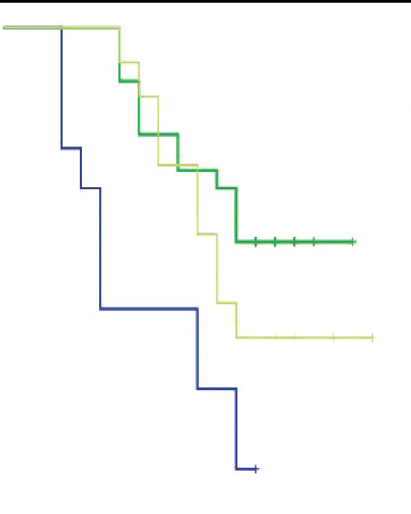
ALK-1, h-TERT in glioblastoma mutiforme • 221
Survival functions Survival functions
1.0 Ki LI 1.0 ALK-1 IHC expression
< 14% Score 0
≥ 14% Score 1
0.8 0.8
< 14%-censored Score 2
Cumulative survival
Cumulative survival
≥ 14%-censored p < .001 Score 3
0.6 p = .022 0.6 Log rank = Score 0-censored
Log rank = 21.654 Score 1-censored
5.267 Score 2-censored
0.4 0.4
Score 3-censored
0.2 0.2
0.0 0.0
0 5 10 15 20 0 5 10 15 20
Time (mo) A Time (mo) B
Survival functions Survival functions
1.0 h-TERT IHC 1.0 ALK gene alterations
expression Negative
Score 1 Rearrangement
0.8 0.8
Score 2 Gain
Cumulative survival
Cumulative survival
Score 3 Amplification
0.6 Score 1-censored 0.6 Negative-censored
Score 2-censored p < .001 Rearrangement-
p = .040 Score 3-censored Log rank = censored
0.4 Log rank = 6.433 0.4
76.436 Gain-censored
Amplification-
0.2 0.2 censored
0.0 0.0
0 5 10 15 20 0 5 10 15 20
Time (mo) C Time (mo) D
Survival functions
1.0 Therapeutic
modalities
RTH only
0.8
CCRT and
Cumulative survival
adjuvant TMZ
0.6 CCRT
RTH only-censored
0.4
p = .004
Log rank =
0.2 14.357
0.0
0 5 10 15 20
Time (mo) E
Fig. 4. Progression-free survival (PFS) for anaplastic lymphoma kinase 1 (ALK-1), human telomerase reverse transcriptase (h-TERT) immu-
nohistochemistry (IHC) expression, and ALK gene alterations. (A) High Ki labeling index (Ki LI) is associated with short PFS. (B) ALK-1 score
3 is associated with poor PFS. (C) TERT score 3 is associated with poor PFS. (D) ALK gene amplification is associated with poor PFS. (E)
Patients treated with adjuvant radiotherapy only had poor PFS compared to those who were treated with concurrent chemoradiotherapy
(CCRT) or CCRT and adjuvant temozolomide (TMZ).
https://doi.org/10.4132/jptm.2021.03.15 https://jpatholtm.org/222 • Elsers D et al.
Table 5. Kaplan-Meier analysis for OS and PFS
OS PFS
Parameter
Log-rank (chi-square) p-value Log-rank (chi-square) p-value
Age (< 50 yr vs. ≥ 50 yr) 3.146 .076 2.419 .120
Sex (male vs. female) 3.326 .119 3.887 .153
Site (CC vs. FP vs. PO vs. TO vs. PS vs. TP) 3.398 .639 4.216 .519
Calcification (absent vs. present) 0.666 .414 0.253 .615
Multiplicity (single vs. multiple) 3.993 .254 3.505 .190
Size ( median) 0.010 .921 0.048 .827
Type of surgical resection (GTR vs. STR vs. biopsy) 0.066 .967 0.168 .919
Ki LI (< 14% vs. ≥ 14%) 5.758 .016 5.267 .022
ALK-1 IHC (score 0 vs. score 1 vs. score 2 vs. score 3) 19.639 < .001 21.654 < .001
h-TERT IHC (score 1 vs. score 2 vs. score 3) 6.919 .031 6.433 .040
ALK gene alterations (negative rearrangement vs. gain amplification) 86.062 < .001 76.436 < .001
Therapeutic modalities (RTH only vs. CCRT vs. CCRT and adjuvant TMZ) 13.797 .002 14.357 .004
Significant at p < 0.5.
OS, overall survival; PFS, progression-free survival; CC, corpus callosum; FP, fronto-parietal; PO, parieto-occipital; TO, tempro-occipital; PS, parasagittal; TP,
tempro-parietal; GTR, gross total resection; STR, subtotal resection; Ki LI, Ki labeling index; ALK-1, anaplastic lymphoma kinase 1; IHC, immunohistochemis-
try; h-TERT, human telomerase reverse transcriptase; RTH, radiotherapy; CCRT, concurrent chemoradiotherapy; TMZ, temozolomide.
Table 6. Multivariate analysis for significant predictors of OS and PFS
OS PFS
B SE p-value HR 95 % CI for Exp (B) B SE p-value HR 95 % CI for Exp (B)
Ki LI 0.125 0.468 .790 1.133 0.453–2.833 –0.001 0.435 .998 0.099 0.426–2.344
ALK-1 protein expression 0.357 0.347 .304 0.429 0.724–2.821 0.505 0.306 .099 1.657 0.909–3.010
h-TERT protein expression –0.368 0.411 .371 0.692 0.310–1.549 –0.352 0.356 .322 0.703 0.350–1.411
ALK gene alterations 2.017 0.421 < .001 7.514 3.292–17.155 1.550 0.338 < .001 4.711 2.429–9.136
Therapeutic modalities 0.050 0.311 .872 1.052 0.571–1.935 –0.007 0.295 .982 0.993 0.558–1.769
Significant at p < 0.5.
OS, overall survival; PFS, progression-free survival; HR, hazards ratio; CI, confident interval; Ki LI, Ki labeling index; ALK-1, anaplastic lymphoma kinase 1; h-
TERT, human telomerase reverse transcriptase.
An interesting finding is the presence of a significant associa- used in the literature.
tion between h-TERT expression and the presence of tumor cal- Overall, there was a strong positive correlation between ALK-1
cification in the present study, which may be attributed to the and h-TERT IHC protein expression in the current research. All
hypoxic state in GBM that leads to activation of hypoxia-induc- GBM cases displayed necrosis and/or microvascular proliferation,
ible factor-1α (HIF-1α) in response to hypoxic status with sub- which is essential for their diagnosis [29]. These findings were
sequent increase in intracellular calcium (Ca2+) and promotion corresponding to a hypoxic state of the microenvironment. ALK
of the Ca2+ signaling pathway [25]. Furthermore, activation of expression was significantly higher in tumor cells in hypervas-
HIF-1α enhances h-TERT transcription [26]. cular lesions as compared to those adjacent to necrotic foci in
Concerning the relationship between Ki LI and ALK-1/h- GBMs, and was positively correlated with the microvascular den-
TERT protein expression, all the aforementioned biomarkers sity as determined by CD34 expression. Overexpression of ALK
had a reportedly positive association with Ki LI [4]. Persson and induced an enhancement of the HIF-1α/vascular endothelial
Englund [11] discerned that high values of Ki LI were not sig- growth factor-A axis through activation of Stat3 [4]. Further-
nificantly associated with high h-TERT staining. The current more, a study done by Marzec et al. [30] reported that ALK-
study showed that high Ki LI had a poor impact on OS and PFS. positive T-cell lymphoma expresses HIF1α. HIF1α mRNA ex-
This is in agreement with Persson and Englund [11], and in con- pression is induced in ALK-positive T-cell lymphoma by the
trast with other studies, which noted no effect of Ki LI on OS transcription of nucleophosmin/ALK tyrosine kinase. NPM/
[11,27,28]. The prognostic value of Ki LI in GBM is still uncer- ALK activates the HIF1α gene through the STAT3 transcrip-
tain, as the distribution of proliferative index was different in vari- tion factor [30].
able areas within the same tumor and different cutoff points are GBMs contain considerable hypoxic areas within the tumor.
https://jpatholtm.org/ https://doi.org/10.4132/jptm.2021.03.15ALK-1, h-TERT in glioblastoma mutiforme • 223
Potharaju et al. [10] suggested that an intratumoral decrease in patients. Subsequent studies with a larger number of GBM cases
oxygen concentration can augment HIF-1α expression and en- are recommended for better evaluation of the role of ALK-1, h-
hance h-TERT transcription and telomerase, which in turn lead TERT, and ALK gene alterations in tumor progression and re-
to proliferation of cancer stem cells [26]. Consequently, both lated mechanisms.
ALK-1 and h-TERT expression are highly expressed in the hy- The current work concluded that high protein expression of
pervascular area of GBM, depending on HIF-1α activation. This ALK-1, h-TERT, and ALK-A had poor impact on the prognosis
may explain the close relationship between ALK and h-TERT of GBM. Accordingly, ALK-1 protein expression, h-TERT pro-
expression in GBM. tein expression, and ALK gene alteration detection could be used
Regarding OS, high ALK-1 expression, high h-TERT expres- as valuable prognostic markers in GBM patients. The type of
sion, and ALK-A were associated with poor OS in the studied therapeutic modality was positively associated with ALK gene
cases. This result was in congruent with a study [4] showing that alterations. The use of ALK-targeting therapy as part of treatment
loss of ALK expression had improved OS in contrast to the ALK- plan for GBM patients with ALK gene alterations is the goal, and
positive cases and another report [10] showing that patients with requires further studies with a larger sample size.
absent or weak h-TERT expression had significantly longer OS
than patients with strong h-TERT expression. However, the cur- Ethics Statement
The research was approved by the Committee of Medical Ethics, Faculty of
rent findings are inconsistent with those of Karagkounis et al. [5] Medicine, Assiut University IRB. No. 17300482. A written informed con-
and Persson and Englund [11], which noted no significant im- sent for participation and publication was obtained from each participant
pact of ALK-1 and h-TERT expression on OS, respectively. after receiving information about the details of the study. Confidentiality of
patients’ records was assured and maintained throughout the study.
Interestingly, we found that ALK gene alteration was an in-
dependent prognostic factor for OS. This in agreement with a Availability of Data and Material
previous observation, which revealed that patients who had ALK- The datasets generated or analyzed during the current study are available
from the corresponding author on reasonable request.
A died within one month from diagnosis [5].
In the current study, the type of therapeutic modality was posi- Code Availability
tively associated with both ALK-1 protein expression and ALK Not applicable.
gene alterations. Patients who were treated with adjuvant radio-
therapy only had poor survival outcomes compared to those who ORCID
Marwa T. Hussien https://orcid.org/0000-0001-8561-8501
were treated with CCRT or CCRT and maintenance TMZ. The
best survival outcome was among patients who were treated with
Author Contributions
adjuvant CCRT and maintenance TMZ. A potential therapeutic Conceptualization: DE, MTH. Data curation: DE, MTH, DFT. Formal
application in patients harboring ALK-A was noted in in vitro analysis: DE, MTH. Methodology: DE, MTH, DFT. Resources: DE, MTH,
DFT. Writing—original draft: DE, MTH. Writing—review & editing:
studies that applied specific ALK inhibitors to cell lines with AMA, AH. Approval of final manuscript: all authors.
ALK-A [7]. Furthermore, Le Rhun et al. [13] suggested that
GBM patients with ALK protein expression and ALK-CNG Conflicts of Interest
could benefit from novel targeted ALK inhibitors (crizotinib). The authors declare that they have no potential conflicts of interest.
Therefore, ALK-targeting therapy may be used for treatment of
Funding Statement
GBM patients with ALK gene alterations, taking into consider- No funding to declare.
ation the h-TERT mutation.
The limitation of this research is that it lacks application of References
IDH-1 IHC for subdivision of GBM into molecular subtypes 1. Stoyanov GS, Dzhenkov D, Ghenev P, Iliev B, Enchev Y, Tonchev
AB. Cell biology of glioblastoma multiforme: from basic science to
according to IDH status and for correlation with ALK-1 and h- diagnosis and treatment. Med Oncol 2018; 35: 27.
TERT IHC, which will add to the research. This point is recom- 2. Louis DN, Perry A, Reifenberger G, et al. The 2016 World Health
mended in future studies. Organization classification of tumors of the central nervous system:
a summary. Acta Neuropathol 2016; 131: 803-20.
In summary, Break-Apart ALK FISH is a reliable diagnostic
3. Chiarle R, Voena C, Ambrogio C, Piva R, Inghirami G. The anaplas-
technique that can be applied with ease on FFPE tissue when- tic lymphoma kinase in the pathogenesis of cancer. Nat Rev Cancer
ever the exact fusion partners are indefinite. Furthermore, ALK 2008; 8: 11-23.
gene alterations have a significant prognostic impact on GBM 4. Chiba R, Akiya M, Hashimura M, et al. ALK signaling cascade con-
https://doi.org/10.4132/jptm.2021.03.15 https://jpatholtm.org/224 • Elsers D et al.
fers multiple advantages to glioblastoma cells through neovascular- 17. Salido M, Pijuan L, Martinez-Aviles L, et al. Increased ALK gene
ization and cell proliferation. PLoS One 2017; 12: e0183516. copy number and amplification are frequent in non-small cell lung
5. Karagkounis G, Stranjalis G, Argyrakos T, et al. Anaplastic lympho- cancer. J Thorac Oncol 2011; 6: 21-7.
ma kinase expression and gene alterations in glioblastoma: correla- 18. Hudson L, Kulig K, Young D, McLendon R, Abemethy A. ALK and
tions with clinical outcome. J Clin Pathol 2017; 70: 593-9. cMET expression in glioblastoma multiforme: implications for ther-
6. Wojas-Krawczyk K, Krawczyk PA, Ramlau RA, et al. The analysis apeutic targeting. Mol Cancer Ther 2011; 10(11 Suppl): A42.
of ALK gene rearrangement by fluorescence in situ hybridization in 19. Kulig K, McLendon RE, Locke SC, et al. MET and ALK in glioblas-
non-small cell lung cancer patients. Contemp Oncol (Pozn) 2013; toma multiforme (GBM): comparison of IHC and FISH. J Clin
17: 484-92. Oncol 2012; 30(15 Suppl): 2021.
7. Zito Marino F, Botti G, Aquino G, et al. Unproductive effects of 20. Peretti U, Ferrara R, Pilotto S, et al. ALK gene copy number gains
ALK gene amplification and copy number gain in non-small-cell in non-small-cell lung cancer: prognostic impact and clinico-path-
lung cancer: ALK gene amplification and copy gain in NSCLC. Int ological correlations. Respir Res 2016; 17: 105.
J Mol Sci 2020; 21: 4927. 21. Lee JS, Lim SM, Rha SY, et al. Prognostic implications of anaplastic
8. Hafezi F, Perez Bercoff D. The solo play of TERT promoter muta- lymphoma kinase gene aberrations in rhabdomyosarcoma; an im-
tions. Cells 2020; 9: 749. munohistochemical and fluorescence in situ hybridisation study. J
9. Leao R, Apolonio JD, Lee D, Figueiredo A, Tabori U, Castelo-Branco Clin Pathol 2014; 67: 33-9.
P. Mechanisms of human telomerase reverse transcriptase (hTERT) 22. Schoppmann SF, Streubel B, Birner P. Amplification but not trans-
regulation: clinical impacts in cancer. J Biomed Sci 2018; 25: 22. location of anaplastic lymphoma kinase is a frequent event in oe-
10. Potharaju M, Mathavan A, Mangaleswaran B, et al. Clinicopatho- sophageal cancer. Eur J Cancer 2013; 49: 1876-81.
logical analysis of HIF-1alpha and TERT on survival outcome in 23. Del Grosso F, De Mariano M, Passoni L, Luksch R, Tonini GP,
glioblastoma patients: a prospective, single institution study. J Can- Longo L. Inhibition of N-linked glycosylation impairs ALK phos-
cer 2019; 10: 2397-406. phorylation and disrupts pro-survival signaling in neuroblastoma
11. Persson A, Englund E. Different assessments of immunohisto- cell lines. BMC Cancer 2011; 11: 525.
chemically stained Ki-67 and hTERT in glioblastoma multiforme 24. Masui K, Komori T, Kato Y, et al. Elevated TERT expression in
yield variable results: a study with reference to survival prognosis. TERT-wildtype adult diffuse gliomas: histological evaluation with a
Clin Neuropathol 2008; 27: 224-33. novel TERT-specific antibody. Biomed Res Int 2018; 2018: 7945845.
12. Stupp R, Mason WP, van den Bent MJ, et al. Radiotherapy plus 25. Leclerc C, Haeich J, Aulestia FJ, et al. Calcium signaling orches-
concomitant and adjuvant temozolomide for glioblastoma. N Engl trates glioblastoma development: facts and conjunctures. Biochim
J Med 2005; 352: 987-96. Biophys Acta 2016; 1863: 1447-59.
13. Le Rhun E, Chamberlain MC, Zairi F, et al. Patterns of response to 26. Nishi H, Nakada T, Kyo S, Inoue M, Shay JW, Isaka K. Hypoxia-in-
crizotinib in recurrent glioblastoma according to ALK and MET ducible factor 1 mediates upregulation of telomerase (hTERT). Mol
molecular profile in two patients. CNS Oncol 2015; 4: 381-6. Cell Biol 2004; 24: 6076-83.
14. Alidousty C, Duerbaum N, Wagener-Ryczek S, et al. Prevalence 27. Alkhaibary A, Alassiri AH, AlSufiani F, Alharbi MA. Ki-67 labeling
and potential biological role of TERT amplifications in ALK trans- index in glioblastoma; does it really matter? Hematol Oncol Stem
located adenocarcinoma of the lung. Histopathology 2021; 78: 578- Cell Ther 2019; 12: 82-8.
85. 28. Tsidulko AY, Kazanskaya GM, Kostromskaya DV, et al. Prognostic
15. Saha R, Chatterjee U, Mandal S, Saha K, Chatterjee S, Ghosh SN. relevance of NG2/CSPG4, CD44 and Ki-67 in patients with glio-
Expression of phosphatase and tensin homolog, epidermal growth blastoma. Tumour Biol 2017; 39: 1010428317724282.
factor receptor, and Ki-67 in astrocytoma: a prospective study in a 29. Abdelzaher E. Glioblastoma multiforme, NOS [Internet]. Bingham
tertiary care hospital. Indian J Med Paediatr Oncol 2014; 35: 149- Farms: PathologyOutlines.com, 2020 [cited 2020 May 27]. Avail-
55. able from: https://www.pathologyoutlines.com/topic/cnstumor-
16. McLeer-Florin A, Moro-Sibilot D, Melis A, et al. Dual IHC and glioblastomagiantcell.html.
FISH testing for ALK gene rearrangement in lung adenocarcino- 30. Marzec M, Liu X, Wong W, et al. Oncogenic kinase NPM/ALK in-
mas in a routine practice: a French study. J Thorac Oncol 2012; 7: duces expression of HIF1alpha mRNA. Oncogene 2011; 30: 1372-8.
348-54.
https://jpatholtm.org/ https://doi.org/10.4132/jptm.2021.03.15You can also read