The Drosophila miR-959-962 Cluster Members Repress Toll Signaling to Regulate Antibacterial Defense during Bacterial Infection
←
→
Page content transcription
If your browser does not render page correctly, please read the page content below

International Journal of
               Molecular Sciences

Article
The Drosophila miR-959–962 Cluster Members Repress Toll
Signaling to Regulate Antibacterial Defense during
Bacterial Infection
Ruimin Li, Xiaolong Yao, Hongjian Zhou, Ping Jin * and Fei Ma *

                                            Laboratory for Comparative Genomics and Bioinformatics & Jiangsu Key Laboratory for Biodiversity and
                                            Biotechnology, College of Life Science, Nanjing Normal University, Nanjing 210046, China;
                                            161201003@njnu.edu.cn (R.L.); 181202093@njnu.edu.cn (X.Y.); 181201007@njnu.edu.cn (H.Z.)
                                            * Correspondence: jinping@njnu.edu.cn (P.J.); mafei01@tsinghua.org.cn (F.M.); Tel.: +86-25-85891852 (P.J.);
                                              +86-25-85891852 (F.M.)

                                            Abstract: MicroRNAs (miRNAs) are a class of ~22 nt non-coding RNA molecules in metazoans
                                            capable of down-regulating target gene expression by binding to the complementary sites in the
                                            mRNA transcripts. Many individual miRNAs are implicated in a broad range of biological path-
                                            ways, but functional characterization of miRNA clusters in concert is limited. Here, we report
                                            that miR-959–962 cluster (miR-959/960/961/962) can weaken Drosophila immune response to bac-
                                            terial infection evidenced by the reduced expression of antimicrobial peptide Drosomycin (Drs)
                                            and short survival within 24 h upon infection. Each of the four miRNA members is confirmed to
                                            contribute to the reduced Drs expression and survival rate of Drosophila. Mechanically, RT-qPCR
                                            and Dual-luciferase reporter assay verify that tube and dorsal (dl) mRNAs, key components of Toll
                                            pathway, can simultaneously be targeted by miR-959 and miR-960, miR-961, and miR-962, respec-
                                            tively. Furthermore, miR-962 can even directly target to the 30 untranslated region (UTR) of Toll.

                                     In addition, the dynamic expression pattern analysis in wild-type flies reveals that four miRNA
                                            members play important functions in Drosophila immune homeostasis restoration at the late stage
Citation: Li, R.; Yao, X.; Zhou, H.; Jin,
P.; Ma, F. The Drosophila miR-959–962
                                            of Micrococcus luteus (M. luteus) infection. Taken together, our results identify four miRNA mem-
Cluster Members Repress Toll                bers from miR-959–962 cluster as novel suppressors of Toll signaling and enrich the repertoire of
Signaling to Regulate Antibacterial         immune-modulating miRNA in Drosophila.
Defense during Bacterial Infection.
Int. J. Mol. Sci. 2021, 22, 886.            Keywords: miR-959–962 cluster; Toll pathway; Toll; tube; dl; Drosophila melanogaster
https://doi.org/10.3390/ijms
22020886

Received: 11 December 2020                  1. Introduction
Accepted: 14 January 2021
                                                 For the host, an appropriate immune response is essential to resist various pathogenic
Published: 17 January 2021
                                            microorganisms and maintain the immune homeostasis. However, uncontrolled immune
                                            response would be detrimental to the host, eventually leading to the acute and chronic
Publisher’s Note: MDPI stays neu-
tral with regard to jurisdictional clai-
                                            inflammatory disorders [1]. Determined by the speed and the specificity of the reaction, the
ms in published maps and institutio-
                                            innate and the adaptive immunities are vital for animal’s survival [2]. While invertebrates,
nal affiliations.
                                            such as Drosophila melanogaster, rely exclusively on innate immunity, as the first-line defense
                                            against microbial invaders [3,4]. The response of the flies to bacterial and fungal infections
                                            involves two main evolutionary conserved signaling pathways, Toll and immune deficiency
                                            (Imd) [5,6] which have been well-established. Upon systemic Gram-positive bacterial or
Copyright: © 2021 by the authors. Li-       fungal infection via septic injury, the Toll pathway is triggered, which involves extracellular
censee MDPI, Basel, Switzerland.            proteolytic cascades activated by secreted recognition molecules (PGRP-SA, PGRP-SD,
This article is an open access article      GNBP1, and GNBP3) [7–11]. Next, the transmembrane receptor Toll is activated and
distributed under the terms and con-
                                            dimerized by the mature proteolytic product Spätzle [12–15], which subsequently causes
ditions of the Creative Commons At-
                                            the recruitment of three intracellular Death domain–containing proteins, MyD88, Tube,
tribution (CC BY) license (https://
                                            and Pelle [16–18]. Then the IκB homologue Cactus is phosphorylated and degraded by the
creativecommons.org/licenses/by/
                                            proteasome, leading to the release of members of the nuclear factor NF-κB family (Dif or
4.0/).

Int. J. Mol. Sci. 2021, 22, 886. https://doi.org/10.3390/ijms22020886                                               https://www.mdpi.com/journal/ijms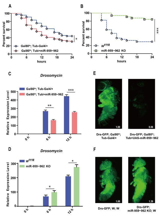
Int. J. Mol. Sci. 2021, 22, 886                                                                                            2 of 17

                                  Dorsal) to translocate to the nucleus [19–21], and activation of genes encoding potent anti-
                                  fungal and anti-bacterial peptides, such as Drosomycin [7,22,23]. In addition, in response
                                  to Gram-negative bacterial infection, the Imd pathway is activated, eventually resulting
                                  that another Drosophila NF-κB family member Relish moves from the cytoplasm to the
                                  nucleus, and the expression of antimicrobial peptide (AMP) genes, such as Diptericin [22,24].
                                  Therefore, the Toll and Imd immune pathways work together and constitute a robust
                                  defense system that protects Drosophila from invading pathogens [5].
                                        The inactivation or overactivation of the immune response could lead to the damage
                                  of the normal tissue. Therefore, the activation and termination of the Toll pathway require
                                  the cooperation of various molecules at multiple stages to establish a complete immune
                                  regulatory system. At present, kinds of modulators have been identified to be involved
                                  in Toll pathway regulation. For example, five serine proteases (ModSP, Grass, Spirit,
                                  Spheroide, and Sphinx1/2), are considered as essential for host resistance to fungal and
                                  Gram-positive infection, which play a vital role in the extracellular proteolytic cascades
                                  linking the signaling recognition proteins and Spz [25,26]. In addition, a highly conserved
                                  protein Pellino, has shown to act as a positive regulator of Toll signaling by interacting
                                  with activated Pelle kinase [27]. Furthermore, in a genome-wide RNAi screens in S2
                                  cells, G Protein-coupled receptor kinase 2 (Gprk2) was identified as a regulator of the Toll
                                  pathway [28], and the transcription factor DEAF-1 is confirmed to be required to induce Toll
                                  pathway target genes at or downstream of Dif/Dorsal [29]. Lastly, a feedback inhibitor is
                                  WntD, which reduces Toll activity by preventing translocation of Dorsal to the nucleus [30].
                                        In addition to the above-mentioned protein regulatory factors, recently, growing
                                  evidences have exhibited that miRNA controls are a critical regulator in the immune
                                  response process via Toll pathway [31]. miRNAs could fine tune gene expression in
                                  diverse cellular and biological processes, through perfect or imperfect base-pairing to the
                                  30 UTR of the target mRNAs, resulting in cleavage or degradation of the target mRNAs
                                  or suppression of their translation [32,33]. For example, the transmembrane receptor Toll
                                  protein is a crucial factor connecting extracellular and intracellular signals, and it has been
                                  reported that miR-8 [34] and miR-958 [35] can target the 30 UTR of its mRNA to negatively
                                  modulate the Toll pathway. Moreover, the nuclear translocation of the transcription factor
                                  Dif or Dorsal and its activation of AMP expression are an indispensable step of the Toll
                                  pathway response. miR-958 [35] and miR-317 [36] have been identified the direct binding
                                  with the 30 UTR of Dif-Ra/b/d and Dif-Rc transcripts, respectively, while miR-8 targets to
                                  the Dorsal mRNA [34]. Last but not least, miR-310–313 family and miR-964 could directly
                                  target to the AMP gene Drosomycin to inhibit its expression [37,38]. Although several
                                  regulators involved in Drosophila Toll-mediated immune response have been identified,
                                  the restoration mechanism of Drosophila immune homeostasis is still largely unknown and
                                  needs for further research.
                                        Especially, in our previous work, we found that the high-expression of four members
                                  of this miR-959–962 cluster could significantly down-regulate Drs expression via RNA-seq
                                  analysis and multiple genetic screening works [37]. Whether Drosophila miR-959–962 cluster
                                  members can synergistically repress Toll signaling to stop an overactive immune response,
                                  which is still not clear. In this study, we further investigated the regulatory mechanism
                                  of miR-959–962 cluster in the Drosophila immune response to bacterial infection. Each in-
                                  dividual miRNA from the miR-959–962 cluster could reduce the survival rate of flies via
                                  inhibiting the expression of AMP Drs. Bioinformatics prediction and in vitro/in vivo ex-
                                  periments verified that four miRNA members (miR-959, miR-960, miR-961, and miR-962)
                                  could negatively regulated the Toll pathway in combination via directly targeting the 30
                                  UTR of tube, dl, or Toll mRNA. In addition, the dynamic expression pattern analysis demon-
                                  strated that four miRNA members were up-regulated at the late stage of M. luteus infection,
                                  revealing their important functions in the immune homeostasis restoration of Drosophila.
                                  Overall, our results have clarified that four miRNA members from miR-959–962 cluster are
                                  novel negative regulators in Drosophila Toll-mediated immune homeostasis restoration and
                                  their aberrant expression seriously influence Drosophila antibacterial defenses.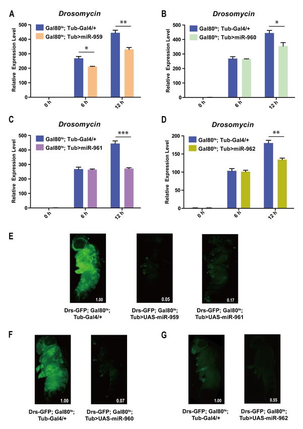
Int. J. Mol. Sci. 2021, 22, 886                                                                                             3 of 17

                                  2. Results
                                  2.1. The miR-959–962 Cluster Could Negatively Regulate Drosophila Toll-Related Immune Response
                                        In order to assess the role of miR-959–962 cluster in Drosophila immune response,
                                  we first observed whether miR-959–962 cluster dysregulation would affect the resistance
                                  of Drosophila in response to lethal Gram-positive bacterial infection, Enterococcus faecalis
                                  (E. faecalis). As shown in Figure 1, the flies transiently overexpressed miR-959–962 cluster
                                  (Gal80ts ; Tub > miR-959–962) under a temperature sensitive control and had a lower sur-
                                  vival rate than the control flies (Gal80ts ; Tub-Gal4/+) (Figure 1A). While the survival rate of
                                  the flies with miR-959–962 cluster knockout (miR-959–962 KO) was obviously increased,
                                  compared with the wild-type flies (w1118 ) (Figure 1B). We also confirmed the exact overex-
                                  pression (Figure S1) and knockout (Figure S2) of each miRNA in the corresponding flies.
                                  These results suggest that the miR-959–962 cluster could weaken Drosophila antibacterial
                                  defense, implying a role for miR-959–962 cluster members in the negative regulation of
                                  Toll signaling.

                                  Figure 1. Cont.
Int. J. Mol. Sci. 2021, 22, 886 4 of 17
Figure 1. The miR-959–962 cluster negatively regulates Drosophila Toll-related immune response.
(A) The changes of the survival rate were observed both in miR-959–962 cluster high-expressing flies
(Gal80ts ; Tub > UAS-miR-959–962) and the control (Gal80ts ; Tub-Gal4/+) flies with E. faecalis infection.
(B) The changes of the survival rate were observed both in miR-959–962 knock-out flies (miR-959–962
KO) and the control (w1118 ) flies upon E. faecalis infection. The expression levels of AMP Drs were
examined in miR-959–962 cluster high-expressing flies (C) and miR-959–962 knock-out flies (D) at 0,
6 and 12 h upon M. luteus infection. (E) The green-fluorescent in miR-959–962 cluster high-expressing
flies (Drs-GFP; Gal80ts ; Tub > UAS-miR-959–962, right) and the controls (Drs-GFP; Gal80ts ; Tub-Gal4/+,
left) carrying with Drs-GFP reporter gene were observed under fluorescent microscope following
infection with M. luteus. (F) The green-fluorescent in miR-959–962 cluster knock-out flies (Drs-GFP;
miR-959–962 KO; w, right) and the controls (Drs-GFP; w; w, left) carrying with Drs-GFP reporter gene
were observed under fluorescent microscope following infection with M. luteus. The levels of GFP
were quantified using Image J software with the default parameters and their relative level values
were marked in the bottom right corner of the image. (* p < 0.05; ** p < 0.01; *** p < 0.001).
To further confirm the effect of miR-959–962 cluster on the Toll pathway, we monitored
the mRNA expression level of the AMP Drs, as the readout of Toll pathway activation,
in the flies with miR-959–962 cluster overexpression and knockout before and after M. luteus
infection. A significant reduction of Drs level was observed in miR-959–962 cluster overex-
pressing flies at 6 h and 12 h under bacterial challenge, compared with the corresponding
control groups (Figure 1C). On the contrary, a higher expression level of Drs was detected
in miR-959–962 KO flies than in wild-type controls (Figure 1D). Likewise, taking advantage
of a Drosomycin–green fluorescent protein (GFP) reporter fly strain (Drs-GFP), we also
observed that overexpression of miR-959–962 cluster inhibited the expression of Drs in live
flies (94%) (Figure 1E), while the knock-out of miR-959–962 cluster increased the expres-
sion of Drs in live flies (39%) (Figure 1F). Our results suggest that four members of the
miR-959–962 cluster may synergistically downregulate Toll signaling response to prevent
overactivation of immune response and maintain Drosophila innate immune homeostasis.
2.2. Each Member from miR-959–962 Cluster Plays a Negative Regulatory Role in Drosophila Toll
Pathway
To further explore the role of each miRNA individual from miR-959–962 cluster in
Drosophila Toll pathway, transgenic lines high-expressing miR-959, miR-960, miR-961 or
miR-962 separately (confirmed using RT-qPCR in Figure S3) were infected with M. luteus.
The expression levels of Drs at 6 h and 12 h post-infection were also detected by RT-qPCR.
Our result revealed that the Drs mRNA levels in the flies with miR-959 high-expression
(Gal80ts ; Tub > miR-959) (Figure 2A), miR-960 high-expression (Gal80ts ; Tub > miR-960)
(Figure 2B), miR-961 high-expression (Gal80ts ; Tub > miR-961) (Figure 2C), or miR-962
high-expression (Gal80ts ; Tub > miR-962) (Figure 2D) were significantly lower than that in
the control flies (Gal80ts ; Tub-Gal4/+) post-infection, respectively. Meanwhile, the corre-Int. J. Mol. Sci. 2021, 22, 886 5 of 17
sponding flies carrying Drs-GFP reporter also suggested a decrease in the level of Drs in
live flies upon infection (95%, 93%, 83% and 45%) (Figure 2E–G). In addition, the survival
situation of individual miRNA high-expressing flies also were observed and recorded
upon E. faecalis infection. Compared with the control groups, their survival ability was
significantly reduced (Figure 3A–D). Taken together, these results indicate that each miRNA
member from miR-959–962 cluster could inhibit the expression of AMP Drs and weaken
the resistance to pathogen, to negatively fine tune Drosophila Toll signaling.
Figure 2. Each of miRNA member from miR-959–962 cluster inhibits the expression of Drs in Drosophila Toll immune
response. The expression levels of AMP Drs were examined in miR-959 high-expressing flies (Gal80ts ; Tub > UAS-miR-959)
(A), miR-960 high-expressing flies (Gal80ts ; Tub > UAS-miR-960) (B), miR-961 high-expressing flies (Gal80ts ; Tub > UAS-
miR-961) (C), and miR-962 high-expressing flies (Gal80ts ; Tub > UAS-miR-962) (D), at 0, 6, and 12 h upon M. luteus
infection. The green-fluorescent in miR-959 high-expressing flies (Drs-GFP; Gal80ts ; Tub > UAS-miR-959, middle) (E), miR-
960 high-expressing flies (Drs-GFP;Gal80ts ; Tub > UAS-miR-960, right) (F), miR-961 high-expressing flies (Drs-GFP;Gal80ts ;
Tub > UAS-miR-961, right) (E) and miR-962 high-expressing flies (Drs-GFP;Gal80ts ; Tub > UAS-miR-962, right) (G), and the
controls (Drs-GFP; Gal80ts ; Tub-Gal4/+, left) (E–G) carrying with Drs-GFP reporter gene were observed under fluorescent
microscope at 12 h upon M. luteus infection. The levels of GFP were quantified using Image J software with the default
parameters and their relative level values were marked in the bottom right corner of the image. (* p < 0.05; ** p < 0.01;
*** p < 0.001).Int. J. Mol. Sci. 2021, 22, 886 6 of 17
Figure 3. Each of miRNA member from miR-959–962 clusters influences the survival of Drosophila. The changes of the
survival rate were observed in miR-959 high-expressing flies (A), miR-960 high-expressing flies (B), miR-961 high-expressing
flies (C) and miR-962 high-expressing flies (D), as well as the control flies upon E. faecalis infection. (* p < 0.05; ** p < 0.01;
*** p < 0.001).
2.3. The Immune-Related Genes Are Potentially Targeted by miRNA Members from miR-959–962
Cluster In Vitro
In order to further determine how miR-959/960/961/962 regulates the Toll pathway,
two algorithms, TargetScan and miRanda, were used to predict the potential target genes
of miR-959, miR-960, miR-961, or miR-962. As described in the method, the intersection
of two algorithms was acquired as the potential targets. Our results showed that miR-
959 and miR-960 could bind with the 30 UTR of tube mRNA, which is the crucial and
indispensable effector molecule in the Toll pathway. Moreover, miR-961 and miR-962 had
the base complementary pairs with the 30 UTR of dl mRNA, a key transcription factor that
activate the transcription of AMP genes. In addition, miR-962 also had a binding with the
30 UTR of Toll mRNA, a transmembrane factor which transduce signals from extracellular
to intracellular. The specific base complementary binding sites are shown in Figure 4A–C.
These results suggest that miRNA members from miR-959–962 cluster may play fine-tuning
functions at different levels of Toll signals transduction.Int. J. Mol. Sci. 2021, 22, 886 7 of 17
Figure 4. The target genes of four miRNA members from miR-959–962 clusters were predicted. The potential binding sites
of miR-959, miR-960, miR-961, and miR-962 in the 30 UTR of tube (A), dl (B) and Toll (C) were present, respectively. The point
mutations (red) at the 30 UTR target sites base pairing to the seed sequence of corresponding miRNA (blue) were performed.
To evaluate the direct targeting relationship between miRNAs and targets, the 30 UTR
sequence of targets (tube, dl, and Toll) was respectively recombined to the downstream of
the luciferase encoding sequence in the pAc 5.1 insect expression vector, as shown in the
Figure 5A,D,G, and the Dual Luciferase Reporter Assay was carried out in Drosophila S2
Cell. The results showed that, compared with the pAc5.1 empty vector, both miR-959 and
miR-960 could significantly reduce the activity of the luciferase reporter containing the 30
UTR of tube (Figure 5B,C). The expression of luciferase reporter carrying with the 30 UTR
of dl could be markedly inhibited both miR-961 and miR-962 (Figure 5E,F). In addition,
miR-962 could lower the luciferase activity of Toll 30 UTR report plasmid (Figure 5H).Int. J. Mol. Sci. 2021, 22, 886 8 of 17
Figure 5. The direct bind between four miRNA members from miR-959–962 cluster and its target
genes were confirmed by Dual luciferase reporter assay in vitro. (A,D,G) The schematic diagrams
of construction of targets 30 UTR and 30 UTR mutation luciferase reporter plasmids were presented.
After co-transfected with miRNA expression plasmid, the corresponding luciferase activity of the
report plasmids without or with mutation sites was determined in Drosophila S2 cell on a Dual
luciferase assay (B,C,E,F,H). (** p < 0.01; *** p < 0.001; and ns, no significance vs. the control).
Furthermore, the target site mutation was performed in the 30 UTR of tube, dl, and Toll
as showed in the Figure 5A,D,G, in which the specific base mutation information is pre-
sented in Figure 4A–C. Dual Luciferase Reporter Assay found that the reporter activity of
tube, dl, or Toll could be restored to the normal level in these cells with co-transfected the
corresponding miRNA expression vector and 30 UTR mutant reporters of tube (Figure 5B,C),
dl (Figure 5E,F), or Toll (Figure 5H), identifying the reliability of the predicted target sites.Int. J. Mol. Sci. 2021, 22, 886 9 of 17
Taken together, our in-vitro results suggest that miR-962 could directly target the 30
UTR of Toll, miR-959, and miR-960 target the 30 UTR of tube, and miR-961 and miR-962
target the 30 UTR of dl, indicating that different miRNA members from miR-959–962 cluster
function on immunity by targeting different or identical immune-related genes.
2.4. The miR-959–962 Member Simultaneously or Serperately Target Key Components of Toll
Pathway (Tube, dl, and Toll) In Vivo
To further confirm the reliability of predicted targets of miR-959, miR-960, miR-961
or miR-962 in Drosophila, we performed RT-qPCR analysis in vivo. Our results found that,
compared with the control flies, the expression levels of tube mRNA in both miR-959 and
miR-960 high-expressing flies were significantly down-regulated upon M. luteus infection
(Figure 6A,B); Meanwhile the expression of dl mRNA in both miR-961 and miR-962 high-
expressing flies also had a lower level than the controls (Figure 6C,D); In addition, the Toll
mRNA level in miR-962 high-expressing flies was a significant reduction (Figure 6E).
These suggest the negative correlations between these four miRNAs and corresponding
targets in Drosophila.
Figure 6. Cont.Int. J. Mol. Sci. 2021, 22, 886 10 of 17
Figure 6. Four miRNA members from miR-959–962 cluster inhibit the expression of its target genes
in vivo. The expression levels of tube were respectively tested in miR-959 high-expressing (A) and
miR-960 high-expressing flies (B). (C,D) The expression levels of dl in miR-961 high-expressing and
miR-962 high-expressing flies were respectively tested. (E) The expression level of Toll in miR-962
high-expressing flies was detected. (* p < 0.05; ** p < 0.01; *** p < 0.001).
2.5. Dynamic Expression Patterns of miR-959–962 Cluster Members in Wild-Type Flies after
M. luteus or PBS Infection
To further explore the important role of this miR-959–962 cluster during Toll pathway
response, we monitored the dynamic expression patterns of Drs, miR-959, miR-960, miR-
961, and miR-962 in wild-type flies with M. luteus infection or PBS (control). Our results
found that the levels of Drs in the M. luteus infected flies were significantly higher than the
PBS-treated groups at 3, 6, 12, 24, 48 h, and peaked at 24 h after infection (Figure 7A). Sub-
sequently, we detected the expression levels of miR-959–962 cluster members, respectively.
We found that miR-959, miR-960, miR-961, and miR-962 (Figure 7B–E) were respectively
significantly increased in the late stage of M. luteus infection. Taken together, we pro-
pose that the miR-959–962 cluster could play a crucial role in restoring Drosophila Toll
immune homeostasis.
Figure 7. Cont.Int. J. Mol. Sci. 2021, 22, 886 11 of 17
Figure 7. The temporal expression patterns of four miRNAs in the wild-type flies prior to and
following M. luteus infection. The dynamic expression changes of Drs (A), miR-959 (B), miR-960
(C), miR-961 (D), miR-962 (E) at six time-points (0, 3, 6, 12, 24, and 48 h) prior to and following
M. luteus or PBS infection, respectively. (* p < 0.05; ** p < 0.01; *** p < 0.001; and ns, no significance vs.
the control).
3. Discussion
Both the deficiency and overactivation of immune response are detrimental to Drosophila.
Therefore, the persistence and intensity of the immune response needs to be strictly con-
trolled to maintain the immune homeostasis [39]. At present, increasing evidences have
demonstrated that some regulators, such as miRNAs, are involved in negatively regulating
the immune signaling to prevent the over-activation of the immune response [34,40,41].
Recently, our group has performed a genome-wide miRNA screening to identify miRNAs
regulating Drosophila Toll-mediated innate immune response, employing small-RNA seq
and transgenic UAS-miRNA library [37]. Several potential miRNAs have been screened
out, followed by in-depth exploration of their regulatory mechanism [35–37]. The current
study found that the high-expression of miR-959–962 cluster in flies suppressed antibacte-
rial defenses, evidenced by lower survival rate and a significant decrease of Drs expression
in the presence of Gram-positive bacterial challenge.
Despite some reports on the contribution of single miRNA to Drosophila innate im-
mune response have emerged, there are limited reports on how cluster of miRNAs work
together. In this study, we demonstrated that each miRNA member of miR-959–962 cluster
contributed to the suppression of antibacterial defense by targeting different components
of Toll signaling pathway in a combinatory or separate manner, such as miR-959/miR-960
targeting tube, miR-961 repressing dl, and miR-962 targeting both dl and Toll. miRNA,
perfectly complementary pairing with its target genes (Figure 4), leads to the cleavage and
degradation of target mRNA to further block the expression of its protein [42,43]. Our re-
sults find that, in the flies with miR-959, miR-960, miR-961, or miR-962 high-expression,Int. J. Mol. Sci. 2021, 22, 886 12 of 17
the corresponding target tube, dl, or Toll in mRNA level have a very significant decrease.
Therefore, although no data are available, we believe that their protein levels is also sure to
be significantly reduced.
MicroRNA clusters widely exist in metazoan genomes, employing with the diversity of
their distribution [44]. Most of clustered miRNAs are located in polycistrons and co-expressed
with adjacent miRNAs, causing the consistent expression patterns and levels [45,46]. On chro-
mosome 2, the mature miR-959–962 cluster are transcribed from an intron of CG31646 gene
and the sequence of miR-963–964 cluster are within neighboring intron in CG31646 gene.
A previous study has showed that the six miRNA members from miR-959–964 cluster are
probably encoded on a single transcription unit and showed a similar phase and ampli-
tude [47]. Moreover, it has been indicated that the miR-959–964 cluster could inhibit Drosophila
immune function against an attenuated strain of Pseudomonas aeruginosa [47]. In our study,
of note that miR-960 may execute antibacterial defense only at late 12 h stage upon infection,
while miR-959 may constantly repress the Drs expression at both 6 h and 12 h (Figure 2A,B
and Figure 3A,B). Meanwhile, miR-961 may contribute more than miR-962 to repress
antibacterial defense (Figure 2C,D; Figure 3C,D and Figure 5E,F). Therefore, we specu-
late that these miRNA of same cluster generated from the same transcripts with similar
spatial-temporal expression pattern might have varied stability of half-lives, thus play
a synergistic regulatory function on the Toll innate immunity via fine-tuning the different
layers of Toll signal transduction (Figure 8).
Remarkably, in our work, the Drs expression and survival analysis shown in Figure 1
were performed under the background of miR-959–962 cluster high-expression or knockout
flies (i.e., non-normal physiological conditions), whereas these dynamic expression patterns
of four miRNA members of miR-959–962 cluster shown in this Figure 7 were performed
under M. luteus infection and PBS treatment in the wild-type flies (i.e., normal physiological
conditions). After the high expression of miR-959–962 cluster, Drs expression was down-
regulated and the survival rate was reduced, implying that the miR-959–962 cluster played
a negative regulator role in the Toll pathway (Figure 1). Thus we suggested that under the
background of the high-expression of miR-959–962 cluster, miR-959/960/961/962 could
inhibit the expression of immune-related target genes (e.g., Toll, tube, and dl) from the
beginning of M. luteus infection, and lead to constant suppression of immune response in
Drosophila. Therefore, compared with the control group, the flies with miR-959–962 cluster
high-expression have an inadequate immune response, and its survival rate has been signif-
icantly reduced. Moreover, we analyzed the dynamic expression patterns of four miRNA
members of the miR-959–962 cluster in wild-type flies to explore the endogenous role of
miR-959/960/961/962 under normal physiological conditions, and we found that com-
pared with PBS treatment groups, all four miRNA members were significantly increased
in the late stage of M. luteus infection (Figure 7). Taken together, our results suggested
that the miR-959–962 cluster plays a negative regulatory role in the later stage of immune
response (Figure 8), i.e., in the early stage of M. luteus infection, the expression levels of
Drs keep rising, and miR-959/960/961/962 is not up-regulated for avoiding the deficiency
of immune response, but in the late stages of infection, in order to avoid the normal tissue
damage caused by over-activation of immune response, miR-959/960/961/962 serve as
negative regulators to down-regulate Drs expression to help Drosophila to restore to a new
immune homeostasis.Int. J. Mol. Sci. 2021, 22, 886 13 of 17
Figure 8. A proposed model. Our results suggested a model in which the miR-959–962 cluster
members (red) play a synergistic regulatory function on the Toll innate immunity via fine-tuning the
different layers of Toll signal transduction. MiR-959 and miR-960 target the 30 UTR of tube; miR-961
and miR-962 target the 30 UTR of dl; and miR-962 also target the 30 UTR of Toll.
In summary, our present studies have revealed the function of miR-959–962 cluster
for inhibiting AMP expression and impairing antibacterial defenses. The functions and
mechanisms of the four miRNAs from this cluster have also been identified, respectively.
Therefore, our results not only identify a new function of miR-959–962 cluster, but also
enrich the repertoire of Toll-related immune-modulating miRNA cluster in Drosophila.
4. Materials and Methods
4.1. Drosophila Stocks and Husbandry
Most flies were obtained from the Bloomington Drosophila Stock Center, includ-
ing UAS-miR-959/960/961/962 (NO.60615), UAS-miR-959 (NO.60614), UAS-miR-961
(NO.41188), miR-959/960/961/962 KO (NO.58944), except UAS-miR-960 (F001954) and
UAS-miR-962 (F001956) from FlyORF. Drosophila was raised on cornmeal-dextrose-yeast
agar medium in a light-dark (12 h cycle) incubator at 25 ◦ C and 60% humidity. To re-
strict miRNA overexpression to adulthood with tubulin-Gal80ts , the flies were reared and
assayed in either 18 ◦ C or 29 ◦ C incubator.Int. J. Mol. Sci. 2021, 22, 886 14 of 17
4.2. Adult Immune Challenge
Control and miRNA mutant adult male flies, aged 2–4 days, were challenged by
Micrococcus luteus (M. luteus), a widely used bacterial strain that activates the Toll-mediated
immune response to induce the expression of the AMP Drs. Flies were firstly incubated at
29 ◦ C for 24 h to activate the overexpression of miRNA. Septic injury was performed by
pricking the thorax of the flies with a pulled glass capillary carrying M. luteus suspension
mounted on a Nanoject apparatus (WPI, Sarasota, FL, UAS) [48], and then the flies were
harvested at specified time points after treatment for RNA extraction and RT-qPCR. For the
survival experiment, flies were infected with Gram-positive lethal bacteria, Enterococcus
faecalis (E. faecalis), and their survival situation was monitored and recorded for 24 h
post-infection [28].
4.3. Quantitative RT–PCR Analysis
Five adult flies per sample group were collected and isolated total RNA with TRIzol
Reagent (Invitrogen, Waltham, MA, USA) according to the manufacturer’s protocol. RNA
concentration and integrity was determined respectively by spectrophotometer and agarose
gel separation. cDNA was synthesized using the HiScript® II Q RT SuperMix for qPCR
(Vazyme, Nanjing, China). Then quantitative PCR analysis was performed with the
StepOnePlus Real-Time PCR System (Applied Biosystems, Foster City, CA, USA) using
AceQ® qPCR SYBR Green Master Mix (High ROX Premixed) (Vazyme, Nanjing, China).
Each experiment was performed in triplicate and the comparative cycle threshold was used
to present a fold change for each specific mRNA/miRNA after normalizing to rp49/U6
snRNA levels. All primers we used in qPCR analyses are listed in Table S1.
4.4. miRNA Targets Prediction
The mature sequences of miR-959/960/961/962 and 30 UTR sequences of all genes
in Drosophila were respectively acquired from miRBase and FlyBase database. Prediction
analysis were carried out locally through employing TargetScan [49] and miRanda [50,51]
software packages, applying their default parameters. To increase confidence and reduce
false positive of the acquired miRNA-targets, the predicted results of TargetScan and
miRanda were overlapped.
4.5. Recombinant Plasmids Generation
pAc5.1/V5-HisA insect expression vector was used for the recombinant plasmids con-
struction. To introduce exogenous miR-959, miR-960, miR-961, and miR-962 in Drosophila
S2 cells, the pre-miR-959, pre-miR-960, pre-miR-961, and pre-miR-962 sequence were am-
plified and cloned into pAc5.1/V5-HisA vector to generate pAc-miR-959, pAc-miR-960,
pAc-miR-961, and pAc-miR-962 plasmids, respectively. The luciferase coding sequence
was subcloned into pAc5.1/V5-HisA to generate pAc-luc. The 30 UTR sequence of the
tube, dl, and Toll transcript was respectively inserted to generate pAc-luc-tube 30 UTR-wt,
pAc-luc-dl 30 UTR-wt and pAc-luc-Toll 30 UTR-wt report plasmids, which were used to
express the firefly luciferase. In addition, we also constructed 5 mutants of the above three
report plasmids, respectively named pAc-luc-miR-959-tube 30 UTR-mut, pAc-luc-miR-960-
tube 30 UTR-mut, pAc-luc-miR-961-dl 30 UTR-mut, pAc-luc-miR-962-dl 30 UTR-mut, and
pAc-luc-miR-962-Toll 30 UTR-mut. In the mutant plasmids, the original binding sites of
corresponding miRNA was replaced with other bases without binding. All primers used
are listed in Table S2.
4.6. Cell Transfection and Luciferase Assays
Drosophila S2 cells were cultured in Drosophila standard medium (Gibco, Waltham,
MA, USA) with 10% fetal bovine serum (Gibco, Waltham, MA, USA) at 28 ◦ C. Before
transfection experiment, cells were firstly seeded in 24-well plates (Corning, NY, USA) and
cultured for 12 h. In each well, 385 ng miRNA expression plasmid, 100 ng pAc-luc-target 30
UTR-wt (or pAc-luc-miRNA-target 30 UTR-mut), and 15 ng pRL were co-transfected intoInt. J. Mol. Sci. 2021, 22, 886 15 of 17
cells by using X-tremeGENE HP DNA Transfection Reagent (Roche, Basel, Switzerland)
according to manufacturer's instructions. The Renilla luciferase expressed by pRL was
used as an internal reference. Dual luciferase assays were performed 48 h post-transfection
with the Dual-Glo luciferase kit (Promega, Madison, WI, USA).
4.7. Data Processing and Statistical Analysis
Results from all experiments are presented as means ± SEM of the data. Statistical
analyses were performed using two-tailed Student’s t-test, while statistical significance of
survival experiment was calculated using the log-rank test (GraphPad Prism 7.04 software).
For all statistical analysis, p < 0.05 was considered significant. All data significantly different
from control values are marked with asterisks, * p < 0.05; ** p < 0.01; *** p < 0.001; and ns,
no significance vs. the control.
Supplementary Materials: The following are available online at https://www.mdpi.com/1422-0
067/22/2/886/s1. Figure S1. The expression levels of miR-959 (A), miR-960 (B), miR-961 (C), and
miR-962 (D) in control flies and miR-959–962 cluster high-expressing fly strains were measured
before M. luteus infection. Figure S2. The expression levels of miR-959 (A), miR-960 (B), miR-961 (C),
and miR-962 (D) were measured in control flies (w1118 ) and miR-959–962 cluster knock-out fly strains
before M. luteus infection. Figure S3. The expression levels of miR-959 in miR-959 high-expressing
flies (A), miR-960 in miR-960 high-expressing flies (B), miR-961 in miR-961 high-expressing flies
(C), and miR-962 in miR-962 high-expressing flies (D) were respectively measured before M. luteus
infection. Table S1. Primers used for quantitative RT-PCR. Table S2. Primers used for transgene
vector construction.
Author Contributions: Conceptualization, R.L., P.J., and F.M.; methodology, R.L., H.Z., and F.M.;
software, X.Y. and H.Z.; validation, R.L. and X.Y.; formal analysis, R.L.; investigation, R.L. and
X.Y.; resources, P.J. and F.M.; data curation, R.L. and F.M.; writing—original draft preparation, R.L.,
P.J., and F.M.; writing—review and editing, R.L., P.J., and F.M.; visualization, R.L., P.J., and F.M.;
supervision, P.J. and F.M.; project administration, P.J. and F.M.; funding acquisition, F.M. All authors
have read and agreed to the published version of the manuscript.
Funding: This research was funded by the National Natural Science Foundation of China (No. 31970477
and No. 31572324), the Natural Science Foundation from Jiangsu Province (No. BK20191368), and
the APC was funded by the Priority Academic Program Development of Jiangsu Higher Education
Institutions.
Institutional Review Board Statement: Not applicable.
Informed Consent Statement: Not applicable.
Data Availability Statement: All relevant data are within the manuscript and its Supporting Materials.
Acknowledgments: We truly appreciate the contributions of all authors to this Original Research.
We also thank all reviewers and editors who assisted us and provided thorough comments and
invaluable suggestions. We are also grateful to the Bloomington Stock Center and FlyORF for
providing fly stocks.
Conflicts of Interest: The authors declare no conflict of interest. The funders had no role in the design
of the study; in the collection, analyses, or interpretation of data; in the writing of the manuscript,
or in the decision to publish the results.
Abbreviations
miRNA microRNA
Drs Drosomycin
dl dorsal
30 UTR 30 untranslated region
imd immune deficiency
AMP antimicrobial peptideInt. J. Mol. Sci. 2021, 22, 886 16 of 17
M. luteus Micrococcus luteus
E. faecalis Enterococcus faecalis
Drs-GFP Drosomycin–green fluorescent protein
References
1. Ayyar, K.K.; Reddy, K.V.R. MAPK and NF-kappaB signalling pathways regulate the expression of miRNA, let-7f in human
endocervical epithelial cells. J. Cell. Biochem. 2018, 119, 4751–4759. [CrossRef] [PubMed]
2. Parkin, J.; Cohen, B. An overview of the immune system. Lancet 2001, 357, 1777–1789. [CrossRef]
3. Kounatidis, I.; Ligoxygakis, P. Drosophila as a model system to unravel the layers of innate immunity to infection. Open Biol.
2012, 2, 120075. [CrossRef] [PubMed]
4. Hoffmann, J.A. The immune response of Drosophila. Nature 2003, 426, 33–38. [CrossRef] [PubMed]
5. Lemaitre, B.; Hoffmann, J. The host defense of Drosophila melanogaster. Annu. Rev. Immunol. 2007, 25, 697–743. [CrossRef]
[PubMed]
6. Hultmark, D. Drosophila immunity: Paths and patterns. Curr. Opin. Immunol. 2003, 15, 12–19. [CrossRef]
7. Valanne, S.; Wang, J.H.; Ramet, M. The Drosophila Toll signaling pathway. J. Immunol. 2011, 186, 649–656. [CrossRef]
8. Michel, T.; Reichhart, J.M.; Hoffmann, J.A.; Royet, J. Drosophila Toll is activated by Gram-positive bacteria through a circulating
peptidoglycan recognition protein. Nature 2001, 414, 756–759. [CrossRef]
9. Bischoff, V.; Vignal, C.; Boneca, I.G.; Michel, T.; Hoffmann, J.A.; Royet, J. Function of the drosophila pattern-recognition receptor
PGRP-SD in the detection of Gram-positive bacteria. Nat. Immunol. 2004, 5, 1175–1180. [CrossRef]
10. Wang, L.; Weber, A.N.; Atilano, M.L.; Filipe, S.R.; Gay, N.J.; Ligoxygakis, P. Sensing of Gram-positive bacteria in Drosophila:
GNBP1 is needed to process and present peptidoglycan to PGRP-SA. EMBO J. 2006, 25, 5005–5014. [CrossRef]
11. Gottar, M.; Gobert, V.; Matskevich, A.A.; Reichhart, J.M.; Wang, C.; Butt, T.M.; Belvin, M.; Hoffmann, J.A.; Ferrandon, D.
Dual detection of fungal infections in Drosophila via recognition of glucans and sensing of virulence factors. Cell 2006, 127,
1425–1437. [CrossRef] [PubMed]
12. Weber, A.N.; Delamasure, S.T.; Hoffmann, J.A.; Lelievre, E.; Gascan, H.; Ray, K.P.; Morse, M.A.; Imler, J.L.; Gay, N.J. Binding of the
Drosophila cytokine Spatzle to Toll is direct and establishes signaling. Nat. Immunol. 2003, 4, 794–800. [CrossRef] [PubMed]
13. Hu, X.; Yagi, Y.; Tanji, T.; Zhou, S.; Ip, Y.T. Multimerization and interaction of Toll and Spatzle in Drosophila. Proc. Natl. Acad. Sci.
USA 2004, 101, 9369–9374. [CrossRef] [PubMed]
14. Morisato, D.; Anderson, K.V. The spatzle gene encodes a component of the extracellular signaling pathway establishing the
dorsal-ventral pattern of the Drosophila embryo. Cell 1994, 76, 677–688. [CrossRef]
15. Schneider, D.S.; Jin, Y.; Morisato, D.; Anderson, K.V. A processed form of the Spatzle protein defines dorsal-ventral polarity in the
Drosophila embryo. Development 1994, 120, 1243–1250.
16. Delamasure, S.T.; Bilak, H.; Capovilla, M.; Hoffmann, J.A.; Imler, J.L. Drosophila MyD88 is required for the response to fungal
and Gram-positive bacterial infections. Nat. Immunol. 2002, 3, 91–97. [CrossRef]
17. Horng, T.; Medzhitov, R. Drosophila MyD88 is an adapter in the Toll signaling pathway. Proc. Natl. Acad. Sci. USA 2001, 98,
12654–12658. [CrossRef]
18. Xiao, T.; Towb, P.; Wasserman, S.A.; Sprang, S.R. Three-dimensional structure of a complex between the death domains of Pelle
and Tube. Cell 1999, 99, 545–555. [CrossRef]
19. Ip, Y.T.; Reach, M.; Engstrom, Y.; Kadalayil, L.; Cai, H.; Gonzalez-Crespo, S.; Tatei, K.; Levine, M. Dif, a dorsal-related gene that
mediates an immune response in Drosophila. Cell 1993, 75, 753–763. [CrossRef]
20. Lemaitre, B.; Meister, M.; Govind, S.; Georgel, P.; Steward, R.; Reichhart, J.M.; Hoffmann, J.A. Functional analysis and regulation
of nuclear import of dorsal during the immune response in Drosophila. EMBO J. 1995, 14, 536–545. [CrossRef]
21. Wu, L.P.; Anderson, K.V. Regulated nuclear import of Rel proteins in the Drosophila immune response. Nature 1998, 392, 93–97.
[CrossRef] [PubMed]
22. Lemaitre, B.; Reichhart, J.M.; Hoffmann, J.A. Drosophila host defense: Differential induction of antimicrobial peptide genes after
infection by various classes of microorganisms. Proc. Natl. Acad. Sci. USA 1997, 94, 14614–14619. [CrossRef] [PubMed]
23. Hetru, C.; Hoffmann, J.A. NF-kappaB in the immune response of Drosophila. Cold Spring Harb. Perspect. Biol. 2009, 1, a000232.
[CrossRef] [PubMed]
24. Myllymaki, H.; Valanne, S.; Ramet, M. The Drosophila imd signaling pathway. J. Immunol. 2014, 192, 3455–3462. [CrossRef]
[PubMed]
25. Kambris, Z.; Brun, S.; Jang, I.H.; Nam, H.J.; Romeo, Y.; Takahashi, K.; Lee, W.J.; Ueda, R.; Lemaitre, B. Drosophila immunity:
A large-scale in vivo RNAi screen identifies five serine proteases required for Toll activation. Curr. Biol. 2006, 16, 808–813.
[CrossRef] [PubMed]
26. Chamy, L.E.; Leclerc, V.; Caldelari, I.; Reichhart, J.M. Sensing of ‘danger signals’ and pathogen-associated molecular patterns
defines binary signaling pathways ‘upstream’ of Toll. Nat. Immunol. 2008, 9, 1165–1170. [CrossRef]
27. Haghayeghi, A.; Sarac, A.; Czerniecki, S.; Grosshans, J.; Schock, F. Pellino enhances innate immunity in Drosophila. Mech. Dev.
2010, 127, 301–307. [CrossRef]Int. J. Mol. Sci. 2021, 22, 886 17 of 17
28. Valanne, S.; Myllymaki, H.; Kallio, J.; Schmid, M.R.; Kleino, A.; Murumagi, A.; Airaksinen, L.; Kotipelto, T.; Kaustio, M.; Ulvila, J.;
et al. Genome-wide RNA interference in Drosophila cells identifies G protein-coupled receptor kinase 2 as a conserved regulator
of NF-kappaB signaling. J. Immunol. 2010, 184, 6188–6198. [CrossRef]
29. Reed, D.E.; Huang, X.M.; Wohlschlegel, J.A.; Levine, M.S.; Senger, K. DEAF-1 regulates immunity gene expression in Drosophila.
Proc. Natl. Acad. Sci. USA 2008, 105, 8351–8356. [CrossRef]
30. Gordon, M.D.; Dionne, M.S.; Schneider, D.S.; Nusse, R. WntD is a feedback inhibitor of Dorsal/NF-kappaB in Drosophila
development and immunity. Nature 2005, 437, 746–749. [CrossRef]
31. Xiao, C.; Rajewsky, K. MicroRNA control in the immune system: Basic principles. Cell 2009, 136, 26–36. [CrossRef] [PubMed]
32. Stark, A.; Brennecke, J.; Russell, R.B.; Cohen, S.M. Identification of Drosophila MicroRNA targets. PLoS Biol. 2003, 1, e60.
[CrossRef] [PubMed]
33. Ha, J.; Kim, H.; Yoon, Y.; Park, S. A method of extracting disease-related microRNAs through the propagation algorithm using
the environmental factor based global miRNA network. BioMed Mater. Eng. 2015, 26 (Suppl. 1), S1763–S1772. [CrossRef]
34. Lee, G.J.; Hyun, S. Multiple targets of the microRNA miR-8 contribute to immune homeostasis in Drosophila. Dev. Comp. Immunol.
2014, 45, 245–251. [CrossRef] [PubMed]
35. Li, S.; Li, Y.; Shen, L.; Jin, P.; Chen, L.; Ma, F. miR-958 inhibits Toll signaling and Drosomycin expression via direct targeting of Toll
and Dif in Drosophila melanogaster. Am. J. Physiol. Cell Physiol. 2017, 312, C103–C110. [CrossRef]
36. Li, R.; Huang, Y.; Zhang, Q.; Zhou, H.; Jin, P.; Ma, F. The miR-317 functions as a negative regulator of Toll immune response and
influences Drosophila survival. Dev. Comp. Immunol. 2019, 95, 19–27. [CrossRef]
37. Li, Y.; Li, S.; Li, R.; Xu, J.; Jin, P.; Chen, L.; Ma, F. Genome-wide miRNA screening reveals miR-310 family members negatively
regulate the immune response in Drosophila melanogaster via co-targeting Drosomycin. Dev. Comp. Immunol. 2017, 68, 34–45.
[CrossRef]
38. Li, S.; Xu, J.; Sun, L.; Li, R.; Jin, P.; Ma, F. Drosophila miR-964 modulates Toll signaling pathway in response to bacterial infection.
Dev. Comp. Immunol. 2017, 77, 252–258. [CrossRef]
39. Li, R.; Zhou, H.; Jia, C.; Jin, P.; Ma, F. Drosophila Myc restores immune homeostasis of Imd pathway via activating miR-277 to
inhibit imd/Tab2. PLoS Genet. 2020, 16, e1008989. [CrossRef]
40. Choi, I.K.; Hyun, S. Conserved microRNA miR-8 in fat body regulates innate immune homeostasis in Drosophila. Dev. Comp.
Immunol. 2012, 37, 50–54. [CrossRef]
41. Chen, C.Z.; Schaffert, S.; Fragoso, R.; Loh, C. Regulation of immune responses and tolerance: The microRNA perspective.
Immunol. Rev. 2013, 253, 112–128. [CrossRef] [PubMed]
42. Sun, W.; Julie Li, Y.S.; Huang, H.D.; Shyy, J.Y.; Chien, S. microRNA: A master regulator of cellular processes for bioengineering
systems. Annu. Rev. Biomed. Eng. 2010, 12, 1–27. [CrossRef] [PubMed]
43. Bartel, D.P. MicroRNAs: Genomics, biogenesis, mechanism, and function. Cell 2004, 116, 281–297. [CrossRef]
44. Zhang, Y.; Zhang, R.; Su, B. Diversity and evolution of MicroRNA gene clusters. Sci. China C Life Sci. 2009, 52, 261–266. [CrossRef]
45. Cullen, B.R. Transcription and processing of human microRNA precursors. Mol. Cell 2004, 16, 861–865. [CrossRef]
46. Baskerville, S.; Bartel, D.P. Microarray profiling of microRNAs reveals frequent coexpression with neighboring miRNAs and host
genes. RNA 2005, 11, 241–247. [CrossRef]
47. Vodala, S.; Pescatore, S.; Rodriguez, J.; Buescher, M.; Chen, Y.W.; Weng, R.; Cohen, S.M.; Rosbash, M. The oscillating miRNA
959-964 cluster impacts Drosophila feeding time and other circadian outputs. Cell Metab. 2012, 16, 601–612. [CrossRef]
48. Neyen, C.; Bretscher, A.J.; Binggeli, O.; Lemaitre, B. Methods to study Drosophila immunity. Methods 2014, 68, 116–128. [CrossRef]
49. Lewis, B.P.; Burge, C.B.; Bartel, D.P. Conserved seed pairing, often flanked by adenosines, indicates that thousands of human
genes are microRNA targets. Cell 2005, 120, 15–20. [CrossRef]
50. Betel, D.; Wilson, M.; Gabow, A.; Marks, D.S.; Sander, C. The microRNA.org resource: Targets and expression. Nucleic Acids Res.
2008, 36, D149–D153. [CrossRef]
51. Enright, A.J.; John, B.; Gaul, U.; Tuschl, T.; Sander, C.; Marks, D.S. MicroRNA targets in Drosophila. Genome Biol. 2003, 5, R1.
[CrossRef] [PubMed]You can also read