PATHOGENIC ANTIBODIES INDUCED BY SPIKE PROTEINS OF COVID-19 AND SARS-COV VIRUSES
←
→
Page content transcription
If your browser does not render page correctly, please read the page content below
Pathogenic antibodies induced by spike proteins of COVID-19 and SARS-CoV viruses Huiru Wang ( wjane60527@yahoo.com ) Huirui Biopharma, Co., Ltd. Qiuchi Chen HuaAn McAb Biotechnology, Co., Ltd. Yue Hu Biolynx Technology (Hangzhou), Co., Ltd. Xiancong Wu HuaAn McAb Biotechnology, Co., Ltd. Lin Dai HuaAn McAb Biotechnology, Co., Ltd. Yuekai Zhang Biolynx Technology (Hangzhou), Co., Ltd. Fang Li HuaAn McAb Biotechnology, Co., Ltd. Jinfeng Lu Biolynx Technology (Hangzhou), Co., Ltd. Yuxing Chen HuaAn McAb Biotechnology, Co., Ltd. Xiaoling Liu ( liuxl@wizybio.com ) HuaAn McAb Biotechnology, Co., Ltd. Research Article Keywords: COVID-19 infection, pathogenic antibodies, anti-spike antibodies, new mechanisms of pathogenesis DOI: https://doi.org/10.21203/rs.3.rs-612103/v1 License: This work is licensed under a Creative Commons Attribution 4.0 International License. Read Full License
Pathogenic antibodies induced by spike proteins of COVID-19 and SARS-CoV viruses

Huiru Wang1*, Qiuchi Chen2, Yue Hu3, Xiancong Wu2, Lin Dai2, Yuekai Zhang3, Fang Li2,

Jinfeng Lu3, and Yuxing Chen2, Xiaoling Liu2*

 1. Huirui Biopharma, Co., Ltd. Hangzhou, China

 2. HuaAn McAb Biotechnology, Co., Ltd. Hangzhou, China

 3. Biolynx Technology (Hangzhou), Co., Ltd. Hangzhou, China

 Abstract
 This study, using a virus-free mouse model, explores the pathogenic roles and novel mechanism of action of
 certain antibodies specific to the spike proteins of highly pathogenic coronaviruses such as the COVID-19 and
 the SARS-CoV viruses. These pathogenic antibodies, induced during a highly pathogenic infection such as the
 COVID-19 infection, target and bind to host vulnerable cells or tissues such as damaged lung epithelium cells,
 initiate a persistent self-attack immune response, and lead to serious conditions including ARDS, cytokine
 storms, and death. Moreover, the pathogenic antibodies may also be responsible for infection-related
 autoimmune diseases, including those experienced by COVID-19 long haulers. Furthermore, the pathogenic
 antibodies can bind to the unmatured fetal tissues and cause abortions, postpartum labors, still births, and
 neonatal deaths of pregnant females. Novel clinical interventions, through disrupting the binding of these
 pathogenic antibodies, can be developed to fight the COVID-19 pandemic. In addition, the new concept explored
 by this study may be applicable to other infectious diseases, such as the highly pathogenic influenza infections.

The pandemic of coronavirus disease 2019 (COVID-19)             cytokine storms played a critical role in the deaths of the
is a major threat to worldwide population health and            infected11,12,13, yet what initiated the cytokine storms
economies1,2. One week after onset of the COVID-19              remains a mystery. We have made the unexpected
infection, the clinical condition of the disease can            discovery in a mouse model that injection of
become severe, progressing with hypoxemia and                   anti-influenza sera into pregnant mice induced lung
dyspnea, and rapidly develop to acute respiratory               inflammation in mouse pups born to the dames14. Thus,
distress syndrome (ARDS) in 17% of patients. 65% of             we suspected that certain antibodies induced by a highly
these patients worsened and died due to multi-organ             pathogenic virus may be pathogenic themselves,
dysfunction3,4,5. Similar clinical characteristics have         through the targeting of host cells or tissues. To prove
also been observed in patients infected with other highly       this hypothesis, we investigated the pathogenic effects
pathogenic respiratory viruses such as the severe acute         of       anti-coronavirus       antibodies,      including
respiratory syndrome (SARS) virus (SARS-CoV)6, the              anti-COVID-19 viral antibodies, in this study.
middle east respiratory syndrome (MERS) virus
(MERS-CoV)7, and the avian H1N5 and H7N9                        Results
influenza viruses8,9. In some cases the viral load was          Anti-COVID-19 antibodies bound to damaged lung
low or undetectable at the time of severe illness10,            epithelium cells
suggesting that there were other causes of death than the       The coronavirus of SARS-CoV-2 is responsible for the
virus alone. It has been reported that over-reacting            COVID-19 infection. Antibodies specific to the
immune responses as well as the accompanying                    SARS-CoV-2 (COVID-19) virus and the SARS-CoV
                                                            1
Table 1. Human monoclonal antibodies isolated from patients with COVID-19 infection
MAb name Fc format Binding antigen NCBI crystal structure site Reference
B38 human IgG1 COVID-19 Spike Protein S1 https://www.ncbi.nlm.nih.gov/Structure/pdb/7BZ5 15
S309 human IgG1 COVID-19 Spike Glycoprotein https://www.ncbi.nlm.nih.gov/Structure/pdb/6WPS 19
4A8 human IgG1 COVID-19 Spike Glycoprotein https://www.ncbi.nlm.nih.gov/Structure/pdb/7C2L 16
CC12.3 human IgG1 COVID-19 Spike Protein S1 https://www.ncbi.nlm.nih.gov/Structure/pdb/6XC4 20
CR3022-b6 human IgG1 COVID-19 Spike Glycoprotein https://www.ncbi.nlm.nih.gov/Structure/pdb/7KZB 18
REGN10987 human IgG1 COVID-19 Spike Protein S1 https://www.ncbi.nlm.nih.gov/Structure/pdb/6XDG 17
REGN10933 human IgG1 COVID-19 Spike Protein S1 https://www.ncbi.nlm.nih.gov/Structure/pdb/6XDG 17
virus were tested for their binding to the human lung anti-SARS-CoV nucleocapsid (N) proteins (anti-SARS
epithelium cell line A549. Seven of the antibodies tested N). These antibodies were commercially available
were monoclonal antibodies specific to the (Bioss Antibodies) and were produced by immunization
SARS-CoV-2 spike protein. These antibodies have been of animals with related recombinant proteins of the
previously reported by others and were isolated from SARS-CoV-2 and SARS-CoV viruses. The polyclonal
the B cells of COVID-19-infected patients15,16,17,18,19,20. anti-SARS-CoV-2 nucleocapsid (N) protein
These seven monoclonal antibodies were reproduced (anti-COVID-19 N) antibodies were obtained by
according to the gene sequences encoding the immunization of rabbits with the recombinant
antibodies, from the NCBI crystal structure site (Table nucleocapsid (N) protein of SARS-CoV-2 virus
1). The other anti-coronavirus antibodies tested include (HuaAn McAb Biotechnology).
polyclonal rabbit anti-SARS-CoV-2 spike one Binding of those antibodies to healthy (intact) or
(anti-COVID-19 S1), rabbit anti-SARS-CoV spike damaged lung epithelium cells were tested with A549
glycoprotein (anti-SARS S), and monoclonal mouse cells. In order to induce damaged cells, A549 cells were
A
B C
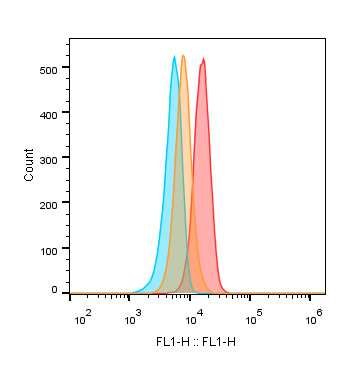
Figure 1. A flow cytometry
Blank Untreated analysis for determination of
Control MAb
Sialidase
the sialic acid levels on the
surface of human lung
epithelium A549 cells that are
either healthy (untreated) or
FL1: WGA-FITC damaged (sialidase treated)
D with missing sialic acid on the
60
3497% Intact cell surface (A); and binding
Positive cells (%)
MAb-B38
In ta c t
D am aged
Damaged of antibodies specific to the
P o s it iv e c e lls ( % )
40
spike (S) or nucleocapsid (N)
20
proteins of COVID-19 virus
0
and SARS virus, to the healthy
A n ti-S A R S -C o v 2 S
Anti-COVID-9 S1
A n ti-S A R S -C o v 2 N
Anti-COVID-9 N (intact) or damaged A549
cells (D-E). B-C: binding of
MAb-REGN10987
E
60
4813% Intact human monoclonal anti-spike
Positive cells (%)
In ta c t
D am aged
Damaged antibodies B38, REGN10987,
P o s it iv e c e lls ( % )
40
and CR3022-b6 (control
20
MAb), to healthy (B) or
damaged (C) A549 cells.
0
A n ti-S A R S S A n ti-S A R S N
Anti-SARS S Anti-SARS N
2treated with neuraminidase (sialidase) according to of SARS-CoV virus (anti-SARS S) strongly bound to
manufacturer’s instructions (Roche) 21. The fluorescent the damaged A549 cells missing sialic acid, but not to
labeled wheat germ agglutinin (WGA), which the healthy A549 cells with sialic acid (Figure 1E). In
specifically binds to N-Acetylneuraminic acid (Vector), addition, the polyclonal antibody specific to the
and a flow cytometry analysis were used to determine nucleocapsid (N) protein of COVID-19 virus
the levels of sialic acid on the surface of A549 cells. The (anti-COVID-19 N) and the antibody specific to the
damaged cells with missing sialic acid on the cell nucleocapsid protein of SARS-CoV virus (anti-SARS N)
surface were used to imitate the in vivo conditions of bound neither significantly to the healthy A549 cells nor
infected lung epithelium cells (sick cells). As shown in to the damaged cells (Figure 1D and 1E). Similar results
Figure 1A, the level of sialic acid on the surface of A549 were also observed with kidney embryonic cells of
cells treated with sialidase was lower than those of HEK-293 (data not shown).
untreated cells (Figure 1A). Taken together, the results of the in vitro assay indicated
The seven human monoclonal antibodies specific to the that certain antibodies specific to the spike proteins of
COVID-19 spike protein (Table 1) were tested for their the COVID-19 and SARS-CoV viruses have the
binding to A549 cells. As shown in Figure 1C, two out potential to mislead the immune system to attack the
of seven (28.6%) such antibodies, REGN1098717 and host by binding to sick cells such as human lung
B3815, strongly bound to the damaged A549 cells with epithelium cells in vivo. The antibody of REGN10987
missed sialic acid. REGN10987 also weakly bound to may a higher potential of activating immune responses
healthy A549 cells while B38 did not bind to the healthy in vivo since it binds not only to sick cells but also to
A549 cells (Figure 1B). The control antibody of healthy cells, albeit at a lower rate.
CR3022-b620 did not bind to the healthy A549 cells nor Anti-COVID-19 spike antibodies alone induced
the damaged cells (Figure 1B and 1C). The other four systemic inflammation and injury in vivo
human monoclonal antibodies bound neither For further confirmation, a timed-pregnant mouse
significantly to healthy A549 cells nor to damaged cells. model without viral infection was used since the surface
Further, the antibodies specific to the spike glycoprotein glycoprotein of fetal organs is not completely developed
E15 E18 E20-E21
A Figure 2. A timed-pregnant mouse
Pregnant Ab IP Ab IP Delivery
model. A: the procedure of injection of
anti-coronavirus antibodies into
Newborn mouse pups Sick newborn pups Dead fetus
pregnant mice. B: representative
images of mouse pups born to the
B dames with antibody injection. C: the
Dead
rates of sickness and death of newborn
mouse pups caused by
anti-coronavirus antibodies, and the
70
60 therapeutic effect of the antibody
50
40 mixtures of anti-S1 plus anti-N, or
C
30
REGN10987 plus CR3022-b6 and
20
10 CC12.3 (2-MAbs).
0
3or unmatured. The purified and endotoxin-free IgG of and a small hemangioma at the side of left eye of one
the anti-COVID-19 S1, anti-COVID-19 N, anti-SARS pup was observed. The pup was born to a dame injected
S, anti-SARS N, human monoclonal antibody of with the polyclonal anti-COVID-19 S1 antibody.
REGN10987 and B38 (Table 1) were used in the mouse Neither the healthy control antibodies nor the
model. The purified and endotoxin-free IgG of healthy anti-COVID-19 N nor the anti-SARS N caused
sera from rabbit, mouse, and human, as well as the significant sickness or death of the newborn mouse pups
monoclonal antibody of CR3022-b6 were used as (Table 2).
controls. Two dosages of each antibody IgG were It was surprising that when the pathogenic polyclonal
injected intraperitoneally (IP) into timed-pregnant mice anti-COVID-19 S1 antibody was mixed with an equal
twice every three days at pregnancy (embryonic) days amount of the non-pathogenic anti-COVID-19 N (50 g
E15 and E18, respectively, as described in the methods + 50 g), the sickness and death rate induced by the
(Figure 2A). antibody mixture was significantly lower than that
The frequencies of sickness and death of the fetus and induced by the anti-COVID-19 S1 alone (Table 2).
newborn mouse pups are summarized in Table 2 and Moreover, the sickness and death rate induced by the
Figure 2C. Injection of REGN10987 into pregnant mice highly pathogenic REGN10987 antibody also decreased
induced significant fetal death and neonatal death of the significantly when a mixture of the antibody and other
mouse pups delivered to those dames (Table 2). The two non-pathogenic antibodies, CR3022-b6 and
fetal death was confirmed by autopsy (Figure 2B). The CC12.3, were injected (Table 2). The mixture consisted
results with this animal model indicated that of 40 g of REGN10987, 20 g of CR3022-b6, and 20
REGN10987 has the highest potential for inducing
g of CC12.3. The data suggested that co-existing of
sickness and death (61.9%), followed by B38 (45.8%)
non-pathogenic antibodies can reduce the pathogenicity
and the polyclonal anti-COVID-19 S1 (45.5%). The
of pathogenic antibodies.
polyclonal anti-SARS S also caused significant sickness
Histology changes. The tissue sections of lungs, brains,
and death in the newborn mouse pups (37.6%). In
hearts, kidneys, intestines, and livers from the newborn
addition, hyperemia at the end of left up and down limbs
Table 2. The sick and death rates of mouse newborns born to the dames with the injection of anti-coronavirus antibodies
Injected IgG of N Sick (%) Death (%) Sick+Death (%) Odds Ratio 95% CI P value
Saline 17 0 5.88 5.88 NA NA NA
Healthy rabbit serum 6 0 16.7 16.7 3.20 0.17 - 61 1.00
Healthy mouse serum 7 0 11.1 11.1 2.67 0.14 - 50 1.00
Healthy human serum pool* 14 0 7.14 7.14 1.23 0.07 - 22 1.00
Anti-COVID-19 N 13 7.69 0 7.69 1.33 0.08 - 24 1.00
Anti-COVID-19 S1 22 27.3 18.2 45.5 13.3 1.5 - 119 0.01
Anti-COVID-19 S1 + N 17 5.88 0 5.88 0.07 0.01 - 0.7 0.01
Anti-SARS N 15 6.67 13.3 20.0 4.00 0.37 - 43 0.30
Anti-SARS S 14 31.3 6.25 37.6 12.0 1.2 - 117 0.03
MAb-CR3022-b6 9 0 11.1 11.1 2.00 0.11 - 36 1.00
MAb-B38 24 33.3 12.5 45.8 13.5 1.5 - 119 0.01
MAb-REGN10987 35 17.1 44.8 61.9 21.3 2.5 - 179 0.006
REGN10987+2MAbs** 11 0 18.2 18.2 0.17 0.03 - 0.9 0.04
*: Normal IgG pool of 4 healthy individuals without infection or vaccination of coronavirus
** MAb-CR3022-b6 and MAb-CC12.3. Fisher Exact Probability Test, two tailed
4mouse pups were stained with hematoxylin-eosin (HE) pups, as mentioned above. The histology of the kidneys
for histology evaluation. The human IgG or rabbit IgG from the mouse pups delivered to the dames with the
bound on the tissues in vivo was detected by an injection of anti-COVID-19 S1, anti-SARS S, B38, and
immunofluorescent staining with fluorescent labeled REGN10987 showed acute tubular injury. Renal tubular
anti-human IgG, or anti-rabbit IgG as secondary epithelial cells showed granular or vacuolar
antibodies. degeneration, dilated or obstructed lumen, renal
Lung inflammation and injury. Acute lung interstitial edema with a small amount of inflammatory
inflammation was observed with the HE stained tissue cells infiltration, and some of the epithelial cells fell off
sections from the mouse pups born to the dames injected (Figures 3 and S2). The kidney injury caused by
with anti-COVID-19 S1, anti-SARS S, REGN10987, REGN10987 was the most significant (Figure 3).
and B38 (Figure 3). The lung lesion included pulmonary Furthermore, small amounts of cerebral hemorrhage or
congestion, alveolar epithelial hyperplasia and inflammatory cell infiltration was observed in the brains
thickening, hemorrhage, alveolar atresia, alveolar of mouse pups delivered to a dame injected with
dilatation, and alveolar fusion (Figure 3 and Figure S1). antibodies of anti-COVID-19 S1, anti-SARS S, B38,
Infiltration of inflammatory cells at the local lesion and REGN10987 (Figure 3 and S2). Additionally,
areas were also observed. There were insignificant or myocardial hemorrhage was observed in the hearts of
minor histological changes with the lungs from the pups the mouse pups delivered to the dames injected with
born to the dames injected with the antibodies of anti-COVID-19 S1, anti-SARS S, and B38 (Figure 3
anti-COVID-19 N, anti-SARS N, CR3022-b6, and the and S2). Lastly, myocardial swelling and inflammatory
control IgGs of human, rabbit, and mouse (Figure 3). cell infiltration were observed in a mouse pup delivered
Other organ inflammation and injury. Inflammatory to a dame injected with antibodies of B38 (Figure 3).
reactions and hemorrhage were also observed with the There were insignificant or minor histological changes
tissues of kidneys, brains, and hearts from the mouse with the tissues from the pups born to the dames
Rabbit IgG Anti-COVID-19 S1 Anti-COVID-19 N Anti-COVID-19 S1+N Anti-SARS S Anti-SARS N
Vehicle Human IgG Control MAb MAb-B38 MAb-REGN10987 REGN10987+2 MAbs
Healthy Kidney REGN10987 Kidney Healthy Brain B38 Brain Healthy Heart B38 Heart
Figure 3. The representative images of the histological changes of lungs (top 2 rows), kidneys, brains, and hearts (bottom
row) from the newborn mouse pups delivered to the dames injected with anti-coronavirus antibodies, and control antibodies
of human IgG, rabbit IgG and human monoclonal antibody (MAb) of CR3022-b6 (Control MAb); or the dames treated with
antibody mixtures. 2MAbs: human monoclonal antibodies of CR3022-b6 and CC12.3.
5injected with the antibodies of anti-COVID-19 N, by the anti-COVID-19 S1 alone (Figure 4). In addition,
anti-SARS N, CR3022-b6, and the control IgGs of the treatment with the antibody mixture comprised of
human, rabbit, and mouse. the pathogenic REGN10987 and two non-pathogenic
Taken together, the in vivo results of the virus-free antibodies of CR3022-b6 and CC12.3 also significantly
animal model proved that certain antibodies specific to reduced the level of MCP-1 (P < 0.001) compared to
the spike proteins of the coronaviruses such as that induced by REGN10987 alone (Figure 4). The
COVID-19 and SARS-CoV viruses can induce levels of other cytokines were not significantly
significant fetal and neonatal deaths and the systemic elevated, probably due to the undeveloped immunity of
inflammation or injury of lung and other organs in vivo. the newborn mouse pups. The results were consistent
The results are consistent with the clinical observations with the results of the sickness and death rates (Table 2)
of COVID-19 patients with severe illnesses5. Therefore, and the histology changes (Figure 3). The data
antibodies of anti-COVID-19 S1, anti-SARS-CoV S, demonstrated that 1) pathogenic antibodies alone can
B38, and REGN10987 are pathogenic and probably induce high levels of inflammatory cytokines and have
responsible for the serious conditions of a severe the potential to induce a cytokine storm or cytokine
COVID-19 infection. On the other hand, the antibodies release syndrome (CRS); and 2) co-existence of
of anti-COVID-19 N, anti-SARS-CoV N, and non-pathogenic antibodies can reduce the inflammatory
CR3022-b6 are non-pathogenic since they did not cytokine release induced by pathogenic antibodies and
induce significant adverse reactions in vivo. prevent the possible cytokine storm or CRS caused by
Inflammatory cytokines the pathogenic antibodies.
As further evidence of the pathogenicity of the anti- In vivo antibody binding to multiple organs of mouse
spike antibodies, the cytokine levels of monocyte pups
chemotactic protein 1 (MCP-1), tumor necrosis As more evidence of the pathogenicity of the
factor- (TNF-), interleukin-4 (IL-4), IL-6, and IL-10 pathogenic anti-COVID-19 spike antibodies, in vivo
in the sera of mouse pups were tested by a multiplex antibody binding to tissues of mouse pups was detected
Luminex assay kit (Millipore) according to using an immunofluorescent staining as described in
manufacturer’s instructions. The results of MCP-1 are methods. The human and rabbit anti-COVID-19 spike
summarized in Figure 4. antibodies were significantly detectable at the
Figure 4. The cytokine levels of MCP-1
in mouse sera from the newborn mouse
pups born to the dames with antibody
injection alone of the anti-coronavirus
antibodies or the dames treated with
antibody mixtures.
Both the pathogenic anti-COVID-19 S1 and inflammatory and lesion areas of the tissues of lungs,
REGN10987 induced significantly higher levels of kidneys, brains, hearts, livers, and intestines from the
MCP-1 (Figure 4). Consistent with the surprising results mouse pups with severe sickness (Figure 5). Those
of the histological changes, the treatment using the mouse pups were delivered to the dames injected with
antibody mixture comprised of the pathogenic the pathogenic antibodies of anti-COVID-19 S1,
anti-COVID-19 S1 and the non-pathogenic anti-SARS-CoV S, REGN10987, and B38 (Figure 5).
anti-COVID-19 N, significantly reduced the cytokine Neither human IgG nor rabbit IgG was significantly
levels of MCP-1 (P < 0.001) compared to that induced detected on the tissues of the pups treated with the
6A
Vehicle
Rabbit-IgG Lung Heart Kidney Brain Liver Intestine
Figure 5. Detection
Anti-Cov19 S1 Anti-SARS S
of anti-coronavirus
antibodies bound in
vivo to the lesion
areas of multiple
organs of the mouse
pups born to the
dames with antibody
B Lung Kidney Heart Brain Liver Intestine injection at E15 and
E18.
CR3022-b6
B38
REGN10987
non-pathogenic antibodies of anti-COVID-19 N, Figure 6. REGN10987 bound to mutiple human fetal
anti-SARS N, and CR3022-b6 (Figure 5). The results tissues of the lungs, heart, kidneys, brain, pancreas,
indicated that certain anti-COVID-19 spike antibodies liver, thymus, and testicles, but not of the esophagus
went through the placenta, bound to the fetal tissues, (Figure 6A). In addition, the REGN10987 also bound to
activated the self-attack immune responses, and led to the fetal tissues of retina and coroid, sclera, and eye ball
the systematic inflammation and injuries of multiple (data not shown). The data indicate that the unmatured
organs such as the lungs, kidneys, and brain. The results fetal tissues are vulnerable to a pathogenic antibody
are consistent with those of histological changes such as REGN10987. In addition, REGN10987 bound
(Figure3) and provided the in vivo evidence for the broadly to the human inflammatory tissues or cancer
pathogenicity of the anti-spike antibodies of COVID-19 tissues of the respiratory, cardiovascular, urinary, and
virus and SARS-CoV virus. digestive systems (Figure 6B-6C). The inflammatory
Binding of pathogenic antibodies to fetal and diseased tissues were from patients suffering from pneumonia,
tissues bronchitis, bronchiectasis, valvular disease, rheumatoid
In order to further evaluate the pathogenicity of the valvular disease, myocarditis, esophagitis,
anti-COVID-19 spike antibodies in humans, the gastritis, collitis, appendicitis, pancreatitis, and
REGN10987 with the highest pathogenic potential was hepatitis. The cancer tissues were from patients with
tested for antibody binding to various human fetal small cell lung carcinoma, kidney clear cell carcinoma,
tissues, or mutiple human diseased tissues from tissue and myxoma. The results indicate that most of the
array slides (US Biomax). The results are shown in actively proliferating cells or tissues such as
7inflammatory tissues or cancer tissues are vulnerable to slide (US Biomax, FDA999w) were used to further
a pathogenic anti-COVID-19 S1 antibody such as evaluate the pathogenicity of REGN10987. The tissue
REGN10987. array slide was comprised of 32 types of normal human
Binding of pathogenic antibodies to human healthy organs encompassing most of human tissues,
tissues representing FDA guidelines for antibody
As more evidence of the pathogenicity of the cross-reactivity testing. The results showed that
pathogenic anti-COVID-19 S1 antibodies, REGN10987 REGN10987 bound significantly to human healthy
and mutiple healthy human tissues from a tissue array tissues of lung, kidney, pancreas, stomach, intestine,
A Lung Kidney Heart B Healthy Pneumonia Bronchitis SCLC
Lung
Pancreas Liver Brain Healthy inflammation inflammation CC carcinoma
Kidney
Esophagus Thymus Testicle Healthy Valvular disease Myocarditis Myxoma
Heart
C Esophagus Stomach Colon Appendix Pancreas Liver
Figure 6. Binding of the human
Healthy
monoclonal anti-COVID-19 S1
antibody, REGN10987, to various
human fetal tissues (A), and
Inflammation
inflammatory and cancer tissues
(B-C). SCLC: small cell lung
carcinoma, CC: kidney clear cell.
Esophagitis Gastritis Colitis Appendicitis Pancreatitis Hepatitis
Lung Kidney Kidney Stomach Small intestine
Figure 7. Binding
of the REGN10987
Pancreas Adrenal gland Peripheral nerve Thyroid gland
to various normal
Spleen
human tissues.
Adenohypophysis Testis Prostate Uterine cervix Bone marrow
8adrenal gland, peripheral nerve, thyroid gland, spleen, anti-COVID-19 spike antibodies inducible by the
adenohypophysis, testicle, prostate, bone marrow, COVID-19 virus are non-pathogenic since the
uterine cervix of cancer adjancent normal tissue (Figure pathogenic antibodies consist of less than 30% of the
7). In addition, REGN10987 also bound to the tissues of total number of antibodies, according to the data of
parathyroid gland, pericardial mesothelium, and monoclonal antibodies in this study.
adjacent normal sclera of eye (data not shown). The data
indicate that certain anti-COVID-19 S1 antibodies, such Discussion
as REGN10987, are highly pathogenic because it has The current study revealed the pathogenic roles and the
the high potential to bind to healthy human tissues, detailed mechanism of action (MOA) of certain
activating self-attacking immune responses and antibodies specific to the spike proteins of
inducing serious adverse reactions in vivo. Based on the coronaviruses such as the COVID-19 virus and the
results, clinical detection of pathogenic antibodies SARS-CoV virus (Figure 8). We had discovered that in
during the COVID-19 infection may be helpful in a mouse model, pre-injection of anti-influenza immune
predicting the consequences of a patient with a serious sera induced more severe infections than the mice
infection. infected with an influenza virus alone14. Wang and
Taken together, the in vitro and in vivo data of the co-workers reported that anti-SARS-CoV spike antisera
current study revealed that certain pathogenic promoted SARS infection through antibody-dependent
antibodies specific to the COVID-19 spike protein can enhancement (ADE) in vitro22. Liu and co-workers
be the cause of a serious COVID-19 infection, and can reported that anti-SARS-CoV spike immune sera
cause serious complications from COVID-19 induced by a SARS-CoV vaccine caused acute lung
infections. Further, the pathogenic antibodies can bind injury by promoting MCP1 and IL-8 production and
to unmatured fetal cells or tissues and cause abortions, monocyte or macrophage recruitment and accumulation
postpartum labors, still births, and neonatal deaths of in SARS-CoV infected macaque models23. The
pregnant females. previously reported mechanism of action (MOA) of
It should be noted that the majority (70% or more) of the these anti-spike antibodies is ADE-based, in that the
Anti-SARS-CoV-2 B cells
antibodies NK cell Neutrophil Macrophage Monocyte Cytokines
T cells
SARS-CoV-2
virus
Healthy Sick wk 1 Sick
wk 2
Lung epithelium cells Ab binding • Cell death & proliferation, more Ab
Misleading binding
• Activating more immune cells
• Releasing more cytokines
Figure 8. Mechanism of action (MOA) of pathogenic anti-spike antibodies of COVID-19 virus.
9antibodies enhance viral infectivity24. The current study, binding could activate and mislead the immune
revealed for the first time, the self-cell targeting MOA response to attack self and induce the injury of multiple
of pathogenic antibodies, in which the antibodies bind organs in vivo. For example, injection of the antibodies
to host vulnerable cells or tissues and mislead immune specific to the COVID-19 S1 or the SARS-CoV S
responses to attack host-self. Our study also explored a proteins to pregnant mice induced fetal and neonatal
new mechanism of pathogenisis (MOP) of highly deaths and the injury of multiple organs of mouse pups
pathogenic viral infections. The MOP is caused by the born to the dames, as shown in Figures 1-4 and Table 2.
pathogenic antibodies inducible by highly pathogenic In contrast, the non-pathogenic antibodies of
viruses such as the COVID-19 virus and SARS-CoV anti-COVID-19 N and anti-SARS N did not induce
virus. Moreover, the pathogenic antibodies may be significant injury in vivo. Neither the anti-COVID-19 N
related to the cause of infection-related autoimmune nor the anti-SARS N antibodies significantly bound to
diseases, including those in COVID-19 long haulers. the healthy A549 cells nor to the damaged A549 cells
Dualistic roles of anti-viral antibodies (Figure 1E). The fetal model was selected because many
Based on the traditional concept, the antibodies induced fetal tissues, including the surface glycoproteins, are
by an infectious pathogen are protective to a host unmatured and vulnerable to the pathogenic antibodies.
because they can neutralize the pathogen and prevent or Seven naturally occurring human monoclonal
treat the infectious disease. Nevertheless, the roles of antibodies specific to the COVID-19 S1 (S-RBD)
such antibodies can be dualistic. Not wishing to be protein15,16,17,18,19,20 were analyzed with the in vitro assay
bound to theory, we noticed that some antibodies can as described above. The results showed that all
cross react to host cells or tissues and trigger immune antibodies except one (85.7%) did not bind to the
reactions to attack the self-cells and self-tissues. The healthy A549 cells. Neverthless, two (28.6%)
data of the current study showed such pathogenic antibodies of B38 and REGN10987 significantly bound
actions of anti-spike proteins of the COVID-19 and to the damaged A549 cells (Figure 1C) and were
SARS viruses. selected as potential pathogenic antibodies for further
Sialic acids are predominant components of the mucous confirmation by the in vivo animal model. The results of
membrane at the outer surface of cell membranes and the virus-free animal experiments showed that the two
mainly act as biological masks or receptors25. Cells or antibodies alone induced significant fetal and neonatal
tissues with sialic acid are recognized as “self” 25. After deaths and the injury of multiple organs of mouse pups
the loss of sialic acids the cellular structures become born to the dames, as shown in Figures 1-4 and Table 2.
“non-self” 25, which can activate immune responses. In contrast, another antibody of the seven monoclonal
Sialic acids are also an important attachment molecule anti-COVID-19 S1 antibodies, CR3022-b6, served as a
of receptors for some viruses, such as coronaviruses and control antibody and did not induce significant injury in
influenza viruses25,21. During an infection of such vivo (Figures 1-4 and Table 2). The data indicated that
viruses, the sialic acid on the infected cells such as lung the in vitro assay is useful for rapid screening of
epithelium cells could be removed or destroyed by the potential pathogenic antibodies, and the virus-free
receptor destroying enzyme (RDE) of a coronavirus animal model is helpful for confirmation of
such as the COVID-19 virus, or the sialidase of pathogenicity of pathogenic antibodies. It should be
influenza viruses. The damaged cells with missing sialic noted that the effect of a pathogenic antibody on the
acid on the cell surface become vulnerable to the mouse fetus could be reduced if the antibody binds to
pathogenic antibodies induced by the virus (Figure 8). the mother’s tissues.
For example, the current study showed that antibodies A pathogenic antibody can be induced during a highly
specific to the COVID-19 spike and the spike pathogenic infection, such as the COVID-19 infection.
glycoprotein of the SARS-CoV virus could The discovery of pathogenic antibodies may solve the
significantly bind to the damaged lung epithelium A549 mystery of the possible MOP of serious infectious
cells (Figure 1) and kidney embryonic HEK-293 cells diseases, serious complications, and sequela of a viral
with missed sialic acid on the cell surface. The antibody infection, particularly of a highly pathogenic viral
10infection such as the COVID-19 infection. This may caused by pathogenic antibodies can lead to serious
also explain the cause of cytokine storms and cytokine conditions such as ARDS, cytokine storm, and death.
release syndrome (CRS), and infection-related The new MOP can explain why the majority of patients
autoimmune diseases (including those suffered by with serious respiratory viral infections such as a
COVID-19 long haulers26), infection- relating cancers, COVID-19 infection or a highly pathogenic influenza
and other possible disorders inducible by pathogenic infection died after week one, especially at weeks 2-3 of
antibodies. The diseases or conditions caused by the infection course3,27, as that matches the time period
pathogenic antibodies further include abortion, of antibody peak levels. The new MOP can also explain
postpartum labor, still birth and neonatal death of why the majority of severe or lethal infections of the
pregnant females related to an infection. 1918 influenza pandemic happened to the young27,
A new pathogenic mechanism of viral infections because younger people could induce higher levels of
The in vitro and in vivo data of the current study support anti-viral antibodies, including pathogenic antibodies.
a new mechanisms of pathogenesis (MOP) of highly Similarly, certain pathogenic antibodies inducible by
pathogenic viruses such as the COVID-19 virus. The other infectious pathogens, such as influenza viruses
MOP includes: 1) a highly pathogenic respiratory virus may also cause serious adverse reactions or
such as the COVID-19 virus causes the initial, primary autoimmune diseases through this MOP (study
injury such as local inflammation and cellular damage ongoing).
with missed sialic acids of its target organ, such as Cells or tissues vulnerable to pathogenic antibodies
lungs, typically within week one of the infection; 2) As shown in Figure 6, the highly pathogenic
anti-viral antibodies, including pathogenic antibodies, REGN10987 antibody bound to the majority of human
elevate from week one. The pathogenic antibodies bind fetal tissues and inflamatory tissues, as well as certain
to the damaged or the inflammatory cells of the target cancer tissues. A common feature of those tissues,
organ (e.g., lungs) (Figure 8) as well as to other organs including the cancer tissues, is that they consist of
with similar injuries (e.g. kidney, brain and heart); 3) actively proliferating cells. Therefore, those cells and
the antibody binding activates and misleads the immune inflamatory tissues are vulnerable to pathogenic
response to attack the self cells or tissues, and induces antibodies, and their interaction with the pathogenic
further damage (the secondary injury); 4) the secondary antibodies can be the cause of 1) serious infections,
injury persistently adds further damage to the primary particularly highly pathogenic viral infections such as
injury, creating a snowball effect, and causes serious the COVID-19 infection; 2) serious complications of
conditions such as ARDS, cytokine storms, and even infections such as ARDS, cytokine storm, or cytokine
death as the antibodies reach peak levels from week one release syndrome (CRS); and 3) infection-relating
to weeks 2-3 (Figure 8); and 5) the self-attacking inflammation such as pneumonia and kidney failure
immune responses misled by the pathogenic antibodies (Figures 1-4). Further, unmatured fetal cells or tissues
can be persistent, and can accumulate after viral are vulnerable to pathogenic antibodies (Figure 6A).
clearance and cause autoimmune diseases as long as the Thus, pathogenic antibodies may be the cause of
antibodies continue to exist. abortions, postpartum labors, still births and neonatal
The primary injury is limited, short, and decreases as the deaths of pregnant females (Figure 1B and 1C).
virus is cleared, as seen in regular influenza infections. Pathogenic antibodies and autoimmune diseases
That means the virus itself is not sufficient to cause Many autoimmune diseases are related to viral
serious conditions such as ARDS, cytokine storms, and infections28, yet the pathogenic mechanisms have
death. On the other hand, the secondary injury caused remained unclear so far. The current study discloses, for
by the pathogenic antibodies is longer, broader, and the first time, that most inflammatory tissues are
additive because antibodies persist much longer than vulnerable to pathogenic antibodies (Figure 6B).
viruses and can bind nonspecifically to other Chronic inflammation is a feature of most autoimmune
inflammatory tissues or organs besides lungs. Thus, the diseases. When an infection occurrs, antibodies
superposition of the self-attack immune reactions including pathogenic antibodies are induced and last
11from months to half a year. The pathogenic antibodies antibody alone (Table 2 and Figure 1C). A similar result
can readily bind to the pre-existing vulnerable was observed with the highly pathogenic REGN10987
inflammatory tissues of an autoimmune disease, and as well with an antibody mixture of the REGN10987
mislead immune responses to attack the body’s own and the two non-pathogenic monoclonal antibodies of
tissues. This process can be repetitive and additive as CR3022-b6 and CC12.3 (Table 2 and Figure 1C). The
antibodies elevate and induces significant reactions. data suggested that co-existence of non-pathogenic
The occurrence time and pathogenic course of an antibodies can reduce the pathogenicity of pathogenic
autoimmune disease caused by pathogenic antibodies antibodies. Thus, a vaccine capable of inducing
can be short or long, depending on the amount of multivalent antibodies may be safer, in which at least
vulnerable cells or tissues and the amount of pathogenic one kind of antibody is of the non-pathogenic kind that
antibodies. The pathogenic antibodies generally persist induce less adverse reactions. One example of such a
for months to half a year, and thus the autoimmune vaccine is the traditional inactivated viral vaccine (e.g.
disease can occur during that period, especially during inactivated COVID-19 vaccine) which induces
the period of antibody peak levels. multivalent antibodies specific for multiple antigens of
COVID-19 long haulers have been reported and the a virus. As another example, a recombinant or mRNA
causes remain a mystery26. The current study provides a COVID-19 vaccine capable of inducing the antibodies
possible pathogenic mechanism of COVID-19 long specific to not only the spike protein but also to the
haulers. Despite the virus being cleared in the recovered nucleocapsid proteins, or a non-spike protein of the
COVID-19 patients, the anti-viral antibodies remained SARS-CoV-2 virus may be safer (patent pending).
and could exist for months to half a year or longer. Despite the mechanism of action being unclear, it is not
Certain pathogenic antibodies could bind to vulnerable likely that the non-pathogenic antibodies affected the
cells or tissues, such as inflammatory cells induced actions of the pathogenic antibodies through
during the COVID-19 infection, and cause persistent competitive binding since neither the anti-COVID-19
adverse reactions such as chronic inflammation of S1 nor the anti-COVID-19 N bind to the same antigens.
multiple organs. Thus, with the same MOA, pathogenic Neither the REGN10987 nor the other two
antibodies can also be responsible for the longer term non-pathogenic antibodies bind to the same epitope of
effects of the COVID-19 infection. For example, a the S1 protein of the COVID-19 virus. We hypothesize
highly pathogenic anti-spike antibody such as that non-pathogenic antibodies affect the action of
REGN10987 can bind to peripheral nerves and may pathogenic antibodies through diluting and interrupting
cause abnormalities of vision and taste. Similarly, the the binding of pathogenic antibodies to vulnerable cells
pathogenic antibodies inducible by other infectious or tissues.
pathogens such as an avian influenza virus can be Novel clinic interventions based on the new
responsible for the adverse reactions or autoimmune mechanism of pathogenesis
diseases caused by the infectious. Additionally, in a Based on the new MOP of the highly pathogenic viral
chronic viral infection (e.g., an HIV infection), infection inducible by pathogenic antibodies, novel
pathogenic antibodies can repeatedly stimulate cellular clinic interventions for treating and preventing a serious
proliferation for a long time and an infection-related condition of the COVID-19 infection may be developed
cancer can occur if the cellular proliferation loses through interrupting the binding of the pathogenic
control. antibodies to host vulnerable cells, tissues, and organs.
Better vaccines For example, the following products and approaches
It was surprising that when the pathogenic may be effective for the treatment of a serious
anti-COVID-19 S1 antibodies was mixed with an equal COVID-19 infection: 1) for patients with serious
amount of the non-pathogenic anti-COVID-19 N conditions, therapies capable of removing the
antibodies, the sickness and death rates caused by the pathogenic antibodies, such as replacing the patient’s
antibody mixture was significantly decreased compared plasma with uninfected healthy human plasma, may be
to the results of the injected anti-COVID-19 S1 effective and should be performed as soon as possible; 2)
12immunoglobulin products, serum or plasma from not (RBD) of the spike protein one (S1) of the
only recovered patients but also from healthy SARS-CoV-2 virus have been reported by
individuals may be helpful for symptom relief through others15,16,17,18,19,20. The seven monoclonal antibodies
diluting and interrupting the binding of pathogenic were reproduced for research use only (HuaAn McAb
antibodies; and 3) pathogen-derived products such as Biotechnology) according to the gene sequences from
viral antigens or antigen fragments, and synthetic the NCBI crystal structure site (Table 1). The features
peptides may be effective (neutralizing pathogenic and information of the antibodies are listed in Table 1.
antibodies). Paired heavy chain and light chain plasmids of
For the diagnosis of a severe COVID-19 infection, anti-Spike human antibody were transiently transfected
determination of the levels of anti-COVID-19 spike into HEK-293FE cells at a mass ratio of 1:2 with
antibodies may be important and may helpful to predict polyetherimide (Polysciences). Transfected cells were
the condition of a patient with a serious infection. This suspension-cultured in a 5% CO2 containing
is because the virus can become undetectable after one atmosphere at 37°C and the condition medium (Gibco)
week, but the antibodies induced by the virus can was refreshed 8 hours after transfection. After 7 days,
elevate from week one and reach peak levels at weeks the supernatants were centrifuged at 10000r per min
2-3, accompanied by the development of serious (rpm) and purified with Protein A prepacked column
conditions such as ARDS, cytokine storms, and death. (Senhui Microsphere Technology). Purified antibodies
Higher levels of the anti-COVID-19 spike antibodies were quantified by Nanodrop or BCA Quantification
may indicate a worsening of the infection. Kit (Beyotime).
Antibody quality control was performed using an
In summary, our study revealed the roles and a novel antigen down ELISA. 1 g/ml antigens of COVID-19
MOA of “pathogenic antibodies” in viral infections. In Spike-RBD-mFc (HuaAn McAb Biotechnology) were
particularly, we explored pathogenic antibodies of coated on ELISA plate at 4℃ for overnight. The plate
highly pathogenic respiratory viral infections, such as was blocked with 1% BSA and coated with anti-spike
COVID-19. The pathogenic antibodies can be induced human antibody as first antibody for 1 hour at 37℃
during an infection, bind to vulnerable cells or tissues successively. Uncombined free antibodies were washed
such as actively growing cells, and initiate a persistent with TBST, the secondary anti-human IgG-HRP
self-attack immune response, leading to serious (Abcam) were added onto the plate, and the plate was
conditions including ARDS, cytokine storms, and death. incubated at 37℃ for 30 minutes. Binding affinity of the
Further, the pathogenic antibodies may also be anti-spike human antibodies were analyzed with the
responsible for infection-related autoimmune diseases, signature of OD450 nm absorption.
including those experienced by COVID-19 long Anti-COVID19 N protein antibody production
haulers. Novel clinical interventions for diagnosis and Two months old New Zealand white rabbits were
treatment, including improved vaccines based on the subcutaneously immunized with 250 μg of purified
new pathogenic mechanisms caused by pathogenic COVID19-N protein ((HuaAn McAb Biotechnology).
antibodies, can be developed. In addition, the concept The rabbits received 3 booster injections at 2-week
and new pathogenic mechanisms explored by this study intervals with 500 g purified COVID19-N protein.
may also be applicable to other infectious diseases, such Serum titer of anti-COVID19-N antibodies were
as highly pathogenic influenza infections (study validated by an antigen down ELISA after each
ongoing). immunization. Rabbit serum were then harvested,
centrifuged at 10,000 rpm for 10 minutes and purified
Methods
with Protein A prepacked column (Senhui Microsphere
Production of human anti-SARC-CoV-2 spike
Technology). Purified antibodies were quantified by
monoclonal antibodies
Nanodrop or BCA Quantification Kit (Beyotime).
Seven naturally occurring human monoclonal
Removal of endotoxin from antibody solutions
antibodies specific to the receptor binding domain
13Antibody solutions were treated with 1% v/v Triton wheat germ agglutinin (WGA), which specifically binds
X-114 in ice for 10 minutes. The solutions were then to sialic acid (Vector), was used as a control (Figure
incubated at 37°C for 5 minutes, whereupon two phases 1A).
formed. The antibody solutions were centrifuged at A timed-pregnant mouse model
12,000 rpm for 5 minutes at room temperature, and A timed-pregnant mouse model without viral infection
carefully aspirate the upper aqueous phase. was developed using the anti-coronavirus antibodies as
Endotoxin-free antibodies were quantified by BCA mentioned above, to evaluate the pathogenic action of
Quantification Kit (Beyotime). anti-coronavirus antibodies. The animal experiments
Other antibodies were performed at the Center of Laboratory Animals of
The rabbit polyclonal antibodies specific for the Hangzhou Normal University. The protocol for the
recombinant spike one (S1) proteins of the animal experiment was approved by the Laboratory
SARS-CoV-2 virus, the recombinant spike proteins of Animal Welfare and Ethics Committee of Hangzhou
SARS-CoV virus, and the mouse monoclonal antibody Normal University. The CALAS No. is 2020244. The
specific for the recombinant nucleocapsid (N) proteins animal experiments were performed three times.
of the SARS-CoV virus were purchased from Bioss Animal SPF-grade C57BL/6J pregnant mice at
Antibodies (Beijing). pregnancy (embryonic) day E13-E14 were purchased
Digestion of A549 cells with neuraminidase from Shanghai SLAC Laboratory Animal Co., Ltd. The
The cells of human lung epithelium cell line A549 were animals were randomly divided into groups as needed,
washed once with the digestion buffer for two pregnant mice for each group at every experiment.
neuraminidase (Roche), the cells were centrifuged at Antibody injection The purified and endotoxin-free
2000 rpm for 5 minutes, and the supernatant was IgG of the anti-coronavirus antibodies used for the
discarded. The digestion buffer was consisted of 50 mM animal model include rabbit polyclonal anti-COVID-19
of sodium acetate, 3% BSA, and 2 mM of CaCl2 in PBS S1, anti-COVID-19 N, anti-SARS-CoV S, mouse
(pH5.5-6.0). The cells were resuspended with the monoclonal anti- SARS-CoV N, and the human
digestion buffer and divided to multiple tubes, each tube monoclonal anti-COVID-19 S1 or S-RBD antibodies of
contained about 106 of cells in 200 l of the digestion B3815, REGN1098717, CC12.318, and CR3022-B620.
buffer containing 50 U of neuraminidase (Roche). The Two dosages of each antibody IgG were injected
tubes were incubated at 37°C for one hour. Then the intraperitoneally (IP) into timed-pregnant mice twice
cells were washed with PBS once and proceeded for every three days at pregnancy (embryonic) day of E15
flow cytometry assay. A549 cells without the treatment (about 26-28 g) and E18 (about 30-32 g) respectively
of neuraminidase were used as controls. (Figure 2A). For each polyclonal antibody, 50 μg
Flow cytometry (microgram) for the first dose (about 2.0 mg/kg) and 60
A549 cell with or without the treatment of μg for the second dose (about 2.0 mg/kg) were
neuraminidase were tested for the binding of administrated. For each monoclonal antibody, 40 μg for
anti-coronavirus antibodies as mentioned above. Each the first dose (about 1.5 mg/kg) and 50 μg for the second
2x10⁵ of cells in 200 l of 1%BSA-PBS were incubated dose (about 1.5 mg/kg) were administrated. In addition,
in ice with each antibody at a concentration of 0.5 the rabbit polyclonal antibody mixture consisted of 50
mg/ml for one hour, washed once with PBS. The cells μg of the anti-COVID-19 S1 and 50 μg of the
anti-COVID-19 N, and the human monoclonal antibody
were resuspended with 200 l of 1%BSA-PBS and 0.25
mixture consisted of 40 μg of the REGN10987, 20 μg of
mg/ml of either PE-labeled goat anti-human IgG
the CC12.3, and 20 μg of the CR3022-B6 were
(Abcam) or FITC-labeled goat anti-rabbit IgG
administered, respectively. The body weight of the
(SouthernBiotech), incubated in ice for 30 minutes, and
pregnant mice was measured every day before and after
washed once with PBS. The cells were resuspended
the antibody injection. The mouse pups were born at
with 200 l of PBS and detected with a flow cytometer
about E20-E21 and the healthy status including clinical
of Accuri 6 (BD Biosciences). The fluorescent labeled
signs of the newborn mouse pups were observed and
14recorded (Figure 2B). The experiment was ended at day H.W. is the shareholder of Huirui Biopharma, X.L. and
1-2 post birth. Y.C. are shareholders of HuaAn McAb Biotechnology,
Sample collection At the end of the day, the blood J.L. is a shareholder of Biolynx Technology. There are
samples were collected from newborn mouse pups, patent applications pending related to this work.
incubated at 4℃ for overnight, centrifuged at 3000 rpm
for 5 minutes, and the supernatant was transferred to a References
new tube. The isolated sera were stored at -80℃ for
cytokine detection. Lungs, hearts, brains, kidneys, 1. Lancet COVID-19 Commissioners, Task Force
livers, and intestines were collected from at least 3 Chairs, and Commission Secretariat. Lancet
mouse pups, fixed in formalin for 48-72 hours, went COVID-19 Commission Statement on the occasion
through gradient alcohol dehydration and embedded in of the 75th session of the UN General Assembly.
paraffin, and tissue sections were processed. Lancet Lond. Engl. 396, 1102–1124 (2020).
HE and immunofluorescent staining The mouse tissue 2. Fauci, A. S., Lane, H. C. & Redfield, R. R. Covid-19
sections of lungs, hearts, brains, kidneys, livers, and - Navigating the Uncharted. N. Engl. J. Med. 382,
intestines were dewaxed and stained with 1268–1269 (2020).
hematoxylin-eosin (HE) for histology evaluation. For 3. Chen, J. et al. Clinical progression of patients with
an immunofluorescent staining, the dewaxed sections COVID-19 in Shanghai, China. J. Infect. 80, e1–e6
were incubated at room temperature with the secondary (2020).
antibodies of PE-labeled goat anti-human IgG (Abcam) 4. Guan, W.-J. et al. Clinical Characteristics of
or FITC-labeled goat anti-rabbit IgG (SouthernBiotech) Coronavirus Disease 2019 in China. N. Engl. J. Med.
for one hour, washed three times with PBS, and 382, 1708–1720 (2020).
examined under an immunofluorescent microscope 5. Mokhtari, T. et al. COVID-19 and multiorgan
(Leica). failure: A narrative review on potential mechanisms.
J. Mol. Histol. 51, 613–628 (2020).
Tissue array Human tissue array slides consisted of
6. Zhong, N. S. et al. Epidemiology and cause of severe
various human fetal tissues (BE01015), or mutiple
acute respiratory syndrome (SARS) in Guangdong,
human diseased tissues of respiratory (LUD151),
People’s Republic of China, in February, 2003.
cardiovesvular (HE1001), urinary (BC07014) and
Lancet Lond. Engl. 362, 1353–1358 (2003).
digestive (BCC000128) system were purchased from
7. Choi, W. S. et al. Clinical Presentation and
US Biomax (Rockville, MD). 4-8 g of biotin-labeled
Outcomes of Middle East Respiratory Syndrome in
REGN10987 antibody was incubated with each of the
the Republic of Korea. Infect. Chemother. 48,
tissues array slide at room temperature for one hour,
118–126 (2016).
washed off the free antibody, stained with FITC-labeled
8. Yu, W. et al. Characteristics of H7N9 avian
strepavidin for 30 minutes at room temperature, washed
influenza pneumonia: a retrospective analysis of 17
three times and examined under an immunofluorescent
cases. Intern. Med. J. 50, 1115–1123 (2020).
microscope (Leica).
9. Abdelrahman, Z., Li, M. & Wang, X. Comparative
Acknowledgement Review of SARS-CoV-2, SARS-CoV, MERS-CoV,
and Influenza A Respiratory Viruses. Front.
We thank Dr. Haifeng Bao for his valuable advice on Immunol. 11, 552909 (2020).
the development of the in vitro cellular assay, Dr. 10. Melo-Vallès, A., Ballesté-Delpierre, C. & Vila,
Xiaohui Zhou from Fudan University for reviewing the J. Review of the Microbiological Diagnostic
manuscript. The study was supported by funding from Approaches of COVID-19. Front. Public Health 9,
Hangzhou HuaAn PuChu Investment Limited 592500 (2021).
Partnership. 11. Wang, C. et al. Alveolar macrophage
dysfunction and cytokine storm in the pathogenesis
Competing Interests
15of two severe COVID-19 patients. EBioMedicine 57, during acute SARS-CoV infection. JCI Insight 4,
102833 (2020). (2019).
12. Fara, A., Mitrev, Z., Rosalia, R. A. & Assas, B. 24. Liu, Y. et al. An infectivity-enhancing site on
M. Cytokine storm and COVID-19: a chronicle of the SARS-CoV-2 spike protein targeted by
pro-inflammatory cytokines. Open Biol. 10, 200160 antibodies. Cell S0092867421006620 (2021)
(2020). doi:10.1016/j.cell.2021.05.032.
13. Sun, X. et al. Cytokine storm intervention in the 25. Schauer, R. & Kamerling, J. P. Exploration of
early stages of COVID-19 pneumonia. Cytokine the Sialic Acid World. in Advances in Carbohydrate
Growth Factor Rev. 53, 38–42 (2020). Chemistry and Biochemistry vol. 75 1–213 (Elsevier,
14. Wang, H. Biological therapeutics for infectious 2018).
or inflammatory diseases or conditions. PCT: 26. Marshall, M. The lasting misery of coronavirus
WO2014/151526. (2014). long-haulers. Nature 585, 339–341 (2020).
15. Wu, Y. et al. A noncompeting pair of human 27. Edwin O. Jordan. Epidemic Influenza: A
neutralizing antibodies block COVID-19 virus survey. Chicago: American Medical Association.
binding to its receptor ACE2. Science 368, (1927).
1274–1278 (2020). 28. Rose, N. R. Prediction and Prevention of
16. Chi, X. et al. A neutralizing human antibody Autoimmune Disease in the 21st Century: A Review
binds to the N-terminal domain of the Spike protein and Preview. Am. J. Epidemiol. 183, 403–406
of SARS-CoV-2. Science 369, 650–655 (2020). (2016).
17. Hansen, J. et al. Studies in humanized mice and
convalescent humans yield a SARS-CoV-2 antibody
cocktail. Science 369, 1010–1014 (2020).
18. Yuan, M. et al. Structural basis of a shared
antibody response to SARS-CoV-2. Science 369,
1119–1123 (2020).
19. Pinto, D. et al. Structural and functional
analysis of a potent sarbecovirus neutralizing
antibody. BioRxiv Prepr. Serv. Biol. (2020)
doi:10.1101/2020.04.07.023903.
20. Rouet, R. et al. Potent SARS-CoV-2 binding
and neutralization through maturation of iconic
SARS-CoV-1 antibodies.
http://biorxiv.org/lookup/doi/10.1101/2020.12.14.42
2791 (2020).
21. Huang, X. et al. Human Coronavirus HKU1
Spike Protein Uses O -Acetylated Sialic Acid as an
Attachment Receptor Determinant and Employs
Hemagglutinin-Esterase Protein as a
Receptor-Destroying Enzyme. J. Virol. 89,
7202–7213 (2015).
22. Wang, S.-F. et al. Antibody-dependent SARS
coronavirus infection is mediated by antibodies
against spike proteins. Biochem. Biophys. Res.
Commun. 451, 208–214 (2014).
23. Liu, L. et al. Anti-spike IgG causes severe acute
lung injury by skewing macrophage responses
16Supplementary Files
This is a list of supplementary les associated with this preprint. Click to download.
FigureS1andS2.pdfYou can also read