Perspectives on monoclonal antibody therapy as potential therapeutic intervention for Coronavirus disease-19 (COVID-19)
←
→
Page content transcription
If your browser does not render page correctly, please read the page content below
REVIEW ARTICLE
                                                                                                Asian Pacific Journal of
                                                                                 Allergy and Immunology

                   Perspectives on monoclonal antibody therapy
                       as potential therapeutic intervention
                     for Coronavirus disease-19 (COVID-19)
            Balamurugan Shanmugaraj,1,2 Konlavat Siriwattananon,1,2 Kittikhun Wangkanont,3 Waranyoo Phoolcharoen1,2

    Abstract

    Last decade witnessed the outbreak of many life-threatening human pathogens including Nipah, Ebola, Chikungunya,
    Zika, Middle East respiratory syndrome coronavirus (MERS-CoV), Severe Acute respiratory syndrome coronavirus
    (SARS-CoV) and more recently novel coronavirus (2019-nCoV or SARS-CoV-2). The disease condition associated with
    novel coronavirus, referred to as Coronavirus disease (COVID-19). The emergence of novel coronavirus in 2019 in
    Wuhan, China marked the third highly pathogenic coronavirus infecting humans in the 21st century. The continuing
    emergence of coronaviruses at regular intervals poses a significant threat to human health and economy. Ironically, even
    after a decade of research on coronavirus, still there are no licensed vaccines or therapeutic agents to treat coronavirus
    infection which highlights an urgent need to develop effective vaccines or post-exposure prophylaxis to prevent future
    epidemics. Several clinical, genetic and epidemiological features of COVID-19 resemble SARS-CoV infection. Hence,
    the research advancements on SARS-CoV treatment might help scientific community in quick understanding of this
    virus pathogenesis and develop effective therapeutic/prophylactic agents to treat and prevent this infection. Monoclo-
    nal antibodies represent the major class of biotherapeutics for passive immunotherapy to fight against viral infection.
    The therapeutic potential of monoclonal antibodies has been well recognized in the treatment of many diseases. Here, we
    summarize the potential monoclonal antibody based therapeutic intervention for COVID-19 by considering the existing
    knowledge on the neutralizing monoclonal antibodies against similar coronaviruses SARS-CoV and MERS-CoV. Further
    research on COVID-19 pathogenesis could identify appropriate therapeutic targets to develop specific anti-virals against
    this newly emerging pathogen.

    Key words: Coronavirus; Emerging threat; Monoclonal Antibody, Immunotherapy, Infectious diseases; Viruses; Zoonoses

    From:                                                                  Corresponding author:
    1
      Research unit for Plant-produced Pharmaceuticals,                    Waranyoo Phoolcharoen
      Chulalongkorn University, Bangkok, Thailand                          E-mail: Waranyoo.P@chula.ac.th
    2
      Department of Pharmacognosy and Pharmaceutical Botany,
      Faculty of Pharmaceutical Sciences, Chulalongkorn University,
      Bangkok, Thailand
    3
      Department of Biochemistry, Faculty of Science,
      Chulalongkorn University, Bangkok, Thailand

Introduction
    In December 2019, cases of pneumonia of unknown cause                  The structural proteins such as membrane (M), envelope (E)
were reported in Wuhan, Hubei Province, China which was lat-               protein, nucleocapsid (N) protein and spike protein (S) play a
er confirmed to be caused by novel coronavirus SARS-CoV-2.                 major role in virus entry and virus replication in the host cell.5-8
The clinical condition caused by novel coronavirus is referred             Two highly pathogenic coronaviruses of zoonotic origin such
to as COVID-19.1-4 Coronaviruses (CoVs) are a large family                 as SARS-CoV and MERS-CoV were identified earlier which
of viruses that are phenotypically and genotypically diverse.              causes widespread epidemics and fatality in many countries.
CoVs are enveloped viruses containing single-stranded pos-                 SARS-CoV-2 is the third known highly pathogenic human
itive-sense RNA that belongs to Coronaviridae family of the                coronavirus infection in the last two decades after MERS-CoV
Orthocoronavirinae subfamily which can cause illness in birds,             and SARS-CoV.3 Although it is believed to be originated from
mammals and humans. The viral genome is about 27-32 kb,                    bats, the exact source of SARS-CoV-2, animal reservoir and
which encodes for both structural and non-structural proteins.             enzootic patterns of transmission still remain uncertain.

                                                                      10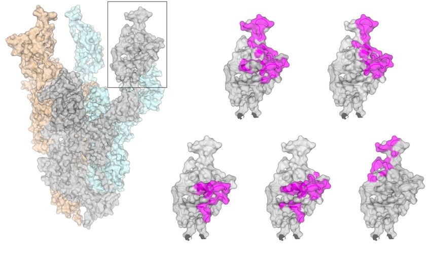
Potential therapeutic intervention for COVID-19

    The COVID-19 symptoms have reportedly ranged from                     intervention strategies including vaccines, monoclonal anti-
mild to severe that can ultimately lead to death. The symp-               bodies, peptides, interferon therapies and small-molecule drugs
toms usually appear 2-14 days after viral exposure which in-              to combat SARS-CoV-2, it may require several months to test
cludes fever, cough, shortness of breath and pneumonia. The               its efficacy in vitro/in vivo and also it largely depends on the
severe cases showed respiratory, hepatic, gastrointestinal and            results of the clinical trials. Even though this virus is newly
neurological complications that can leads to mortality. The               identified, the clinical and genetic features showed similarity
transmission of COVID-19 is reported to be human-to-human                 with SARS-CoV.3 Their similarities would make it easier to uti-
transmission via., respiratory droplets or direct contact with            lize the existing knowledge and adapt the available vaccines or
the infected patients.1-3,6,9-13 The virus spreads to more than 20        therapeutic models developed against other coronaviruses to
countries within short period and nearly 73,000 infected cas-             target the unique aspects of SARS-CoV-2.
es of COVID-19 with a total of 1,870 deaths were reported as                  Immunotherapy is regarded as an effective method for
of February 18, 2020. The numbers of infected cases and death             clinical treatment of infectious diseases. The use of monoclo-
associated with COVID-19 is increasing daily. More number                 nal antibodies is a new era in infectious disease prevention
of infected cases has been reported in China, followed by Sin-            which overcomes many drawbacks associated with serum
gapore, Hong Kong Thailand, Korea, Japan, Taiwan, Malaysia,               therapy and intravenous immunoglobulins preparations in
Vietnam, Australia, Germany, USA, France, UAE, UK, Cana-                  terms of specificity, purity, low risk of blood-borne pathogen
da, Italy, Philippines, India, Spain, Finland, Sweden, Belgium,           contamination and safety. Monoclonal antibodies are versatile
Nepal, Sri Lanka, Egypt, and Cambodia.13                                  class of pharmaceuticals that have been successfully used by
    Significant efforts have been made to develop therapeutic             pharmaceutical industry which can provide an efficient ther-
interventions against coronavirus infection. Major research               apeutic intervention with a highly specific treatment against
has been focused on identifying anti-viral molecules targeting            particular disease.20-23 Many monoclonal antibodies against
the spike protein as it mediates viral entry, and their potential         viruses are developed in recent years and some are in clinical
in inducing host immune responses and eliciting protective                pipeline.24-26
antibody responses in infected individuals. In this review, we                CoV infection starts with the interaction of receptor bind-
highlight the therapeutic potential of neutralizing antibodies            ing domain located in the S protein and target receptor on the
that showed promising efficacy against SARS-CoV or MERS-                  host cell surface such as Angiotensin converting enzyme 2
CoV which might have the potential for the therapy and pro-               (ACE2) for SARS-CoV and dipeptidyl peptidase-4 (DPP4) for
phylaxis of SARS-CoV-2.                                                   MERS-CoV.27 The effective treatment options against SARS-
                                                                          CoV-2 can either based on the use of broad-spectrum anti-vi-
Therapeutic Intervention for COVID-19                                     ral drugs or by using specific therapeutic molecules that can
    Neither an effective vaccines nor anti-viral therapeutic              directly interrupt any stages of the viral lifecycle or the recep-
agents have been approved to treat COVID-19 or any other                  tor proteins located in the host cell surface to restrain the vi-
human CoV infection till date. The current approach to coro-              rus binding thereby blocking the virus attachment and entry.
navirus disease management focuses on supportive care. Rap-               This can be achieved by using peptidic fusion inhibitors, anti-
id public health interventions with antibodies, anti-virals or            SARS-CoV-2 neutralizing monoclonal antibodies, anti-ACE2
novel vaccine strategies are highly essential to contain the              monoclonal antibodies and protease inhibitors. The spike pro-
virus and disease transmission. Passive antibody therapy can              tein present on the viral membrane plays a vital role in virus
be considered as a way to limit COVID-19 epidemics. Pas-                  entry and is the principal antigenic component responsible for
sive immunization of antibody that can recognize epitopic                 inducing host immune response.28 Hence, it has been consid-
regions in the foreign virus particle can reduce the virus rep-           ered as a key target to develop potential effective therapeutics
lication and disease severity. Antibodies for passive immuno-             against coronavirus infection. The receptor-binding motif lo-
therapy can be isolated from the blood of the infected patients           cated in the receptor-binding domain (RBD) of S1 sub-unit of
or it can be manufactured in the laboratory. Immunotherapy                spike protein interacts with the cell receptor and mediates the
by transferring the convalescent sera to infected patients may            virus attachment with the host cells.29 Similar to SARS-CoV,
be effective in humans in neutralizing the virus and prevent              SARS-CoV-2 utilizes host receptor, angiotensin-converting
further infection. Based on the existing evidence and prior               enzyme 2 (ACE2) for its attachment and entry (Figure 1).30-32
experience in treating other viral infections such as influenza,          Hence the therapies for SARS-CoV can be extrapolated to use
SARS, MERS and Ebola, the early administration of convales-               for SARS-CoV-2. The specific neutralizing monoclonal anti-
cent plasma or hyper-immune immunoglobulin from patients                  bodies either against receptor-binding domain (RBD) in spike
that contains significant antibody titers can likely reduce the           protein or specific antibody that binds to ACE2 could effec-
viral load and disease mortality.14-19 However, the key chal-             tively block the virus entry (Figure 2). The structure of SARS-
lenges such as availability of sufficient donors, clinical condi-         CoV-2, SARS-CoV and MERS-CoV spike protein and mono-
tion, viral kinetics, and host interactions of SARS-CoV-2 needs           clonal antibody interaction sites are shown in figures 3, 4, and
to be elucidated before considering convalescent plasma as a              5. The protein structure figures were generated by PyMOL.
therapeutic option. However, there is an urgent need to con-              As both SARS-CoV and SARS-CoV-2 uses same host cell sur-
sider novel therapies for treating clinically advanced condi-             face receptor, potential blocking agents or strategies tested to
tions in order to reduce mortality, virus spread and to mitigate          prevent SARS entry could be evaluated against SARS-CoV-2.
the potential future outbreaks. Although researchers are in               Coughlin and Prabhakar, (2012) reported a series of human
the process of developing specific preventive and therapeutic             monoclonal antibodies targeting the RBD region of S protein

                                                                     11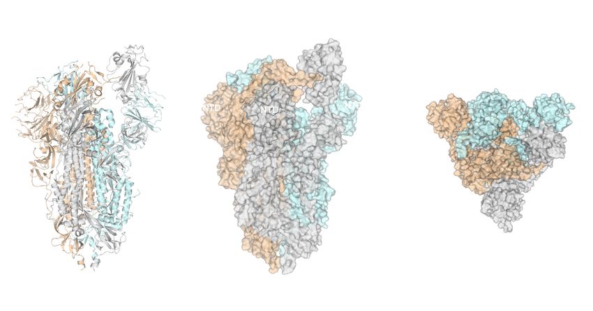
Asian Pac J Allergy Immunol 2020;38:10-18 DOI 10.12932/AP-200220-0773

                           SARS-CoV                         MERS-CoV                        SARS-CoV-2

                       Receptor ACE2                     Receptor DPP4                    Receptor ACE2

Figure 1. Graphical representation of SARS-CoV, MERS-CoV, SARS-CoV-2 and its cellular receptor.
The schematic representation shows the envelope spike proteins of SARS-CoV and MERS-CoV that binds to host receptor angio-
tensin-converting enzyme 2 (ACE2) and dipeptidyl peptidase 4 (DPP4), respectively. Similar like SARS-CoV, novel coronavirus
SARS-CoV-2 uses ACE2 as its receptor for host entry. Binding between receptor binding domain in spike protein and the cellular
receptor mediates membrane fusion and initiate the virus life cycle.

                                      SARS-CoV-2                                    Neutralizing mAb
                                   Spike Protein                                   Receptor ACE2

                                                                                                            Host cell

                                                                                    Viral RNA release

                         Fusion

                                                                               Endosomal membrane
                                                                               fusion

Figure 2. Schematic representation of SARS-CoV-2 neutralization mechanism.
Interaction of spike protein and the cellular receptor is required for membrane fusion and entry into the target cell. The monoclonal
antibodies targeting spike protein of SARS-CoV-2 could potentially inhibit the virus binding to its cellular receptor thereby prevent-
ing its entry into the cell.

                                                                  12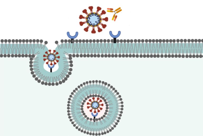
Potential therapeutic intervention for COVID-19
Host Cell
A) B) RBD C)
RBD RBD
(in)
(in) (out)
RBD
(in)
NTD NTD
NTD NTD NTD
90°
RBD
(out)
RBD
(in)
NTD
SARS-CoV-2 Spike Protein Viral Envelope
Ectodomain
Figure 3. Structure of SARS-CoV-2 spike protein ectodomain (PDB ID 6VSB)71.
A) Ribbon diagram of the trimeric spike protein.
B) Surface representation (side view) of the trimeric spike protein.
C) The surface facing the host cell consists of the N-terminal domain (NTD) and the receptor binding domain (RBD). The RBD can
be in either the in or out conformation. The out conformation is proposed to interact with the host receptor ACE2.
RBD
ACE2 80R
F26G1 m396 S230
SARS-CoV Spike Protein
Ectodomain
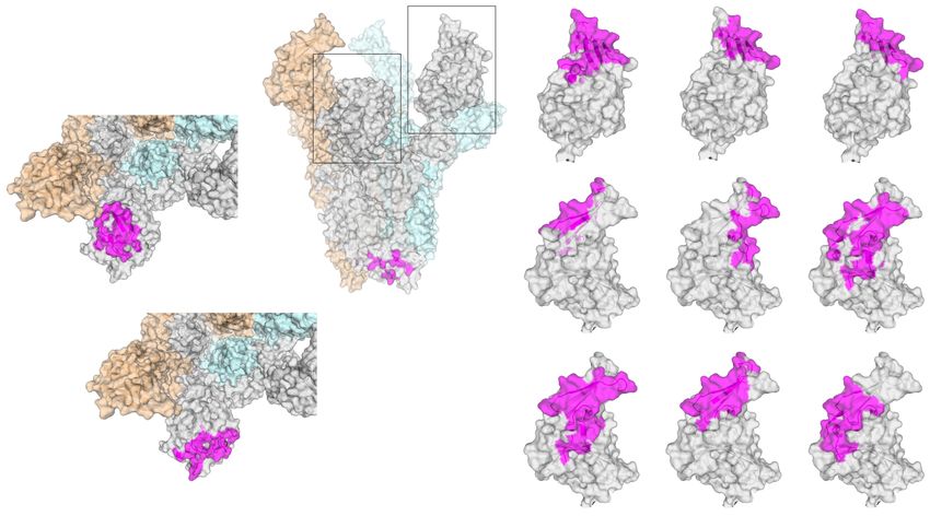
Figure 4. Structure of the trimeric SARS-CoV spike protein ectodomain in the RBD out conformation (PDB ID 6NB7)51.
The binding surface of the ACE2 receptor (PDB ID 6CS2)72 and the following antibodies are shown in magenta: 80R (PDB ID
2GHW)73, F26G1 (PDB ID 3BGF)74, m396 (PDB ID 2DD8)75, and S230 (PDB ID 6NB7)51.
13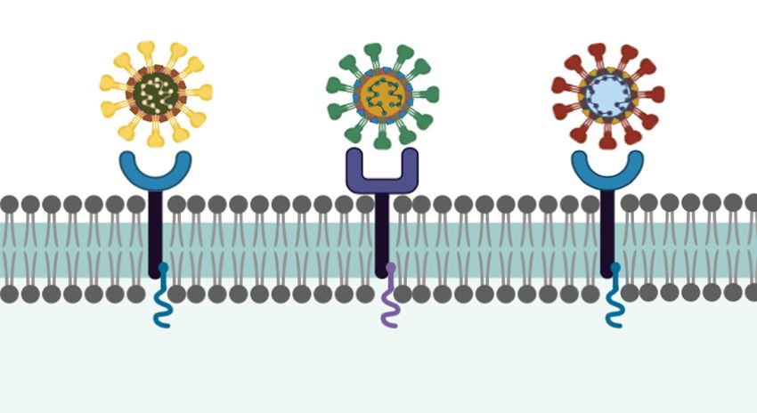
RBD                      DPP4                     m336                  CDC2-C2
                MERS-CoV Spike Protein
                     Ectodomain

                                                                   NTD

                                                     90°
                                                                                                   120°

                                                                                                            DPP4                   MERS-4                         D12

              7D10
                                                                                 G4

14
                                                                         Variable Loop
                                                                             of S2

                                                                                                          JC57-14                     MCA1                      LCA60
                                                                                                                                                                                     Asian Pac J Allergy Immunol 2020;38:10-18 DOI 10.12932/AP-200220-0773

                              G2

     Figure 5. Structure of the trimeric MERS-CoV spike protein ectodomain in the RBD out conformation (PDB ID 5X59)76.
     The DPP4 binding surface (magenta, PDB ID 4KR0)77 in the RBD is shown in two orientations (120° apart) for ease of comparison with the antibody binding surfaces.Three re-
     gions in which targeting antibodies have been reported are the RBD, NTD, and the variable loop of the S2 connector domain. All the interaction surfaces are shown in magenta.
     RBD: m336 (PDB ID 4XAK)78, CDC2-C2 (PDB ID 6C6Z)64, MERS-4 (PDB ID 5YY5)56, D12 (PDB ID 4ZPT)62, JC57-14 (PDB ID 6C6Y)64, MCA-1 (PDB ID 5GMQ)67, andLCA60
     (PDB ID 6NB4)51. NTD: 7D10 (PDB ID 6J11)69 and G2(PDB ID 6PXH)70. Variable loop of the S2 connector domain: G4 (PDB ID 5W9I).63
Potential therapeutic intervention for COVID-19
of SARS-CoV.33 The monoclonal antibodies targeting spike For effective disease prevention, the combination of differ-
protein in SARS-CoV and MERS-CoV showed promising re- ent monoclonal antibodies that recognizes different epitopes on
sults in vitro and in vivo that could be potentially effective the viral surface could be assessed to neutralize wide range of
against SARS-CoV-2 are listed in the table 1 and 2. isolates including escape mutants and best candidates could be
Table 1. Neutralizing monoclonal antibodies targeting SARS-CoV and their mechanism of action
Monoclonal
Mechanism of action References
antibody
• Binding to the conformational epitope (amino acid residues 426-492) on S1 fragment of SARS-CoV.
80R • Blocking the interaction of S1 subunit protein with cellular receptor ACE2 using 6 complementary determining region 11,20,41,42
(CDR) in vitro and in vivo (Mouse).
• Binding to the amino acid residues 318-510 and amino acid residue 565 with high affinity on S1 fragment of SARS-CoV.
CR3014 43-45
• Blocking the interaction of S1 subunit protein with cellular receptor ACE2 in vitro and in vivo (Ferret).
• Binding to the amino acid residues 318-510 on S1 fragment of SARS-CoV.
CR3022 44
• Blocking the interaction of S1 subunit protein (RBD) with cellular receptor ACE2 in vitro.
• Binding to the linear epitope (amino acid residues 460-476) on S1 fragment of SARS-CoV.
F26G18 42
• Blocking the interaction of S1 subunit protein (RBD) with cellular receptor ACE2 in vitro.
• Binding to the conformational epitope (amino acid residues 359-362, 391-392, 424-427, and 486-492) on S1 fragment of
F26G19 SARS-CoV. 42
• Blocking the interaction of S1 subunit protein (RBD) with cellular receptor ACE2 in vitro.
• Binding to the conformational epitope (amino acid residues 482-491) on S1 fragment of SARS-CoV.
m396 42,46
• Blocking the interaction of S subunit protein using CDR loops H1, H2, H3, and L3 with cellular receptor ACE2 in vitro.
• Binding to the Heptad repeat (HR) loops including heptad repeat 1 (HR1) and heptad repeat 1 (HR2) domain on S2 frag-
1A9 ment of SARS-CoV. 47,48
• Blocking the interaction of S2 subunit protein (amino acid residues 1111-1130) with cellular receptor in vitro.
• Binding to the amino acid residues 490-510 on S1 fragment of SARS-CoV.
201 33,49
• Blocking the interaction of S1 subunit protein with cellular receptor ACE2 in vitro and in vivo (Mouse Syrian Hamster).
68 • Binding to the amino acid residues 130-150 of SARS-CoV in vitro and in vivo (Mouse) 33,49
• Binding to the amino acid residues 12-261 of SARS-CoV and N-terminal of RBD
4D4 33,50
• Inhibiting the post-interaction in the viral penetration in vitro.
• Binding to epitopes partially overlapping with receptor binding motifs on B domain of SARS-CoV.
S230 51
• Blocking the interaction of S1 subunit protein with cellular receptor ACE2 in vitro
Table 2. Neutralizing monoclonal antibodies targeting MERS-CoV and their mechanism of action
Monoclonal
Mechanism of action References
antibody
• Binding to the C-terminal segment of the β5-β6, β6-β7 and β7-β8 loops on the receptor-binding subdomain in RBD of
MERS-CoV with no overlap DPP4 binding surface.
MERS-4 52-55
• Blocking the interaction of S1 subunit protein with cellular receptor DPP4 in vitro by inducing β5-β6 shallow groove on
the RBD.
• Binding to the C-terminal segment of the β6-β7 loop and β7 strand on RBD of MERS-CoV and overlap with the DPP4
MERS-27 binding surface. 52-57
• Blocking the interaction of S1subunit protein with cellular receptor DPP4 in vitro.
• Binding to the C-terminal segment of the β6-β7 loop and β7 strand on RBD of MERS-CoV and overlap with the DPP4
4C2 binding surface. 52,53,56,58
• Blocking the interaction of S1 subunit protein with cellular receptor DPP4 in vitro and in vivo (Mouse).
• Binding to the C-terminal segment of the β5-β8 strands, β5-β6 loop and β6-β7 loop in RBD of MERS-CoV and overlap
with the DPP4 binding surface.
m336 52,53,56,59-61
• Blocking the interaction of S1 subunit protein with cellular receptor DPP4 by mimicking the interaction between RBD and
DPP4 in the similar binding angle in vitro and in vivo (Mouse and rabbit).
G4 • Binding to the glycosylated surface on the S2 subunit protein in vitro. 52,62,63
• Binding to the C-terminal segment of the β6-β7 loop and β7 strand on RBD of MERS-CoV and overlap with the DPP4
D12 binding surface. 52,56,63,64
• Blocking the interaction of S1 subunit protein with cellular receptor DPP4 in vitro.
15Asian Pac J Allergy Immunol 2020;38:10-18 DOI 10.12932/AP-200220-0773
Table 2. (Continued)
Monoclonal
Mechanism of action References
antibody
• Binding to the C-terminal segment of the β6-β7 loop and β7 strand on RBD of MERS-CoV and overlap with the DPP4
JC57-14 binding surface. 52,56,64
• Blocking the interaction of S1 subunit protein with cellular receptor DPP4 in vitro.
• Binding to the C-terminal segment of the β5-β8 strands, β5-β6 loop and β6-β7 loop in RBD of MERS-CoV.
MERS-GD27 • Blocking the interaction of S1 subunit protein with cellular receptor DPP4 by mimicking the interaction between RBD and 52,65
DPP4 in the same binding angle in vitro and in vivo (Mice).
• Binding to the C-terminal segment of the β5-β8 strands, β5-β6 loop and β6-β7 loop in RBD of MERS-CoV.
MERS-GD33 • Blocking the interaction of S1 subunit protein with cellular receptor DPP4 mimicking the interaction between RBD and 52,66
DPP4 in the same binding angle in vitro.
• Binding to the C-terminal segment of the β8 strand, β6-β9 loop, and β6-β8 loop on RBD of MERS-CoV.
LCA60 51
• Blocking the interaction of S1 subunit protein with cellular receptor DPP4 in vitro.
• Binding to RBD with 6 complementarity-determining regions
MCA1 67,68
• Blocking the interaction of S1 subunit protein with cellular receptor DPP4 in vitro and in vivo (Mouse).
CDC2-C2 • Blocking the interaction of S1 subunit protein with cellular receptor DPP4 in vitro and in vivo (Mouse). 64
• Binding to N-terminal domain of S protein of MERS-CoV
7D10 69
• Blocking the interaction of S1 subunit protein with cellular receptor DPP4 in vitro and in vivo (Mouse).
• Binding to N-terminal domain of S protein of MERS-CoV
G2 69,70
• Blocking the interaction of S1 subunit protein with cellular receptor DPP4 in vitro.
used for passive immunotherapy. Monoclonal antibody cock- MERS and SARS. Further detailed understanding of the virus
tail may exhibit more potent anti-virus activity that could in- pathogenesis might increase the opportunities for the realistic
crease the effectiveness of the treatment and prevent the viral design of therapeutics specific to novel coronavirus.
escape.34-36 Although, several monoclonal antibodies showed
promising result in neutralizing SARS-CoV and MERS-CoV Author Contributions
infection, the large-scale production of monoclonal antibodies All authors have made a considerable, direct and intellectual
is labor intensive, expensive and time consuming which out- contribution to the work, and approved it for publication.
weighs the monoclonal antibody clinical application especially
monoclonal antibodies against emerging pathogen. The recent
advancement in the therapeutic protein production platforms Acknowledgement
could make the monoclonal antibody production at lower Author (BS) would like to acknowledge the Second Century
production costs and affordable. The sequences of monoclonal Fund (C2F), Chulalongkorn University, for providing the fel-
antibodies that are effective against SARS-CoV could be cloned lowship.
and expressed in suitable expression system such as mamma-
lian, yeast or plant and recombinant monoclonal antibodies Conflict of Interest
could be tested against SARS-CoV-2. Plant expression system The authors declare that no conflict of interest.
could be considered for the rapid production of monoclonal
antibodies in a short time with the affordable cost which is
one of the major advantages to be considered especially during
References
1. Wang D, Hu B, Hu C, Zhu F, Liu X, Zhang J, et al. Clinical characteristics
epidemic situation.37-40 of 138 hospitalized patients with 2019 novel coronavirus-infected
pneumonia in Wuhan, China. JAMA[Preprint]. 2020 [cited 2020 Feb 18]:
[9 p.]. Available from: https//doi.org/10.1001/jama.2020.1585
Concluding Remarks 2. Li Q, Guan X, Wu P, Wang X, Zhou L, Tong Y, et al. Early transmission
The need to treat the emerging novel coronavirus that caus- dynamics in Wuhan, China, of novel coronavirus–infected pneumonia.
es global impact throws spotlight on developing monoclonal N Engl J Med [Preprint]. 2020 [cited 2020 Feb 16]: [9 p.]. Available from:
https://doi.org/10.1056/NEJMoa2001316
antibody-based passive immunotherapy to provide a quick 3. Gralinski LE, Menachery VD. Return of the coronavirus: 2019-nCoV.
response. Even though there is a major progress towards the Viruses. 2020;12(135).
development of monoclonal antibody therapy for coronavirus 4. World Health Organization [Internet]. Geneva; World Health Organization;
infection, no monoclonal antibodies have yet been success- c2020 [cited 2020 Feb 18]. Coronavirus disease (COVID-19) outbreak;
[about 2 screens]. Available from: https://www.who.int/emergencies/
fully marketed. The increasing understanding on MERS-CoV diseases/novel-coronavirus-2019
and SARS-CoV in recent years might galvanize the research 5. Masters PS. The molecular biology of coronaviruses. Adv Virus Res.
community to make significant progress in the COVID-2019 2006;66:193-292.
therapeutic design in an accelerated time by utilizing the ex- 6. Banerjee A, Kulcsar K, Misra V, Frieman M, Mossman K. Bats and
Coronaviruses. Viruses. 2019;11(41).
isting anti-viral regimen that showed promising results against
16Potential therapeutic intervention for COVID-19
7. Li F. Structure, function, and evolution of coronavirus spike proteins. Annu 28. Du L, Yang Y, Zhou Y, Lu L, Li F, Jiang S. MERS-CoV spike protein: a key
Rev Virol. 2016;3(1):237-61. target for antivirals. Expert Opin Ther Targets. 2017;21(2):131-43.
8. Kruse RL. Therapeutic strategies in an outbreak scenario to treat the novel 29. Song Z, Xu Y, Bao L, Zhang L, Yu P, Qu Y, et al. From SARS to MERS,
coronavirus originating in Wuhan, China. F1000Res. 2020;9(72). thrusting coronaviruses into the spotlight. Viruses. 2019;11(1)(59).
9. Carlos WG, Cruz CSD, Cao B, Pasnick S, Jamil S. Novel Wuhan 30. Li W, Moore MJ, Vasilieva N, Sui J, Wong SK, Berne MA, et al.
(2019-nCoV) coronavirus. Am J Respir Crit Care Med. 2020;201(4):7-8. Angiotensin-converting enzyme 2 is a functional receptor for the SARS
10. Lai C-C, Shih T-P, Ko W-C, Tang H-J, Hsueh P-R. Severe acute respiratory coronavirus. Nature. 2003;426(6965):450-4.
syndrome coronavirus 2 (SARS-CoV-2) and coronavirus disease-2019 31. Li F, Li W, Farzan M, Harrison SC. Structure of SARS coronavirus spike
(COVID-19): the epidemic and the challenges. Int J Antimicrob Agents. receptor- binding domain complexed with receptor. Science. 2005;
2020; 105924. 309(5742):1864-8.
11. Zhang L, Liu Y. Potential interventions for novel coronavirus in China: a 32. Wan Y, Shang J, Graham R, Baric RS, Li F. Receptor recognition by
systematic review. J Med Virol [Preprint]. 2020 [cited 2020 Feb 18]: [ 36 p.]. novel coronavirus from Wuhan: An analysis based on decade-long
Available from: https://doi.org/10.1002/jmv.25707 structural studies of SARS. J Virol [Preprint]. 2020 [cited 2020 Feb 18]:
12. Coleman CM, Frieman MB. Coronaviruses: important emerging human [25 p.] Availbale from: https://jvi.asm.org/content/jvi/early/2020/01/23/
pathogens. J Virol. 2014;88(10):5209-12. JVI.00127-20.full.pdf
13. World Health Organization [Internet]. Geneva; World Health Organization; 33. Coughlin MM, Prabhakar BS. Neutralizing human monoclonal antibodies
c2020 [cited 2020 Feb 18]. Coronavirus disease 2019 (COVID-19) situation to Severe acute respiratory syndrome coronavirus: target, mechanism of
report – 29; [7 screens]. Available from: https://www.who.int/docs/default action and therapeutic potential. Rev Med Virol. 2012;22(1):2-17.
-source/coronaviruse/situation-reports/20200218-sitrep-29-covid-19.pdf 34. Schroeder HW Jr, Cavacini L. Structure and function of immunoglobulins.
?sfvrsn=6262de9e_2 J Allergy Clin Immunol. 2010;125:41-52.
14. Mupapa K, Massamba M, Kibadi K, Kuvula K, Bwaka A, Kipasa M, et 35. Dimitrov DS. Therapeutic proteins. Methods Mol Biol. 2012;899:1-26.
al. Treatment of Ebola hemorrhagic fever with blood transfusions from 36. Sparrow E, Friede M, Sheikh M, Torvaldsen S. Therapeutic antibodies for
convalescent patients. International Scientific and Technical Committee. J infectious diseases. Bull World Health Organ. 2017;95(3):235-7.
Infect Dis. 1999;179:18-23. 37. Demurtas OC, Massa S, Illiano E, Martinis DD, Chan PKS, Bonito PD, et al.
15. Yeh K-M, Chiueh T-S, Siu LK, Lin J-C, Chan PKS, Peng M-Y, et al. Antigen production in plant to tackle infectious diseases flare up: the case
Experience of using convalescent plasma for severe acute respiratory of SARS. Plant Sci. 2016;7(54).
syndrome among healthcare workers in a Taiwan hospital. J Antimicrob 38. Hiatt A, Whaley KJ, Zeitlin L. Plant-derived monoclonal antibodies for
Chemother. 2005;56(5):919-22. prevention and treatment of infectious disease. Microbiol Spectr. 2014;2(1).
16. Luke TC, Kilbane EM, Jackson JL, Hoffman SL. Meta-analysis: convalescent 39. Sainsbury F. Innovation in plant-based transient protein expression for
blood products for Spanish influenza pneumonia: a future H5N1 treatment? infectious disease prevention and preparedness. Curr Opin Biotechnol.
Ann Intern Med. 2006;145(8):599-609. 2019;6(61):110-5.
17. Chan K-H, Chan JF-W, Tse H, Chen H, Lau CC-Y, Cai J-P, et al. 40. Shanmugaraj B, Malla A, Phoolcharoen W. Emergence of Novel
Cross-reactive antibodies in convalescent SARS patients’ sera Coronavirus 2019-nCoV: Need for Rapid Vaccine and Biologics
against the emerging novel human coronavirus EMC (2012) by both Development. Pathogens. 2020;9(148).
immunofluorescent and neutralizing antibody tests. J Infect. 2013;67(2): 41. Sui J, Li W, Murakami A, Tamin A, Matthews LJ, Wong SK, et al. Potent
130-40. neutralization of severe acute respiratory syndrome (SARS) coronavirus
18. Mair-Jenkins J, Saavedra-Campos M, Baillie JK, Cleary P, Khaw F-M, Lim by a human mAb to S1 protein that blocks receptor association. Proc Natl
WS, et al. The effectiveness of convalescent plasma and hyperimmune Acad Sci U S A. 2004;101(8):2536-41.
immunoglobulin for the treatment of severe acute respiratory infections of 42. Berry JD, Hay K, Rini JM, Yu M, Wang L, plummer FA, et al. Neutralizing
viral etiology: a systematic review and exploratory meta-analysis. J Infect epitopes of the SARS-CoV S-protein cluster independent of repertoire,
Dis. 2015;211(1):80-90. antigen structure or mAb technology. MAbs. 2010;2(1):53-66.
19. Arabi Y, Balkhy H, Hajeer AH, Bouchama A, Hayden FG, Al‐Omari A, et 43. van den Brink EN, ter Meulen J, Cox F, Jongeneelen MAC, Thijsse A,
al. Feasibility, safety, clinical, and laboratory effects of convalescent plasma Throsby M, et al. Molecular and biological characterization of human
therapy for patients with Middle East respiratory syndrome coronavirus monoclonal antibodies binding to the spike and nucleocapsid proteins
infection: a study protocol. Springerplus. 2015;4(709). of Severe acute respiratory syndrome coronavirus. J Virol. 2005;79(3):
20. Sui J, Li W, Roberts A, Matthews LJ, Murakami A, Vogel L, et al. 1635-44.
Evaluation of human monoclonal antibody 80R for immunoprophylaxis of 44. ter Meulen J, van den Brink EN, Poon LLM, Marissen WE, Leung CSW,
severe acute respiratory syndrome by an animal study, epitope mapping, Cox F, et al. Human monoclonal antibody combination against SARS
and analysis of spike variants. J Virol. 2005;79(10):5900-6. coronavirus: synergy and coverage of escape mutants. PLoS Med.
21. Bayry J, Lacroix-Desmazes Sb, Kazatchkine MD, Kaveri SV. Monoclonal 2006;3(7):1071-9.
antibody and intravenous immunoglobulin therapy for rheumatic 45. ter Meulen J, Bakker ABH, van den Brink EN, Weverling GJ, Martina BEE,
diseases: rationale and mechanisms of action. Nat Clin Pract Rheumatol. Haagmans BL, et al. Human monoclonal antibody as prophylaxis for SARS
2007;3(5):262-72. coronavirus infection in ferrets. Lancet. 2004;363(9427):2139-41.
22. Both L, Banyard AC, Dolleweerd CV, Wright E, Ma JK-C, Fooks AR. 46. Zhu Z, Chakraborti S, He Y, Roberts A, Sheahan T, Xiao X, et al. Potent
Monoclonal antibodies for prophylactic and therapeutic use against viral cross-reactive neutralization of SARS coronavirus isolates by human
infections. Vaccine. 2013;31(12):1553-9. monoclonal antibodies. Proc Natl Acad Sci U S A. 2007;104(29):12123-8.
23. Marasco WA, Sui J. The growth and potential of human antiviral 47. Ng O-W, Keng C-T, Leung CS-W, Peiris JSM, Poon LLM, Tan Y-J.
monoclonal antibody therapeutics. Nat Biotechnol. 2007;25(12):1421-34. Substitution at aspartic acid 1128 in the SARS coronavirus spike
24. Davey RT Jr, Dodd L, Proschan MA, Neaton J, Neuhaus Nordwall J, glycoprotein mediates escape from a S2 domain-targeting neutralizing
Koopmeiners JS, et al. A randomized, controlled trial of ZMapp for Ebola monoclonal antibody. PLoS One. 2014;9(7).
Virus infection. N Engl J Med. 2016;375(15):1448-56. 48. Lip K-M, Shen S, Yang X, Keng C-T, Zhang A, Oh H-LJ, et al. Monoclonal
25. Gupta P, Kamath AV, Park S, Chiu H, Lutman J, Maia M, et al. Preclinical antibodies targeting the HR2 domain and the region immediately upstream
pharmacokinetics of MHAA4549A, a human monoclonal antibody to of the HR2 of the S protein neutralize in vitro infection of Severe acute
influenza A virus, and the prediction of its efficacious clinical dose for respiratory syndrome coronavirus. J Virol. 2006;80(2):941-50.
the treatment of patients hospitalized with influenza A. MAbs. 2016;8(5): 49. Greenough TC, Babcock GJ, Roberts A, Hernandez HJ, Thomas WD,
991-7. Coccia JA, et al. Development and characterization of a Severe acute
26. Caskey M, Klein F, Lorenzi JCC, Seaman MS, West AP Jr, Buckley N, et respiratory syndrome-associated coronavirus-neutralizing human
al. Viraemia suppressed in HIV-1-infected humans by broadly neutralizing monoclonal antibody that provides effective immunoprophylaxis in mice. J
antibody 3BNC117. Nature. 2015;522(7557):487-91. Infect Dis. 2005;191(4):507-14.
27. Raj VS, Mou H, Smits SL, Dekkers DH, Muller MA, Dijkman R, et al. 50. Elshabrawy HA, Coughlin MM, Baker SC, Prabhakar BS. Human
Dipeptidyl peptidase 4 is a functional receptor for the emerging human monoclonal antibodies against highly conserved HR1 and HR2 domains
coronavirus-EMC. Nature. 2013;495(7440):251-4. of the SARS-CoV spike protein are more broadly neutralizing. PLoS One.
2012;7(11).
17Asian Pac J Allergy Immunol 2020;38:10-18 DOI 10.12932/AP-200220-0773
51. Walls AC, Xiong X, Park Y-J, Tortorici MA, Snijder J, Quispe J, et al. 66. Niu P, Zhang S, Zhou P, Huang B, Deng Y, Qin K, et al. Ultrapotent human
Unexpected Receptor Functional Mimicry Elucidates Activation of neutralizing antibody repertoires against Middle East respiratory syndrome
Coronavirus Fusion. Cell. 2019;176(5):1026-39. coronavirus from a recovered patient. J Infect Dis. 2018;218(8):1249-60.
52. Xu J, Jia W, Wang P, Zhang S, Shi X, Wang X. Antibodies and vaccines 67. Chen Z, Bao L, Chen C, Zou T, Xue Y, Li F, et al. Human Neutralizing
against Middle East respiratory syndrome coronavirus. Emerg Microbes Monoclonal Antibody Inhibition of Middle East Respiratory Syndrome
Infect. 2019;8(1):841-56. Coronavirus Replication in the Common Marmoset. J Infect Dis. 2017;
53. Rabaan AA, Alahmed SH, Bazzi AM, Alhani HM. A review of candidate 215(12):1807-15.
therapies for Middle East respiratory syndrome from a molecular 68. de Wit E, Rasmussen AL, Falzarano D, Bushmaker T, Feldmann F,
perspective. J Med Microbiol. 2017;66(9):1261-74. Brining DL, et al. Middle East respiratory syndrome coronavirus (MERS-
54. Jiang L, Wang N, Zuo T, Shi X, Poon K-MV, Wu Y, et al. Potent CoV) causes transient lower respiratory tract infection in rhesus macaques.
neutralization of MERS-CoV by human neutralizing monoclonal Proc Natl Acad Sci U S A. 2013;110(41):598-603.
antibodies to the viral spike glycoprotein. Sci Transl Med. 2014;6(234): 69. Zhou H, Chen Y, Zhang S, Niu P, Qin K, Jia W, et al. Structural definition
234-59. of a neutralization epitope on the N-terminal domain of MERS-CoV spike
55. Ying T, Li H, Lu L, Dimitrov DS, Jiang S. Development of human glycoprotein. Nat Commun. 2019;10(3068).
neutralizing monoclonal antibodies for prevention and therapy of 70. Wang N, Rosen O, Wang L, Turner HL, Stevens LJ, Corbett KS, et
MERS-CoV infections. Microbes Infect. 2015;17(2):142-8. al. Structural Definition of a Neutralization-sensitive Epitope on the
56. Zhang S, Zhou P, Wang P, Li Y, Jiang L, Jia W, et al. Structural definition MERS-CoV S1-NTD. Cell Rep. 2019;28(13):3395-405.
of a unique neutralization epitope on the Receptor-binding domain of 71. Wrapp D, Wang N, Corbett KS, Goldsmith JA, Hsieh C-L, Abiona
MERS-CoV spike glycoprotein. Cell Rep. 2018;24(2):441-52. O, et al. Cryo-EM structure of the 2019-nCoV spike in the prefusion
57. Yu X, Zhang S, Jiang L, Cui Y, Li D, Wang D, et al. Structural basis for the conformation. BioRxiv [Preprint]. 2020 [cited 2020 Feb 20]: [30 p.]. Avail-
neutralization of MERS-CoV by a human monoclonal antibody MERS-27. able from: https://www.biorxiv.org/content/10.1101/2020.02.11.944462v1.
Sci Rep. 2015;5(13133). full.pdf
58. Li Y, Wan Y, Liu P, Zhao J, Lu G, Qi J, et al. A humanized neutralizing 72. Kirchdoerfer RN, Wang N, Pallesen J, Wrapp D, Turner HL, Cottrell CA,
antibody against MERS-CoV targeting the receptor-binding domain of the et al. Stabilized coronavirus spikes are resistant to conformational changes
spike protein. Cell Res. 2015;25(11):1237-49. induced by receptor recognition or proteolysis. Sci Rep. 2018;8(1).
59. van Doremalen N, Falzarano D, Ying T, de Wit E, Bushmaker T, 73. Hwang WC, Lin Y, Santelli E, Sui J, Jaroszewski L, Stec B, et al. Structural
Feldmann F, et al. Efficacy of antibody-based therapies against Middle East basis of neutralization by a human anti-severe acute respiratory syndrome
respiratory syndrome coronavirus (MERS-CoV) in common marmosets. spike protein antibody, 80R. J Biol Chem. 2006;281(45):34610-6.
Antiviral Res. 2017;143:30-7. 74. Pak JE, Sharon C, Satkunarajah M, Auperin TC, Cameron CM, Kelvin
60. Houser KV, Gretebeck L, Ying T, Wang Y, Vogel L, Lamirande EW, et DJ, et al. Structural insights into immune recognition of the severe acute
al. Prophylaxis with a Middle East respiratory syndrome coronavirus respiratory syndrome coronavirus S protein receptor binding domain.
(MERS-CoV)-specific human monoclonal antibody protects rabbits from J Mol Biol. 2009;388(4):815-23.
MERS-CoV infection. J Infect Dis. 2016;213(10):1557-61. 75. Prabakaran P, Gan J, Feng Y, Zhu Z, Choudhry V, Xiao X, et al. Structure
61. Ying T, Du L, Ju TW, Prabakaran P, Lau CCY, Lu L, et al. Exceptionally of Severe Acute Respiratory Syndrome Coronavirus Receptor-binding
potent neutralization of Middle East respiratory syndrome coronavirus by Domain Complexed with Neutralizing Antibody. J Biol Chem. 2006;
human monoclonal antibodies. J Virol. 2014;88(14):796-805. 281(23):25829-15836.
62. Wang L, Shi W, Joyce MG, Modjarrad K, Zhang Y, Leung K, et al. 76. Yuan Y, Cao D, Zhang Y, Ma J, Qi J, Wang Q, et al. Cryo-EM structures
Evaluation of candidate vaccine approaches for MERS-CoV. Nat Commun. of MERS-CoV and SARS-CoV spike glycoproteins reveal the dynamic
2015;6(7712). receptor binding domains. Nat Commun. 2017;8(15092).
63. Pallesena J, Wang N, Corbett KS, Wrapp D, Kirchdoerfer RN, Turner HL, 77. Lu G, Hu Y, Wang Q, Qi J, Gao F, Li Y, et al. Molecular basis of binding
et al. Immunogenicity and structures of a rationally designed prefusion between novel human coronavirus MERS-CoV and its receptor CD26.
MERS-CoV spike antigen. Proc Natl Acad Sci U S A. 2017;114(35):7348-57. Nature. 2013;500(7461):227-31.
64. Wang L, Shi W, Chappell JD, Joyce MG, Zhang Y, Kanekiyo M, et al. 78. Ying T, Prabakaran P, Du L, Shi W, Feng Y, Wang Y, et al. Junctional and
Importance of neutralizing monoclonal antibodies targeting multiple allele-specific residues are critical for MERS-CoV neutralization by an
antigenic sites on the Middle East respiratory syndrome coronavirus spike exceptionally potent germline-like antibody. Nat Commun. 2015;6(8223).
glycoprotein to avoid neutralization escape. J Virol. 2018;92(10).
65. Niu P, Zhao G, Deng Y, Sun S, Wang W, Zhou Y, et al. A novel human
mAb (MERS-GD27) provides prophylactic and postexposure efficacy in
MERS-CoV susceptible mice. Sci China Life Sci. 2018;61(10):1280-2.
18You can also read