Recognition of potential Covid-19 drug treatments through the study of existing protein-drug and protein-protein structures: an analysis of ...
←
→
Page content transcription
If your browser does not render page correctly, please read the page content below
Recognition of potential Covid-19 drug treatments through the
study of existing protein-drug and protein-protein structures: an
analysis of kinetically active residues
                                             Ognjen Perišić1
          (1) Big Blue Genomics, Vojvode Brane 32, 11000 Belgrade, Serbia, ognjen.perisic@gmail.com

                                             May 13, 2020

Abstract
We report results of our study of approved drugs as potential treatments for COVID-19, based on
the application of various bioinformatics predictive methods. The drugs studied include
Chloroquine, Ivermectin, Remdesivir and α-difluoromethylornithine (DMFO). Our results indicate
that these small drug molecules selectively bind to stable, kinetically active residues and residues
adjoining them on the surface of proteins and inside protein pockets, and that some prefer
hydrophobic over other active sites. Our approach is not restricted to viruses and can facilitate
rational drug design, as well as improve our understanding of molecular interactions, in general.

Introduction
The Coronaviridae positive stranded RNA virus family includes a substantial number of members,
many of whom are known to cause a broad range of illnesses from common cold to sever
diseases like Severe Acute Respiratory Syndrome (SARS), Middle East Respiratory Syndrome
(MERS), etc. [1, 2]. The latest worldwide rapidly spreading disease, COVID-19, is caused by a
new member of this virus family, SARS-COV-2. The disease originally emerged in China in
December 2019 with most common symptoms being fever and cough, as well as shortness of
breath, sore throat, headache, muscles ache, nausea, and diarrhea [1]. In some cases initial
symptoms also involve a still unexplained loss of smell and taste [3]. The SARS-COV-2 virus
spreads through respiratory droplets, directly via physical contact, or through contact with
contaminated objects, and can severely affect patients with immune systems weakened by pre-
existing conditions, such as hypertension, diabetes mellitus or cardiovascular diseases [4]. The
virus has lower fatality rate than SARS and MERS, but spreads more easily due to high binding
affinity between the virus spike glycoprotein (S) and the host receptor [5-8], making it, potentially,
more deadly. The virus is therefore a primary cause or initiator of a significant number of deaths
all over the world. By restricting access to work and slowing down supply lines the virus also
directly affects the global economy, which experiences a significant decline in gross national
products worldwide and unemployment rates reaching levels not encountered since the Great
depression.

As this manuscript is being prepared there are several efforts and clinical trials underway to
develop a vaccine and evaluate potential drugs for COVID-19, but such investigations usually
take months or even years to yield a successful treatment. Drug repurposing, on the other hand,
may offer an immediate solution, because it considers already approved compounds as potential
treatments for COVID-19. There are two paths toward a viral treatment. One path directly attacks
the virus and interrupts its replication machinery or its ability to attack host cells [9]. This path is

                                                                                                      1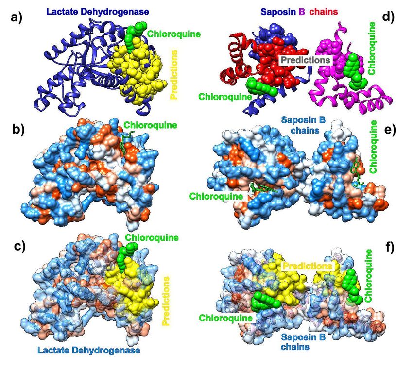
often hard to implement due to rapid emergence of new viral strains with acquired resistance to
implemented drugs. The second path should therefore aim to block the host-viral interactions on
the host side due to difficulties viral single point mutations should have in recovering the loss of
host factors [10]. A recent study of human-virus protein-protein interactions (PPIs) detected 332
high-confidence SARS-CoV-2-human PPIs [11]. The study showed that 40% of SARS-CoV-2
proteins interact with endomembrane compartments or vesicle trafficking pathways, and that viral
proteins also interact with multiple innate immune pathways, the host translation machinery,
bromodomain proteins, enzymes involved in ubiquitination regulation, and Cullin ubiquitin ligase
complex. Importantly, it showed that the SARS-COV-2 human PPI map is very similar to the
interaction maps of West Nile Virus (WNV) and Mycobacterium tuberculosis (Mtb). Among the
human proteins involved in interactions with viral proteins, the study detected 66 druggable
human (host) proteins targeted by 69 compounds (29 FDA-approved drugs, 12 drugs in clinical
trials, and 28 preclinical compounds). It identified two groups of compounds with noticeable
antiviral activity: inhibitors of mRNA translation/protein biogenesis (zotatifin, ternatin-4, PS3061,
and plitidepsin), and predicted regulators of the Sigma1 and Sigma2 receptors (Haloperidol,
PB28, PD-144418, Hydroxychloroquine, Clemastine, Cloperastine, Progesterone, and the clinical
molecule Siramesine). The first group of compounds directly affects the viral cap-dependent
mRNA translation because coronaviruses use the host translation machinery for their own mRNA
translation. The compounds affecting the second group of proteins are approved and long
established human therapeutics [11]. As much as they are informative, such screening
associative studies rarely offer detailed insights into mechanisms of molecular interactions,
whereas structural studies [5-8, 12] give snapshots into residue and atom level physical
interactions between molecules, but cannot offer general principles of molecular interactions.

To facilitate the drug screening we undertook a comparative study of binding modes of four
antiviral candidate drugs. We analyzed compounds that bind to parasitic and to human proteins.
We use those results to anticipate their binging affinities. The drugs we have studied so far include
(hydroxyl)chloroquine, Ivermectin, Remdesivir (and sofosbuvir), and α-difluoromethylornithine
(DMFO) (see Table 1).

Chloroquine [13] and its less toxic derivative hydroxychloroquine [14] are drugs used to
prevent and treat acute attacks of malaria. They are also used to treat discoid or systemic lupus
erythematosus and rheumatoid arthritis in patients whose symptoms have not improved with other
treatments. These drugs are subject of a number of clinical trials worldwide as potential treatment
for Covid-19 [15, 16]. Interestingly, the study mentioned above [11] showed that PB28 was ~20
times more potent viral inhibitor than hydroxychloroquine.

Ivermectin is a medication used to treat many various types of parasite infestations [17]. They
include, but are not limited to, head lice, scabies, river blindness (onchocerciasis),
strongyloidiasis, trichuriasis, ascariasis, and lymphatic filariasis. Depending on the kind of
treatment, the drug is taken by mouth or applied to the skin for external infestations. Ivermectin
molecular structure is rather complex and made of a set of macrocyclic lactone isomers.
Ivermectin binds to glutamate-gated chloride channels and increases the permeability of chloride
ions. The drug was shown to inhibit the replication of SARS-COV-2 in vitro [18] and is currently
the subject of clinical trials as a potential COVID-19 treatment [19].

Remdesivir is a nucleoside analog RNA-dependent RNA Polymerase (RdRp) inhibitor initially
developed to treat Ebola and Marburg virus diseases [9, 20]. The drug decreases the viral RNA
production by affecting the function of RdRp and proofreading by viral exoribonuclease (ExoN).
Remdesivir is a subject of clinical trials as a potential COVID-19 treatment [21], as it was shown
to reduce the lung viral load and improve pulmonary function with SARS infection [9].

                                                                                                   2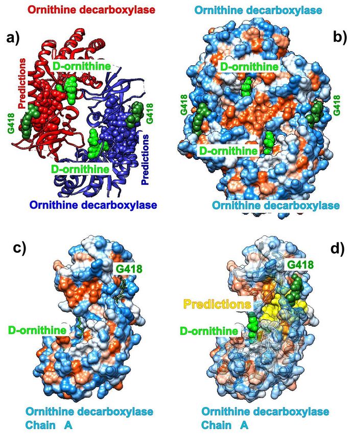
α-difluoromethylornithine (DMFO), is a medication primary used to treat African
trypanosomiasis (sleeping sickness) and excessive facial hair in women [22]. Specifically, it is
used for the second stage of sleeping sickness caused by T. b. gambiense and may be used with
nifurtimox [23]. It is used by injection or applied to the skin. The drug prevents binding of the
natural product ornithine to the active site of ornithine decarboxylase. We did not find any record
of this drug ever being tried, so far, for COVID-19. However, since it is a halogenated organic
molecule with somehow similar active sites as Chloroquine we decided to study it towards
treatment of COVID-19.

Mass media, internet and even professional publications are currently immersed in controversial
debates over the effectiveness, or lack of it, of some of those drug for the treatment of COVID-19.
We avoid getting involved in such discussions and only report our research findings based on the
existing bioinformatics predictive methods and tools [24, 25] and the method which we have
implemented to recognize binding patches through the analysis of normal modes in proteins [26,
27], Self Adjustable Gaussian Network Model (SAGNM) [28]. The method predicts binding areas
without any information on the binding partner’s properties, position or orientation.

Results
The analysis of chloroquine binding patterns to human lysosomal protein saposin B (pdb id 4v2o)
and plasmodium falciparum lactate dehydrogenase (pdb id 1cet) reveals that chloroquine binds
to kinetically active sites recognized by the SAGNM algorithm [28] which are mostly hydrophobic
(Figure 1). Other kinetically active sites although exposed to solvent are not binding targets. The
analysis of Chloroquine’s nondiscriminatory binding to human and parasitic proteins may offer an
explanation of its efficiency against parasitic infections as well offer a glimpse into its toxicity. Our
results suggest that Chloroquine’s binding to Covid-19 proteins should follow the same patterns
of attachment to residues which are both hydrophobic and kinetically active (or close to kinetically
active sites).

The drug Ivermectin binds glutamate-gated chloride channels and thus increases their
permeability to chloride ions. We analyzed the Ivermectin’s binding to the human glycine receptor
alpha-3 (pdb id 5vdh). This structure, besides Ivermectin, also has glycine and the potentiator
AM-3607 (7c6) bound to the glycine receptor. The receptor is a pentamer, so we only analyzed
the binding to its chain A. The analysis shown in Figure 2 reveals that all three compounds bind
to kinetically active and adjoining residues [28], some of which are highly hydrophobic, but
Ivermectin binds to almost exclusively hydrophobic residues. That means that this drug well seek
similar sites on the surface of the Covid-19 proteins.

We used the recently cryo-EM determined structure of SARS-COV-2 RdRp with double-stranded
template-primer RNA and Remdesivir (pdb id 7bv2, [12]) to analyze the RNA and drug binding to
residues in RdRp. The structure reveals that the double stranded RNA is inserted into RdRp’s
central channel and that the active triphosphate form of Remdesivir is covalently bound to the
primer strand at the first replicated base, which effectively terminated the chain elongation. It
shouldbe noted that the prodrug form of Remdesivir does not have any inhibitory effect on the
polymerization activity of the purified enzyme [12]. Our analysis reveals that the residues
recognized via the fastest two normal modes (corresponding to kinetically active residues [28])
delineate the central channel (Figure 3). The enzymatically important residues K500, S501, K545
and R555 are all recognized by the SAGNM algorithm using just the fastest normal mode, while

                                                                                                       3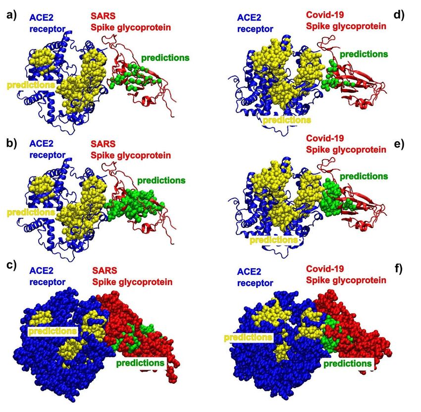
the residue D761 of the catalytic center (out of residues 759-761 that form the catalytic center) is
also emphasized with the two fastest modes. Residues K545 and R555 are important because
they stabilize the incoming nucleotide in the correct position for catalysis. The crystal structure
shows that the catalytic center of RdRp, NSP12 protein (Non Structural Protein 12), does not
have any contacts with base pairs of RNA emphasizing RdRp’s sequence-agnostic
polymerization ability [12]. This is in concordance with our coarse grained analysis, based on the
positions of C-α atoms only, that shows that stable, kinetically active residues outline the
enzyme’s central channel.
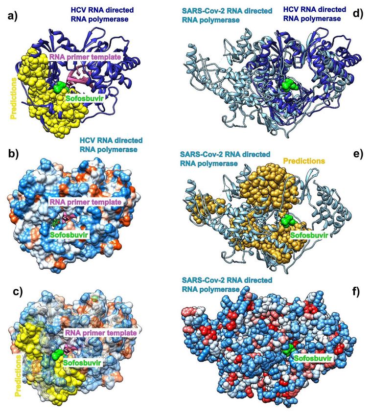
We also performed a comparative analysis of the Hepatitis C virus (HCV) RdRp (chain A in pdb
id 4wtg; the structure is give with the drug sofosbuvir bound to it) and the Covid-19 RdRp (chain
A in pdb id 6m71). We followed the steps of Y. Gao and collaborators [29] and attempted to
compare predictions of the binding residues in HCV RdRp to sofosbuvir, to binding residues
predictions in Covid-19 RdRp. The binding residues in HCV are buried deep inside the
polymerase catalytic core. Our analysis shows that they are generally delineated by the kinetically
active residues and are thus stable and characterized by the two fastest normal modes (Figure
S1a), but they are not explicitly hydrophobic (Fig. S1b-c). The structural alignment of HCV and
Covid-19 RdRp (Figure S1d) shows that they share the structure of the binding pocket, and also
reveals that the catalytic cores in both are bounded by kinetically active residues, but the overall
distribution of residues is partially different between the two proteins (Figure S1e). The similarities
suggest that the interior of the RdRps in coronaviruses are attractive binding spot for small
compounds in general.
We also analyzed the binding patterns of the three proteins that form the SARS-COV-2 RdRp
(pdb id 6m71). The main enzymatic unit is NSP12, which mostly keeps its conformation between
RNA free and RNA bound structures [12]. Figure S2 shows that cofactors NSP7 and NSP8 seek
patches with kinetically active residues on the surface of NSP12. However, they are also in
contact with kinetically less active areas. This should be analyzed in the light of fact that SARS-
COV-2 RdRp (NSP12) cannot perform its function without NSP7 and NSP8 [12]. The distribution
of kinetically very active and kinetically dormant residues may be important for the overall stability
of NSP12, and also act as stochastic transformer that translates random fluctuations of solvent
and proteins into a regular vibrations that produce a regular rhythm of translation (i.e. act as a
regular clock/oscillator).
The analysis of the contact patterns between the ACE2 receptor and the spike glycoprotein
receptor binding domains (RBD) in SARS (pdb id 6cs2) and SARS-COV-2 (pdb id 6m0j) reveals
a difference in the distribution of kinetically active residues important for binding between RBD
and ACE2 (Figure 4). SARS-RBD has a smaller number of kinetically active and adjoining
residues in direct contact with ACE2 (Figure 4a-c), while kinetically active residues in Covid-19
RBD are directly oriented and are in contact with the active residues in ACE2 (Figure 4d-f). In
SARS active residues are mostly perpendicular to the interfacial plane (compare the distributions
of C-alpha atoms in Figs. 4a and 4d). That should make the binding affinity between the
Covid-19-RBD and ACE2 receptor stronger than between the SARS-RBD and ACE2 receptor. In
both cases the predicted residues are recognized via the fastest vibrational mode (see [28]).
The analysis of kinetically active and adjoining residues in the SARS monomer (pdb id 6nb6)
reveals that they are attractive binding spots for glycans (Figure S3). Glycans form the glycan
shield, which was already suggested to assist in immune evasion similarly to the HIV-1 envelope
trimer [30]. The kinetically active residues recognized by the SAGNM algorithm [28] can be used
as target areas for drugs aimed at removing/disrupting the viral glycan shield. Those residues are
4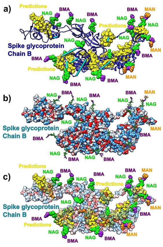
not particularly hydrophobic and should be targeted by drugs that bind to hydrophilic patches, and
electrostatically complementary.

We also analyzed the kinetically active residues in the structure formed by the SARS Spike
Glycoprotein RBD and human neutralizing S230 antibody FAB fragment (pdb id 6nb6). The
analysis reveals that S230 antibody binds to kinetically active residues in SRAS RBD, while heavy
and light chains in S230 communicate via kinetically active residues (see Figure S4). The binding
residues are mostly neutral to hydrophilic, thus any potential drug should be able to bind to similar
surfaces (neutral/hydrophilic and stable).

The drug α-difluoromethylornithine (Eflornithine) prohibits binding of the natural non-coded amino
acid ornithine to the active site on the surface of Trypanosoma brucei ornithine decarboxylase
(ODC, pdb id 1njj). The binding of this drug should follow the binding patterns of ornithine. Figure
S5. shows that SAGNM algorithm accurately detects binding sites for both ornithine and G418
(Geneticin), an aminoglycoside antibiotic. In contrary to chloroquine, both compounds bind
preferably to the hydrophilic sites on the surface of ODC. If applied to treat Covid-19, the drug
Eflornithine should bind to similar sites on the surface of Covid-19 proteins (hydrophobic and
kinetically active, i.e. stable).

Conclusion
Covid-19 is the first severe global pandemic caused by a coronavirus, and there are no
guarantees that it will be the last. We therefore need not only to develop an effective and efficient
treatment for the current pandemic, but also have to design a set of protocols to address all future,
similar pandemics. In this manuscript we presented our strategy to recognize potential drug
binding residues in human and viral proteins. We analyzed four currently approved drugs
(Chloroquine, Ivermectin, Remdesivir, and Eflornithine). Our results indicate that small, drug like
compounds preferentially bind to kinetically active and adjoining residues, thus seeking stable
residues characterized by fast normal modes with small amplitude of fluctuations [28]. Some
drugs preferentially seek active patches that are hydrophobic (Chloroquine, Ivermectin), while
others prefer hydrophilic surfaces (Remdesivir, Sofosbuvir, Eflornithine). We can postulate that in
water environment drugs binding to hydrophilic patches will be more stable, as their removal will
lead toward the reduction in structural entropy, but a full account of this proposition will require
calculations of binding free energy differences based on full atom molecular dynamics, using, for
instance, steered molecular dynamics simulations (SMD) [31]. We can also propose that the
drugs/small molecules that bind to deep pockets will be more stable, and thus more effective. Our
algorithm accurately recognizes such pockets as binding spots for drugs (Figure 3), and small
peptides (see, in particular, Figure 6a in [28]).

Multidrug cocktails are frequently used to treat viral diseases [32]. Our analysis shows that in
designing antiviral drug cocktails, binding affinity to and between kinetically active (stable) sites
should be combined with the information on their hydrophobic and hydrophilic properties to avoid
conflicts, increase drug cocktail efficiency, and reduce toxicity and other unwanted side effects.

In our analysis we used both viral-parasitic, as well as human proteins. The analysis shows that
kinetically active residues exist in both human and non-human proteins/enzymes and that drugs
bind indiscriminately to them regardless of their origin. The compounds that bind to human

                                                                                                   5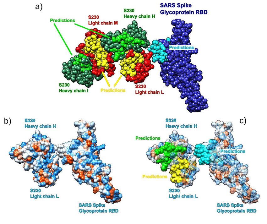
proteins potentially offer longer lasting treatments as host cells and tissues have less chance of
developing drug resistance through single point mutations.

The protocol depicted here may also help in accessing drug toxicity. Binding spots in various
proteins can be very effectively predicted with our SAGNM approach and accessed with other
bioinformatics tools for charge and shape complementarity, binding affinity, atomic mass and
other properties as well.

Recent advances in machine learning helped advance our ability to predict and design protein
structures [33], but the full theoretical foundation is still lacking. The quality of the machine learning
protocol directly depends on the quality and size of training data sets and thus in many ways
follows classical methods based on statistical potentials and homology modeling [34]. Our results
can also help in that respect as they offer interpretation on how residue packing inside protein
segments guides their assemblage.

The procedure we described here is fast and effective, and can analyze protein structure much
faster than computationally demanding molecular dynamics simulations. The only requirement is
the protein structure. Its advantage is not in its efficiency, but also in its ability to suggest general
binding patterns between proteins and drugs or small peptides. It can be used to filter binding
areas on protein surfaces and thus facilitate preclinical stages in drug design.

Acknowledgement: The author would like to thank G.A. Mansoori for his encouragements to
undertake this activity and reading the manuscript.

References:
[1]       W.-j. Guan, Z.-y. Ni, Y. Hu, W.-h. Liang, C.-q. Ou, J.-x. He, L. Liu, H. Shan, C.-l. Lei, D.S.C. Hui, B. Du, L.-j.
Li, G. Zeng, K.-y. Yuen, R.-c. Chen, C.-l. Tang, T. Wang, P.-y. Chen, J. Xiang, S.-y. Li, J.-l. Wang, Z.-j. Liang, Y.-x.
Peng, L. Wei, Y. Liu, Y.-h. Hu, P. Peng, J.-m. Wang, J.-y. Liu, Z. Chen, G. Li, Z.-j. Zheng, S.-q. Qiu, J. Luo, C.-j. Ye,
S.-y. Zhu, and N.-s. Zhong. Clinical Characteristics of Coronavirus Disease 2019 in China. New England Journal of
Medicine, 382(18):1708–1720, 2020.
[2]       C. Liu, Q. Zhou, Y. Li, L.V. Garner, S.P. Watkins, L.J. Carter, J. Smoot, A.C. Gregg, A.D. Daniels, S. Jervey,
and D. Albaiu. Research and development on therapeutic agents and vaccines for covid-19 and related human
coronavirus diseases. ACS Central Science, 6(3):315–331, 2020. PMID: 32226821.
[3]       C.H. Yan, F. Faraji, D.P. Prajapati, C.E. Boone, and A.S. DeConde. Association of chemosensory dysfunction
and covid-19 in patients presenting with influenza-like symptoms. International Forum of Allergy & Rhinology, n/a(n/a).
[4]       L. Palmieri, X. Andrianou, P. Barbariol, A. Bella, S. Bellino, and E. Benelli et al. Characteristics of COVID-19
patients dying in Italy Report based on available data on April 2th, 2020. (Report). Istituto Superiore di Sanità, 3 April
2020.
[5]       R. Yan, Y. Zhang, Y. Li, L. Xia, Y. Guo, and Q. Zhou. Structural basis for the recognition of SARS-CoV-2 by
full-length human ACE2. Science, 367(6485):1444–1448, 2020.
[6]       D. Wrapp, N. Wang, K.S. Corbett, J.A. Goldsmith, C.-L. Hsieh, O. Abiona, B.S. Graham, and J.S. McLellan.
Cryo-EM structure of the 2019-nCoV spike in the prefusion conformation. Science, 367(6483):1260–1263, 2020.
[7]       J. Lan, J. Ge, J. Yu, S. Shan, H. Zhou, S. Fan, Q. Zhang, X. Shi, Q. Wang, L. Zhang, and X. Wang. Structure
of the SARS-CoV-2 spike receptor-binding domain bound to the ACE2 receptor. Nature, 2020.
[8]       J. Shang, G. Ye, K. Shi, Y. Wan, C. Luo, H. Aihara, Q. Geng, A. Auerbach, and F. Li. Structural basis of
receptor recognition by SARS-CoV-2. Nature, 2020.
[9]       T.P. Sheahan et al. Comparative therapeutic efficacy of Remdesivir and combination Lopinavir, Ritonavir, and
Interferon beta against MERS-CoV. Nature Communications, 11(222), 2020.
[10]      A. Prussia, P. Thepchatri, J.P. Snyder, and R.K. Plemper. Systematic Approaches towards the Development
of Host-Directed Antiviral Therapeutics. Int J Mol Sci., 12(6):4027–4052, 2011.
[11]      D.E. Gordon, G.M Jang G.M., and M. Bouhaddou et al. A SARS-CoV-2 protein interaction map reveals targets
for drug repurposing. Nature, https://doi.org/10.1038/s41586-020-2286-9 (null), 2020.

                                                                                                                          6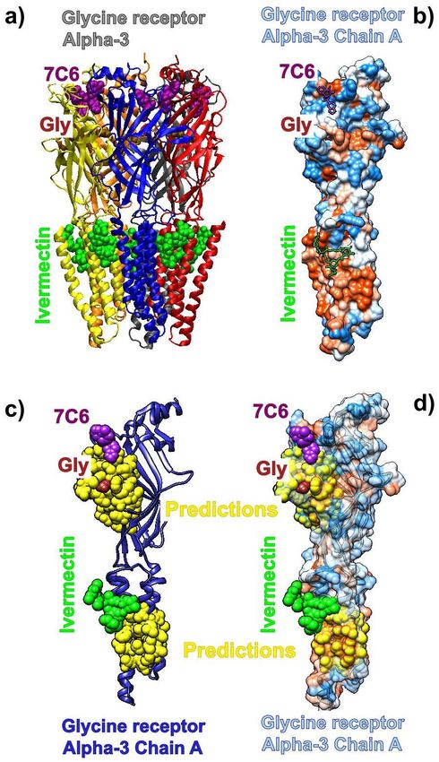
[12]       W. Wanchao, C. Mao, X. Luan, D.-D. Shen, Q. Shen, H. Su, X. Wang, F. Zhou, W. Zhao, M. Gao, S. Chang,
Y.-C. Xie, G. Tian, H.-W. Jiang, S.-C. Tao, J. Shen, Y. Jiang, H. Jiang, Y. Xu, S. Zhang, Y. Zhang, and H.E. Xu.
Structural basis for inhibition of the RNA-dependent RNA polymerase from SARS-CoV-2 by Remdesivir. Science, 2020.
[13]       U.S. National Library of Medicine. Chloroquine. https://druginfo.nlm.nih.gov/drugportal/name/chloroquine.
[14]       U.S.                     National Library                   of Medicine.                      Hydroxychloroquine.
https://druginfo.nlm.nih.gov/drugportal/name/hydroxychloroquine.
[15]       U.S.                National Library                of Medicine.                  Chloroquine               trials.
https://clinicaltrials.gov/search/intervention=CHLOROQUINE+.
[16]       U.S.               National Library            of Medicine.                 Hydroxychloroquine              trials.
https://clinicaltrials.gov/search/intervention=hydroxychloroquine.
[17]       U.S. National Library of Medicine. Ivermectin. https://druginfo.nlm.nih.gov/drugportal/name/ivermectin.
[18]       L. Caly, J.D. Druce, M.G. Catton, D.A. Jans, and K.M. Wagstaff. The FDA-approved Drug Ivermectin inhibits
the replication of SARS-CoV-2 in vitro. Antiviral Research, https://doi.org/10.1016/j.antiviral.2020.104787, 2020.
[19]       MedinCell has launched a COVID-19 research initiative based on its experience to formulate long-acting
injectable Ivermectin. https://www.medincell.com/wp-content/uploads/2020/04/PR_MedinCell-Covid19-EN.pdf, 2020.
[20]       U.S. National Library of Medicine. Remdesivir. https://druginfo.nlm.nih.gov/drugportal/name/remdesivir.
[21]       U.S. National Library of Medicine. Remdesivir trials. https://clinicaltrials.gov/search/intervention=GS-5734.
[22]       U.S. National Library of Medicine. Eflornithine. https://druginfo.nlm.nih.gov/drugportal/rn/70052-12-9.
[23]       U.S. National Library of Medicine. Nifurtimox. https://druginfo.nlm.nih.gov/drugportal/name/nifurtimox.
[24]       E.F. Pettersen, T.D. Goddard, C.C. Huang, G.S. Couch, D.M. Greenblatt, E.C. Meng, and T.E. Ferrin. UCSF
Chimera–a visualization system for exploratory research and analysis. J. Comput. Chem., 25(13):1605–12, 2004.
[25]       W. Humphrey, A. Dalke A, and K. Schulten. Journal of Molecular Graphics, 14:33–38, 1996.
[26]       T. Haliloglu, I. Bahar, and B. Erman. Gaussian Dynamics of Folded Proteins. Physical Review Letters,
79(16):3090–3093, 1997.
[27]       M. C. Demirel, A. R. Atiligan, R. L. Jernigan, B. Erman, and I. Bahar. Identification of kinetically hot residues
in proteins. Protein Science, 7:2522–2532, 1998.
[28]       O. Perišić. Heterodimer binding scaffolds recognition via the analysis of kinetically hot residues. MDPI
Pharmaceuticals, 11(29), 2018.
[29]       Y. Gao, L. Yan, Y. Huang, F. Liu, Y. Zhao, L. Cao, T. Wang, Q. Sun, Z. Ming, L. Zhang, J. Ge, L. Zheng,
Y. Zhang, H. Wang, Y. Zhu, C. Zhu, T. Hu, T. Hua, B. Zhang, X. Yang, J. Li, H. Yang, Z. Liu, W. Xu, L.W. Guddat,
Q. Wang, Z. Lou, and Z. Rao. Structure of the RNA-dependent RNA polymerase from COVID-19 virus. Science, 2020.
[30]       X. Xiong, M.A. Tortorici, J. Snijder, C. Yoshioka, A.C. Walls, W. Li, A.T. McGuire, F.A. Rey, B.-J. Bosch, and
D. Veesler. Glycan shield and fusion activation of a deltacoronavirus spike glycoprotein fine-tuned for enteric infections.
Journal of Virology, 92(4), 2018.
[31]       O. Perišić and H. Lu. On the improvement of free-energy calculation from steered molecular dynamics
simulations using adaptive stochastic perturbation protocols. PLoS ONE, e101810. doi:10.1371/journal.pone.0101810,
2014.
[32]       A. Moscona. Global transmission of oseltamivir-resistant influenza. The New England journal of medicine,
360(10):953–956, 2009.
[33]       A.W. Senior, R. Evans, J. Jumper, J. Kirkpatrick, L. Sifre, T. Green, C. Qin, A. Židek, A.W.R. Nelson,
A. Bridgland, H. Penedones, S. Petersen, K. Simonyan, S. Crossan, P. Kohli, D.T. Jones, D. Silver, K. Kavukcuoglu,
and D. Hassabis. Improved protein structure prediction using potentials from deep learning. Nature, 577(7792):706–
710, 2020.
[34]       N. Eswar, M. A. Marti-Renom, B. Webb, M. S. Madhusudhan, D. Eramian, M. Shen, U. Pieper, and A. Sali.
Comparative Protein Structure Modeling With MODELLER. Current Protocols in Bioinformatics, John Wiley & Sons,
Inc., Supplement 15, 5.6.1-5.6.30, 2006.

                                                                                                                            7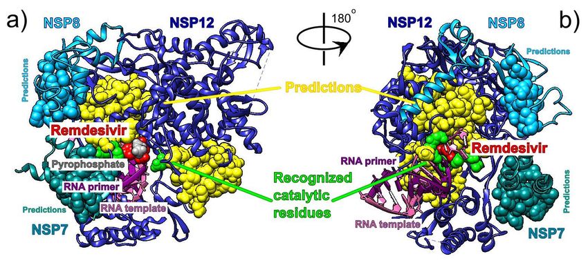
Dosage in                                     Precautions in patients with complications
                                         individuals                           Side
         Drug               Indication                   Effectiveness
                                          aged ≥ 12                           effects    Cardio-
                                            years                                                     Renal     Hepatic    Retinal
                                                                                        Pulmonary
                                                       Malaria, Amebiasis,
                            Treatment    500-600 mg
      Chloroquine                                      Porphyria Cutanea      Serious      Yes         Yes       Yes        Yes
                            Prevention     weekly
                                                               Tarda
                            Treatment      3-15 mg            Parasitic        Mild-
       Ivermectin                                                                           No         Yes       Yes         No
                            Prevention       once           infestations      Serious
                                         100-200 mg      Ebola, Marburg
      Remdesivir            Treatment                                          Mild         No         Yes        No         No
                                             daily        virus diseases
                                           300-400      Trypanosomiasis,
                                                                               Mild-
α-Difluoromethylornithine   Treatment    mg/kg/day,     reduction of facial                 No          No       Yes         No
                                                                              Serious
                                            cream         hair in women
Table 1. Comparison of existing drugs currently being tested for the antiviral treatment and prevention of
Covid-19 through drug repurposing.

                                                                                                                       8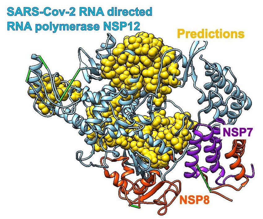
Figures

Figure 1. Chloroquine and its target proteins. Images on the left depict chloroquine bound to cofactor
binding site of Plasmodium Falciparum Lactate Dehydrogenase (pdb id 1cet). Images on the right depict
chloroquine bound to Saposin B (pdb id 4v2o).

                                                                                                    9
Figure 2. Ivermectin and its target protein, Human glycine receptor Alpha-3 (pdb id 5vdh).
10Figure 3. Remdesivir bound to the primer RNA inside the central channel of SARS-COV-2 RNA dependent
RNA polymerase (RdRp), NSP12) (pdb id 7bv2 described in [12]). The dashed lines represent protein
segments missing from the deposited structure.
11Figure 4. SARS spike glycoprotein chain B RBD and ACE2 receptor (pdb id 6cs2) in comparison to Covid-
19 spike glycoprotein chain A RBD and ACE2 receptor (pdb id 6m0j). In a) and d) spike glycoprotein’s
predictions are shows through C-alpha atoms only. In b, c, e and f predictions are shown through all atoms.
12Recognition of potential Covid-19 drug treatments through the
study of existing protein-drug and protein-protein structures: an
analysis of kinetically active residues
Ognjen Perišić1
(1) Big Blue Genomics, Vojvode Brane 32, 11000 Belgrade, Serbia, ognjen.perisic@gmail.com
May 13, 2020
Supplementary material
Methods and Materials
Our aim was to analyze presently available structures existing drugs bound to parasitic and
human proteins and predict their binding patterns, as well as the binding patterns of SARS and
SARS-COV-2 binding patterns to the human ACE2 receptor. To predict binding residues in protein
we applied our Self Adjustable interpretation of Gaussian Network Model (SAGNM) [28]. The
structure alignment, visualization and analyses were performed with programs Chimera [24] and
VMD [25].
We focused our study on pdb structures with the listed drugs present as ligands. For chloroquine
we analyzed 2 structures (pdb ids 1cet and 4v2o). For Ivermectin we analyzed the binding pattern
of the drug to the human glycine receptor alpha-3 (pdb id 5vdh). For Remdesivir we analyzed its
binding patterns in the recently released structure [12] (pdb id 7bv2). We also performed the
analysis of binding pattern of drug Sofosbuvir to the hepatitis C virus (HCV) RdRp (pdb id 4wtg)
and compared them to the Covid-19 RdRp predictions (pdb id 6m71). Sofosbuvir was already
analyzed in light of similarities between HCV and SARS-COV-2 RdRp and similarities between
Remdesivir’s and Sofosbuvir’s [29]. For α-Difluoromethylornithine we analyzed a structure of
Trypanosoma brucei ornithine decarboxylase (ODC) with D-ornithine bound to it (pdb id 1njj). α-
Difluoromethylornithine binds to the active site of ODC and inhibits ornithine binding to it. We
performed the comparative analysis of the binding patterns between the ACE2 human receptor
and the spike glycoproteins from SARS (pdb id 6cs2) and SARS-COV-2 (pdb id 6moj). We also
analyzed the binding patterns between the SARS RBD with S230 human neutralizing antibody,
and between SARS RBD and glycan shield (pdb id 6nb6).
13Supplementary material figures
Figure S1. Comparative analysis of HCV (pdb id 4wtg, chain A, figures of the left) and Covid-19 RNA
directed RNA polymerase (pdb id 6m71, chain A, figures on the right). HCV RNA directed RNA polymerase
has sofosbuvir bound to it. With Covid-19 RNA polymerase sofosbuvir’s position corresponds to the position
it has when bound to HCV RNA polymerase.
14Figure S2. Covid-19 RNA directed RNA polymerase with cofactors NSP7 and NSP8 (pdb id 6m71). The
algorithm predictions are yellow. The green sticks represent missing segments.
15Figure S3. SARS-Cov spike glycoprotein (Chain B, pdb id 6nb6) and glycans (NAG, BMA, MAN) bound to
it. The algorithm predictions are yellow. The predictions accurately predict binding spots for glycans. They
can be used as target areas for drugs aimed at removing/disrupting the virus glycan shield. Cyan bars
(Figure S4a) represent missing segments.
16Figure S4. Receptor binding domain (RBD) of SARS-COV spike glycoprotein (Chain A, pdb id 6nb6) with
human neutralizing S230 antibody FAB fragment. a) SRAS-COV RBD (blue, chain A) with heavy (green,
chains H and I) and light chains (red, chains L and M). Predictions are cyan (SARS), yellow (S230 light)
and light green (S230 heavy). b) Hydrophobic surface of SARS RBD and S230 (chains H and L). c)
Transparent hydrophobic surface of SARS RBD and S230 (chains H and L) with predictions.
17Figure S5. D-ornithine and its target protein Trypanosoma brucei ornithine decarboxylase (pdb id 1njj).
Lower figures depict ornithine decarboxylase monomer.
18You can also read