Cellulose hydrogel skeleton by extrusion 3D printing of solution - De Gruyter
←
→
Page content transcription
If your browser does not render page correctly, please read the page content below
Nanotechnology Reviews 2020; 9:345–353

Research Article

Xiangzhou Hu, Zhijie Yang, Senxian Kang, Man Jiang*, Zuowan Zhou, Jihua Gou, David Hui, and
Jing He

Cellulose hydrogel skeleton by extrusion 3D
printing of solution
https://doi.org/10.1515/ntrev-2020-0025
Received January 16, 2020; accepted January 24, 2020
                                                                         1 Introduction
Abstract: Cellulose is the most abundant natural polymer                 3D printing is an important additive manufacturing (AM)
on earth, which has obtained increasing interest in the                  method, which can produce complex geometries accord-
field of functional materials development for its renew-                 ing to the computer design. During the past years, some
able, high mechanical performance and environmental be-                  revolutionary techniques have been created to realize
nign. In this study, the traditional processing method (wet              rapid and scalable 3D printing with high resolution by
spinning and film production) of cellulose-based materi-                 new stereolithography, tomographic reconstruction tech-
als was applied by using cellulose solution for 3D printing,             niques [1–3]. The development of the materials in tissue
which can directly build complex 3D patterns. Herein, a                  engineering [4–6] provides opportunities for 3D printing,
natural cellulose is dissolved in an effective mixed aque-               since it is able to construct high precision and complex
ous solution of dimethyl sulfoxide (DMSO) and tetrabuty-                 shapes [7–9]. The 3D bio-printing has also obtained great
lammonium hydroxide (TBAH). The cellulose solution ex-                   progress in building components of human organs [10–
trusion was controlled by a modified fused deposition                    14]. As a biomass derived natural organic polymer, cellu-
modeling (FDM) 3D printer. During the controlled extru-                  lose has been considered an attractive material for the
sion 3D printing process, the viscous cellulose solution                 fabrication of bio-compatible and bio-degradable multi-
will gelifies and further solidifies into a predetermined 3D             functional products. But the processes are always com-
pattern at room temperature in air. Subsequently, a cellu-               plex and time consuming. It is particularly difficult to dis-
lose hydrogel skeleton was obtained, when the 3D pattern                 solve the cellulose in common solvents and it cannot be
was solvent-exchanged with deionized water. Finally, the                 melted with heating. Because of the recalcitrant property,
mechanical and swelling performance of the cellulose hy-                 it is difficult to efficiently extract the cellulose, in order to
drogel scaffold was improved by a cross-linking agent treat-             apply it. Hence, the lignocellulose biomass has also been
ment method. With treatment of the 3D printed scaffolds in               refined into bioethanol and biodiesel by chemical or bi-
0.8 wt% cross-linking agent solution, the obtained cellu-                ological catalysis process [15–17], or been prepared into
lose hydrogel could absorb 28 g/g water, and the compres-                biomass based carbon materials for supercapacitor elec-
sion strength was 96 kPa. This work provided an efficient                trode [18, 19]. A specific cellulose such as nanocrystal [20–
way to prepare natural cellulose hydrogel by 3D printing                 22] or nanofibers cellulose hydrogels [23–25] has been ap-
under room temperature.                                                  plied for 3D printing to fabricate 3D structures.
Keywords: cellulose, solution, 3D printing, hydrogel skele-
ton

                                                                         School of Materials Science and Engineering, Southwest Jiaotong Uni-
                                                                         versity, Chengdu, 610031, China
                                                                         Jihua Gou: Department of Mechanical and Aerospace Engineering,
                                                                         University of Central Florida, Orlando, FL 32816, United States of Amer-
                                                                         ica
*Corresponding Author: Man Jiang, Key Laboratory of Advanced             David Hui: Composite Material Research Laboratory, Department of
Technologies of Materials (Ministry of Education), School of Materials   Mechanical Engineering, University of New Orleans, New Orleans, LA
Science and Engineering, Southwest Jiaotong University, Chengdu,         70148, United States of America
610031, China; e-mail: jiangman1021@swjtu.edu.cn                         Jing He: Key Laboratory of Development and Application of Rural
Xiangzhou Hu, Zhijie Yang, Senxian Kang, Zuowan Zhou: Key Lab-           Renewable Energy, Biogas Institute of Ministry of Agriculture and
oratory of Advanced Technologies of Materials (Ministry of Education),   Rural Affairs, Chengdu 610041, China

  Open Access. © 2020 Xiangzhou Hu et al., published by De Gruyter.                 This work is licensed under the Creative Commons Attribution
4.0 License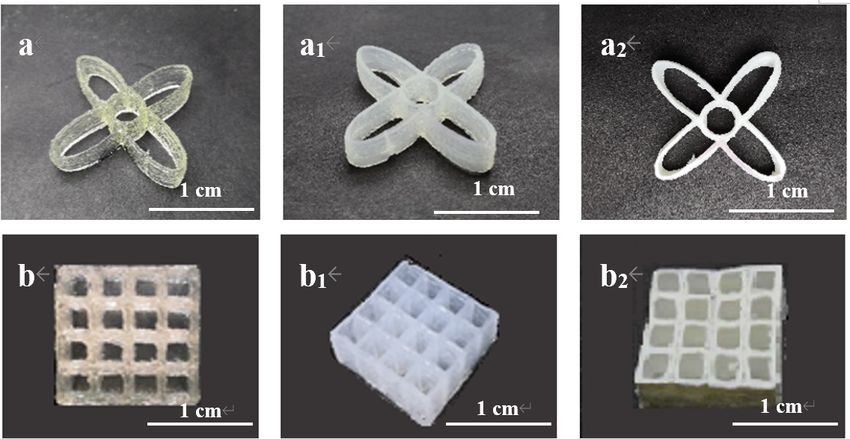
346 | Xiangzhou Hu et al.

     The challenges still exist for direct utilization of nat-   was supplied by Chengdu Haihong Chemical Reagent Co.,
ural cellulose for 3D printing. Except the stubborn prop-        Ltd (Sichuan, China). All chemicals used in this work were
erties of cellulose itself, the obstacle also comes from the     of an analytical grade, and were applied without further
work modes of present 3D printer. 3D printer with fused          purification.
deposition modeling (FDM) is commonly used for thermo-
plastic polymer filaments 3D printing, such as polylactic
acid (PLA) [26–29], acrylonitrile butadiene styrene (ABS)        2.1 Preparation of viscous cellulose solution
[30–33], and polycarbonate (PC) [34, 35]. Those reactive
thermal-setting polymers like epoxy [36, 37] are also ap-        The refined 50wt% TBAH aqueous solution was
plied for 3D printing, cured by irradiation of UV, laser,        mixed with DMSO to make the mixed solvent of
or heat. Besides, other 3D printers of different work prin-      DMSO/TBAH/H2 O in the weight ratio of 8:1:1. The 6.3 wt%
ciples include powder bed and ink-jet head 3D printing           and 6.7 wt% cellulose solutions were prepared by adding
(3DP), stereolithography (SLA), 3D plotting/direct-write,        the certain amount of cellulose into the solvent under
and selective laser sintering (SLS) has been detailed sum-       stirring at 1600 rpm for 12 minutes at room temperature
marized [38]. In the field of shape memory polymer com-          into transparent solution. Before 3D printing, the cellulose
posite, 3D printing also presents promising advantages for       solution was further conducted with vacuum defoaming
obtaining complicated three-dimensional structures [39–          treatment.
41].
     In this work, the mixed aqueous solution consisting
of dimethyl sulfoxide and tetrabutylammonium hydroxide           2.2 3D printing
(DMSO/TBAH/H2 O) has been adopted as a solvent for nat-
ural cellulose. It has been discovered as an outstanding         The 3D printer applied in this work was a modified plas-
room temperature solvent for cellulose in our previous re-       tic 3D printer MakerBot Replicator 2X with a solution ex-
search [42]. The favorable rheological properties required       truder, which replaced the plastic extruder. The solution
for 3D printing can be provided by convenient tuning the         extruder was combined with a syringe filled with cellu-
concentration of the natural cellulose solution. The gela-       lose solution and was powered by an injection pump. The
tion of the cellulose solution during the extrusion pro-         modified printer is shown in Figure 1. The Simplify 3D soft-
ceeds 3D printing in the air to produce a solid shape, which     ware was used to control the printing routes. A CAD file in
was then conducted by a solvent exchange with water to           stl mode was converted to a g code file to be read by the
obtain the cellulose hydrogel skeleton. Different from the       printer. A nozzle with diameter of 564µm was selected to
widely used nanocrystal or nanofibril cellulose hydrogel in      conduct the printing with an injecting speed of 30 µl/s to
recent days, a facile procedure is provided to realize the di-   match the printing speed of 2 mm/s.
rect 3D printing of natural cellulose viscous solution.

2 Materials and methods
The cotton cellulose was provided by Xinxiang Chemi-
cal Fiber Co., Ltd (Henan, China), which was dried at
105∘ C for 4 hours and smashed into cotton fibers for dis-
solving. The degree of polymerization (DP) of this cot-
ton cellulose was measured by the gel permeation chro-
matography (GPC) in our previous work [43] and the re-
sult was DPGPC = 731. Dimethyl sulfoxide (DMSO, Mw =
78.13 g·mol−1 ) purchased from Chengdu Kelong Chemical
Reagent Co., Ltd (Sichuan, China) and Tetrabutyl ammo-
nium hydroxide (TBAH, 15wt% aqueous solution) was pur-
chased from Runjing Chemical Co. Ltd (Jiangsu, China),           Figure 1: The viscous solution extrusion 3D printer for printing
which was condensed into 50wt% under reduced pressure.           cellulose hydrogel scaffolds. A is the controllable pump fixed with a
N, N’-Methylenebisacrylamide (MBA, Mw = 154.17 g·mol−1 )         syringe and needle to pipe out the cellulose solution by extrusion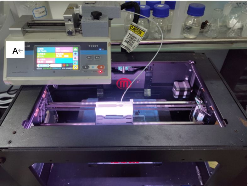
Cellulose hydrogel skeleton by extrusion 3D printing of solution | 347
2.3 Processing of cellulose hydrogel cross section part of the scaffolds, which were dipped in
scaffolds liquid N2 before cutting. All the samples were coated with
gold powder by spraying for 60 seconds at 10 mA.
The 6.7 wt% cellulose solution was adopted for the 3D
printing. The 3D printed scaffold was put into deion-
ized water (DI water) to conduct solvent exchange to ob- 3.4 The aggregation structure analysis
tain the cellulose hydrogel scaffold. For improving the re-
swellability of the 3D printed scaffold, before the solvent To compare the aggregation structures with and without
exchange in DI water, the 3D printed scaffold was kept in crosslinking treatment of the 3D printed scaffolds, the XRD
the MBA/DMSO solution with different concentrations for was performed on a X-ray diffractometer (X’pert PRO, PAN
12 hours at 50∘ C to form chemical cross-linkage inside the alytical, Holland) with copper radiation (λ = 0.154056 nm).
cellulose scaffold. The final 3D printed cellulose hydrogel The scan range was from 5 to 60∘ , with a scanning speed
scaffolds were freeze-dried to observe the morphology by of 10∘ /min.
SEM.
3.5 Mechanical properties
3 Characterization To evaluate the mechanical properties of the cellulose hy-
drogel scaffolds, the compression tests were performed on
3.1 The rheological property analysis an electronic universal testing machine (CMT4000, Sansi-
taijie, China) with a compressing speed of 5 m/min. The
To find out the suitable concentration of the cellulose so-
average value of five samples was taken as the final result.
lution for 3D printing, the rheological behavior was stud-
ied. The rheological property was tested by a Control Stress
Rheometer (TA instruments, America). The viscosity of
3.6 Re-swelling performance
the cellulose solution was measured in steady state with
the shear rate ranging from 0 to 100 s−1 . To test the stor-
The re-swelling performance of cellulose hydrogel scaf-
age moduli (G′ ) and loss moduli (G′′ ), a logarithmic stress
folds was evaluated by drying the samples and then dip-
sweep was plotted at a frequency of 1 Hz. The frequency
ping into DI water to swell, and calculate the swelling ra-
sweep was performed in the range from 0 to 100 Hz, at a
tion (SR) using the Eq. (1),
constant strain rate of 1%. This strain rate was chosen after
performing the linear viscoelastic region sweep. SR(%) = (Ws − W d )/W d · 100% (1)
where Ws is the wet weight of hydrogel after absorbing DI
3.2 Chemical structure characterization water to a swelling balance, and W d is the weight of the
dried 3D printed cellulose scaffolds. The average value of
To analyze the interactions between the functional groups three testing results was taken as the final result.
of the components consisted in the hydrogel scaffolds,
with and without crosslink agent treatment, Fourier trans-
form infrared spectroscopy (FT-IR) was recorded on a FT-IR
spectrometer equipped with ATR accessory (Nicolet 6700,
4 Results and discussion
Nicolet, America). The spectra were recorded in hydrogel
samples with a resolution of 4 cm−1 and an accumulation 4.1 The rheological property of the cellulose
of 50 scans in the spectral range of 400-4000 cm−1 at room solution
temperature.
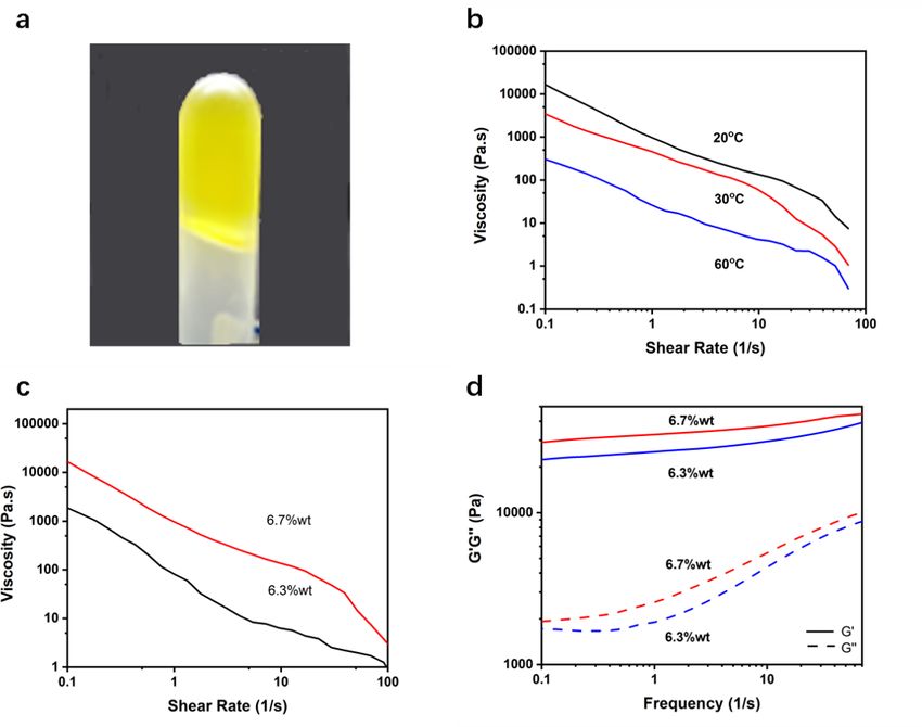
The rheological properties of cellulose solutions were
tested, and the results were collected in Figure 2. As shown
3.3 Morphology analysis in Figure 2a, the 6.7 wt% cellulose solution appeared as a
semisolid. The rheological properties of the cellulose so-
The morphology of the freeze-dried 3D printed hydrogel lutions under different temperatures (Figure 2b, 6.7 wt%
scaffolds was studied with a scanning electron microscope cellulose solution) and with different concentrations (Fig-
(QUANPA200, FEI, Holland). To observe the morphology of ure 2c, 20∘ C) were tested separately as a function of shear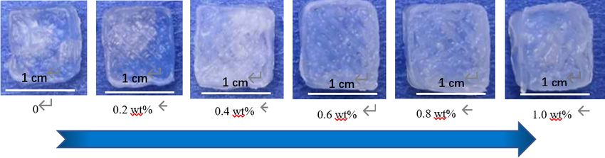
348 | Xiangzhou Hu et al.

Figure 2: The rheological properties of the viscous cellulose solutions: (a) optical picture of the 6.7 wt% cellulose solution; (b) the viscosity
of the 6.7 wt% cellulose solution as a function of shear rate under different temperatures; (c) the viscosity of the cellulose solutions with
different concentrations (6.3 wt% and 6.7 wt%) as a function of shear rate; (d) the storage modulus (G′ , the hard line) and the loss modulus
(G′′ , the dotted line) of the cellulose solutions with different concentrations (6.3 wt% and 6.7 wt%) under room temperature

rate. With the increasing of the shear rate, the viscosity                would be produced for the molecular chains to move eas-
of the cellulose solution decreased obviously, which pre-                 ier. Such rheological properties of the cellulose viscous so-
sented a strong non-Newtonian shear thinning behavior.                    lutions provided the necessities their 3D printing to keep
The characteristics of the viscous cellulose solution was                 the stable dimension and precise shape.
beneficial for the extrusion 3D printing under room tem-
perature. They were easy to be extruded out through the
printing nozzle and then changed into solid-like to form                  4.2 The morphology of the 3D printed
a prescribed shape. As summarized in the Figure 2c, the                       cellulose hydrogel scaffolds
solid lines, presented the storage modulus (G′ ), were above
the loss modulus (G′′ ) within measuring range. The rhe-                  The 6.7 wt% cellulose solution was applied for the 3D
ological behavior of the cellulose solution was more like                 printing under room temperature. The 3D printed scaffolds
a solid, so they were suitable for conducting 3D printing                 were immersed in DI water to conduct regeneration and
under room temperature. It could be explained that the                    solvent exchange to produce the aim cellulose hydrogel
entanglement force and hydrogen bond between molec-                       scaffolds, as shown in Figure 3. As it could been seen
ular chains decreased, and the molecular chains would                     that the 3D printed scaffolds, both the as printed samples
move easily with increasing the shear rate. In the mean-                  (a and b) and the hydrogels (a1 and b1 ) after solvent ex-
time, with increasing the temperature, more free volume                   change with DI water kept the prescribed shape according
Cellulose hydrogel skeleton by extrusion 3D printing of solution | 349
Figure 3: The digital pictures of the 3D printed cellulose scaffolds: a, b – as-printed samples; a1 , b1 – the hydrogel scaffolds; a2 , b2 – the
freeze-dried samples
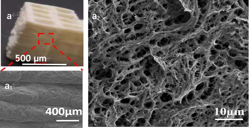
Figure 4: SEM images of the freeze-dried 3D printed cellulose scaffold: (a) the layer by layer printed 3D grid shape; (a1 ) the morphology of
the surface of the layers; (a2 ) the cross section
to the designed CAD models. While, the freeze-dried sam- in Figure a2 , the regenerated cellulose fibers in different
ples (a2 and b2 ) showed some shrinkage. diameters agglomerated and formed hierarchical pores,
In order to take observation into the morphology of the which contributed micro and macro chambers to contain
layer by layer printed 3D cellulose scaffolds, the SEM was water.
applied, the images were shown in Figure 4. As shown in
Figure 4a and 4b, the side view presented apparent layer
by layer stacked structure. The cross section was shown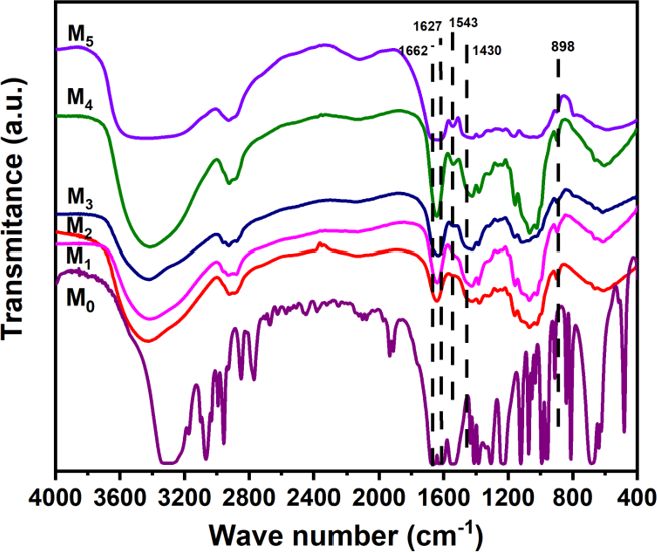
350 | Xiangzhou Hu et al.

Figure 5: The re-swelled 3D printed cellulose hydrogel samples treated by cross linking agent solutions with different concentrations

Table 1: The re-swelling property of the 3D printed cellulose hydrogel scaffolds treated by MBA/DMSO solution.

  MBA content (wt%)                  0                 0.2                0.4                0.6                 0.8              1.0
      RS (g/g)                       14                19                 22                 24                  28               26

4.3 The impact of the crosslinking agent
    treatment on the properties of the 3D
    printed cellulose hydrogel scaffolds
The re-swelling ability is one of the most important prop-
erty for hydrogel materials. While, the re-swelling ratio of
the as prepared cellulose hydrogel scaffold is only 14 wt%.
To improve the re-swelling ability, the crosslinking agent
(N, N’-Methylenebisacrylamide, MBA) was introduced into
the 3D printed cellulose hydrogel scaffolds, by immersing
the printed cellulose scaffolds into the MBA/DMSO solu-
tion with concentrations ranging from 0 to 1.0 wt%. The
pictures of the re-swelled samples were shown in Figure 5.
The sample treated in the 0.8 wt% cross link agent solution
had the best re-swelling property, which adsorbed 28 g/g
DI water of its own weight. With increasing the concentra-
                                                                       Figure 6: The compression strength of the cross-linking agent
tion of the MBA content to 1.0%, the re-swelling ability of
                                                                       treated cellulose hydrogel scaffolds
the 3D printed cellulose hydrogel decreased to 24 g/g. The
re-swelling results of the samples treated by MBA/DMSO
solution with different concentrations were summarized in              strength considerably increased from 45 to 75 kPa when
Table 1, and the photos were shown in Figure 5.                        the concentration of MBA in the cross-linking medium in-
                                                                       creased from 0.4 to 0.6 wt%.

4.4 The compression properties of the
    cross-linking agent treated 3D printed                             4.5 Chemical structure analysis by FTIR
    cellulose hydrogel scaffolds                                           spectra

The mechanical property is important for the practical uti-            In order to understand the interact of the cross-linking
lization of the hydrogel materials, which has also been                agent with the cellulose, the MBA treated cellulose hydro-
tested, taking the reported method [44] as reference. As               gel scaffolds were freeze dried and analyzed by FTIR, as
shown in Figure 6, with increasing the concentration of                shown in Figure 7.
the MBA in the solution, the compression strength was in-                   The strong adsorption at 1662 and 1627 cm−1 , ascribed
creased from 36 to 103 kPa. Especially, the compression                to the stretching vibration of C=C and C=O in MBA, respec-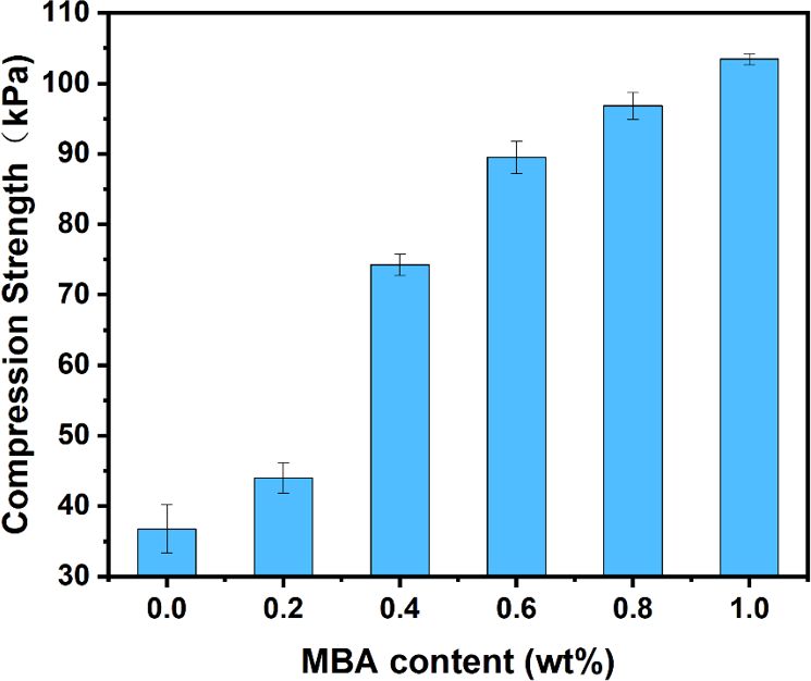
Cellulose hydrogel skeleton by extrusion 3D printing of solution | 351

Figure 7: FTIR spectra of the cross-linking agent treated cellulose
                                                                        Figure 8: XRD patterns of the cross-linking agent treated cellulose
hydrogel scaffolds. M0 : the MBA, M1 -M5 : the cellulose hydrogel
                                                                        scaffolds. N0 -N5 corresponding to the cellulose scaffolds treated
scaffolds treated with 0.2, 0.4, 0.6, 0.8, and 1.0 wt% MBA solutions,
                                                                        with 0, 0.2, 0.4, 0.6, 0.8, 1.0 wt% MBA solutions, respectively
respectively

tively. The peak at 1543 cm−1 and 1430 cm−1 represented to              crease of hydrogen bonds, and hence decreased the crys-
the bending vibration of N-H and C-N in MBA [44]. Those                 tallinity.
typical vibrations presented amide structure of MBA. As
shown in the M1 -M5 of Figure 7, with the increasing of the
MBA in the cross-linking agent solution, the representa-                5 Conclusion
tive adsorptions at 1543 cm−1 and 1430 cm−1 became en-
hanced. The characteristic adsorption of β-1,4-D-glycosidic             In summary, the natural cotton cellulose solution in
bond in cellulose at 897 cm−1 were obvious in the M1 -M5 of             dimethyl sulfoxide and tetrabutylammonium hydroxide
Figure 7. Additionally, with the introduction of cross-link             aqueous solution (DMSO/TBAH/H2 O) was found to be
agent, the stretching vibration in about 3400 cm−1 became               suitable for extrusion 3D printing for its special rheo-
wider and some red shift, which indicated the hydrogen                  logical properties. It presented solid-like behavior un-
bonds formed between the carbonyl groups of MBA and                     der room temperature, and a strong non-Newtonian
the hydroxyl groups of cellulose. As a result, the MBA was              shear thinning property. With introduction of N, N’-
successfully introduced into the cellulose scaffolds.                   Methylenebisacrylamide (MBA) into the 3D printed cellu-
                                                                        lose hydrogel scaffolds, the compression as well as the
                                                                        re-swelling properties were considerably improved. This
4.6 The crystal structure analysis by XRD                               work provides a new way for construct complex cellulose
    diffraction                                                         hydrogel scaffolds for practical application by 3D printing.

The cross-linking agent treated cellulose scaffolds were                Acknowledgement: The authors acknowledge the finan-
freeze dried to conduct the XRD analysis to study the effect            cial support from the International Cooperation Project of
of the MBA on the crystal structures of cellulose, as shown             Sichuan Province (NO.2018HH0087) and the Sichuan Ma-
in Figure 8.                                                            jor Science and Technology Project (NO.2019ZDZX0018).
     According to the XRD diffraction patterns in Figure 8,
with the increase of MBA content in the cross-linking solu-
tion, the diffraction peaks of the cellulose in the 110 and
200 crystal planes decreased in intensity, while the 110
                                                                        References
crystal plane gradually disappeared. As mentioned before,
                                                                        [1]   Kelly BE, Bhattacharya I, Heidari H, Shusteff M, Spadaccini CM,
the cross-linking interaction between cellulose and MBA                       Taylor HK. Volumetric additive manufacturing via tomographic
consumed the -OH at C6 in cellulose, which caused the de-                     reconstruction. Science. 2019 Mar;363(6431):1075–9.
352 | Xiangzhou Hu et al.
[2] Saha SK, Wang D, Nguyen VH, Chang Y, Oakdale JS, Chen SC. structures via multi-materials-multi-methods printing. Addit.
Scalable submicrometer additive manufacturing. Science. 2019 Manuf. 2019;28:14–22.
Oct;366(6461):105–9. [22] Hausmann MK, Rühs PA, Siqueira G, Läuger J, Zimmermann T,
[3] Walker DA, Hedrick JL, Mirkin CA. Rapid, large-volume, thermally Studart AR. 3D printed cellulose nanocrystal composites through
controlled 3D printing using a mobile liquid interface. Science. digital light processing. ACS Nano. 2018;12(7):6926–37.
2019 Oct;366(6463):360–4. [23] Hakansson KM, Henriksson IC, Vazquez CD, Kuzmenko V, Mark-
[4] Kelly CN, Miller AT, Hollister SJ, Guldberg RE, Gall K. Design stedt K, Enoksson P, et al. Solidification of 3D Printed Nanofibril
and Structure-Function Characterization of 3D Printed Synthetic Hydrogels into Functional 3D Cellulose Structures, Adv. Mater
Porous Biomaterials for Tissue Engineering. Adv Healthc Mater. Technol. 2016;1(7):1600096.
2018 Apr;7(7):e1701095. [24] Xu C, Zhang Molino B, Wang X, Cheng F, Xu W, Molino P, et al.
[5] Li RQ, McCarthy A, Zhang YS, Xie JW. Decorating 3D Printed Scaf- 3D printing of nanocellulose hydrogel scaffolds with tunable
folds with Electrospun Nanofiber Segments for Tissue Engineer- mechanical strength towards wound healing application. J Mater
ing. Adv. Biosys. 2019;3(12):1900137. Chem B Mater Biol Med. 2018 Nov;6(43):7066–75.
[6] Richards DJ, Tan Y, Jia J, Yao H, Mei Y. 3D Printing for Tissue Engi- [25] Huang L, Du X, Fan S, Yang G, Shao H, Li D, et al. Bacterial
neering. Isr J Chem. 2013 Oct;53(9-10):805–14. cellulose nanofibers promote stress and fidelity of 3D-printed
[7] Pearre BW, Michas C, Tsang JM, Gardner TJ, Otchy TM. Fast micron- silk based hydrogel scaffold with hierarchical pores. Carbohydr
scale 3D printing with a resonant-scanning two-photon micro- Polym. 2019 Oct;221:146–56.
scope. Addit. Manuf. 2019;30:100887. [26] Giordano RA, Wu BM, Borland SW, Cima LG, Sachs EM, Cima
[8] Kalms M, Narita R, Thomy C, Vollertsen F, Bergmann RB. New MJ. Mechanical properties of dense polylactic acid structures
approach to evaluate 3D laser printed parts in powder bed fusion- fabricated by three dimensional printing. J Biomater Sci Polym
based additive manufacturing in-line within closed space. Addit. Ed. 1996;8(1):63–75.
Manuf. 2019;26:161–5. [27] Kiendl J, Gao C. Controlling toughness and strength of FDM 3D-
[9] Gelber MK, Hurst G, Comi TJ, Bhargava R. Model-guided design printed PLA components through the raster layup, Compos. Pt.
and characterization of a high-precision 3D printing process for B-Eng. 2020;180:107562.
carbohydrate glass. Addit. Manuf. 2018;22:38–50. [28] Kaygusuz B, Ozerinc S. Improving the ductility of polylactic acid
[10] Grigoryan B, Paulsen SJ, Corbett DC, Sazer DW, Fortin CL, Zaita parts produced by fused deposition modeling through polyhy-
AJ, et al. Multivascular networks and functional intravascu- droxyalkanoate additions. J Appl Polym Sci. 2019;136(43):48154.
lar topologies within biocompatible hydrogels. Science. 2019 [29] Tian XY, Liu TF, Wang QR, Dilmurat A, Li DC, Ziegmann G. Recy-
May;364(6439):458–64. cling and remanufacturing of 3D printed continuous carbon fiber
[11] Lee A, Hudson AR, Shiwarski DJ, Tashman JW, Hinton TJ, Yerneni reinforced PLA composites. J Clean Prod. 2017;142(4):1609–18.
S, et al. 3D bioprinting of collagen to rebuild components of the [30] Torrado AR, Shemelya CM, English JD, Lin YR, Wicker RB, Rober-
human heart. Science. 2019 Aug;365(6452):482–7. son DA. Characterizing the effect of additives to ABS on the me-
[12] Bose S, Vahabzadeh S, Bandyopadhyay A. Bone tissue engineer- chanical property anisotropy of specimens fabricated by material
ing using 3D printing. Mater Today. 2013;16(12):496–504. extrusion 3D printing. Addit. Manuf. 2015;6:16–29.
[13] Murphy SV, Atala A. 3D bioprinting of tissues and organs. Nat [31] Dilberoglu UM, Simsek S, Yaman U. Shrinkage compensation ap-
Biotechnol. 2014 Aug;32(8):773–85. proach proposed for ABS material in FDM process. Mater Manuf
[14] Mannoor MS, Jiang Z, James T, Kong YL, Malatesta KA, Soboyejo Process. 2019;34(9):993–8.
WO, et al. 3D printed bionic ears. Nano Lett. 2013 Jun;13(6):2634– [32] Samykano M, Selvamani SK, Kadirgama K, Ngui WK, Kanagaraj G,
9. Sudhakar K. Mechanical property of FDM printed ABS: influence
[15] Rai M, dos Santos JC, Soler MF, Marcelino PR, Brumano LP, In- of printing parameters. Int J Adv Manuf Technol. 2019;102(9-
gle AP, et al. Strategic role of nanotechnology for production of 12):2779–96.
bioethanol and biodiesel. Nanotechnol Rev. 2016;5(2):231–50. [33] Levenhagen NP, Dadmun MD. Improving Interlayer Adhesion
[16] Lemée L, Kpogbemabou D, Pinard L, Beauchet R, Laduranty J. in 3D Printing with Surface Segregating Additives: Improving
Biological pretreatment for production of lignocellulosic biofuel. the Isotropy of Acrylonitrile-Butadiene-Styrene Parts. ACS Appl.
Bioresour Technol. 2012 Aug;117:234–41. Polym. Mater. 2019;1(4):876–84.
[17] Zhao X, Luo K, Zhang Y, Zheng Z, Cai Y, Wen B, et al. Improving [34] Park SJ, Lee JE, Park JH, Lyu MY, Park K, Koo MS, et al. FDM 3D
the methane yield of maize straw: focus on the effects of pre- Printing of Environmental Friendly and High Strength Bio-based
treatment with fungi and their secreted enzymes combined with PC Filaments for Baby Toys. Elastom. Compos. 2017;52(2):99–
sodium hydroxide. Bioresour Technol. 2018 Feb;250:204–13. 104.
[18] Li ZH, Xu K, Pan YS. Recent development of Supercapaci- [35] Yap YL, Toh W, Koneru R, Lin KH, Yeoh KM, Lim CM, et al.
tor Electrode Based on Carbon Materials. Nanotechnol Rev. A non-destructive experimental-cum-numerical method-
2019;8(1):35–49. ology for the characterization of 3D-printed materials-
[19] Gao T, Xu C, Li R, Zhang R, Wang B, Jiang X, et al. Biomass-Derived polycarbonate-acrylonitrile butadiene styrene (PC-ABS). Mech
Carbon Paper to Sandwich Magnetite Anode for Long-Life Li-Ion Mater. 2019;132:121–33.
Battery. ACS Nano. 2019 Oct;13(10):11901–11. [36] Shi Q, Yu K, Kuang X, Mu XM, Dunn CK, Dunn ML, et al. Recyclable
[20] Sultan S, Mathew AP. 3D printed scaffolds with gradient porosity 3D printing of vitrimer epoxy. Mater Horiz. 2017;4(4):598–607.
based on a cellulose nanocrystal hydrogel. Nanoscale. 2018 [37] Pierson HA, Celik E, Abbott A, De Jarnette H, Gutierrez LS, Johnson
Mar;10(9):4421–31. K, et al. Mechanical Properties of Printed Epoxy-Carbon Fiber
[21] Li VC, Kuang X, Hamel CM, Roach D, Deng YL, Qi HJ. Cellulose Composites. Exp Mech. 2019;59(6):843–57.
nanocrystals support material for 3D printing complexly shapedCellulose hydrogel skeleton by extrusion 3D printing of solution | 353
[38] Wang X, Jiang M, Zhou ZW, Gou JH, Hui D. 3D printing of polymer [42] Cao J, Wei W, Gou GJ, Jiang M, Cui YH, Zhang SL, et al. Cel-
matrix composites: A review and prospective, Compos. Pt. B-Eng. lulose films from the aqueous DMSO/TBAH-system. Cellulose.
2017;110:442–58. 2018;25(3):1975–86.
[39] Lei M, Chen Z, Lu HB, Yu K. Recent progress in shape mem- [43] Bu D, Hu X, Yang Z, Yang X, Wei W, Jiang M, et al. Elucidation of the
ory polymer composites: methods, properties, applications and Relationship between Intrinsic Viscosity and Molecular Weight
prospects. Nanotechnol Rev. 2019;8(1):327–51. of Cellulose Dissolved in Tetra-N-Butyl Ammonium Hydrox-
[40] Yang H, Leow WR, Wang T, Wang J, Yu J, He K, et al. 3D Printed ide/Dimethyl Sulfoxide. Polymers (Basel). 2019 Oct;11(10):E1605.
Photoresponsive Devices Based on Shape Memory Composites. [44] Geng H. A one-step approach to make cellulose-based hydrogels
Adv Mater. 2017 Sep;29(33):1701627. of various transparency and swelling degrees. Carbohydr Polym.
[41] Wu W, Ye W, Wu Z, Geng P, Wang Y, Zhao J. Influence of Layer 2018 Apr;186:208–16.
Thickness, Raster Angle, Deformation Temperature and Recovery
Temperature on the Shape-Memory Effect of 3D-Printed Polylactic
Acid Samples. Materials (Basel). 2017 Aug;10(8):E970.You can also read