A comparative morphological study of titanium dioxide surface layer dental implants
←
→
Page content transcription
If your browser does not render page correctly, please read the page content below

Open Chemistry 2021; 19: 189–198
Rapid Communication
Dragos Vladimir Budei, Danut-Ionel Vaireanu*, Petronela Prepelita, Gianina Popescu-Pelin,
Mihaela Mincu, Ioana-Alina Ciobotaru
A comparative morphological study of titanium
dioxide surface layer dental implants
https://doi.org/10.1515/chem-2021-0197 aforementioned implants, providing them with a tool in
received February 10, 2020; accepted November 28, 2020 choosing the proper dental implant to suit their needs. It
Abstract: Most dental implants used in dental practices are was found that, as the technology evolved and the costs
made of titanium or titanium alloys so that the essential were reduced, there is a net preference for using pure tita-
differences promoted by the various manufacturers are nium or its alloys in the manufacture of dental implants
at the level of their surface; through specific surface treat- versus the stainless steel titanium alloys, considered now
ments, the aim is to obtain improved results regarding a thing of the past.
osseointegration. This study attempts to identify the differ- Keywords: dental materials, implants surface, surface
ences between a series of used brands of dental implants by analysis
analyzing the chemical composition and the morphology of
their surface and is particularly significant for the potential
users as it highlights the manner of performances of the
1 Introduction
A significant number of researchers sought to identify
* Corresponding author: Danut-Ionel Vaireanu, Inorganic
over the time a number of biocompatible materials which
Chemistry, Physical Chemistry and Electrochemistry Department,
Faculty of Applied Chemistry and Material Sciences, Polytechnic can be used to replace and/or restore the missing or
University of Bucharest, 1-7 Gh Polizu St. s1, Bucharest, 011061, injured anatomical structures. It is considered that a mate-
Romania, e-mail: di_vaireanu@yahoo.co.uk, tel: +40-21-402-3939, rial must meet at least three conditions in order to be used
fax: +40-21-402-3939 for implantation: to be biocompatible, to have a balance of
Dragos Vladimir Budei: Dentix Millennium S.R.L., Str. Crinului 2,
physical and mechanical properties so that it performs as
Sabareni, 087153, Giurgiu, Romania; Inorganic Chemistry, Physical
expected, and the medical device should be relatively easy
Chemistry and Electrochemistry Department, Faculty of Applied
Chemistry and Material Sciences, Polytechnic University of to produce in large volumes, to the same standard [1].
Bucharest, 1-7 Gh Polizu St. s1, Bucharest, 011061, Romania, Biotolerant materials (Co–Cr alloys, stainless steel,
e-mail: budei.dragos@dentixmillennium.ro tantalum, or some polyols), bioinert materials (titanium
Petronela Prepelita: National Institute for Laser, Plasma and and its alloys, zirconia), or even bioactive materials
Radiation Physics, Department of Lasers, 409 Atomistilor Street,
(hydroxyapatite) have been identified, but the constraints
PO Box MG-36, 077125, Magurele, Ilfov, Romania,
e-mail: petronela.garoi01@gmail.com
due to the small size of devices and the increased
Gianina Popescu-Pelin: National Institute for Laser, Plasma and mechanical stress they are subjected to led over the
Radiation Physics, Department of Lasers, 409 Atomistilor Street, time to a preference for bioinert materials such as tita-
PO Box MG-36, 077125, Magurele, Ilfov, Romania, nium and its alloys, which have proven to represent an
e-mail: gianina.popescu@inflpr.ro optimal solution for dental implants. Their enhanced bio-
Mihaela Mincu: Dentix Millennium S.R.L., Department of Quality
compatibility and other desired properties and qualities
Assurance, Str. Crinului 2, Sabareni, 087153, Giurgiu, Romania;
University of Agronomic Sciences and Veterinary Medicine of made the manufacturers to choose titanium and its alloys
Bucharest, Bucharest, Romania, as raw materials for dental implants over the stainless
e-mail: quality@dentixmillennium.ro steel titanium alloys [2–15]. Titanium reacts with many
Ioana-Alina Ciobotaru: Inorganic Chemistry, Physical Chemistry and other chemical elements (Ag, Al, V, Zn etc.) to form
Electrochemistry Department, Faculty of Applied Chemistry and
alloys. The American Society for Testing and Materials
Material Sciences, Polytechnic University of Bucharest, 1-7 Gh
Polizu St. s1, Bucharest, 011061, Romania,
organized titanium and its alloys on a scale of 1–39.
e-mail: ioanaalinaciobotaru@yahoo.com, tel: +40-21-402-3939, The first four grades are of pure (non-alloy) titanium
fax: +40-21-402-3939 types and the others for alloys. The alloys are organized
Open Access. © 2021 Dragos Vladimir Budei et al., published by De Gruyter. This work is licensed under the Creative Commons Attribution
4.0 International License.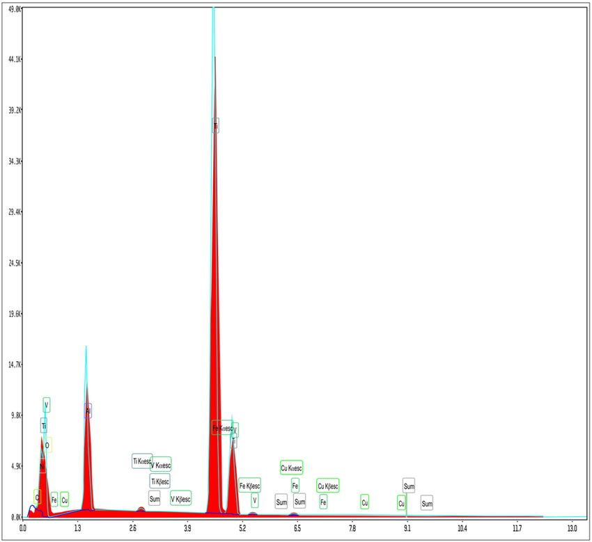
190 Dragos Vladimir Budei et al.
into three broad categories, Alpha, Alpha Beta, and Beta, with oxygen is important. The formation of a TiO2 layer on
and their characteristics, properties, preferences, and uses the surface of titanium and titanium alloys cannot be
differ widely based on the particular type of alloy chosen avoided; this layer forms spontaneously, upon contact
[14–23]. Alpha alloys are most commonly made of aluminum with oxygen from atmospheric air [4,5,21,22].
and tin, which makes them ductile, with high hardness,
gives them good mechanical properties at low tempera-
tures, has the highest corrosion resistance, cannot be 2 Materials and methods
forged at high temperatures, but they can be welded.
Alpha Beta alloys are more durable, but Alpha alloys In this research, we compared a Dentix SLA implant
are strong enough to make them a preferred choice in (sample 10) as well as a Dentix Nano implant (sample
chemical or aeronautical equipment. Beta alloys have 11), produced by the company Dentix Millennium in
medium to high hardness, can be forged or processed Romania with nine other implants from well-known
at high temperatures, they are weldable, and are used reputable manufacturers of dental implants, purchased,
in aeronautics, in the development of prosthetic ele- and sealed in the original packaging from local distribu-
ments, and in marine equipment. Their elevated density tors (Adin, Alpha-Bio, Ankylos, Israel OEM, Megagen,
makes them particularly suitable for the production of Nobel Biocare, Osstem, Ritter, Straumann, Zimmer) ran-
certain parts which must maintain their shape and struc- domly named from S1 to S9. All the samples were sub-
ture even at the highest pressures. jected to electron microscopy investigations. In order to
Titanium Grade 2, Titanium Grade 4, and Ti6Al4V- limit the variable to a minimum possible, all the investi-
ELI alloy (Titanium Grade 23) are the most widely used gated implants had a diameter between 4.0 and 4.3 mm
materials in the production of dental implants [15]. In and a length between 12.5 and 13 mm. The average sur-
Table 1, a comparison of the technical data of these raw face area of these implants, measured on the 3D model,
materials [2,3,15], namely, the chemical composition and was 209.2486 mm2. This surface area is increased by the
the associated physical characteristics for the aforemen- processes of roughness modification. The morphological
tioned materials, is presented. characterization of samples was carried out using scanning
It can be observed that the values for fracture tough- electron microscopy (SEM) with an FEI Inspect S50 apparatus
ness are close for TiCP4 to Ti6Al4V-ELI alloy (Ti Grade 23). (FEI, Hillsboro, OR, USA), used with energy-dispersive X-ray
The metallographic analysis reveals the lack of homo- spectroscopy (EDS) system produced by EDAX (Mahwah, NJ
geneity in the titanium alloys [20]: the presence of alloying 07430, USA), with fixed silicon detector and Peltier element
elements can induce microscopic defects. Last but not integrated as a cooling system. The SEM analyses were per-
least, the reactivity to the ambient temperature of titanium formed at an acceleration voltage of 20–30 kV, in the magni-
fication range of 50–50,000×, at the working distance of
10 cm, with a spot beam 2. For EDS, the beam spot used
Table 1: Chemical composition and physical characteristics of
was 5.5–6, the working distance was 10 cm, and the dead
TiCP2, TiCP4, and Ti6Al4V-ELI (Ti Grade 23)
time was 33. Prior to microscope insertion and visualization,
Ti Grade 2 Ti Grade 4 Ti6Al4V-ELI
the samples were washed in isopropyl alcohol and then
rinsed with double distilled water. A scratch was made on
Maximum content of Al — — 5.5–6.5% the surface of each dental implant, in order to detect the
Maximum content of V — — 3.5–4.5%
differences in composition between the treated surface of
Maximum content of C 0.08% 0.08% 0.08%
Maximum content of N 0.03% 0.05% 0.05% the implant and its interior.
Maximum content of O 0.25% 0.4% 0.13%
Maximum content of Fe 0.3% 0.5% 0.25% Ethical approval: The conducted research is not related to
Maximum content of H 0.015% 0.015% 0.012% either human or animal use.
Maximum content of 0.4% 0.4% 0.4%
other elements in total
Minimum content of Ti 98.825% 98.455% 88.078%
Density (g/cm3) 4.51 4.51 4.42 3 Results and discussion
Elasticity modulus (GPa) 103–107 105 105–116
Melting point (C) 1,665 1,660 1,655 The results of the samples examined under the electron
Fracture 66 99–140 100 microscope, their surface morphology, the associated che-
toughness (MPa)
mical composition, and the corresponding SEM images are
Shear modulus (GPa) 45 40 44
presented in Figures 1–12.Study of titanium dioxide surface layer dental implants 191
Figure 1 shows the image of sample S1 at a magnifi-
cation of 20k×. At magnifications of 200×, 1,000× we
observe the uniform appearance, with fine structure of
holes on the surface of the material, with an appearance
similar to the surface corroded with acid. The surface,
analyzed in detail, has a certain degree of roughness,
its appearance changing from uneven wells, with thin,
irregular, corroded (5,000×) wells and walls toward
ridges and concavities of different shapes with several
microns, 1–4 µm in the image. Similar results were found
with other implants with their surface sample treated
with acids, presented in detailed images (20,000×),
where deep and lacy pores with diameters of 3–5 μm
can be noticed. In Figure 2, the surface treatment origin-
ally applied to sample S2 ensured a homogeneous,
Figure 3: SEM image of sample S3 at a magnification of 20k×.
Figure 1: SEM image of sample S1 at a magnification of 20k×. Figure 4: SEM image of sample S4 at a magnification of 20k×.
Figure 2: SEM image of sample S2 at a magnification of 20k×. Figure 5: SEM image of sample S5 at a magnification of 20k×.192 Dragos Vladimir Budei et al.
Figure 6: SEM image of sample S6 at a magnification of 20k×. Figure 9: SEM image of sample S9 at a magnification of 20k×.
abrasive appearance throughout the implant. At 2,000×,
there are rounded honeycomb-type alveoli in character-
istic assemblies with an appearance similar to the acid-
corroded surface. At higher magnifications (20,000×), the
rounded alveoli have the appearance of deep pores with
diameters of 1.5–3 microns, some joined, rounded but
irregular, with unevenly corroded walls. Figure 3 shows
the image of sample S3 at a magnification of 20k×. At
magnifications over 1,000, the investigated implant pre-
sents a relatively uneven, flattened appearance, a struc-
ture with ridges and valleys, resembling a honeycomb,
consisting of asperities, grains, and pores (5,000×) of 1–2
microns, circular (image 20k×), sometimes rectangular.
Figure 4 shows the image of sample S4 at 20k× magnifi-
cation. The SEM image at low magnification (200×) indi-
Figure 7: SEM image of sample S7 at a magnification of 20k×. cates a uniform appearance of the implant, with dunes
Figure 8: SEM image of sample S8 at a magnification of 20k×. Figure 10: SEM image of sample S10 at a magnification of 5k×.Study of titanium dioxide surface layer dental implants 193
samplebly subjected: sand blasting and acid corrosion
(SLA): rough relief, with craters, irregular inclusions,
grooves (200× and 2,000×), all generating an expanded
surface for easier cell attachment. In detail, we can distin-
guish inclusions of different angular and/or semi-rounded
shapes and micron grains embedded in the irregular,
uneven surface, full of pores (200×, 5,000×, 20,000×) of
various diameters (0.5–4 μm). Figure 7 shows the image of
S7 sample at a magnification of 20k×. Observed at low
magnifications (200×), the surface of this implant has a
similar appearance to the blast. Furthermore, at 2,000×
the unevenly corroded appearance becomes visible, with
untouched areas (with a size of 1–20 microns) juxtaposed
with the corroded ones. Going forward with the enlarge-
ment of the images (5,000×), the structure is revealed as
Figure 11: SEM image of sample S11 at a magnification of 5k×.
very porous, attacked deeper, consisting of islands of
uncorroded material between relatively circular depths
with attacked walls, full of formations samplebly given
by the corrosion with the acids used in the treatment.
The rough structure is spread from depressions of 15–30
microns (2,000×) to the finest pores, even submicron
(20,000×), of different sizes. In Figure 8, it is observed
that this dental implant presents a continuous appearance
with rounded alveolar structures (1.7–2.5 microns). Figure 9
shows the SEM image at 20k× of sample S9. The surface is
characterized by a slightly rough, smooth, homogeneous
appearance, with periods of ∼10 μm (200× and 1,000×).
The extended surface has numerous, relatively flattened
edges. In detail, a rugged, rocky structure (2,000× and
5,000×) is highlighted. A topography cauliflower-like is
also distinguished. At 20,000 magnifications, the material
appears loose, with pores of approximately 1 micron or
submicron. Figure 10 shows the image of S10 sample
Figure 12: SEM image of sample S11 at a magnification of 20k×. (Dentix Nano implant) at a magnification of 20k×. The
TiO2 nanostructure can be seen all over the surface, cov-
ering the Ti microstructure. The TiO2 nanostructure is
between 10 and 20 μm, with small grains. At higher mag- composed of a layer of TiO2 nanotubes, perpendicularly
nifications (5,000×), the corrugated relief, sand dune oriented on the implant surface. The TiO2 nanotubes on
type, is preserved, noticing concavities of 10–18 μm in the surface of the Dentix Nano implant have an average
diameter, with irregular holes and fine grains, of lengths of 100–200 nm in height, 60–80 nm outer diameter,
of ∼1 μm (20,000×), stuck in the walls of the recesses. The 40–60 nm inner diameter, and 10–15 nm wall thickness.
surface type looks similar to acid corroded surface (SLA). The EDS analysis also provides a graphical represen-
In Figure 5, the SEM analysis at 20k× magnification tation of the peaks of the spectra emitted by the various
shows simple (0) and joined (double – 8/∞) tubular chemical elements in the composition of all samples ana-
cylindrical structures, rarely triple, with depth aspect, lyzed. In Figures 13 and 14, one may see two representative
with outer diameters 4–5 microns and inner diameters graphs for sample S4 and sample S6 depicting the ele-
1.5 to 2 microns. Distinct deep tubular pores with walls ments with highest mass percentages. For sample S4, in
∼2 microns in diameter. On the implant analyzed the ratio Figure 13, the graph shows the highest mass percentage for
of enlarged tubes/pore surface (shorter tubes) is about titanium, which was expected, followed by oxygen, a
1:1. In Figure 6, the appearance of this dental implant is direct proof of titanium dioxide formation, followed by
characteristic of the two treatments to which it was vanadium and aluminum, confirming the existence of194 Dragos Vladimir Budei et al.
copper; as different manufacturers use different alloy
compositions, this technique may be used as a useful
tool in differentiating between two very similar implants
and making correlations also between the chemical sur-
face composition and their osseointegration behavior.
The results of EDS investigations for S1–S11 samples
and the interpretations associated with these analyses
are presented in Table 2.
Most dental implants have surfaces with similar mor-
phology; the roughness is between 1 and 10 μm, and their
appearance is specific to the surfaces obtained by acid
attack (19) in the SLA-type surface treatment – treatment
that has already confirmed its performance in the dec-
ades since it is used. Some of the manufacturers, who
tried to differentiate by applying anodizing techniques,
obtained surfaces with different appearance, showing
craters instead of acid corrosion wells or fields of nano-
tubes. The presence of TiO2 nanotube layer increases the
surface area exposed to the body fluids by six times,
offering increased potential for body response and bone
integration. The TiO2 nanotubes on the surface of the
Dentix Nano implants have an average of 100–200 nm
Figure 13: EDS of sample S4.
in height, 60–80 nm outer diameter, 40–60 nm inner dia-
meter, and 10–15 nm wall thickness. The nanotubes cover
the entire surface of the implant, being joined together,
like a honeycomb. The ratio between the inner surface of
the cylinder and that covered by a nanotube is (the
average values were used) as follows:
150 × 50 nm × 3.14/(70 × 70 nm)/ 4 × 3.14
= 23, 550 nm2 / 3846.5 nm2 = 6.12.
The chemical composition of the materials from which
dental implants are made must be analyzed in the context
of the homogeneity of the respective material. If the raw
material is not homogeneous, dental implants, being a
very small device (a medium-sized implant – 4.2 × 11 mm,
weighs 0.4 g), may end up having marginally different
chemical compositions, even if they come from the same
bar of raw material with potential repercussions on the
behavior from the mechanical point of view, e.g., fatigue
resistance and/or microbial-induced corrosion resis-
tance. This is an additional reason for using pure tita-
Figure 14: EDS of sample S6. nium instead of titanium alloys, thereby reducing the
effects of compositional non-homogeneities, as well as
the alloying elements and traces of carbon, iron, and the creation of a localized galvanic reaction that can
chlorine. induce galvanic corrosion. The phenomenon of fracture
Figure 14 corresponding to EDS for sample S6 shows of dental implants is quite rare [18]. The percentage of
also the highest peak for titanium, followed by oxygen implants that fracture in the initial stages (at insertion or
and high values for aluminum and vanadium; one may in the first days) is about 1% after some authors [6], but
also notice the presence, besides carbon and iron of we consider this figure to be too high. The mechanicalStudy of titanium dioxide surface layer dental implants 195
Table 2: EDS compositional analysis of the studied samples
Sample EDS analysis results Description
Surface Weight (%) Atomic (%) Error (%)
element
S1 CK 0.00 0.00 99.99 EDS analysis indicates a titanium majority implant (67.91% mass,
OK 27.74 53.26 10.54 43.55% atomic), with an important contribution of oxygen (predictable
Ca K 3.48 2.67 3.07 on the surface of oxidized titanium layer, on the surface, 27.64% mass,
Ti K 67.91 43.55 1.39 53.26% atomic) and a significant calcium content (3.48%, 2.67%
VK 0.86 0.52 5.08 atomic). Treatment of this type of implant involves an extremely fine
layer (monoatomic, most likely) of Ca, in order to improve the
osseointegration
S2 CK 0.13 0.44 14.13 Titanium appears to be the only major chemical constituent of this
OK 10.57 26.05 10.58 implant (89.30% mass, 73.51% atomic) in quantifying EDS, next to
Ti K 89.30 73.51 1.31 oxygen (10.57% mass, 26.05% atomic), of which we can assume that it
exists only in the oxidized layer from the surface. Carbon samplebly
appears as a contaminant. In contrast, the compositional analysis in the
scratch has fine traces of calcium (0.14% atomic), aluminum (0.12%),
and silicon (1.51% atomic), samplebly due to the surface treatment with
the processing substances
S3 OK 14.55 34.51 10.73 The EDS compositional analysis confirms a titanium alloy (80–85% by
Al K 0.08 0.11 21.35 mass) with zirconium (6–5% by mass) and the remaining oxygen
Zr L 6.02 2.51 1.91
Ti K 89.30 73.51 1.31
S4 CK 2.96 8.47 9.12 EDS analysis confirms Ti–Al–V alloy (Titan Grade 5) as a material of this
NK 0.00 0.00 7.76 dental implant: Ti 73.11, Al 3.66, V 5.58, in mass percentages. Iron
OK 14.10 30.27 11.18 (0.40% by mass) and carbon (2.96% by mass) are also present as a
Al K 3.66 4.66 6.70 trace and as a minority element. On the scratched surface, the more the
Cl K 0.18 0.18 15.80 scratch is very fine, the ratio of the elements is about the same. The
Ti K 73.11 52.41 1.36 difference consists only in a larger amount of oxygen in the scratch
VK 5.58 3.76 2.53 (21.62%, compared to 14.10% by mass)
Fe K 0.40 0.25 22.45
S5 OK 33.11 EDS analysis indicates an implant based on pure, oxidized titanium
Ti K 66.09 (most likely on the surface: 66.09% atomic Ti with 33.11% atomic O, on
the surface, respectively, ∼74% atomic Ti with 25.2% atomic O). The
difference in oxygen between the implant surface and the inside
(scratch) may suggest that the tubular structures on the surface,
removed in the case of scratches, are those that contain more oxygen, in
accordance with the structure declared by the manufacturer, anodized
titanium, with titanium dioxide and phosphorus
S6 CK 0.04 0.10 72.63 EDS analysis shows the implant is made of Ti–Al–V alloy: in mass
NK 0.00 0.00 8.97 percent Ti 58.31, Al 8.32, V 4.83. Significant amount of oxygen, 28.28%
OK 28.18 51.95 10.46 by mass, 51.95% atomic, indicates the oxidation and passivation of the
Al K 8.32 9.09 6.07 titanium support. There are also fine traces of iron (0.16% atomic),
Ti K 58.31 35.90 1.37 possibly also carbon and copper (below 0.04% atomic, with an error
VK 4.83 2.97 2.54 margin over 50%)
Fe K 0.30 0.16 18.04
Cu K 0.02 0.01 58.11
S7 CK 1.15 3.50 9.64 EDS analysis indicates a titanium alloy based on Al and V as the implant
NK 0.00 0.00 6.92 material. Unexpectedly, the content of V (5.2–6.8% by mass) seems to
OK 12.32 28.21 10.94 be higher than that of Al (∼4% by mass). The phenomenon of surface
Al K 4.02 5.46 6.39 oxidation (passivation) is also highlighted by the significant oxygen
Ti K 76.98 58.89 1.31 content (∼12.3% by mass), both on its surface and in the applied
VK 5.18 3.72 2.08 scratches. On the surface, the sample also suffered a slight carbon
Cu K 0.35 0.20 9.44 contamination (1.15% mass, 3.50% atomic), most likely accidental. Fine
traces of Fe and Cu are also distinguished
S8 OK 7.52 19.56 11.80 EDS analysis shows mostly titanium content (91.89% mass, 79.79%
Al K 0.03 0.05 66.31 atomic per thread), without other alloying elements, with incorporated196 Dragos Vladimir Budei et al.
Table 2: Continued
Sample EDS analysis results Description
Surface Weight (%) Atomic (%) Error (%)
element
Si K 0.05 0.08 60.26 oxygen (7.52% mass, respectively, 19.56% atomic) and traces of Ca
Ca K 0.50 0.52 6.22 (0.50%), only on the surface. The titanium percentage increases and the
Ti K 91.89 79.79 1.33 oxygen decreases in the scratch, as the scratch deepens, as the
biocompatible layer of TiO2 forms at the surface
S9 CK 0.00 0.00 99.99 EDS analysis applied to the OEM implant indicates that it is made of
OK 21.47 43.28 10.50 Ti–Al–V (mass percentages: Ti 64.02; Al 7.94; V 4.92). The
Na K 0.00 0.00 99.99 compositional percentages differ from the Ti Grade 5 alloy recipe, most
Mg K 0.00 0.00 99.99 samplebly due to the fact that passivation oxygen appears from the
Al K 7.94 9.49 6.17 implant surface (21.47% by mass) and elements specific to the surface
Si K 0.04 0.05 43.89 treatment (And samplebly by the blasting process 0.04% by mass,
Ca K 0.35 0.28 8.09 possible as 0.35%) as well as possible contaminants (Cl, Fe, Ni, Cu, W).
Ti K 64.02 43.11 1.37 The traces of iron, nickel, and copper can come from the composition of
VK 4.92 3.11 2.21 the alloy. The relatively high percentage of Al could come from blasting
Fe K 0.94 0.54 8.37 with alumina particles
Ni K 0.05 0.03 57.84
Cu K 0.15 0.08 38.96
WL 0.12 0.02 56.20
S10 Ti K 99.83 99.85 1.15 EDS analysis indicates that the Dentix SLA implant is made of pure
Fe K 0.17 0.15 44.63 titanium (99.83% mass percentage), without alloying elements. The
presence of iron is lower than that declared by the producer of the raw
material
S11 OK 20.85 43.72 11.18 EDS analysis shows that the Dentix Nano implant is made of pure
Al K 1.52 1.89 8.72 titanium, without any other alloying elements. Aluminum and silicon are
Si K 0.06 0.08 45.69 samplebly derived from the blasting material, and the iron is below the
Ti K 77.39 54.20 1.18 limits declared by the raw material manufacturer. The oxygen is present
Fe K 0.18 0.11 31.83 in a high concentration, above the average of the compared implants,
demonstrating the abundance of the nanostructured layer of TiO2
properties of the least resistant material (Ti Grade 2 and Ti at identifying nanostructuring methods of the TiO2 layer
Grade 4) are more than sufficient for the dental implants which, when formed spontaneously, is in an amorphous
to fully respond to the proposed purpose [17], a fact state. The nanostructuring of the TiO2 layer will open
demonstrated by the fatigue studies carried out by our new opportunities leading the development of medical
team. The risk of fracture of dental implants is not a real devices to the next level. Regarding the chemical compo-
reason for manufacturers to opt for one material or sition of the titanium and its alloys, we consider some
another. Neither osseointegration performance justifies aspects as being particularly important and which were
the choice of Ti6Al4V alloy in favor of pure titanium not given the true importance: the purity and homo-
[11]. Due to the high reactivity of titanium with oxygen, geneity of the material from which dental implants are
as well as surface treatment procedures, all dental produced [7] and the biological effect of the presence of
implants have a layer of TiO2 that chemically stabilizes the alloying chemical elements. The low purity and non-
the surface (passivation). Titanium oxide plays a protec- homogeneity of the material from which dental implants
tive role, preventing contact of the titanium with other are produced is the main cause of their fracture. The
chemical elements, thus explaining the corrosion resis- process begins with microcracks observable under the
tance of this material. In fact, titanium dioxide is due to electron microscope, microcracks that either exist in
the biocompatibility of dental implants: the bone tissue the raw material, or occur at the first mechanical expo-
comes in close contact with the TiO2 layer, not with pure sure on the implant, due to the different resistance at
or allied titanium [12]. Recent concerns of the research different points of the piece. Even so, a number of factors
departments of dental implant manufacturers are aimed are needed for a fracture to occur, and this happens whenStudy of titanium dioxide surface layer dental implants 197
we are dealing with a small implant, where the tech- compositional differences have a beneficial effect on the
nology has been pushed to the limit. In our studies, we osseointegration of the implants and their durability over
encountered fractured implants, with a size of 3.3 mm × time. It was found that by comparing the chemical com-
8 mm, at which the minimum wall thickness (where it position of the various brands of dental implants with the
also failed) was 60 microns. It happened that in that chemical composition of the raw materials used, it is
area, the material was not homogeneous, and thus, the possible to construct an image of possible contamination
accumulation of factors occurred. The biological effect of (most samplebly of the surface) with other elements not
the chemical elements with which titanium alloying has initially existing in the raw material, as well as of the
been achieved is insufficiently studied. Aluminum and efficiency of the cleaning processes. As far as the prefer-
vanadium are not known to be chemical elements with ence of pure titanium versus its alloys is concerned, there
beneficial effects on the body [8–10]. The cell viability of is no need for an increased mechanical strength of alloys,
binary alloys (TiAl and TiV) is lower than that of pure these bringing about more negative issues and offsetting
titanium, TiCP [16]. Vanadium is cytotoxic at concentra- its relative advantages as long as pure titanium has suffi-
tions above 4% in alloys and neurotoxic [13]. We consider ciently good mechanical properties and better osseointe-
the use of vanadium too risky, given that the theoretical gration characteristics.
percentage is exactly at the limit of cytotoxicity (in Ti6Al4V,
vanadium has 4%). The metallurgical procedures for Acknowledgement: This research was supported by Dentix
obtaining titanium alloys, made on batches of tonnes, Millennium SRL through the project “Dental implants sur-
cannot have the precision of laboratory procedures, so face functionalization for improved osseointegration – an
that there is a guarantee that the vanadium level does not innovative method” (Metoda inovativă pentru funcţionalizarea
exceed 4% at all points in the volume of a batch. As evi- suprafeţelor implanturilor dentare cu scopul îmbunătăţirii
dence, the alloy manufacturers claim the presence of these osteointegrării) code MySMIS 104809, contract no. ANCSI
chemical elements with ranges of values, not with exact 73/8.09.2016. Dragos Vladimir Budei is a PhD student
values, and the chemical compositions presented above enrolled at the Politehnica University of Bucharest and
by EDS analysis also demonstrate different concentrations Mihaela Mincu is a PhD student enrolled at the University
in reality. All these risks are assumed free of charge as long of Agronomic Sciences and Veterinary Medicine of Bucharest.
as the benefits are not tailored, and above all, they are not
required. The increased mechanical strength of the Ti6Al4V Funding information: This research was supported by
alloy is not required, and the shortcomings that an alloy can Dentix Millennium SRL through the project “Dental
bring can negatively offset its relative advantages. Pure implants surface functionalization for improved osseo-
materials have sufficiently good properties, and statistics integration – an innovative method” (“Metoda inovativă
do not show that pure titanium implants fracture more often pentru funcţionalizarea suprafeţelor implanturilor den-
than alloy implants. tare cu scopul îmbunătăţirii osteointegrării”) code MySMIS
104809, contract no. ANCSI 73/8.09.2016. Dragos Vladimir
Budei is a PhD student enrolled at the Politehnica
University of Bucharest and Mihaela Mincu is a PhD stu-
4 Conclusions dent enrolled at the University of Agronomic Sciences
and Veterinary Medicine of Bucharest.
Following the investigations made on the 11 dental
implant samples from different providers, it emerges as Author contributions: All authors made substantial con-
a general conclusion the preference for the use of pure tributions to all of the following: (1) the conception and
titanium or its alloys in the manufacture of dental design of the study, acquisition of data, interpretation of
implants. The most commonly used materials are Ti data; (2) drafting the article or revising it critically; and
Grade 4 and Ti6Al4V-ELI alloy. However, there are differ- (3) final approval of the version to be submitted.
ences from producer to producer, some manufacturers
preferring custom-made alloys, by adding a certain ele- Conflict of interest: The authors state no conflict of
ment, such as Zr with possible positive contributions to interest.
product performance. However, only clinically conducted
studies (these are costly and they stretch over the years, Data availability statement: The datasets generated during
requiring investigations on human subjects) are able to and/or analyzed during the current study are available from
demonstrate for good whether or not these marginal the corresponding author on reasonable request.198 Dragos Vladimir Budei et al.
References [12] Yang WE, Huang HH. Improving the biocompatibility of titanium
surface through formation of a TiO 2 nano-mesh layer. Thin Solid
[1] Soumya N, Banerjee R. Fundamentals of medical implant Films. 2010;518/24:7545–50. doi: 10.1016/j.tsf.2010.05.045.
materials. ASM handbook, vol 23. Materials for medical [13] Fatola OI, Olaolorun FA, Olopade FE, Olopade JO. Trends in
devices. Cleveland: ASM International; 2012. p. 6–14. vanadium neurotoxicity. Brain Res Bull. 2019;145:75–80.
[2] Saini M, Singh Y, Arora P, Arora V, Jain K. Implant biomaterials: doi: 10.1016/j.brainresbull.2018.03.010.
a comprehensive review. World J Clin Cases. 2015;3(1):52–7. [14] ASTM B265-15 standard specification for titanium and titanium
doi: 10.12998/wjcc.v3.i1.52. alloy strip, sheet, and plate. West Conshohocken, PA: ASTM
[3] Carpenter dynamet titanium grade 2, grade 4 and Ti6Al4V-ELI International; 2015. www.astm.org
data sheets. [15] Froes F, Qian M. Titanium in medical and dental applications.
[4] Vaquila I, Vergara LI, Passeggi Jr MCG, Vidal RA, Ferro´n J. Cambridge: Woodhead Publishing; 2018. p. 495–504.
Chemical reactions at surfaces: titanium oxidation. Surf Coat [16] Liu X, Chen S, Tsoi JKH, Matinlinna JP. Binary titanium alloys as
Technol. 1999;122:67–71. dental implant materials – a review. Regen Biomater.
[5] Donachie MJ. Titanium: a technical guide, vol 132. West 2017;315–23. doi: 10.1093/rb/rbx027.
Conshohocken, PA: ASM International; 2000. p. 125–6. [17] Felli F, Pilone D, Scicutelli A. Fatigue behaviour of titanium dental
[6] Sanivarapu S, Moogla S, Kuntcham RS, Kolaparthy LK. endosseous implants. Frat ed Integrità Strutt. 2011;18:14–22.
Implant fractures: rare but not exceptional. J Indian Soc doi: 10.3221/IGF-ESIS.18.02.
Periodontol. 2016;20(1):6–11. doi: 10.4103/0972- [18] Shemtov-Yona K, Rittel D. Fatigue of dental implants: facts and
124X.154190. fallacies. Dent J. 2016;4:16. doi: 10.3390/dj4020016.
[7] Stoicănescu M, Buzamet E, Budei DV, Craciun V, Budei R, [19] Elias CN, Fernandes DJ, de Souza FM. Mechanical and clinical
Cosnita M, et al. Possible causes in breaking of dental properties of titanium and titanium-based alloys (Ti G2, Ti G4
implants research. Mater Sci Forum. 2017;907:104–18. cold worked nanostructured and Ti G5) for biomedical appli-
[8] Klotz K, Weistenhöfer W, Neff F, Hartwig A, van Thriel C, cations. J Mater Res Technol. 2018;8(1):1060–9. doi: 10.1016/
Drexler H. The health effects of aluminum exposure. Dtsch j.jmrt.2018.07.016.
Arztebl Int. 2017;114(39):653–9. doi: 10.3238/ [20] Gammon LM, Briggs RD, Packard JM, Batson KW, Boyer R,
arztebl.2017.0653. Domby CW. Metallography and microstructures of titanium and its
[9] U.S. Department of Health and Human Services – Public alloys. ASM handbook, vol 9. Metallography and microstructures.
Health Service Agency for Toxic Substances and Disease Cleveland: ASM International; 2004. p. 899–917. doi: 10.1361/
Registry. Toxicological profile vanadium, vol 44; 2012. asmhba0003779.
p. 23–27. [21] Lutjering G, Williams JC, Gysler A. Microstructure and mechan-
[10] U.S. Department of Health and Human Services – Public Health ical properties of titanium alloys. Singapore: World Scientific;
Service Agency for Toxic Substances and Disease Registry. 2000. p. 1–77. Chapter 1. doi: 10.1142/9789812793959_0001.
Public Health Statement Alum CAS#7429-90-5; 2012. [22] Byvaltsev SV. The mechanism of oxide film formation for drawing
[11] Shah FA, Trobos M, Thomsen P, Palmquist A. Commercially titanium. Amsterdam: Elsevier Ltd; 2019. doi: 10.1016/
pure titanium (cp-Ti) versus titanium alloy (Ti6Al4V) materials j.matpr.2019.07.048.
as bone anchored implants – is one truly better than the other? [23] Namavar F, Sabirianov RF, Marton D, Rubinstein A, Garvin KL.
Mater Sci Eng C Mater Biol Appl. 2016;62:960–6. doi: 10.1016/ Why is titanium biocompatible. ORS 2012 annual meeting.
j.msec.2016.01.032. Poster no. 0981.You can also read