Effects of bioactive glass incorporation into glass ionomer cement on demineralized dentin - Nature
←
→
Page content transcription
If your browser does not render page correctly, please read the page content below
www.nature.com/scientificreports
OPEN Effects of bioactive glass
incorporation into glass ionomer
cement on demineralized dentin
Hyun‑Jung Kim1, Han Eul Bae2, Ji‑Eun Lee2, In‑Seong Park2, Hee‑Gyun Kim2,
Jiyoung Kwon2 & Duck‑Su Kim3*
The effects of the incorporation of sodium-free bioactive glass into glass ionomer cement (GIC) on
the demineralized dentin are studied. Four experimental groups with various amounts of BAG in GIC
were considered: BG0 group: 0 wt% (control); BG5 group: 5 wt%; BG10 group: 10 wt%; BG20 group:
20 wt%. The GIC surface and GIC-approximated demineralized dentin surfaces were evaluated using
field emission scanning electron microscopy (FE–SEM). X-ray diffraction (XRD) analysis was performed
to evaluate the chemical changes in the GIC-approximated dentin surface. In addition, a shear bond
strength test was performed to evaluate the effects of BAG incorporation on the bond strength of
GIC. FE–SEM analysis indicated that BAG-incorporated GICs formed distinct precipitates on their
surface. Precipitates were also formed on the GIC-approximated demineralized dentin surface. It was
more obvious when the amount of BAG increased. In the XRD analysis, fluorapatitie (FAP) peaks were
detected in the BG5, BG10, and BG20 groups. There was no significant difference in the shear bond
strength among all experimental groups. BAG-incorporated GIC precipitated FAP crystals underlying
demineralized dentin surface without affecting bond strength. This study suggests the possibility of
BAG as a beneficial additive in GIC.
Glass ionomer cement (GIC) was developed by Wilson and Kent in 1972. Since then, it has been used widely in
dental practices to manufacture dental liners, bases, and restorative and luting agents, and it is recommended
for adoption as a restoration material in high-caries-risk lesions. GIC is mainly composed of fluoride-contain-
ing alumino-silicate glass powder and polyacrylic acid-based liquid. After the two components are mixed, an
acid–base reaction occurs to form a solid mass. GIC has various advantages, such as biocompatibility, tooth adhe-
sion, fluoride release, minimal microleakage, and a coefficient of thermal expansion similar to that of teeth1–5.
Among them, fluoride release is the most important because it contributes to its anti-cariogenic effects, reduces
demineralization, enhances remineralization, and inhibits plaque f ormation6–8.
The acid released during pathological processes (such as erosion or caries progression) or phosphoric acid-
etching causes demineralization of d entin9. To prevent the cariogenic effect and induce dentin remineralization,
fluoride is incorporated into GIC. Fluoride promotes the formation of fluorapatite (FAP) in the presence of
calcium and phosphate i ons10. It inhibits the demineralization of enamel and dentin while enhancing reminer-
alization at crystal surfaces when sufficient calcium and phosphate ions are available11. The chemical exchange
between GIC and dentin has been proven to be effective in previous study12. Therefore, GIC has the potential to
inhibit demineralization13,14 and promote the remineralization of dentin through the formation of FAP on the
adjacent enamel and dentin15.
Bioactive glass (BAG) was invented by Dr. Larry Hench in 196916. When BAG is immersed in aqueous solu-
tions, such as artificial saliva (AS) or simulated body fluid (SBF), a rapid cation exchange of Na+ and/or Ca2+ with
H+ ions from the solution occurs owing to surface hydrolysis. Phosphate is also leached from BAG. The solution
acidity increases gradually, and a silica-rich region forms on the surface of BAG. Soluble silica is lost in the form
of Si(OH)4 to the solution and repolymerizes in a silica-rich layer. Ca2+ and PO43− migrate back from the solution
to the surface, forming an amorphous calcium phosphate layer on the silica-rich layer. Finally, hydroxyl ions and
carbonate are incorporated from the solution, and calcium phosphate crystallizes to hydroxyapatite (HAP). When
BAG contains fluorine or free fluoride ions are available in the surrounding environment, BAG causes the forma-
tion of FAP, which is proven to be more resistant to acidic dissolution when compared with H AP17,18. Previous
1
Department of Conservative Dentistry, Kyung Hee University Dental Hospital, Seoul, Korea. 2Department of
Conservative Dentistry, Graduate School, Kyung Hee University, Seoul, Korea. 3Department of Conservative
Dentistry, School of Dentistry, Kyung Hee University, 26 Kyungheedae‑ro, Dongdaemun‑gu, Seoul 02447,
Korea. *email: dentist96@khu.ac.kr
Scientific Reports | (2021) 11:7016 | https://doi.org/10.1038/s41598-021-86481-y 1
Vol.:(0123456789)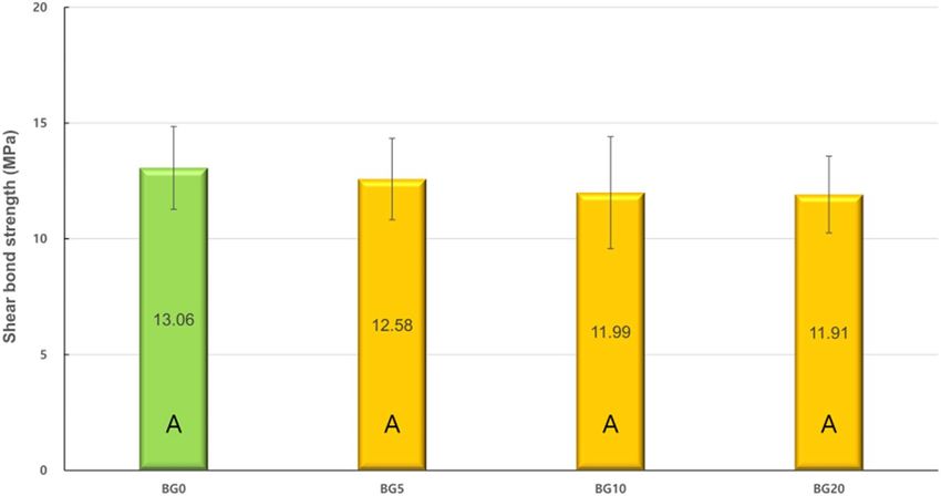
www.nature.com/scientificreports/
Materials Product and manufacturer Composition
Powder: Alumino-fluorosilicate glass and other ingredients
Glass ionomer cement (GIC) FUJI-IX GP; GC, Tokyo, Japan
Liquid: Polyacrylic acid and proprietary ingredients
Bioactive glass (BAG) 63S BAG; Bonding Chemical, Katy, TX, USA 63% SiO2, 31% CaO, 6% P2O5, > 99%, < 20 μm
CaCl2 (0.7 mM/L), M
gCl2·6H20 (0.2 mM/L), K
H2PO4 (4.0 mM/L) KCl, NaN3 (0.3 mM/L), and
Artificial saliva (AS)
HEPES buffer (20 mM/L)
Table 1. Composition of materials used in this study.
Groups Description
BG0 (control) Glass ionomer cement (GIC) powder and liquid
BG5 5 wt% BAG incorporated GIC powder and liquid
BG10 10 wt% BAG incorporated GIC powder and liquid
BG20 20 wt% BAG incorporated GIC powder and liquid
Table 2. Experimental groups for this study.
studies have indicated that BAG could remineralize e namel19–21 and d entin22,23. In addition, BAG-containing
24 25,26 27–31
dental materials, such as adhesives , composite resins , and GICs , have exhibited the potential to cause
dentin remineralization. However, recent studies have evaluated BAG-containing GICs to validate the claims
that such a combination will improve tooth bioactivity, regeneration capacity, and restoration32,33. Furthermore,
there is an ever-increasing interest in the application of bioactive materials in the dental field in an attempt to
remineralize the caries-affected d entin34.
Based on the results of previous studies, there is a possibility of remineralization synergy if BAG is incorpo-
rated into GIC because this material induces coordinated remineralization through the flouride release from GIC
and calcium and phosphate supplements from BAG. Choi et al. reported that the addition of BAG to conven-
tional GIC led to the formation of a mineral phase on the surface of GIC, demonstrating the in vitro bioactivity
of BAG-incorporated GIC. However, this increases the setting time of G IC29. Yli-Urpo et al. reported that the
BAG-containing conventional and resin-modified GICs enhanced the in vivo mineralization effect, but nega-
tively affected the compressive strength and surface micro-hardness27,28,30. Valanezhad et al. reported that the
addition of BAG to resin-modified GIC affected the flexural s trength31,35 of GIC and reduced cell c ytotoxicity35.
Although numerous studies on BAG-incorporated GIC have confirmed its bioactivity and ability to change
the mechanical properties of GIC, its impact on the adjacent demineralized dentin has not been proven directly.
Therefore, this study investigated the effect of BAG-incorporated GIC on demineralized dentin. To enhance the
bioactivity of BAG and minimize the adverse effect on the mechanical properties of GIC, a sodium-free, sol–gel
derived 63S BAG was used in this study. The null hypothesis of this study is that BAG incorporation into GIC
does not affect the bioactivity of GIC.
Materials and methods
Materials. Eighty-seven caries-free, human mandibular premolars were used in this study.
The conventional strontium-based GIC, FUJI-IX GP (GC, Tokyo, Japan), was used in this study. Sol–gel
derived, sodium-free BAG was purchased from Bonding Chemical (63S BAG; Katy, TX, USA). For specimen
storage, AS was prepared and used as per the procedure presented by Garcia-Godoy et al36. The materials used in
this study are listed in Table 1. There were four experimental groups based on the amount of BAG: BG0 group: 0
wt% (control), BG5 group: 5 wt%, BG10 group: 10 wt%, and BG20 group: 20 wt%. The BAG and GIC powders
were mixed for 2 min in a sealed capsule that contained 50 metal balls with a diameter of 2 mm. These groups
are summarized in Table 2.
Methods. FE–SEM analysis of GIC surface. To evaluate the changes on the surface of each experimen-
tal group, experimental GIC powder and liquid were mixed according to the manufacturer’s instructions. The
mixed GICs were placed in a silicon mold with a volume of 6 × 6 × 4 mm3 to produce blocks. Five blocks were
produced for each experimental group and were stored in AS for two weeks. AS was replaced every two days. The
blocks were then rinsed with phosphate-buffered saline (PBS) at pH 7.4 for 1 h with three changes followed by
rinsing with distilled water for 1 min. The blocks were dehydrated using 25, 50, 75, 95, and 100% ethanol for 20,
20, 20, 30, and 60 min, respectively, and analyzed using field-emission scanning electron microscopy (FE–SEM;
Apreo S; Thermo Fisher Scientific, Waltham, MA, USA).
FE–SEM analysis of dentin surface. Four experimental groups, namely, BG0, BG5, BG10, and BG20 groups,
and one control group (ED group) that evaluated demineralized dentin only were used. Three premolars were
used in each group. Three GIC blocks of the same volume (as in “FE–SEM analysis of GIC surface”) were fabri-
cated for each group except for the ED group. Superficial enamel was removed, and the sound dentin surface was
exposed to a high-speed diamond saw (Isomet 5000; Buehler Ltd., Lake Bluff, IL, USA). Exposed dentin surfaces
Scientific Reports | (2021) 11:7016 | https://doi.org/10.1038/s41598-021-86481-y 2
Vol:.(1234567890)www.nature.com/scientificreports/
were demineralized using a 17% ethylene diamine tetra acetic acid (EDTA) solution (Vista Dental Products,
Racine, WI, USA) for 6 h. The GIC blocks were approximated to the demineralized dentin surface as closely as
possible. They were fastened using orthodontic bands and stored in AS for two weeks, and AS was changed every
two days. After storing, GIC was removed and the dentin surface was washed with deionized water (DW) for
3 min. Dentin specimens were treated as described by Perdigao et al.37 The specimens were immediately fixed in
a 2.5% glutaraldehyde solution (Sigma-Aldrich, St. Louis, MO, USA) for 12 h. After fixation, the specimens were
rinsed with phosphate buffered saline (PBS) for 1 h with three changes followed by rinsing with distilled water
for 1 min. The specimens were dehydrated using 25%, 50%, 75%, 95%, and 100% ethanol for 20, 20, 20, 30, and
60 min, respectively. After dehydration, the specimens were immersed in hexamethyldisilazane (Sigma-Aldrich)
for 10 min and examined through FE–SEM (Apreo S; Thermo Fisher Scientific).
XRD analysis of the dentin surface. X-ray diffraction (XRD) analysis was performed to evaluate the chemical
changes in all experimental groups. Three GIC blocks and premolars were processed as described in “FE–SEM
analysis of dentin surface” for each experimental group. The dentin surface of each premolar was demineralized
using 17% EDTA for 6 h. GIC-approximated dentin specimens were stored in AS for two weeks. After storing,
the GIC blocks were removed, and dentin surfaces were rinsed with DW for 3 min. XRD was performed at three
different times for the same dentin surface (before demineralization, after demineralization, and after approxi-
mation of BAG-containing GIC for two weeks) with an X-ray diffractometer (D8 Advance; Bruker, Billerica,
MA, USA) under 40 kV Cu K α radiation using a Ni filter. The diffraction intensities were measured by scanning
in the 2θ range of 20°–60° in 0.02° steps at a scanning speed of 0.1 s per step.
Shear bond strength test. Shear bond strength (SBS) test was performed to evaluate the impact of BAG incorpo-
ration on the bond strength of GIC. Fifteen premolars were used in each in experimental group. The superficial
enamel was removed, and the sound dentin surface was exposed using a high-speed diamond saw (Isomet 5000).
The exposed dentin surfaces were ground using a 600-grit silicon carbide paper to prepare a standard smear
layer and were treated with a dentin conditioner (GC) for 20 s to remove the smear layer. Next, the experimental
GIC mixture was applied to the dentin surface using an SBS specimen jig (Ultradent Bonding Clamp and Bond-
ing Mold Inserts; Ultradent, South Jordan, UT, USA). After storing in DW for 24 h, SBS was measured using a
universal testing machine (AGS-X; Shimadzu, Tokyo, Japan) at a crosshead speed of 1 mm/min.
Statistical analysis. The SBS test results were analyzed with one-way analysis of variance (ANOVA) to deter-
mine their statistical significance. Tukey’s HSD test was used for post-hoc comparison tests. The level of signifi-
cance was set at α = 0.05.
Ethical standards. Ethical approval. The study design was approved by the institutional review board at
Kyung Hee University Dental Hospital (KH-DT 19007). All procedures performed in studies involving human
participants were in accordance with the ethical standards of the institutional and/or national research commit-
tee and with the 1964 Helsinki declaration and its later amendments or comparable ethical standards.
Informed consent. For this type of study, formal consent is not required.
Institutional review board approval. The IRB of this study was approved by Kyung Hee University Dental Hos-
pital under IRB No. KH-DT19007. In our institution, a comprehensive consent is obtained when a patient’s
teeth are extracted in the OMS part. It states that the extracted teeth might be used for research in the future.
Therefore, the individual patient’s consent is not required in the studies using only previously extracted teeth.
Results
FE–SEM analysis of GIC surface. Figure 1 depicts the GIC surface of all experimental groups. All
groups exhibited some cracks on the surface of GIC. There was no surface change in the GIC in the BG0 group
(Fig. 1A,B). However, some precipitates were observed on the surface of the GIC in the BG5 group (Fig. 1C,D).
These precipitates were more prominent in the BG10 and BG20 groups (Fig. 1E–H).
FE–SEM analysis of dentin surface. Figure 2 depicts the dentin surfaces of all the experimental groups.
In the ED group, the smear layer was removed, and dentinal tubules were almost opened (Fig. 2A,B). Further-
more, collapsed collagen networks were observed on the dentin surface. The FE–SEM images of the BG0 group
were similar to those of the ED group (Fig. 2C,D). However, some precipitates were present on the dentin surface
and dentinal tubules that were partly covered with precipitates in the BG5 group (Fig. 2E,F). In the BG10 group,
more precipitates were observed compared to the BG5 group (Figs. 2G,H). In addition, the dentin surface was
almost covered with numerous precipitates in the BG20 group (Fig. 2I,J).
XRD analysis of dentin surface. The representative results for the XRD analyses of all the experimen-
tal groups are depicted in Fig. 3. There were intense diffraction lines at 26° (002) and 31.8° (211) reflections
on the dentin surface before demineralization (Fig. 3A,D,G,J). These peaks are representative of FAP or HAP
crystals38,39. These exhibited a conspicuous decrease after demineralization (Fig. 3B,E,H,K). In the BG0 group,
the peaks rarely increased despite GIC approximation (Fig. 3C). However, the BG5, BG10, and BG20 groups
exhibited an increased intensity at both peaks (Fig. 3F,I,L). In particular, the increases in the BG10 and BG20
groups were prominent.
Scientific Reports | (2021) 11:7016 | https://doi.org/10.1038/s41598-021-86481-y 3
Vol.:(0123456789)www.nature.com/scientificreports/
Figure 1. Representative FE–SEM images of GIC surface. (A,B) BG0 group; unreacted alumino-fluorosilicate
glass is observed on the surface. Some crack lines are observed on the surface (black arrow). (C,D) BG5 group;
some surface precipitates as well as crack lines (black arrow) are observed. (E,F) BG10 group; precipitates
formed on the surface, covering the cracks. (G,H) BG20 group; the observed rough surface of GIC caused by an
abundance of precipitate formation. The cracks are hardly observed owing to abundant crystal precipitations.
SBS tests. Figure 4 depicts the SBS values of all the experimental groups. The BG0 group exhibited the high-
est SBS; however, there were no significant differences when compared to those of the BG5, BG10, and BG20
groups (p > 0.05).
Scientific Reports | (2021) 11:7016 | https://doi.org/10.1038/s41598-021-86481-y 4
Vol:.(1234567890)www.nature.com/scientificreports/
Figure 2. Representative FE–SEM images of dentin surfaces. (A,B) ED group (control); demineralized,
collapsed collagen network is observed. Opened dentinal tubules are clearly seen (white arrow). (C,D) BG0
Group; demineralized collagen network and opened dentinal tubules are observed (white arrow). (E,F) BG5
group; some dentinal tubules are partly covered with precipitates (black arrow). (G,H) BG10 group, some
dentinal tubules are almost covered with precipitates (black arrow). (I,J) BG20 group; most dentinal tubules and
dentin surfaces are covered with precipitates (black arrow).
Scientific Reports | (2021) 11:7016 | https://doi.org/10.1038/s41598-021-86481-y 5
Vol.:(0123456789)www.nature.com/scientificreports/
Figure 3. Representative XRD graph of dentin surface. (A–C) BG0 group. (D–F) BG5 group. (G–I) BG10
group. (J–L) BG20 group. (A,D,G, and K are representative of dentin surfaces before demineralization. B,E,H,
and K are representative of dentin surfaces after demineralization. C,F,I, and L are representative of dentin
surfaces after GIC approximation). The gray dotted line: 26° (002) and 31.8° (211) peak represent fluorapatite
(FAP) or hydroxyapatite (HAP) crystal.
Discussion
This study investigated the effects of BAG-incorporated GIC on demineralized dentin. The FE–SEM results indi-
cate that the incorporation of BAG into GIC can induce precipitates on the surface of demineralized dentin. The
Scientific Reports | (2021) 11:7016 | https://doi.org/10.1038/s41598-021-86481-y 6
Vol:.(1234567890)www.nature.com/scientificreports/
Figure 4. Shear bond strengths of experimental groups. The same capital indicates no statistical difference
(p > 0.05).
precipitates were believed to be calcium fluorapatite or strontium fluorapatite on the basis of the XRD analyses.
In addition, BAG incorporation did not adversely affect the bond strength of G IC40,41.
As described in “Introduction”, GIC has the ability to inhibit demineralization and promote remineralization
of teeth owing to the presence of fluoride ions. Fluoride is released from the GIC surface, replaces the hydroxyl
ion of HAP, and forms FAP on the surface of teeth, thereby enhancing the resistance against acid attacks. This
ability is developed in the presence of adequate levels of calcium and phosphate ions, and the availability of these
ions can be a limiting factor for the net tooth r emineralization40. During the formation of FAP, for every two
fluoride ions, ten calcium ions, and six phosphate ions are required to form a unit cell of FAP ( Ca10(PO4)6F2)40.
FAP formation is preferred over the production of HAP in a fluoride-containing environment because FAP
is chemically more stable and less dissolvable at low pH c onditions18,41. We believe that the precipitates were
FAP even though both crystals have similar peaks in the XRD a nalysis42. In addition, it was also presumed that
the strontium-mixed FAP was possibly formed because FUJI-IX GP contains strontium and strontium may
be substituted for calcium with virtually no change in the s tructure43. One study revealed that it is impossible
to remineralize HAP-depleted d entin44. Therefore, we assume that the incorporation of BAG might provide a
mineral reservoir and supply calcium and phosphate ions into the interface between GIC and demineralized
dentin, which is a prerequisite for crystal precipitation.
In the FE–SEM analysis, all the GIC surfaces exhibited cracks, which may be attributed to dehydration
during specimen preparation. In the BG0 and BG5 groups, surface cracks were obvious. The BG10 and BG20
groups exhibited more precipitates in the FE-SEM images (Fig. 1). When the amount of BAG increased in GIC,
more precipitates were observed on the dentin surface (Fig. 2). This finding is consistent with those of previ-
ous studies27–29,35. Yli-Urpo et al. suggested that more calcium ions are present in BAG-containing GIC, as was
evidenced by an amorphous calcium phosphate layer27. They also reported in vivo mineral deposition around
BAG-containing GIC restorations28. Choi et al. suggested other evidence for the same finding by weighing their
specimens29. The XRD analysis confirmed the intense peaks representing FAP or HAP crystals in the BG5, BG10,
and BG20 groups (Fig. 3). This means that these crystals are formed on the dentin surface because of GIC incor-
poration. The precipitates formed on the BAG-containing GIC surfaces were thought to be a calcium phosphate
layer in this study. Calcium phosphate layer is regarded as a precursor of HAP, and it can bind the demineral-
ized collagen and form HAP crystals. Notably, peak changes became significantly prominent with an increasing
amount of BAG. In addition, it was confirmed that the incorporation of BAG into GIC did not adversely affect
the SBS of GIC to dentin in this study (Fig. 4).
Although this study confirmed the benefits of incorporating BAG into GIC, the incorporation of BAG into
GIC has some disadvantages also. Yli-Urpo et al. stated that the reason for the decreased mechanical properties
of BAG-containing GIC is a loose attachment of BAG particles to the GIC matrix27. It could be the formation
of sodium polyacrylate salt that inhibits the cross-linking reaction of polyalkenoic acid with A l3+ and Ca2+45.
Therefore, we used a sodium-free BAG for this study to exclude the deteriorating effect of sodium. Based on a
study by Choi et al., the setting time for BAG-containing GIC increases proportionally with the amount of B AG29.
The reason for this is that the cationic release of BAG is lower than that of alumino-fluorosilicate glass, thereby
compromising the acid–base reaction of G IC31.
The viscosity of GIC is a crucial factor because it can hinder the clinical application of GIC in oral cavities.
In this study, the viscosity of BAG-incorporated GIC tended to increase with an increase in the amount of BAG.
This increase in viscosity may be related to silica, which is one of the main components of BAG. Silica tends to
self-agglomerate and can increase the viscosity of the GIC mixture. For clinical use and commercialization of
the BAG-incorporated GIC, it is necessary to determine the optimum ratio of the BAG to GIC powder, which
can satisfy both the remineralization capacity and handling properties.
It is not easy to evaluate the morphological changes on the dentin surface when GIC is bonded. Therefore,
GIC blocks were approximated to demineralized dentin surfaces in this study. This method has been used in a
Scientific Reports | (2021) 11:7016 | https://doi.org/10.1038/s41598-021-86481-y 7
Vol.:(0123456789)www.nature.com/scientificreports/
previous study and proven to be e ffective25. AS was used as the storage solution in this study, although SBF can
also be used. However, it may cause an autogenous precipitation of calcium phosphates on the surface of dentin.
In this study, dentin demineralization was performed with 17% EDTA because it is a strong chelating agent for
dentin and can simulate harsh, mineral-depleted dentin surfaces. According to Kawasaki et al., EDTA rarely
leaves minerals on the surface of dentin46. The FE–SEM analysis of the ED and BG0 groups also indicated col-
lapsed collagen networks on the dentin surface (Fig. 2B,D). Vollenweider et al. reported that the demineralized
dentin with EDTA might cause this situation in addition to the lack of nuclei on the dentin surface23. Despite
this harsh environment, BAG-incorporated GIC induced remarkable apatite crystal precipitation on the demin-
eralized dentin surface in this study. The FE–SEM analyses of the BG5, BG10, and BG20 groups indicated some
precipitates that partly covered the dentin surface. The presence of the precipitate was more prominent when
the amount of BAG increased. This phenomenon was similar to that of the GIC surface.
The initial acidity of the GIC setting reaction, involving a prolonged period at pH below 3, affects not only
the pH of the setting cement but also the quantity of the available acid at the dentin interface47. In the first
reaction stage, BAG can uptake H+ or H3O+ ions from the solution, causing hydrolysis of the silica groups and
enabling the release of calcium and phosphate ions48. The pH of the solution increases as result of H + ions in
the solution being replaced by cations. The pH changes in the BAG reaction might increase the pH of the GIC
setting environment. One study suggests that the GIC-BAG composite increases the pH value in an aqueous
environment49. The increased pH may affect the mineralizing property of the BAG-incorporated GIC and impart
an antimicrobial effect to it.
XRD analyses were performed to evaluate the chemical changes on the dentin surface with or without the
approximation of BAG-incorporated GIC. The present study differs from the previous ones in that it evaluated
the same tooth at three different times: before demineralization, after demineralization, and after approximation
of BAG-incorporated GIC for two weeks. The reason for using the same tooth was that the histological variations
in different teeth might result in analytical differences. Although no statistical analysis was not performed, the
BG10 and BG20 groups indicated an increased FAP peaks after the approximation. This was consistent with the
results of the FE–SEM analysis.
SBS test was performed to evaluate the effects of BAG incorporation on GIC adhesion. Although the micro-
tensile bond strength test is used at present, it is not an appropriate method to evaluate the low bond strength
of GIC. In this study, a dentin conditioner was used to increase the bond strength and minimize the pre-testing
failure. The dentin conditioner contains a diluted solution of polyacrylic acid and removes superficial smear
layers on the surface of the dentin, thereby improving chemical a dhesion50. Ionic bond, between the calcium
ions of HAP or FAP and the carboxylate groups of GIC, is the mechanism through which GIC adheres to the
dentin51. In this study, BAG incorporation did not adversely affect the SBS of GIC regardless of the amount of
BAG, indicating that BAG did not affect the adhesion mechanism of GIC. The use of sodium-free BAG was
required to maintain the SBS of the BAG-incorporated GIC.
This study was designed to evaluate the effects of BAG-incorporated GIC on demineralized dentin. Based on
the results of this study, BAG-incorporated GIC exhibited the ability to induce crystal precipitation on demin-
eralized dentin. Further studies are necessary to support the results of this study and investigate the changes in
mechanical properties on the dentin surface and long-term effect of BAG-incorporated GIC.
Conclusion
Within the limitation of this study, we conclude that BAG-incorporated GIC could precipitate FAP crystals
underlying demineralized dentin without hampering the bond strength. BAG is an advantageous supplement
to GIC, and its incorporation up to 20 wt% is beneficial for FAP crystal precipitation of demineralized dentin.
This study suggests the possibility of using BAG as a functional additive in GIC for clinical settings.
Received: 10 January 2021; Accepted: 11 March 2021
References
1. Forss, H., Jokinen, J., Spets-Happonen, S., Seppa, L. & Luoma, H. Fluoride and mutans streptococci in plaque grown on glass
ionomer and composite. Caries Res. 25, 454–458. https://doi.org/10.1159/000261410 (1991).
2. Preston, A. J., Mair, L. H., Agalamanyi, E. A. & Higham, S. M. Fluoride release from aesthetic dental materials. J. Oral Rehabil. 26,
123–129 (1999).
3. Tyas, M. J. & Burrow, M. F. Adhesive restorative materials: a review. Aust. Dent. J. 49, 112–121 (quiz 154) (2004).
4. Nicholson, J. W. Chemistry of glass-ionomer cements: a review. Biomaterials 19, 485–494 (1998).
5. Nicholson, J. W. Polyacid-modified composite resins (“compomers”) and their use in clinical dentistry. Dent. Mater. 23, 615–622.
https://doi.org/10.1016/j.dental.2006.05.002 (2007).
6. Komatsu, H., Shimokobe, H., Kawakami, S. & Yoshimura, M. Caries-preventive effect of glass ionomer sealant reapplication: Study
presents three-year results. J. Am. Dent. Assoc. 1939(125), 543–549. https://doi.org/10.14219/jada.archive.1994.0082 (1994).
7. Mukai, M. et al. Fluoride uptake in human dentine from glass-ionomer cement in vivo. Arch. Oral Biol. 38, 1093–1098. https://
doi.org/10.1016/0003-9969(93)90171-h (1993).
8. Retief, D. H., Bradley, E. L., Denton, J. C. & Switzer, P. Enamel and cementum fluoride uptake from a glass ionomer cement. Caries
Res. 18, 250–257. https://doi.org/10.1159/000260773 (1984).
9. Rodríguez-Sendra, J., Torres, I., Jiménez, N., Sauro, S. & Camarena, F. Ultrasonic monitoring of dentin demineralization. IEEE
Trans. Ultrason. Ferroelectr. Freq. Control https://doi.org/10.1109/TUFFC.2020.3015668 (2020).
10. Hemagaran, G. & Neelakantan, P. Remineralization of the tooth structure-the future of dentistry. Int. J. Pharmtechnol. Res. 6,
487–493 (2014).
11. Featherstone, J., Glena, R., Shariati, M. & Shields, C. Dependence of in vitro demineralization of apatite and remineralization of
dental enamel on fluoride concentration. J. Dent. Res. 69, 620–625 (1990).
Scientific Reports | (2021) 11:7016 | https://doi.org/10.1038/s41598-021-86481-y 8
Vol:.(1234567890)www.nature.com/scientificreports/
12. Ngo, H. C., Mount, G., Mc Intyre, J., Tuisuva, J. & Von Doussa, R. J. Chemical exchange between glass-ionomer restorations and
residual carious dentine in permanent molars: an in vivo study. J. Dent. 34, 608–613. https://doi.org/10.1016/j.jdent.2005.12.012
(2006).
13. Dionysopoulos, P., Kotsanos, N., Koliniotou-Koubia, E. & Tolidis, K. Inhibition of demineralization in vitro around fluoride
releasing materials. J. Oral Rehabil. 30, 1216–1222 (2003).
14. Francci, C. et al. Fluoride release from restorative materials and its effects on dentin demineralization. J. Dent. Res. 78, 1647–1654.
https://doi.org/10.1177/00220345990780101001 (1999).
15. ten Cate, J. M. & van Duinen, R. N. Hypermineralization of dentinal lesions adjacent to glass-ionomer cement restorations. J. Dent.
Res. 74, 1266–1271. https://doi.org/10.1177/00220345950740060501 (1995).
16. Jones, J. R. Review of bioactive glass: From Hench to hybrids. Acta Biomater. 9, 4457–4486. https://doi.org/10.1016/j.actbio.2012.
08.023 (2013).
17. Brauer, D. S., Karpukhina, N., O’Donnell, M. D., Law, R. V. & Hill, R. G. Fluoride-containing bioactive glasses: Effect of glass design
and structure on degradation, pH and apatite formation in simulated body fluid. Acta Biomater. 6, 3275–3282 (2010).
18. Mneimne, M., Hill, R. G., Bushby, A. J. & Brauer, D. S. High phosphate content significantly increases apatite formation of fluoride-
containing bioactive glasses. Acta Biomater. 7, 1827–1834 (2011).
19. Farooq, I., Majeed, A., AlShwaimi, E. & Almas, K. Efficacy of a novel fluoride containing bioactive glass based dentifrice in rem-
ineralizing artificially induced demineralization in human enamel. Fluoride 52, 447–455 (2019).
20. Kanwal, N. et al. In-vitro apatite formation capacity of a bioactive glass-containing toothpaste. J. Dent. 68, 51–58 (2018).
21. Bakry, A. S. et al. A novel fluoride containing bioactive glass paste is capable of re-mineralizing early caries lesions. Materials 11,
1636 (2018).
22. Jung, J. H. et al. Dentin sealing and antibacterial effects of silver-doped bioactive glass/mesoporous silica nanocomposite: An
in vitro study. Clin. Oral Investig. 23, 253–266. https://doi.org/10.1007/s00784-018-2432-z (2019).
23. Vollenweider, M. et al. Remineralization of human dentin using ultrafine bioactive glass particles. Acta Biomater. 3, 936–943.
https://doi.org/10.1016/j.actbio.2007.04.003 (2007).
24. Jun, S. K. et al. Multi-functional nano-adhesive releasing therapeutic ions for MMP-deactivation and remineralization. Sci. Rep.
8, 5663. https://doi.org/10.1038/s41598-018-23939-6 (2018).
25. Jang, J. H. et al. Effect of bioactive glass-containing resin composite on dentin remineralization. J. Dent. 75, 58–64. https://doi.org/
10.1016/j.jdent.2018.05.017 (2018).
26. Tezvergil-Mutluay, A. et al. Effects of composites containing bioactive glasses on demineralized dentin. J. Dent. Res. 96, 999–1005.
https://doi.org/10.1177/0022034517709464 (2017).
27. Yli-Urpo, H., Lassila, L. V., Narhi, T. & Vallittu, P. K. Compressive strength and surface characterization of glass ionomer cements
modified by particles of bioactive glass. Dent. Mater. 21, 201–209. https://doi.org/10.1016/j.dental.2004.03.006 (2005).
28. Yli-Urpo, H., Narhi, M. & Narhi, T. Compound changes and tooth mineralization effects of glass ionomer cements containing
bioactive glass (S53P4), an in vivo study. Biomaterials 26, 5934–5941. https://doi.org/10.1016/j.biomaterials.2005.03.008 (2005).
29. Choi, J. Y., Lee, H. H. & Kim, H. W. Bioactive sol-gel glass added ionomer cement for the regeneration of tooth structure. J. Mater.
Sci. Mater. Med. 19, 3287–3294. https://doi.org/10.1007/s10856-008-3464-8 (2008).
30. Khoroushi, M., Mousavinasab, S. M., Keshani, F. & Hashemi, S. Effect of resin-modified glass ionomer containing bioactive glass
on the flexural strength and morphology of demineralized dentin. Oper. Dent. 38, E1-10. https://doi.org/10.2341/11-325-l (2013).
31. Kim, D. A. et al. Sol-gel-derived bioactive glass nanoparticle-incorporated glass ionomer cement with or without chitosan for
enhanced mechanical and biomineralization properties. Dent. Mater. 33, 805–817. https://doi.org/10.1016/j.dental.2017.04.017
(2017).
32. Yli-Urpo, H., Närhi, M. & Närhi, T. Compound changes and tooth mineralization effects of glass ionomer cements containing
bioactive glass (S53P4), an in vivo study. Biomaterials 26, 5934–5941 (2005).
33. Ana, I. D., Matsuya, S., Ohta, M. & Ishikawa, K. Effects of added bioactive glass on the setting and mechanical properties of resin-
modified glass ionomer cement. Biomaterials 24, 3061–3067 (2003).
34. Khoroushi, M. & Keshani, F. A review of glass-ionomers: From conventional glass-ionomer to bioactive glass-ionomer. Dent. Res.
J. 10, 411 (2013).
35. Valanezhad, A. et al. Modification of resin modified glass ionomer cement by addition of bioactive glass nanoparticles. J Mater.
Sci. Mater. Med. 27, 3. https://doi.org/10.1007/s10856-015-5614-0 (2016).
36. Garcia-Godoy, F. et al. Degradation of resin-bonded human dentin after 3 years of storage. Am. J. Dent. 20, 109 (2007).
37. Perdigao, J., Lambrechts, P., Van Meerbeek, B., Vanherle, G. & Lopes, A. L. Field emission SEM comparison of four postfixation
drying techniques for human dentin. J. Biomed. Mater. Res. 29, 1111–1120. https://doi.org/10.1002/jbm.820290911 (1995).
38. Brundavanam, R. K., Poinern, G. E. J. & Fawcett, D. Modelling the crystal structure of a 30 nm sized particle based hydroxyapatite
powder synthesised under the influence of ultrasound irradiation from x-ray powder diffraction data. Am. J. Mater. Sci. 3, 84–90.
https://doi.org/10.5923/j.materials.20130304.04 (2013).
39. Aljabo, A., Abou Neel, E. A., Knowles, J. C. & Young, A. M. Development of dental composites with reactive fillers that promote
precipitation of antibacterial-hydroxyapatite layers. Mater. Sci. Eng. C 60, 285–292. https://doi.org/10.1016/j.msec.2015.11.047
(2016).
40. Reynolds, E. C. Calcium phosphate-based remineralization systems: scientific evidence?. Aust. Dent. J. 53, 268–273 (2008).
41. Shah, F. A. Fluoride-containing bioactive glasses: Glass design, structure, bioactivity, cellular interactions, and recent developments.
Mater. Sci. Eng. C 58, 1279–1289 (2016).
42. Zhao, J. et al. Solution combustion method for synthesis of nanostructured hydroxyapatite, fluorapatite and chlorapatite. Appl.
Surf. Sci. 314, 1026–1033 (2014).
43. Stamboulis, A., Law, R. V. & Hill, R. G. Characterisation of commercial ionomer glasses using magic angle nuclear magnetic
resonance (MAS-NMR). Biomaterials 25, 3907–3913 (2004).
44. Kim, Y. K. et al. Failure of a glass ionomer to remineralize apatite-depleted dentin. J. Dent. Res. 89, 230–235. https://doi.org/10.
1177/0022034509357172 (2010).
45. De Barra, E. & Hill, R. Influence of alkali metal ions on the fracture properties of glass polyalkenoate (ionomer) cements. Bioma-
terials 19, 495–502 (1998).
46. Kawasaki, K., Ruben, J., Tsuda, H., Huysmans, M. & Takagi, O. Relationship between mineral distributions in dentine lesions and
subsequent remineralization in vitro. Caries Res. 34, 395–403 (2000).
47. Smith, D. C. & Ruse, N. D. Acidity of glass ionomer cements during setting and its relation to pulp sensitivity. J. Am. Dent. Assoc.
112, 654–657 (1986).
48. Rabiee, S. M., Nazparvar, N., Azizian, M., Vashaee, D. & Tayebi, L. Effect of ion substitution on properties of bioactive glasses: A
review. Ceram. Int. 41, 7241–7251 (2015).
49. Yli-Urpo, H., Söderling, E., Vallittu, P. K. & Närhi, T. pH changes induced by bioactive glass ionomer cements. Key Eng. Mater.
254, 599–602 (2004).
50. Powis, D. R., Follerås, T., Merson, S. A. & Wilson, A. D. Improved adhesion of a glass ionomer cement to dentin and enamel. J.
Dent. Res. 61, 1416–1422. https://doi.org/10.1177/00220345820610120801 (1982).
51. Yoshida, Y. et al. Evidence of chemical bonding at biomaterial-hard tissue interfaces. J. Dent. Res. 79(709–714), 2000. https://doi.
org/10.1177/00220345000790020301 (2000).
Scientific Reports | (2021) 11:7016 | https://doi.org/10.1038/s41598-021-86481-y 9
Vol.:(0123456789)www.nature.com/scientificreports/
Acknowledgements
This study was supported by the National Research Foundation of Korea (NRF) grant funded by the Korea
government (MSIT) (No. 2019R1F1A1057615).
Author contributions
All authors contributed to the concept and design of this study. Material preparation and data collection were
performed by H.E.B., I-S.P., J-E.L., and D-S.K. Data analysis was performed by H-G. K. and J.K. The first draft
of the manuscript was written by H.E.B. and H.-J. K. All authors reviewed the draft manuscript, revised it, and
approved the final manuscript.
Competing interests
The authors declare no competing interests.
Additional information
Correspondence and requests for materials should be addressed to D.-S.K.
Reprints and permissions information is available at www.nature.com/reprints.
Publisher’s note Springer Nature remains neutral with regard to jurisdictional claims in published maps and
institutional affiliations.
Open Access This article is licensed under a Creative Commons Attribution 4.0 International
License, which permits use, sharing, adaptation, distribution and reproduction in any medium or
format, as long as you give appropriate credit to the original author(s) and the source, provide a link to the
Creative Commons licence, and indicate if changes were made. The images or other third party material in this
article are included in the article’s Creative Commons licence, unless indicated otherwise in a credit line to the
material. If material is not included in the article’s Creative Commons licence and your intended use is not
permitted by statutory regulation or exceeds the permitted use, you will need to obtain permission directly from
the copyright holder. To view a copy of this licence, visit http://creativecommons.org/licenses/by/4.0/.
© The Author(s) 2021
Scientific Reports | (2021) 11:7016 | https://doi.org/10.1038/s41598-021-86481-y 10
Vol:.(1234567890)You can also read