Molecular Biotyping, Phylogenetic Analysis of Indole Acetic Acid Producing Rhizobium Sp. CU8 and Co-inhabitant Non-rhizobial Nodule Associated ...
←
→
Page content transcription
If your browser does not render page correctly, please read the page content below
Molecular Biotyping, Phylogenetic Analysis of
Indole Acetic Acid Producing Rhizobium Sp. CU8
and Co-inhabitant Non-rhizobial Nodule Associated
Bacteria From Mimosa Pudica L.
Maya R.
University of Calicut
YUSUF AKKARA ( akkara69@yahoo.co.in )
University of Calicut https://orcid.org/0000-0002-1931-5278
Research Article
Keywords: Nitrogen fixers, Phylogenetic analysis, Plant growth promoters, Root nodule, 16S rRNA
Posted Date: August 23rd, 2021
DOI: https://doi.org/10.21203/rs.3.rs-810699/v1
License: This work is licensed under a Creative Commons Attribution 4.0 International License.
Read Full License
Page 1/15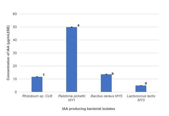
Abstract
The interaction between rhizobia and other nodule associated bacteria assist to mitigate nutrient stress in
leguminous plants by fixing atmospheric nitrogen and synthesizing plant growth regulators. Beneficial
effects of microbial inoculants emphasize the need for further research and their use in modern
agriculture. The present work describes the isolation, identification, plant growth promoting properties
and phylogenetic analysis of nodule associated bacteria from Mimosa pudica L. Isolation and
characterization of nodule associated bacteria were done according to standard procedures. Molecular
characterization of the isolates was performed using 16S ribosomal RNA. Plant growth promoting ability
was analyzed by quantifying the levels of Indole acetic acid. Evolutionary distance and relatedness was
analyzed using neighbor joining method. Rhizobium sp. CU8 and three other co-resident non-rhizobial
nodule associated bacteria (Bacillus cereus MY5, Ralstonia pickettii MY1 and Lactococcus lactis MY3)
exhibiting nitrogen fixation, plant growth promotion and other potential microbial activities were
characterized. Phylogenetic analysis revealed the genetic relatedness and evolutionary significance of
Rhizobium sp. CU8 and other co-inhabitant non-rhizobial nodule associated bacteria from M. pudica.
Present study identified the four isolates as potential biofertilizers due to their nitrogen fixing and growth
promoting characteristics. L. lactis MY3 is the first co-resident nitrogen fixer and plant growth promoter
reported from the root nodules of M. pudica.

Introduction
The members of Leguminosae, are associated with endophytic, root nodule associated bacteria (NAB)
which ameliorates nutrient stress by fixing atmospheric nitrogen (N2) and producing plant growth
promoters. The genus Mimosa received considerable attention in the recent years because of its potential
to fix atmospheric nitrogen. Biological nitrogen fixation is ecologically important, contributing ~ 100–
290 million tons of nitrogen annually to the natural ecosystem and enhancing the growth of
agronomically important forage and crop plants. Biological nitrogen fixation (both symbiotic and non-
symbiotic) reduces the use of synthetic nitrogen fertilizers.

Plant growth promoting bacteria (PGPB) enhance plant growth by fixing atmospheric nitrogen and its
assimilation to the plant, production of siderophores to chelate iron and its absorption, solubilization of
minerals such as phosphorus, synthesis of phytohormones, augmenting plant nutrient uptake (Glick
1995; Kloepper 1992) and the production of substances like antibiotics (Glick 1995). In addition, the PGP
microbes express abiotic stress tolerance like extreme temperature, drought, salinity, pH, heavy metal and
pesticide pollution (Gopalakrishnan et al. 2015).

PGPB has beneficial effects on legume growth and some strains enhance the nodulation and nitrogen
fixation by effective interaction between plant and rhizobia (Parmar and Dadarwal 1999). Most of the
nodulating bacteria are free-living rhizobacteria, however, some are intracellular or intercellular
endophytes (Sturz et al. 2000) and gain advantage of being protected from environmental stresses and
microbial competition (Kobayashi and Palumbo 2000). The endophytes and epiphytes are the two

                                                   Page 2/15
different types of plant growth promoting rhizobia associated with host tissue. There are many
endophytic and epiphytic bacteria which are directly or indirectly involved in plant growth and
development. Endophytic bacteria live in plant tissues without affecting the normal metabolism of the
host or gaining any benefit other than a noncompetitive environment inside the host. It has been
demonstrated that bacterial endophytes have beneficial effects on host plants, such as growth promotion
and biological control of pathogens (Downing and Thomson 2000; Ryu et al. 2005; Sturz et al. 1999).
Legume root nodules may contain microbes other than rhizobia (Martínez-Hidalgo and Hirsch 2017;
Zgadzaj et al. 2015), however, the function of these co-residents in the nodules is yet to be fully
elucidated, their main role might be to assist rhizobia during the nodule infection process and to promote
plant growth (Chibeba et al. 2020; Peix et al. 2015).
Modern agriculture faces challenges, such as loss of soil fertility, fluctuating climatic factors and
increasing pathogen and pest attacks. Sustainability and environmental safety of agricultural production
relies on eco-friendly approaches like use of biofertilizers, biopesticides and crop residue recycling. From
the agricultural and ecological viewpoints, “how can we increase food quality and quantity, and to
improve the sustainable plant productivity, while maintaining environmental quality?” the importance of
nitrogen fixing and plant growth promoting bacteria, their gene conservation can contribute better in
sustainable agriculture and 16S ribosomal RNA typing is used to identify microorganisms. The 16S rRNA
based phylogenetic analysis revealed the distribution of genotypes in Rhizobium sp., B. cereus, R. pickettii
and L. lactis. Our understanding of microbial interactions in the rhizosphere must be complemented by
combining the basic and applied studies.
The beneficial effects of microbial inoculants, particularly nitrogen fixing and plant growth promoters
(PGP), from the root nodule of M. pudica accentuate the need for research and its application in modern
agriculture. The present study is focused on the isolation, identification, characterization, screening and
comprehensive evaluation on phylogenetic relationship based on 16S rRNA gene of potential nitrogen
fixers and plant growth promoters from root nodule of M. pudica.
Materials And Methods
Isolation of nodule associated bacteria
Bacteriae were isolated from the root nodules of M. pudica grown in different locations in the University
of Calicut campus (geographic coordinates: 11°08'01.0"N, 75°53'19.0"E; 11°08'00.4"N, 75°53'17.5"E). The
nodule associated bacteria (NAB), were isolated from healthy pink coloured root nodules, washed
thoroughly using running tap water, surface sterilized using 70% (v/v) ethanol for 30s, 0.1% (w/v) HgCl2
for 2 min and washed thrice with sterile double distilled water under aseptic condition for one min
(Rajendran et al. 2012). Nodules were crushed using a sterile glass rod and the extracts were plated on to
Yeast Mannitol agar medium (pH 6.8), supplemented with Congo red dye (0.025 gl-1). The cultures were
incubated at 28±2ºC for 24-48hrs. Single colony-forming units were checked for purity by repeated
Page 3/15transfer on to nutrient agar medium pH-7(Vincent 1970). Pure cultures were maintained on nutrient agar
medium with regular subculturing and used for analysis.
CHNS analysis
Soil samples collected from the rhizosphere of M. pudica was used for CHNS elemental analysis using a
Euro Vector CHNS Analyser Model No: Ea 3000. Instrument parameters were carrier pressure 100kPa,
purge flow 80 mL/min, oxygen flow 20 mL and run for 7 min. Temperature of front furnace was 980ºC
and GC oven temperature was 105ºC. Sample (8.5-9.5 mg) was used for the analysis.
Phenotypic characterization
Bacterial isolates grown in nutrient agar medium were subjected to phenotype characterization based on
the morphological and biochemical characters examined using Bergey’s manual of systematic
bacteriology (Bergeys 2001). The morphological characters were analysed using gram staining, motility
test by hanging drop method and endospore staining by malachite green method using a phase contrast
microscope. Biochemical analysis was performed using indole production, hydrolysis of urea, methyl red
(MR) test, voges proskauer (VP), citrate utilization and nitrate reduction test. The Intrinsic antibiotic
resistance of the isolates was determined by disc method with Ampicillin (Amp) (10 mcg/disc),
Tetracycline (TE) (30 mcg/disc), and Penicillin G (PG) (10 IU/disc) (Cappuccino and Sherman 1983).
Molecular characterization
DNA extraction, 16S ribosomal RNA typing and sequencing
Genomic DNA of the three species was extracted and purified using CTAB method (Ausubel et al. 1995).
The purified DNA was quantified using a Nanodrop 2000 spectrometer (UV scanning Thermo scientific).
PCR amplification of the 16S rRNA gene fragment was carried out using the universal primers 1-27F
(AGAGTTTGATCCTGGCTCAG) and 1495R (CTACGGCTACCTGTTACGA) (Lane 1991) at an annealing
temperature of 50⁰C. The band size was verified using agarose gel electrophoresis. The PCR products
were cleaned and sequenced from Agrigenome Lab Pvt Ltd, Cochin, Kerala. Cloned 16S rRNA sequences
were minimally edited and manually aligned using Bioedit software. Species identification and homology
between the sequences were identified using BLAST (https://www.ncbi.nlm.nih.gov/BLAST/). The cloned
sequences were submitted to GenBank, NCBI.
Characterization of plant growth promoting potential of bacterial isolates
Plant growth enhancement potential of the four isolates was verified by the production of indole acetic
acid, organic acid and capacity to fix atmospheric nitrogen in plants.
(a). Production of indole acetic acid (IAA)
The isolates with IAA production capacity was identified using bacterial culture grown in nutrient broth
supplemented with 0.1% L-Tryptophan(w/v) incubated at 30⁰C for 48hrs. Indole acetic acid (IAA)
Page 4/15production was analysed using colorimetric method of Gordon and Weber (1951). IAA in the culture was
quantified using standard calibration curve prepared using gradient concentrations of IAA.
(b). Production of organic acid
Assessed by growing bacterial culture in calcium carbonate agar [CaCO3 5 gl-1, glucose 50 gl-1, yeast
extract 5 gl-1, agar 15 gl-1] medium and the clear zone around the colony confirmed the production of
organic acid.
(c). Assessment of nitrogen fixation
The N2 fixation capacity was evaluated by growing the cultures in nitrogen free malate containing
bromothymol blue medium [DL- Malic acid 5 gl-1, KOH 4 gl-1, K2HPO4 0.5 gl-1, FeSO4.7H2O 0.05 gl-1,
MnSO4.7H2O 0.01 gl-1, MgSO4 0.01 gl-1, NaCl 0.02 gl-1, CaCl2 0.01 gl-1, Na2MoO4 0.002 gl-1, Yeast extract
0.05 gl-1, Bromothymol Blue 2 mLl-1 for 24- 48hrs at 30ºC in 120 rpm on a rotary shaker (Day and
Dobereiner 1976). Colour change of the medium from pale green to pale blue indicates the ability to fix
atmospheric N2.
16S rRNA based phylogenetic analysis
Phylogenetic analysis based on 16S rRNA sequence were performed using the MEGA 7.0 (Kumar et
al. 2016) based on neighbor joining statistical method (Felsenstein 1981) and the branching support of
1000 bootstrap (Felsenstein 1985). The phylogenetic tree construction based on, 16S rRNA was initially
performed using the cloned sequence along with sequences retrieved from GenBank and aligned with
ClustalW. The model selection was performed using MEGA 7(Kumar et al. 2016) based on the lowest
Bayesian Information Criterion (BIC) value (Schwarz 1978). To compare the similarity or diversity, the
nucleotide sequences of 40 selected Rhizobium, Bacillus, Ralstonia, Lactococcus species from different
geographical regions were retrieved from NCBI, GenBank (http:/www.ncbi.nlm.nih.gov/ ). The list of
sequences retrieved from NCBI with strain name, accession number and locations are given in online
resource 1.
Statistical analysis
Using the SPSS software (27.0V, SPSS, Chicago, USA), one way ANOVA was performed to analyze the
concentration of IAA in the isolates after 48hrs. Statistical analysis was carried out according to Turkey’s
test (P≤0.05). The data were an average of 4 separate experiments with three replicates (n=3).
Results
Isolation and CHNS elemental analysis
Page 5/15Four nodules associated bacteria were isolated from the healthy nodules of M. pudica. The isolates were
purified and subcultured on nutrient agar medium (pH-7). CHNS analysis of the rhizosphere soil showed
nitrogen (0.274%), carbon (1.534%), hydrogen (2.094%) and sulphur (0%). The soil was acidic to slightly
neutral with a mean pH value of 6.49 ± 0.1.
Phenotypic characterization
Phenotypic characteristics of the four isolates are presented in Table 1.
Table 1
Morphological, biochemical and physiological features of the four isolates extracted from root nodules of
Mimosa pudica
Phenotypic Rhizobium sp. Bacillus cereus Ralstonia pickettii Lactococcus
characterization CU8 MY5 MY1 lactis MY3
Morphological Features
Gram staining gram negative gram positive gram negative gram negative
Motility motile motile motile non-motile
Spore production non sporulating non sporulating non sporulating
sporulating
Biochemical Features
Indole production + + + +
Hydrolysis of urea + + + +
Methyl red (MR) test - - - +
Voges proskauer + + + -
(VP)
Citrate utilization + + + +
Nitrate reduction test + + - -
Antibiotic TE,PG, Amp TE, PG, Amp TE, PG TE, PG, Amp
susceptibility
(+/− indicates test is positive or negative. TE− tetracycline, PG− penicillin−G, Amp− ampicillin)
Molecular characterization
DNA extraction, 16S ribosomal RNA typing and sequencing
Genomic DNA was extracted from the four isolates using CTAB method. The 260/280 ratio of the DNA
samples were 1.8-2 indicating the purity of the samples. Molecular characterization by 16S rRNA
confirmed that the four isolates are Rhizobium sp. CU8, B. cereus MY5, R. pickettii MY1 and L. lactis MY3.
Page 6/15The 16S rRNA gene from the Rhizobium sp. CU8, B. cereus MY5, R. pickettii MY1 and L. lactis MY3
possessed 99–100% homology with the nucleotide sequence of other species retrieved from NCBI. L.
lactis MY3 from the root nodules of M. pudica are not reported earlier. GenBank accession numbers were
provided for the 16S rRNA gene sequence of Rhizobium sp. CU8 (MN744368), B. cereus MY5
(MH997483), R. pickettii MY1(MH997486) and L. lactis MY3 (MW132401).
Characterization of plant growth promoting activities
(a). IAA production potential of the isolates
IAA production during the 48hr of growth was quantified in Rhizobium sp. CU8, B. cereus MY5, R. pickettii
MY1 and L. lactis MY3 using Salkowski reagent. R. pickettii MY1 and Rhizobium sp. CU8 developed
colour reaction immediately after the addition of reagents indicating the formation of IAA and better IAA
production was observed when the cultures were incubated for 25 min in dark. The highest quantity of
IAA was produced in R. pickettii MY1 (49.8630 ± 0.1779 µg/mL) followed by B. cereus MY5 (13.5159 ±
0.2416 µg/mL), Rhizobium sp. CU8 (11.6895 ± 0.1837 µg/mL) and L. lactis MY3 (4.9315 ± 0.0790
µg/mL) (Fig. 1) after 48hrs of incubation, which was significant at P < 0.05.
(b). Production of organic acid
Among the four isolates, L. lactis MY3 showed a clear zone after 24hrs of incubation due to the
degradation of calcium carbonate leading to the production of organic acid. Other three isolates doesn’t
show any clear zone around the colony.
(c). Nitrogen fixing potential of the isolates
The four isolates, Rhizobium sp. CU8, B. cereus MY5, R. pickettii MY1 and L. lactis MY3 exhibited N2
fixing ability grown in nitrogen free malate medium containing bromothymol blue as indicator. The
Rhizobium sp. CU8, B. cereus MY5 and R. pickettii MY1 showed significant colour change from pale
green to pale blue indicating N2 fixing ability within 24hrs. However, L. lactis MY3 developed the colour
change only after 48hrs.
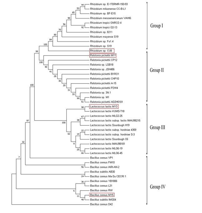
Phylogeny based on 16S rRNA gene
The cloned sequence and the sequences retrieved from the GenBank were used to construct the
phylogenetic tree using Neighbor Joining (NJ) method with 1000 bootstraps. Models with the lowest BIC
scores were considered to describe the best nucleotide substitution pattern. The TN93 + G (Tamura Nei
Model) displayed the lowest BIC scores (10125.617- online resource 2) to construct consensus NJ tree
from the aligned sequences (Fig. 2). The multiple sequence alignment based phylogram using MEGA 7.0
and TN93 + G model based on bootstrap analysis of 1000 replicates was performed to estimate the
confidence of the tree topologies. The phylogenetic position of the Rhizobium sp. CU8, B. cereus MY5, R.
pickettii MY1 and L. lactis MY3 in relation to other species of this genus is illustrated in Fig. 2; the
numbers adjacent to nodes are the statistical frequency of the indicated species.
Page 7/15The genus Rhizobium and Ralstonia fall under the same phylum proteobacteria classified as subclass α
and β proteobacteria. Based on 16S rRNA homology, both genera are placed in separate group originated
from a single node. L. lactis and B. cereus were grouped as a separate clade.
Phylogenetic tree revealed that Rhizobium sp. CU8 and B. cereus MY5, showed highest relatedness with
other members of the genus. Rhizobium sp. CU8 shows closest relatedness with Indian Rhizobium sp.
S19. B. cereus MY5 shows highest similarity to B. subtilis IMG04 from India among other members of
this genus. In the case of R. pickettii MY1 and L. lactis MY3, the maximum similarity of these two native
strains were shown to R. pickettii CP12 from China and L. lactis KUMS-T18 from Iran.
Group I Rhizobium sp. CU8 and Group II Ralstonia pickettii MY1 showed 100% bootstrap support within
the genus level. L. lactis MY3 in Group III is tightly clustered with L. lactis KUMS-T18 with bootstrap
support 98%. In Group IV, B. cereus MY5 clustered with B. subtilis IMG04 with bootstrap support (> 50%).
However, none of the isolates that clustered in different phylogenetic trees based on 16S rRNA analysis.
Discussion
The interaction between rhizobia and other nodule associated bacteria are of relevance in the
improvement of N2 fixation and plant growth promotion in leguminous plants (Barea et al. 2005; Ryu et
al. 2005). The isolation, characterization and phylogenetic relationship of newly identified bacteria from
root nodule of M. pudica illustrated the N2-fixation and PGP activity of these isolates. The isolation
procedure consists of nodule surface sterilization specifically aimed to allow the obtention of nodule
endophytes (Rajendran et al. 2008) resulting in the isolation of three non-rhizobial bacteria from M.
pudica nodule. In the past, bacteria isolated from the nodules with dissimilar growth and appearance to
that of typical rhizobia were considered as contaminants and discarded, however, recent studies
convincingly demonstrated the occurrence of non-rhizobial bacteria in the nodules and their role on the
host plants, rhizobial strains or the symbiosis are under investigation (Martínez-Hidalgo and Hirsch
2017). It is now well recognized that non-rhizobial bacteria can promote plant growth by an array of
mechanisms including solubilization and mobilization of nutrients (Srivastwa et al. 2014), N2-fixation
(Castellano-Hinojosa et al. 2016), production of phytohormones (Chinnaswamy et al. 2018), along with
microbial processes. Nodule endophytes belonging to the genera Bacillus, Burkholderia, Pseudomonas
and Enterobacter have been isolated from different legumes (Dudeja et al. 2012; Martínez-Hidalgo and
Hirsch 2017).
The morphological and microscopic features of the isolates were in congruence with the earlier reports.
All the isolates were sensitive to standard antibiotics suggesting their prevention mechanisms. The
isolates, R. pickettii MY1, L. lactis MY3, B. cereus MY5 and Rhizobium sp. CU8 exhibited important agro-
biotechnological properties, such as nitrate reduction, production of organic acid, indole acetic acid and
ability to fix N2. Interestingly, urease activity was also observed in the isolates, indicating the importance
of consortia with N2 fixing bacteria as a requirement for survival (Chibeba et al. 2020).
Page 8/15The present result confirms the nitrogen fixation ability of R. pickettii MY1, B. cereus MY5, Rhizobium sp.
CU8 and L. lactis MY3. Zhao et al. (2011) reported endophytic non-rhizobial Bacillus cereus and Ralstonia
spp. are potent N2 fixers (Bulut 2013). The genus Rhizobium is the first bacteria participating in nitrogen
fixation in legumes (Lindström and Mousavi 2020).
L. lactis MY3 is a rare observation from the root nodule of M. pudica and can be used as an agent for
plant growth promotion (Lamont et al. 2017). According to Higdon et al. (2020), Lactococcal bacteria
exists as a diazotroph in maize without nifHDKENB homologs, and hypothesized that L. lactis isolates
from the mucilage microbiota of Sierra Mixe maize possess genes enabling BNF activity and elucidated
that all the important genes for the BNF trait in L. lactis underpinning the ability to fix atmospheric
nitrogen present in the mucilage-derived lactococci, which supports the hypothesis that lactococci can
exist as diazotrophs. L. lactis MY3 develop organic acid indicating the interactions between PGPR and
plants can enhance the secretion of organic acids, which play an important role in the process of the
activation and absorption of insoluble nutrients by plants (Pii et al. 2015).
A diverse group of microbes, including free living, epiphytic and tissue colonizing bacteria synthesize IAA
(Patten and Glick 1996). The four strains produced considerable quantity of IAA, which is comparable
with earlier studies on various bacteria including Rhizobium sp., B. cereus, R. pickettii and L. lactis (
Kumar and Ram 2012; Kuklinsky-Sobral et al. 2004; Mohite 2013; Strafella et al. 2021). According to
Datta and Basu (2000), most of the studies reported that IAA producing organisms are gram negative,
however, few Bacillus known to produce IAA which is gram positive strains (Wahyudi et al. 2011). Present
study showed that B.cereus MY5 is IAA producing gram positive bacteria.
The evolutionary history was derived using the neighbor joining method based on the Tamura-Nei model
(Tamura and Nei 1993). The bootstrap consensus tree developed from 1000 replicates represented the
evolutionary history of the taxa analyzed (Felsenstein 1985). Branches corresponding to partitions
reproduced in less than 50% bootstrap replicates are collapsed. The percentage of replicate trees in which
the associated taxa clustered together in the bootstrap test (1000 replicates) is shown next to the
branches (Felsenstein 1985). Rhizobium sp. CU8 and B. cereus MY5 showed highest similarity with
native strains.
Conclusions
This study reports the isolation, molecular identification, characterization and phylogenetic relationship
of the Rhizobium sp. CU8, R. pickettii MY1, L. lactis MY3, and B. cereus MY5 from the root nodule of M.
pudica. The biochemical analysis confirms the nitrogen fixing potential, plant growth promotion and
other potential microbial activities of the obtained isolates. The bacteria with N2 fixing capacity act as
plant growth promoters hence can be used as biofertilizers. L. lactis strain MY3 is a new report from the
root nodule of M. pudica with plant growth promotion and N2 fixation capacity. Phylogenetic analysis
using neighbor joining method showed the relatedness and evolutionary position of the isolates with
native strains as well as other geographical locations retrieved from NCBI. The analysis showed that non-
Page 9/15rhizobial bacteria, L. lactis MY3 and B. cereus MY5 may co-exist with Rhizobium sp. CU8 and R. pickettii
MY1 in the root nodule of M. pudica.
However, it requires further studies to assess the role of these isolates in N2 fixation and plant growth
promotion under pot culture as well as in field condition and these can be used as a potential biofertilizer.
References
1. Ausubel FM, Brent R, Kingston RE, Moore DD, Seidman JG, Smith JA, Struhl K (1995) Short Protocols
in Molecular Biology. 3rd edn. John Wiley & Sons Inc. 2.11- 2.12.
2. Barea JM, Pozo MJ, Azco’n R, Azco´n-Aguilar C (2005) Microbial co-operation in the rhizosphere. J
Exp Bot 56:1761-1778. https://doi.org/10.1093/jxb/eri197
3. Bergey DH (2001) Bergey's Manual® of Systematic Bacteriology. Springer Science & Business Media
2.
4. Bulut S (2013) Evaluation of efficiency parameters of phosphorous-solubilizing and N-fixing bacteria
inoculations in wheat (Triticum aestivum L.). TURK J AGRIC FOR 37(6):734-743.
https://doi.org/10.3906/tar-1301-112
5. Cappuccino JG, Sherman N, Microbiology A (1983) A laboratory manual. Addision-1999.
6. Castellano-Hinojosa A, Correa-Galeote D, Palau J, Bedmar EJ (2016) Isolation of N2-fixing
rhizobacteria from Lolium perenne and evaluating their plant growth promoting traits. J Basic
Microbiol 56(1):85-91. https://doi.org/10.1002/jobm.201500247
7. Chibeba AM, Pereira CS, Antunes JEL, Ribeiro RA, de Almeida Lopes AC, Gomes RLF, Hungaria M,
Araujo ASF (2020) Polyphasic characterization of nitrogen-fixing and co-resident bacteria in nodules
of Phaseolus lunatus inoculated with soils from Piauí State, Northeast Brazil. SYMBIOSIS 80(3): 279-
292. https://doi.org/10.1007/s13199-020-00672-1
8. Chinnaswamy A, Coba de la Peña T, Stoll A, de la Peña RD, Bravo J, Rincón A, Lucas M, Pueyo J
(2018) A nodule endophytic Bacillus megaterium strain isolated from Medicago
polymorpha enhances growth, promotes nodulation by Ensifer medicae and alleviates salt stress in
alfalfa plants. Ann Appl Biol 172(3):295-308. https://doi.org/10.1111/aab.12420
9. Datta C, Basu PS (2000) Indole acetic acid production by a Rhizobium species from root nodules of
a leguminous shrub, Cajanus cajan. Microbiol Res 155(2):123-127. https://doi.org/10.1016/S0944-
5013(00)80047-6
10. Döbereiner J, Day JM (1976) Associative symbioses in tropical grasses: characterization of
microorganisms and dinitrogen-fixing sites. In Proceedings of the 1st international symposium on
nitrogen fixation, Washington State University Press, Pullman 2:518-538
11. Downing KJ, Thomson JA (2000) Introduction of the Serratia marcescens chiA gene into an
endophytic Pseudomonas fluorescens for the biocontrol of phytopathogenic fungi. Can J Microbiol
46:363-369. https://doi.org/10.1139/w99-147
Page 10/1512. Dudeja S, Giri R, Saini R, Suneja-Madan P, Kothe E (2012) Interaction of endophytic microbes with
legumes. J Basic Microbiol 52(3):248-260. https://doi.org/10.1002/jobm.201100063
13. Felsenstein J (1981) Evolutionary trees from gene frequencies and quantitative characters: finding
maximum likelihood estimates. Evolution 1229-1242. https://doi.org/10.2307/2408134
14. Felsenstein J (1985) Confidence limits on phylogenies: An approach using the bootstrap. Evol
39:783-791. https://doi.org/10.1111/j.1558-5646.1985.tb00420.x
15. Glick BR (1995) The enhancement of plant growth by free living bacteria. Can J Microbiol 41:109-
117. https://doi.org/10.1139/m95-015
16. Gopalakrishnan S, Sathya A, Vijayabharathi R, Varshney RK, Gowda CL, Krishnamurthy L (2015)
Plant growth promoting rhizobia: challenges and opportunities. 3 Biotech 5(4):355-377.
https://doi.org/10.1007/s13205-014-0241-x
17. Gordon SA, Weber RP (1951) Colorimetric estimation of indole acetic acid. Plant physiol 26(1):192.
https://dx.doi.org/10.1104%2Fpp.26.1.192
18. Higdon SM, Huang BC, Bennett AB, Weimer BC (2020) Identification of Nitrogen Fixation Genes in
Lactococcus Isolated from Maize Using Population Genomics and Machine Learning.
Microorganisms 8(12):2043. https://doi.org/10.3390/microorganisms8122043
19. Higdon SM, Pozzo T, Kong N, Huang BC, Yang ML, Jeannotte R, Weimer BC et al (2020) Genomic
characterization of a diazotrophic microbiota associated with maize aerial root mucilage. PLoS one
15(9):e0239677. https://doi.org/10.1371/journal.pone.0239677
20. Kloepper JW (1992) Plant growth-promoting rhizobacteria as biological control agents. Soil
microbial ecology: applications in agricultural and environmental management 255-274
21. Kobayashi DY, Palumbo JD (2000) Bacterial endophytes and their effects on plants and uses in
agriculture. Microbial endophytes 19:199-233
22. Kuklinsky‐Sobral J, Araújo WL, Mendes R, Geraldi IO, Pizzirani‐Kleiner AA, Azevedo JL (2004)
Isolation and characterization of soybean‐associated bacteria and their potential for plant growth
promotion. Environ Microbiol 6(12):1244-1251. https://doi.org/10.1111/j.1462-2920.2004.00658.x
23. Kumar PR, Ram MR (2012) Production of indole acetic acid by Rhizobium isolates from Vigna
trilobata (L) Verdc. Afr J Microbiol Res 6(27):5536-5541. https://doi.org/10.5897/AJMR11.105
24. Kumar S, Stecher G, Tamura K (2016) MEGA7: molecular evolutionary genetics analysis version 7.0
for bigger datasets. Mol Biol Evol 33(7):1870-1874. https://doi.org/10.1093/molbev/msw054
25. Lamont JR, Wilkins O, Bywater-Ekegärd M, Smith DL (2017) From yogurt to yield: Potential
applications of lactic acid bacteria in plant production. Soil Biol Biochem 111:1-9.
https://doi.org/10.1016/j.soilbio.2017.03.015
26. Lane DJ (1991) 16S/23S rRNA sequencing. Nucleic acid techniques in bacterial systematics 115-175
27. Lindström K, Mousavi SA (2020) Effectiveness of nitrogen fixation in rhizobia. Microb Biotechnol
13(5):1314-1335. https://doi.org/10.1111/1751-7915.13517
Page 11/1528. Martínez-Hidalgo P, Hirsch AM (2017) The nodule microbiome: N2-fixing rhizobia do not live alone.
Phytobiomes 1(6):7082. https://doi.org/10.1094/PBIOMES-12-16-0019-RVW
29. Mohite B (2013) Isolation and characterization of indole acetic acid (IAA) producing bacteria from
rhizospheric soil and its effect on plant growth. Soil Sci Plant Nutr 13(3):638-649.
http://dx.doi.org/10.4067/S0718-95162013005000051
30. Parmar N, Dadarwal KR (1999) Stimulation of nitrogen fixation and induction of flavonoid-like
compounds by rhizobacteria. J Appl Microbiol 86:36-64. https://doi.org/10.1046/j.1365-
2672.1999.00634.x
31. Patten CL, Glick BR (1996) Bacterial biosynthesis of indole-3-acetic acid. Can J Microbiol 42(3):207-
220. https://doi.org/10.1139/m96-032
32. Peix A, Ramírez-Bahena MH, Velázquez E, Bedmar EJ (2015) Bacterial associations with legumes.
CRC Crit Rev Plant Sci 34(1-3):17-42. https://doi.org/10.1080/07352689.2014.897899
33. Pii Y, Penn A, Terzano R, Crecchio C, Mimmo T, Cesco S (2015) Plant-microorganism-soil interactions
influence the Fe availability in the rhizosphere of cucumber plants. Plant Physiol Biochem 87:45-52.
https://doi.org/10.1016/j.plaphy.2014.12.014
34. Rajendran G, Patel MH, Joshi SJ (2012) Isolation and characterization of nodule-associated
Exiguobacterium sp. from the root nodules of fenugreek (Trigonella foenum-graecum) and their
possible role in plant growth promotion. Int J Microbiol. https://doi.org/10.1155/2012/693982
35. Rajendran G, Sing, F, Desai AJ, Archana G (2008) Enhanced growth and nodulation of pigeon pea by
co-inoculation of Bacillus strains with Rhizobium spp. Bioresour Technol 99(11):4544-4550.
https://doi.org/10.1016/j.biortech.2007.06.057
36. Ryu CM, Kim JW, Choi OH, Park SY, Park SH, Park CS (2005) Nature of a root associated
Paenibacillus polymyxa from field-grown winter barley in Korea. J Microbiol Biotechnol 15:984-991
37. Schwarz G (1978) Estimating the dimension of a model. Ann Stat 6(2):461-464.
https://doi.org/10.1214/aos/1176344136
38. Srivastwa PK, Kanhaiyaji V, Nishi K (2014) Growth promotion of plant by nutrient mobilizing PGPR of
salt-affected soil. Asian J Soil Sci 9(1):126-129
39. Strafella S, Simpson DJ, Yaghoubi Khanghahi M, De Angelis M, Gänzle M, Minervini F, Crecchio C
(2021) Comparative Genomics and In Vitro Plant Growth Promotion and Biocontrol Traits of Lactic
Acid Bacteria from the Wheat Rhizosphere. Microorganisms 9(1):78.
https://doi.org/10.3390/microorganisms9010078
40. Sturz AV, Christie BR, Matheson BG, Arsenault WJ, Buchanan NA(1999) Endophytic bacterial
communities in the periderm of potato tubers and their potential to improve resistance to soil-borne
plant pathogens. Plant Pathol 48(3):360-369. https://doi.org/10.1046/j.1365-3059.1999.00351.x
41. Tamura K, Nei M (1993) Estimation of the number of nucleotide substitutions in the control region of
mitochondrial DNA in humans and chimpanzees. Mol Biol Evol 10:512-526
https://doi.org/10.1093/oxfordjournals.molbev.a040023
Page 12/1542. Vincent JM (1970) A manual for the practical study of the root-nodule bacteria. Blackwell scientific
publication, Oxford.
43. Wahyudi AT, Astuti RP, Widyawati A, Mery A, Nawangsih AA (2011) Characterization of Bacillus sp.
strains isolated from rhizosphere of soybean plants for their use as potential plant growth for
promoting rhizobacteria. J Microbiol Antimicrob 3(2):34-40. https://doi.org/10.5897/JMA.9000020
44. Zgadzaj R, James EK, Kelly S, Kawaharada Y, de Jonge N, Jensen DB, Madsen LH, Radutoiu S (2015)
A legume genetic framework controls infection of nodules by symbiotic and endophytic bacteria.
PLoS Genet 11(6):e1005280. https://doi.org/10.1371/journal.pgen.10052803
45. Zhao L, Xu Y, Sun R, Deng Z, Yang W, Wei G (2011) Identification and characterization of the
endophytic plant growth prompter Bacillus cereus strain MQ23 isolated from Sophora
alopecuroides root nodules. Braz J Microbiol 42(2):567-575. https://dx.doi.org/10.1590%2FS1517-
838220110002000022
Figures
Figure 1
The quantity of IAA produced in different bacteria spp. isolated during 48hrs of culture. The different
letters indicates the significant difference at the PFigure 2
Neighbor joining tree constructed using 16S rRNA gene sequences of isolates and the homologous gene
sequences retrieved from NCBI with homologous sequences obtained from NCBI. The species
represented in boxes indicates the isolates from the study
Supplementary Files
This is a list of supplementary files associated with this preprint. Click to download.
supplementarymaterial2.doc
Page 14/15suppllementarymaterial1.doc
Page 15/15You can also read