CHARACTERIZATION OF THE SARS-COV-2 CORONAVIRUS X4-LIKE ACCESSORY PROTEIN
←
→
Page content transcription
If your browser does not render page correctly, please read the page content below
Durojaye et al. Egyptian Journal of Medical Human Genetics                 (2021) 22:48
https://doi.org/10.1186/s43042-021-00160-1
                                                                                                                     Egyptian Journal of Medical
                                                                                                                                Human Genetics

 RESEARCH                                                                                                                                           Open Access

Characterization of the SARS-CoV-2
coronavirus X4-like accessory protein
Olanrewaju Ayodeji Durojaye1, Nkwachukwu Oziamara Okoro2,3 and Arome Solomon Odiba2,4,5*

  Abstract
  Background: The novel coronavirus SARS-CoV-2 is currently a global threat to health and economies. Therapeutics
  and vaccines are in rapid development; however, none of these therapeutics are considered as absolute cure, and
  the potential to mutate makes it necessary to find therapeutics that target a highly conserved regions of the viral
  structure.
  Results: In this study, we characterized an essential but poorly understood coronavirus accessory X4 protein, a core
  and stable component of the SARS-CoV family. Sequence analysis shows a conserved ~ 90% identity between the
  SARS-CoV-2 and previously characterized X4 protein in the database. QMEAN Z score of the model protein shows a
  value of around 0.5, within the acceptable range 0–1. A MolProbity score of 2.96 was obtained for the model
  protein and indicates a good quality model. The model has Ramachandran values of φ = − 57o and ψ = − 47o for
  α-helices and values of φ = − 130o and ψ = + 140o for twisted sheets.
  Conclusions: The protein data obtained from this study provides robust information for further in vitro and in vivo
  experiment, targeted at devising therapeutics against the virus. Phylogenetic analysis further supports previous
  evidence that the SARS-CoV-2 is positioned with the SL-CoVZC45, BtRs-BetaCoV/YN2018B and the RS4231 Bat SARS-
  like corona viruses.
  Keywords: Coronavirus, COVID-19, SARS-CoV-2, X4 protein

Background                                                                             status including remdesivir [5], dexamethasone, convales-
World Health Organization (WHO) declared the novel                                     cent plasma, and monoclonal antibodies (MABs). Several
coronavirus 2019-nCoV previously referred to as Wuhan-                                 vaccine candidates are in the final stages of clinical
Hu-1, and now officially named SARS-CoV-2 the cause of                                 trials from pharmaceutical companies including
the COVID-19 outbreak a public health emergency of                                     Johnson & Johnson, Novavax (NVAX), AstraZeneca’s
international concern in January, 2020 [1, 2]. COVID-19                                (AZN), Moderna (MRNA), and Pfizer (PFE). Two of
has become a major threat to health and economies                                      these pharmaceutical companies, Pfizer (PFE) and
around the world. More so, a second wave of spikes has                                 Moderna (MRNA), recently announced their vaccines
been recorded across Europe, USA, and South America                                    to the over 90% and 94.5% safe and are currently be-
recently. Since the isolation of SARS-CoV-2 in 2019, la-                               ing administered under EUA. So far, none of the
boratories have been in the race for therapeutics and vac-                             current therapeutics in use, or vaccine candidates, has
cines in many countries [3, 4]. This race has yielded many                             been certified to be an absolute cure. One of the
drugs currently with Emergency Use Authorization (EUA)                                 major reasons amongst many of the possible causes
                                                                                       for this setback may be based on very recent evidence
* Correspondence: arome.odiba@unn.edu.ng                                               that the coronavirus undergoes quick mutation in its
2
 Guangxi Bioscience and Technology Research Centre, Guangxi Academy of
Sciences, Nanning 530007, People’s Republic of China
                                                                                       genome [6], as strains genetically different from the
4
 Department of Molecular Genetics and Biotechnology, Faculty of Biological             originally sequenced strain have been isolated. Tack-
Sciences, University of Nigeria, Nsukka, Enugu State 410001, Nigeria                   ling this challenge will require targeting a highly
Full list of author information is available at the end of the article

                                       © The Author(s). 2021 Open Access This article is licensed under a Creative Commons Attribution 4.0 International License,
                                       which permits use, sharing, adaptation, distribution and reproduction in any medium or format, as long as you give
                                       appropriate credit to the original author(s) and the source, provide a link to the Creative Commons licence, and indicate if
                                       changes were made. The images or other third party material in this article are included in the article's Creative Commons
                                       licence, unless indicated otherwise in a credit line to the material. If material is not included in the article's Creative Commons
                                       licence and your intended use is not permitted by statutory regulation or exceeds the permitted use, you will need to obtain
                                       permission directly from the copyright holder. To view a copy of this licence, visit http://creativecommons.org/licenses/by/4.0/.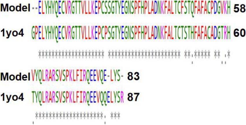
Durojaye et al. Egyptian Journal of Medical Human Genetics (2021) 22:48 Page 2 of 11
conserved and stable region of the virus core struc- amino acid residues with homology to the SARS corona
ture as the bedrock for the design of new virus X4 protein, and it is the sequence of interest for
therapeutics. further studies.
Viruses have a relatively small genome and usually
need a host to suitable execute their life cycle. The Coro- Homology modeling
naviridae have a genome spanning 26 to 32 kb positive- The homology modeling of the SARS-CoV-2 aligned
sense RNA [7–9]. Coronaviruses (CoVs) like the severe segment was done using the SWISS-MODEL (http://
acute respiratory syndrome (SARS) and Middle-East re- swissmodel.expasy.org) for automated comparative mod-
spiratory syndrome (MERS) viruses are primarily zoo- eling of three-dimensional (3D) protein structures [15].
notic [10]. Humans are a complex species in terms of QMEAN (Qualitative Model Energy Analysis) was used
genome; however, the human system is highly suscep- for the assessment of the model protein quality [16]. A
tible to this “respiratory-philic” pathogenic virus, which considerable number of alternative models were pro-
if untreated is fatal. These class of viruses have a con- duced, from which subsequently the final model was se-
served small integral membrane CoV envelope protein lected based on produced scores. We employed
necessary for budding, packaging, envelope formation, as MolProbity (version 4.4) to evaluate the model global and
well as a contributing factor to its pathogenesis [9]. Un- local protein quality [17–19], and Ramachandran plot for
derstanding the biochemistry and molecular structure of torsion angles between residues. In sequence order, φ is
this highly conserved structure is a major factor needed the N(i − 1), C(i), Ca(i), N(i) torsion angle and ψ is the
to kill the pathogen, as designing therapeutics is totally C(i), Ca(i), N(i), C(i + 1) torsion angle. The φ values were
dependent on understanding the structural composition. plotted on the x-axis while the ψ values on y-axis.
Members of this group of coronaviruses have four struc-
tural proteins namely, membrane (M), spike (S), nucleo- 3D structure comparison
capsid (N), and envelope (E) [11]. They also have the X4 The 3D modeling of the SARS-CoV-2 genome translated
(ORF7a) accessory proteins, but their functions are still segment was followed by a structural comparison with the
not yet well understood. The coronavirus X4 protein is X4 protein 3D structure (PDB: 1YO4) using the UCSF
vital to the survival and replication of the coronavirus as Chimera [20]. High-quality images were generated and
recent studies show that X4 is involved during the repli- presented using amino Pymol molecular visualizer [21].
cation cycle of the SARS-CoV [12]. Targeting this pro-
tein with suitable binding moieties that could interrupt Protein physiochemical parameters
the function of this protein may support other existing Calculation of the physiochemical parameters of proteins
strategies to treat this infection. In this study, we did not is a sub-function of the ExPASy server, basically for pro-
focus on targeting the X4 protein rather, we characterized tein identification, and was used for determining the
molecular the structure of the SARS-CoV-2 X4 protein, physiochemical parameters such as theoretical isoelectric
alongside some predicted biochemical features as a bed- point, molecular weight, amino acid composition, extinc-
rock for further studies; providing valuable information tion coefficient, and instability index [22].
for the design of therapeutics. We also further compared
it with other homologues in other species as supportive Phylogenetic analysis
evidence for its lineage amongst the Coronaviridae. We employed Tamura-Nei model for phylogenetic ana-
lysis and is based on the maximum likelihood using
Methods MEGA5 program [23].
Sequence data and alignment
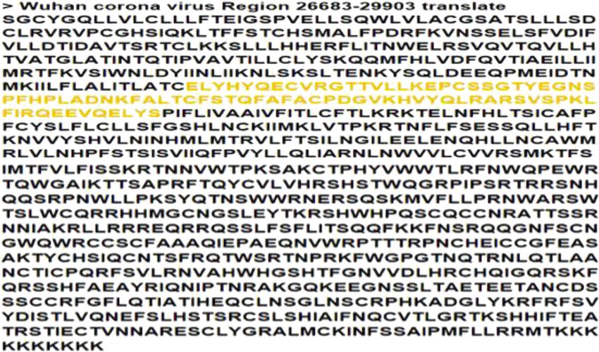
The genome sequence data of the isolated SARS-CoV- Results
2virus was sourced from the GenBank database The full genome of the SARS-CoV-2 consists of 29,
(MN908947.3, which has 100% homology with NC_ 903 nucleotides but here, nucleotides between 26,683
045512.2). We considered the nucleotide sequence be- and 29,903 were considered as the portion coding
tween 26,683 and 29,903 as the region within which to for the group of proteins from which we intended to
find the location of the X4 protein, since based on previ- find the particular protein of our interest, and direct
ous studies, the X4 sequence is located in this region translation of this segment of nucleotides produced a
coding for several of the accessory proteins. EMBOSS sequence of 1004 amino acids after the deletion of
transeq and backtranseq were used for sequence transla- existing stop codons (Fig. 1). The deletion of stop
tion and back translation, respectively [13]. Clustal codons was necessary as the 3D homology tool used
Omega software package was used for all alignments be- for the modeling of the reference protein of interest
tween SARS corona virus X4 protein and SARS-CoV-2 does not recognize them. We used the highlighted
[14]. Within this sequence, we found a portion of the 83 segment in Figs. 1 and 2 for the predicted 3D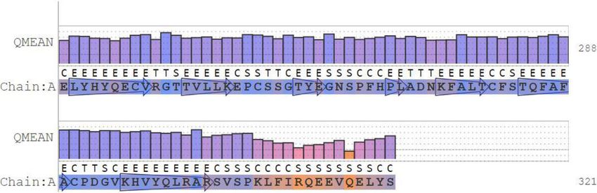
Durojaye et al. Egyptian Journal of Medical Human Genetics        (2021) 22:48                                                         Page 3 of 11

 Fig. 1 Translated sequence of the SARS-CoV-2 corona virus nucleotide sequence with the highlighted segment forming the model protein coding
 sequence of interest

 Fig. 2 Sequence alignment between the amino acid sequence of the model protein and the SARS related corona virus X4 protein. As depicted,
 few homology differences were noticed. Single asterisk (*) represents regions with complete conservation, while colon (:) represents conservation
 between amino acid residues with similar properties. Period (.) represents conservation between amino acids with less similar properties. The
 non-conserved regions are empty space
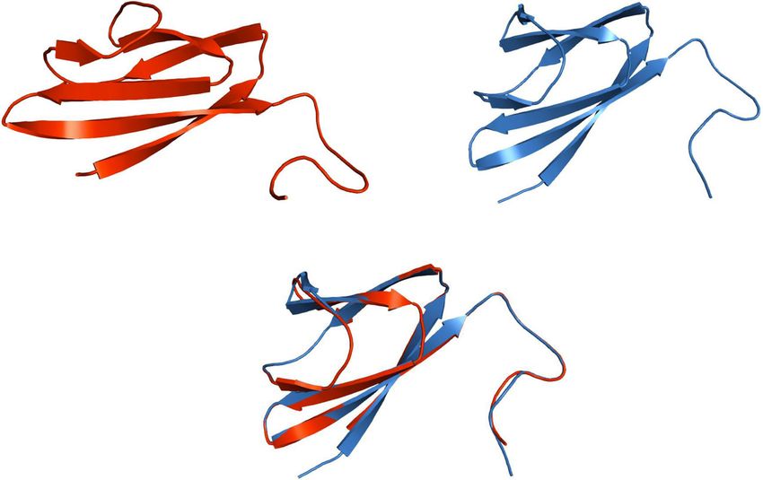
Durojaye et al. Egyptian Journal of Medical Human Genetics       (2021) 22:48                                                        Page 4 of 11

 Fig. 3 3D structures of the model and template protein with the structural comparison. Model protein is presented in red while the template in
 blue. The matching together of the two was depicted in the mixed picture beneath for comparison

structure modeling in comparison with the X4 pro-                          SARS-CoV-2 full genome (Fig. 5). This back-
tein 3D structure (Fig. 3).                                                translated sequence alignment shows that the hom-
  The amino acid sequence of the model protein was                         ology between the model protein sequence and the
back-translated (Fig. 4) to generate the corresponding                     SARS-CoV-2 complete genome is located between 27,
nucleotide sequence which was then aligned with the                        439 and 27,684.

 Fig. 4 Back-translation of the model protein amino acid sequence to generate the corresponding nucleotide sequence
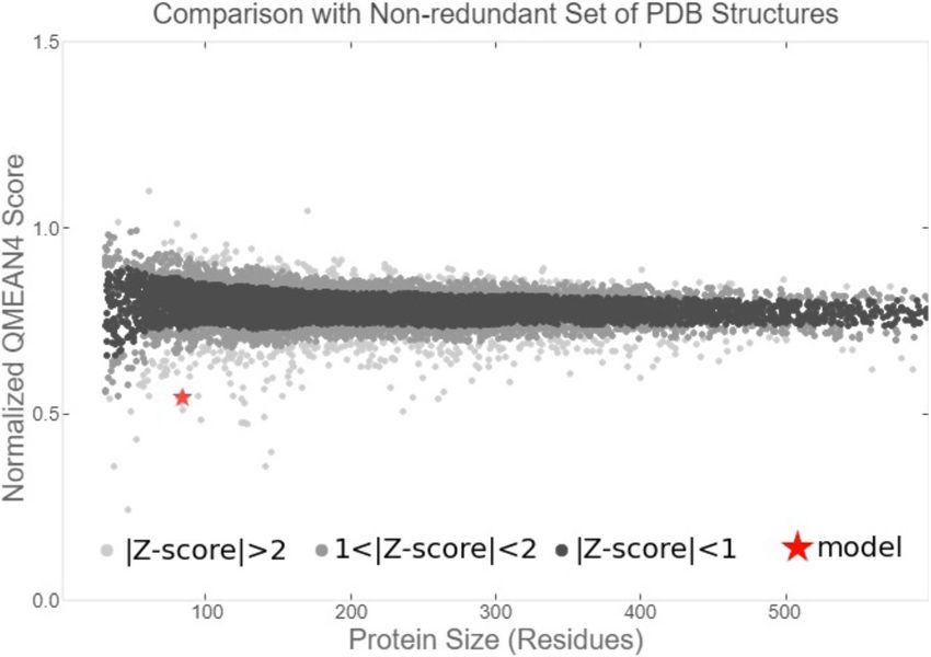
Durojaye et al. Egyptian Journal of Medical Human Genetics        (2021) 22:48                                                        Page 5 of 11

 Fig. 5 Sequence alignment between the model protein nucleotides and the 27,439 to 27,684 nucleotide region of the SARS-CoV-2 complete
 genome. Single asterisk (*) represents regions with complete conservation, while colon (:) represents conservation between amino acid residues
 with similar properties. Period (.) represents conservation between amino acids with less similar properties. The non-conserved regions are
 empty space

  The result of the QMEAN parameter scores of the                           global scores are originally in a range [0,1] with one being
model protein based on the composite scoring function                       good. By default they are transformed into Z scores to re-
(which evaluates several structural features of the model                   late them with what we would expect from high resolution
protein) are presented in Figs. 6, 7, and 8 and Table 1.                    X-ray structures. The local scores are a linear combina-
The absolute quality estimate of the model is expressed                     tions of the 4 statistical potential terms as well as the
in terms of how well the model score agrees with the ex-                    agreement terms evaluated on a per residue basis. They
pected values from a representative set of high-                            are as well in the range [0,1] with one being good (Fig. 7).
resolution experimental structures (Fig. 6). There are                        When compared to the set of non-redundant protein
two global score values, QMEAN4 (for linear combin-                         structures, the QMEAN Z scores as shown in Fig. 8 were
ation of statistical potential) and QMEAN6 (assessing                       close to 0. Good models have scores < 1 and are often
prediction-based consistency of structural features). Both                  located in the dark zone.

 Fig. 6 Residue quality chart which depicts the absolute quality of the model protein on the basis of individual amino acid residue
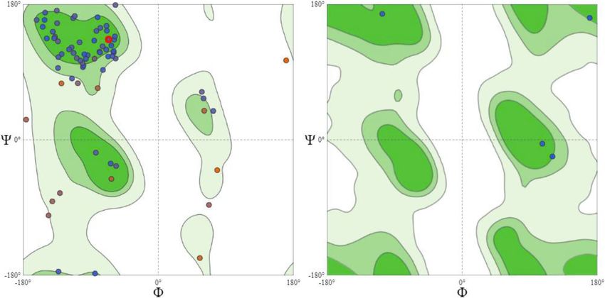
Durojaye et al. Egyptian Journal of Medical Human Genetics        (2021) 22:48                                                          Page 6 of 11

 Fig. 7 Local quality estimate graph showing the values of the predicted local similarity to target plotted against the model protein
 residue number

  The restriction of the Ramachandran angles in the                         for both the vertical and horizontal axis. This is a con-
protein to certain values is visible in the Ramachandran                    venient presentation and allows clear distinction of the
plot in (Fig. 9). The plot shows that each type of second-                  characteristic regions of α-helices and β-sheets. An ex-
ary structure elements occupies its characteristic range                    ception from the principle of clustering around the α-
of φ and ψ angles. The horizontal axis shows φ values,                      and β-regions can be seen on the right plot of Fig. 9. In
while the vertical shows ψ values. Each dot on the plot                     this case, the Ramachandran plot shows torsion angle
shows the angles for an amino acid. The counting starts                     distribution for one single residue, glycine. Glycine does
in the left hand corner from − 180 and extend to + 180                      not have a side chain, which allows high flexibility in the

 Fig. 8 Graphical presentation of estimation of absolute quality of the model protein
Durojaye et al. Egyptian Journal of Medical Human Genetics (2021) 22:48 Page 7 of 11
Table 1 Z score for the individual components of QMEAN for this study, the target protein was modeled using the
the model protein SARS-CoV protein X4 as template. This selection was
Components Scores based on the high resolution and its identity with the
QMEAN score − 4.18 target protein which is as high as 91.57%. The SARS-
Interaction energy of C_β − 1.22 CoV-2 nucleotides between 26,683 and 29,903 were
considered as the portion coding for a group of pro-
Pairwise energy of all atoms − 1.31
tein, of which our target protein of interest is found,
Solvation energy − 1.30
and directly translated to produce a sequence of 1004
Torsion angle energy − 3.47 amino acids (Fig. 1). Structural differences were no-
ticed when alignment analysis was carried out on the
sequence (Figs. 2 and 3). The percentage amino acid
polypeptide chain, making forbidden rotation angles ac- sequence identity between the model and the tem-
cessible. This explains why glycine is often found in loop plate protein shows a high level of conservation, with
regions, where the polypeptide chain needs to make a 90% identity observed between both sequences, show-
sharp turn. This is further depicted in the model protein ing that the conserved domains are predominant.
secondary structures (Fig. 10). Model and template pro- Also, the alignment between the back-translated
tein comparative physiochemical parameters ProtParam model protein nucleotides and the 27,439 to 27,684
were obtained from the amino acid sequences of the in- nucleotide portion of the SARS-CoV-2 complete gen-
dividual proteins (Tables 3 and 4). ome shows that the model protein coding sequence is
The phylogeny tree with the highest log likelihood (− located between 27,439 and 27,684 nucleotides of the
80762.5778) based on the model protein sequence is viral genome (Figs. 4 and 5).
shown in Fig. 11. The percentage of trees in which the The absolute quality estimate of the model is
associated taxa clustered together is shown next to the expressed in terms of how well the model score
branches as conducted in MEGA5. agrees with the expected values from a representa-
tive set of high-resolution experimental structures
Discussion (Fig. 6). The QMEAN scores were transformed into
Proteins that share a high sequence similarity are Z scores to decipher the model of a high resolution
likely to have very similar three-dimensional struc- X-ray structure, and the values are within range
tures and by implication similar function [24, 25]. In (Fig. 7). Our study shows the Z score of the model
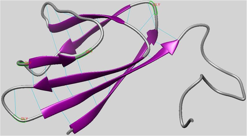
Fig. 9 Presented here are two Ramachandran plots. The plot on the left hand side is hand side shows the general torsion angles for all the
residues in the model protein while the plot on the right hand side is specific for the glycine residues of the proteinDurojaye et al. Egyptian Journal of Medical Human Genetics (2021) 22:48 Page 8 of 11 Fig 10 The model protein secondary structures with the inter model hydrogen bonds. Regions of beta sheets and loops are shown in purple and grey colors, respectively. Labeled in red are the glycine residues of the loops protein has a value of around 0.5, which falls value as compared to the Ramachandran outliers within the acceptable range 0–1, as indicated in and rotamer outliers individual values of 4.94% and Fig. 8 and such a score is an indication of a rela- 27.03%, respectively, we arrived at a MolProbity tively good model as it is close to zero which is the score of 2.96. This value is low enough to indicate average Z score for a good model [26]. Lower Mol- the quality of a good model in the experimental Probity (MP scores) clash score values are expected protein [17]. to be an indication of good models as proven by The repetitive nature of secondary structures is the clash score value (Table 2) exhibited by the due to the repetitive conformation of the residues experimental protein that was modeled for the pur- and, ultimately, repetitive values of φ and ψ. The pose of this study [17–19]. Rotamer outliers asymp- varied secondary conformations can be differentiated tote to a value of < 1% at high resolution, a by their φ and ψ values with the values of different general-case Ramachandran outliers to < 0.05%, and secondary conformations mapping to different areas Ramachandran favored to 98%. With a 3.07 clash of the Ramachandran plot [27]. The Ramachandran score, and a 76.54% Ramachandran favored region plot peptides have points clustered about the values Fig. 11 Bootstrap consensus phylogenetic tree based on the model protein sequence
Durojaye et al. Egyptian Journal of Medical Human Genetics       (2021) 22:48                                                  Page 9 of 11

Table 2 The individual parameters and scores as calculated by             leucine, and valine). It may be regarded as an indica-
MolProbity                                                                tion for the increase in thermostability of globular
Parameters                                                   Scores       proteins. The aliphatic index of the experimental pro-
MolProbity score                                             2.96         teins were calculated according to the following for-
Clash score                                                  3.07         mula [35].
                                                                            Aliphatic index = X(Ala) + a × X(Val) + b × [X(Ile) + X(Leu)]
Ramachandran favored                                         76.54%
                                                                            where X(Ala), X(Val), X(Ile), and X(Leu) are mole per-
Ramachandran outliers                                        4.94%
                                                                          cent (100 × mole fraction) of alanine, valine, isoleucine,
Rotamer outliers                                             27.03%       and leucine. The “a” and “b” coefficients are the relative
                                                                          volume of valine side chain with a value of a = 2.9 and
                                                                          of Leu/Ile side chains b = 3.9 to the side chain of ala-
of φ = − 57o and ψ = − 47o which are the average                          nine. The aliphatic index calculated for the experimental
values for α-helices while the plot for twisted beta                      protein shows a higher thermostability for the model
sheets have points clustered about the values of φ =                      protein than the template.
− 130o and ψ = + 140o which are the average values                          It has been shown that α-helices are more stable,
for twisted sheets. The core regions (green in Fig. 9)                    robust to mutations and designable than β-strands in
contain the most favorable combinations of φ and ψ                        natural proteins [36]. The template and model pro-
and contain the greatest number of points. The re-                        teins respectively have a total of 87 and 83 amino
sult also shows a small third core region in the                          acid residues (Table 4) with the composition of indi-
upper right quadrant. This is called the allowed re-                      vidual residues shown in Table 3. As shown in Fig.
gion and can be situated around the core regions or                       10, the model protein which shares a structural
unassociated with a core region and it contains                           homology with the template is predominantly occu-
fewer data points than the core regions [27]. The                         pied by residues forming beta sheets and coils, with
remaining areas of the plot are considered disallowed.                    none forming helices. The instability observed for
Since glycine residues have only one hydrogen as side                     these two proteins from their physiochemical charac-
chain and has φ and ψ values of + 55o and− 116o, respect-                 teristics show that the unavailability of residues
ively which does not exhibit steric hindrance and for that                forming alpha helix may be the accountable factor.
reason positioned in the disallowed region of the Rama-                   In this study, we also compared a genome of interest
chandran plot as shown in the right hand side plot (Fig.                  to similar genomes in the GenBank database to pre-
9). The extinction coefficient reveals how much light                     dict the evolutionary relationships between homolo-
a protein absorbs at a certain wavelength. It is useful                   gous genes represented in the genomes of each
to have an estimation of this coefficient for monitor-                    divergent species [8, 23, 24]. Organisms with com-
ing a protein in a spectrophotometer when purifying                       mon ancestors were positioned in the same mono-
it, and estimating the molar extinction coefficient de-                   phyletic group in the tree and the same clade where
termined from the amino acid composition [28]                             the genome of interest (SARS-CoV-2) is positioned
which is shown in Table 3.                                                with the SL-CoVZC45, BtRs-BetaCoV/YN2018B, and
   It has been shown that the identity of the N-                          the RS4231, all which are Bat SARS-like corona vi-
terminal residue of a protein plays an important role                     ruses [37]. This shows that the four viral strains
in determining its stability in vivo [29–32]. A protein                   share a common source with shorter divergence
with instability index smaller than 40 is predicted as                    period. TW1 virus, a SARS corona virus is the most
stable; and above 40 is considered unstable [33, 34].                     distantly related based on its branch length and as
The comparative instability index values for the tem-                     such can be regarded as an outlier in the tree.
plate and model proteins were 66.61 and 56.58, re-
spectively, showing both are unstable proteins. A                         Conclusions
protein’s aliphatic index is the relative volume occu-                    We modeled the target protein using the hypothetical
pied by aliphatic side chains (isoleucine, alanine,                       protein X4 as template based on a high similarity index

Table 3 Amino acid composition table for both the template and model proteins
               Amino acid residues in one letter codes
Proteins       A     R     N      D      C      Q     E      G       H      I    L     K     M     F     P     S     T     W       Y     V
Template       6     6     2      2      4      5     7      5       4      1    8     3     0     5     6     6     8     0       5     4
Model          5     4     2      2      4      5     7      4       3      1    8     4     0     6     5     7     5     0       5     6
Durojaye et al. Egyptian Journal of Medical Human Genetics (2021) 22:48 Page 10 of 11
Table 4 Calculated physiochemical properties by the ExPASy ProtParam server
Calculated parameters Template protein Model protein
Molecular weight 9896.10 9478.71
Theoretical pI 7.06 6.32
Amino acid composition (total) 87 83
Atomic composition C438H667N123O132S4 C426H644N112O126S4
Extinction coefficient 7700 7700
Estimated half-life 30 h (mammalian reticulocytes, in vitro). 30 h (mammalian reticulocytes, in vitro).
> 20 h (yeast, in vivo). > 20 h (yeast, in vivo).
> 10 h (Escherichia coli, in vivo). > 10 h (Escherichia coli, in vivo).
Instability index 66.61 56.58
Aliphatic index 60.57 69.28
GRAVY − 0.569 − 0.343
of 91.57%, as revealed by sequence analysis where the writing—original draft preparation, reviewing, and editing. All authors read
percentage amino acid sequence identity between the and approved the final version of the manuscript.
model and the template protein shows a high level of Funding
conservation. The QMEAN value show that the model Not applicable
generated for study here is within the acceptable stand-
Availability of data and materials
ard and amenable to structural analysis, including X-ray Data are available on the appropriate databases cited.
resolution. All the predicted structural parameters for
this model protein studied such as the MolProbity (MP Declarations
scores) clash score, staggered χ angles, Ramachandran
Ethics approval and consent to participate
values (φ and ψ), all demonstrate a protein that is suit- Not Applicable.
able for further study and a potential target for thera-
peutics and vaccines. However, the comparative Consent for publication
Not applicable.
instability index values for the template and model pro-
teins were 66.61 and 56.58, respectively, suggesting that Competing interests
the protein may be too sensitive for in vitro studies. On The authors declare that they have no competing interests.
the other hand, the aliphatic index shows that the ther- Author details
mostability of the model protein is higher than the tem- 1
Department of Molecular and Cell Biology, University of Science and
plate and may withstand more harsh conditions during Technology of China, Hefei, People’s Republic of China. 2Guangxi Bioscience
and Technology Research Centre, Guangxi Academy of Sciences, Nanning
experimental studies. Our results supporting previous 530007, People’s Republic of China. 3Department of Pharmaceutical and
studies, show that the SARS-CoV-2is positioned with Medicinal Chemistry, Faculty of Pharmaceutical Sciences, University of
other Bat SARS-like corona viruses including SL- Nigeria, Nsukka 410001, Nigeria. 4Department of Molecular Genetics and
Biotechnology, Faculty of Biological Sciences, University of Nigeria, Nsukka,
CoVZC45, BtRs-BetaCoV/YN2018B, and the RS4231. Enugu State 410001, Nigeria. 5Department of Biochemistry, College of Life
Science and Technology, Guangxi University, Nanning 530007, People’s
Abbreviations Republic of China.
SARS-CoV-2: Severe acute respiratory syndrome coronavirus 2; 2019-
nCoV: 2019 novel coronavirus; SARS-CoV: Severe acute respiratory syndrome Received: 17 December 2020 Accepted: 24 March 2021
coronavirus; MERS: Middle East respiratory syndrome; SARS: Severe acute
respiratory syndrome; pI : Isoelectric point; DB: Protein Data Bank; ARD: Acute
respiratory disease; DNA: Deoxyribonucleic acid; RNA: Ribonucleic acid; References
hCoV: Human coronavirus; NCBI: National Center for Biotechnology 1. Johnson M (2020) Wuhan 2019 novel coronavirus - 2019-nCoV. Mater
Information; INSDC: International Nucleotide Sequence Database Methods 10:1–5. https://doi.org/10.13070/mm.en.10.2867
Collaboration; QSQE: Quaternary structure quality estimate; CFSSP: Chou and 2. Harapan H, Itoh N, Yufika A, Winardi W, Keam S, Te H et al (2020)
Fasman Secondary Structure Prediction; MEGA: Molecular Evolutionary and Coronavirus disease 2019 (COVID-19): a literature review. J Infect Public
Genetics Analysis; QMEAN: Qualitative Model Energy Analysis; GRAVY: Grand Health 13(5):667–673. https://doi.org/10.1016/j.jiph.2020.03.019
average of hydropathy 3. Caddy S (2020) Developing a vaccine for covid-19. BMJ 369:1–2. https://doi.
org/10.1136/bmj.m1790
Acknowledgements 4. Bollyky TJ, Gostin LO, Hamburg MA. The Equitable Distribution of COVID-19
Not applicable Therapeutics and Vaccines. JAMA. 2020;323(24):2462–3. https://doi.org/10.1
001/jama.2020.6641.
Authors’ contributions 5. Williamson BN, Feldmann F, Schwarz B, Meade-White K, Porter DP, Schulz J,
OAD: conceptualization, methodology, data curation, software, van Doremalen N, Leighton I, Yinda CK, Pérez-Pérez L, Okumura A, Lovaglio
writing—original draft preparation. ONO: visualization, validation, J, Hanley PW, Saturday G, Bosio CM, Anzick S, Barbian K, Cihlar T, Martens C,
writing—reviewing and editing. OAS: visualization, investigation, supervision, Scott DP, Munster VJ, de Wit E (2020) Clinical benefit of remdesivir in rhesusDurojaye et al. Egyptian Journal of Medical Human Genetics (2021) 22:48 Page 11 of 11
macaques infected with SARS-CoV-2. Nature 585(7824):273–276. https://doi. density information. BMC Struct Biol 9(1):1–17. https://doi.org/10.1186/1472-
org/10.1038/s41586-020-2423-5 6807-9-35
6. Sahin AR (2020) 2019 novel coronavirus (COVID-19) outbreak: a review of 27. Patil VM, Balasubramanian K, Masand N (2018) Dengue virus polymerase: a
the current literature. Eurasian J Med Oncol 4:1–7. https://doi.org/10.14744/ crucial target for antiviral drug discovery. Elsevier Inc. https://doi.org/10.101
ejmo.2020.12220 6/B978-0-12-815422-9.00014-0
7. Maringer K, Fernandez-Sesma A. Since January 2020 Elsevier has created a 28. Gill SC, von Hippel PH (1989) Calculation of protein extinction coefficients
COVID-19 resource centre with free information in English and Mandarin on from amino acid sequence data [published erratum appears in anal
the novel coronavirus COVID-19. The COVID-19 resource centre is hosted on Biochem 1990 Sep;189(2):283]. Anal Biochem 182(2):319–326. https://doi.
Elsevier Connect, the company’s public news and information 2020. org/10.1016/0003-2697(89)90602-7
8. Campillo-Balderas JA, Lazcano A, Becerra A (2015) Viral genome size 29. Apel W, Schulze WX, Bock R (2010) Identification of protein stability
distribution does not correlate with the antiquity of the host lineages. Front determinants in chloroplasts. Plant J 63(4):636–650. https://doi.org/10.1111/
Ecol Evol 3. https://doi.org/10.3389/fevo.2015.00143 j.1365-313X.2010.04268.x
9. Schoeman D, Fielding BC (2019) Coronavirus envelope protein: current 30. Fantini D, Vascotto C, Marasco D, D’Ambrosio C, Romanello M, Vitagliano L,
knowledge. Virol J 16(1):1–22. https://doi.org/10.1186/s12985-019-1182-0 Pedone C, Poletto M, Cesaratto L, Quadrifoglio F, Scaloni A, Radicella JP, Tell
10. Van Den Brand JMA, Smits SL, Haagmans BL (2015) Pathogenesis of Middle G (2010) Critical lysine residues within the overlooked N-terminal domain of
East respiratory syndrome coronavirus. J Pathol 235(2):175–184. https://doi. human APE1 regulate its biological functions. Nucleic Acids Res 38(22):
org/10.1002/path.4458 8239–8256. https://doi.org/10.1093/nar/gkq691
11. Satarker S, Nampoothiri M (2020) Structural proteins in severe acute 31. Arnesen T (2011) Towards a functional understanding of protein N-terminal
respiratory syndrome Coronavirus-2. Arch Med Res 51(6):482–491. https:// acetylation. PLoS Biol 9(5):e1001074. https://doi.org/10.1371/journal.pbio.1
doi.org/10.1016/j.arcmed.2020.05.012 001074
12. Hänel K, Stangler T, Stoldt M, Willbold D (2006) Solution structure of the X4 32. Deng S, Marmorstein R (2020) Protein N-terminal acetylation: structural
protein coded by the SARS related coronavirus reveals an immunoglobulin basis, mechanism, versatility, and regulation. Trends Biochem Sci 46(1):1–13.
like fold and suggests a binding activity to integrin I domains. J Biomed Sci https://doi.org/10.1016/j.tibs.2020.08.005
13(3):281–293. https://doi.org/10.1007/s11373-005-9043-9 33. Mohan R (2012) Computational structural and functional analysis of
13. Li W, Cowley A, Uludag M, Gur T, McWilliam H, Squizzato S, Park YM, Buso hypothetical proteins of Staphylococcus aureus. Bioinformation 8(15):722–
N, Lopez R (2015) The EMBL-EBI bioinformatics web and programmatic 728. https://doi.org/10.6026/97320630008722
tools framework. Nucleic Acids Res 43(W1):W580–W584. https://doi.org/10.1 34. Sahay A, Piprodhe A, Pise M (2020) In silico analysis and homology
093/nar/gkv279 modeling of strictosidine synthase involved in alkaloid biosynthesis in
14. Sievers F, Higgins DG (2018) Clustal omega for making accurate alignments catharanthus roseus. J Genet Eng Biotechnol 18(1):44. https://doi.org/10.11
of many protein sequences. Protein Sci 27(1):135–145. https://doi.org/10.1 86/s43141-020-00049-3
002/pro.3290 35. Gasteiger E, Hoogland C, Gattiker A, Duvaud S, Wilkins MR, Appel RD et al
15. Schwede T, Kopp J, Guex N, Peitsch MC (2003) SWISS-MODEL: an (2005) The proteomics protocols handbook. Proteomics Protoc Handb:571–
automated protein homology-modeling server. Nucleic Acids Res 31(13): 608. https://doi.org/10.1385/1592598900
3381–3385. https://doi.org/10.1093/nar/gkg520 36. Abrusán G, Marsh JA (2016) Alpha helices are more robust to mutations
16. Benkert P, Tosatto SCE, Schomburg D (2008) QMEAN: a comprehensive than Beta strands. PLoS Comput Biol 12(12):1–16. https://doi.org/10.1371/
scoring function for model quality assessment. Proteins Struct Funct Genet journal.pcbi.1005242
71(1):261–277. https://doi.org/10.1002/prot.21715 37. Chan JFW, Yuan S, Kok KH, KKW T, Chu H, Yang J et al (2020) A familial
17. Chen VB, Arendall WB, Headd JJ, Keedy DA, Immormino RM, Kapral GJ et al cluster of pneumonia associated with the 2019 novel coronavirus indicating
(2010) MolProbity: all-atom structure validation for macromolecular person-to-person transmission: a study of a family cluster. Lancet
crystallography. Acta Crystallogr Sect D Biol Crystallogr 66(1):12–21. https:// 395(10223):514–523. https://doi.org/10.1016/S0140-6736(20)30154-9
doi.org/10.1107/S0907444909042073
18. Davis IW, Leaver-Fay A, Chen VB, Block JN, Kapral GJ, Wang X, Murray LW, Publisher’s Note
Arendall WB, Snoeyink J, Richardson JS, Richardson DC (2007) MolProbity: all- Springer Nature remains neutral with regard to jurisdictional claims in
atom contacts and structure validation for proteins and nucleic acids. Nucleic published maps and institutional affiliations.
Acids Res 35(Web Server):375–383. https://doi.org/10.1093/nar/gkm216
19. Davis IW, Murray LW, Richardson JS, Richardson DC (2004) MolProbity:
structure validation and all-atom contact analysis for nucleic acids and their
complexes. Nucleic Acids Res 32(Web Server):615–619. https://doi.org/10.1
093/nar/gkh398
20. Huang CC, Meng EC, Morris JH, Pettersen EF, Ferrin TE (2014) Enhancing
UCSF chimera through web services. Nucleic Acids Res 42(W1):478–484.
https://doi.org/10.1093/nar/gku377
21. Yuan S, Chan HCS, Hu Z (2017) Using PyMOL as a platform for
computational drug design. Wiley Interdiscip Rev Comput Mol Sci 7(2).
https://doi.org/10.1002/wcms.1298
22. Cash P (1999) 2-D proteome analysis protocols. Cell Biol Int 23(5):385.
https://doi.org/10.1006/cbir.1999.0355
23. Tamura K, Peterson D, Peterson N, Stecher G, Nei M, Kumar S (2011) MEGA5:
molecular evolutionary genetics analysis using maximum likelihood,
evolutionary distance, and maximum parsimony methods. Mol Biol Evol
28(10):2731–2739. https://doi.org/10.1093/molbev/msr121
24. Kaczanowski S, Zielenkiewicz P (2010) Why similar protein sequences
encode similar three-dimensional structures? Theor Chem Accounts 125(3-
6):643–650. https://doi.org/10.1007/s00214-009-0656-3
25. Zhang B, Jaroszewski L, Rychlewski L, Godzik A (1997) Similarities and
differences between nonhomologous proteins with similar folds: evaluation
of threading strategies. Fold Des 2(5):307–317. https://doi.org/10.1016/S13
59-0278(97)00042-4
26. Benkert P, Schwede T, Tosatto SC (2009) QMEANclust: estimation of protein
model quality by combining a composite scoring function with structuralYou can also read