Vivid biofluorescence discovered in the nocturnal Springhare (Pedetidae) - Nature
←
→
Page content transcription
If your browser does not render page correctly, please read the page content below
www.nature.com/scientificreports
OPEN Vivid biofluorescence discovered
in the nocturnal Springhare
(Pedetidae)
Erik R. Olson1*, Michaela R. Carlson1, V. M. Sadagopa Ramanujam2, Lindsay Sears3,
Sharon E. Anthony1, Paula Spaeth Anich1, Leigh Ramon4, Alissa Hulstrand1,
Michaela Jurewicz1, Adam S. Gunnelson1, Allison M. Kohler1,5 & Jonathan G. Martin1
Biofluorescence has been detected in several nocturnal-crepuscular organisms from invertebrates to
birds and mammals. Biofluorescence in mammals has been detected across the phylogeny, including
the monotreme duck-billed platypus (Ornithorhyncus anatinus), marsupial opossums (Didelphidae),
and New World placental flying squirrels (Gluacomys spp.). Here, we document vivid biofluorescence
of springhare (Pedetidae) in both museum specimens and captive individuals—the first documented
biofluorescence of an Old World placental mammal. We explore the variation in biofluorescence across
our sample and characterize its physical and chemical properties. The striking visual patterning and
intensity of color shift was unique relative to biofluorescence found in other mammals. We establish
that biofluorescence in springhare likely originates within the cuticle of the hair fiber and emanates,
at least partially, from several fluorescent porphyrins and potentially one unassigned molecule
absent from our standard porphyrin mixture. This discovery further supports the hypothesis that
biofluorescence may be ecologically important for nocturnal-crepuscular mammals and suggests that
it may be more broadly distributed throughout Mammalia than previously thought.
As our understanding of the visual capacities of various species develops1,2, we are realizing that many species
are capable of seeing the world through a different lens, and that, in addition to or as an alternative to visible
light (approximately, 400–700 nm), other wavelengths of light may be ecologically important. For example, some
species use cues from ultraviolet (UV) light for sexual s election3, predator a voidance4, and f oraging5. Many mam-
mals are sensitive to UV wavelengths of light1,2,6, especially m arsupials7 (New World opossums), a m onotreme8
(platypus, Ornithorhynchus anatinus), and some r odents7 (Sciurognathi).
Biofluorescence, the absorption of short wavelengths of light and re-emission of longer wavelengths of light,
has been increasingly observed in a wide range of i nvertebrates9, fishes10,11, reptiles and a mphibians12–16 and birds
3,17,18
. Within mammals, biofluorescence has been observed in New World placental flying s quirrels19 (Glaucomys
spp.), New World marsupial opossums20,21 (Didelphidae), and the monotreme duck-billed platypus (O. anati-
nus) of Australia and T asmania22. These groups, inhabit three continents and a diverse array of ecosystems and
are widely distributed across the mammalian family tree. All of these biofluorescent mammalian species are
nocturnal-crepuscular19–22. Thus, biofluorescence in mammals may be of significance to nocturnal-crepuscular
mammals that are active in low light environments9,22. Here we document biofluorescence in another noctur-
nal mammal, springhare (Pedetidae)—the first well-documented biofluorescence of an Old World eutherian
mammal.
Like other biofluorescent mammals, springhares (Pedetidae) are nocturnal. Apart from their nocturnal life-
style, springhares are not ecologically similar to other known fluorescent species and are rather distant relatives of
the biofluorescent rodents within Sciuridae19,23. Both species of springhares are endemic to Africa: the springhare
(Pedetes capensis) lives in southern Africa; and the East African springhare (P. surdaster) inhabits portions of
Kenya and Tanzania23. Both species are nocturnal, fossorial grazers that inhabit semi-arid areas24,25. Springhares
are mainly solitary, sheltering in their own burrows during the day and preferentially foraging individually in
short-grass environments at night24. Although short-grass environments provide springhares with minimal cover
1
Departments of Environmental Sciences and Natural Resources, Northland College, 1411 Ellis Avenue,
Ashland, WI 54806, USA. 2Department of Preventive Medicine and Population Health, The University of Texas
Medical Branch, Galveston, TX 77555, USA. 3Omaha’s Henry Doorly Zoo and Aquarium, 3701 South 10th Street,
Omaha, NE 68107, USA. 4Mesker Park Zoo and Botanic Garden, 1545 Mesker Park Drive, Evansville, IN 47720,
USA. 5Natural Resources Ecology Laboratory, Colorado State University, Fort Collins, CO 80523, USA. *email:
eolson@northland.edu
Scientific Reports | (2021) 11:4125 | https://doi.org/10.1038/s41598-021-83588-0 1
Vol.:(0123456789)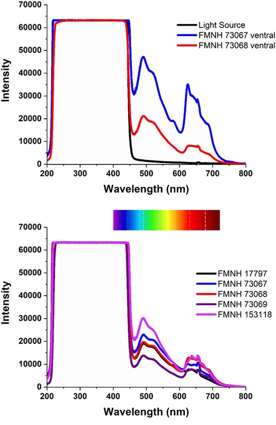
www.nature.com/scientificreports/
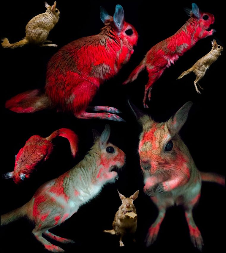
Figure 1. Biofluorescence in East African springhare museum specimens. A male Pedetes surdaster museum
specimen (FMNH 73067) collected from the Central Province of Kenya in 1952, photographed under visible
light (first and third image from left) and 395 nm ultraviolet light using a 470 nm longpass filter. Orange to red
biofluorescence (~ 650 nm) is observed on the dorsal (leftmost images) and ventral surfaces (rightmost images)
of this museum specimen from the Field Museum of Natural History (FMNH) in Chicago, Illinois, USA.
from predators, like mongooses (Herpestidae) and jackals (Canidae), these open areas provide springhares with
clear lines of sight for the detection of predators and few obstacles to escape via explosive, bipedal leaps25. Here
we document vivid biofluorescence in Pedetidae, its variation, its underlying chemical nature, and its potential
ecological implications.
Results
While studying biofluorescence in New World flying squirrels (Glaucomys spp.) and members of Anomaluromor-
pha at the Field Museum of Natural History (FMNH) in Chicago, Illinois, USA, in April 201819 and November
201922, we discovered springhare biofluorescence and subsequently documented the trait in multiple specimens
and captive individuals.
We examined a total of 14 museum specimens, including eight museum specimens of P. capensis collected
from Angola (n = 2) and Botswana (n = 6), and six specimens of P. surdaster collected from Kenya (n = 3) and
Tanzania (n = 3) (Fig. 1, Supplementary Table S1). We examined individuals of both sexes, including four males
(P. capensis, n = 2; P. surdaster, n = 2) and ten females (P. capensis, n = 6; P. surdaster, n = 4), and specimen collec-
tion dates ranged from 1905 to 1963. For museum specimens, we observed and photographed biofluorescence
on both the dorsal and ventral side of each specimen following the methods of Anich et al.22 (Canon EOS 50D,
Canon USA Inc., Melville, NY, USA; Sigma 17–70 mm f 2.8–4 DC Macro) under visible light (Canon Speedlite
430EX) and then separately under 395 nm UV light (LED UV flashlight, iLumen8 100 LED). We photographed
a subset of specimens using a 470 nm longpass filter (K&F Concept, Guangdong Sheng, China; Tiffen Yellow 2
#8, Hauppauge, New York, USA) under UV illumination to absorb any residual light in the blue wavelengths.
We also captured UV reflectance using a Nurugo SmartUV camera (Union community Co., Ltd., Seoul, Republic
of Korea), which suggested that very little UV light was being absorbed on either dorsal or ventral surfaces of a
subset of springhare specimens. Following the methods of Anich et al.22 for fluorescence spectroscopy (Ocean
Optics USB2000+, Largo, Florida, USA), we identified two peaks of fluorescence at 500 and 650 nm for a section
of highly fluorescent fur on the ventral surface (Figs. 1 and 2).
We also observed and photographed biofluorescence on five living captive-bred P. capensis individuals (three
males and two females) at Omaha’s Henry Doorly Zoo and Aquarium in Omaha, Nebraska and one deceased
(due to natural causes) individual (female) at the Mesker Park Zoo & Botanic Garden in Evansville, Indiana
(Fig. 3; Supplementary Table S2, Canon EOS 6D, Canon USA Inc., Melvill, New York, USA; Canon 17–40 mm
f 2.8–4; LED 395 UV flood light; Tiffen Yellow 2 #8, Hauppauge, New York, USA; white balance corrected
from DGK Color Tools WB card, DGK Color Tools LLC, Waban, Massachusetts, USA). Using compound light
microscopy (Eclipse E2300, with DSFI2 camera, Nikon Corporation, Tokyo, Japan), we examined hair samples
from the recently deceased captive individual springhare under visible and UV light with 4× magnification. We
photographed a human hair under the same conditions for comparison. We observed fluorescence of individual
springhare hair fibers and variation in the presence of fluorescence within individual hair fibers, suggesting that
the fluorescence is distributed through the thickness of the cuticle and absent from the core and tips of hair fib-
ers (Fig. 4). Washing the hair or fur with Dawn dish soap (Cincinnati, Ohio, USA) did not remove or diminish
fluorescence or result in a transfer of the fluorescence.
We used thin layer chromatography to separate fluorescent extracts from hair samples. Using high perfor-
mance liquid chromatography (HPLC), we revealed the presence of fluorescent porphyrin species including
uroporphyrin-I, uroporphyrin-III, heptacarboxylporphyrin, hexacarboxylporphyrin, and coproporphyrin-I
(Fig. 5; Supplementary Figs S1 and S2). We also detected an unassigned molecule absent from our standard
Scientific Reports | (2021) 11:4125 | https://doi.org/10.1038/s41598-021-83588-0 2
Vol:.(1234567890)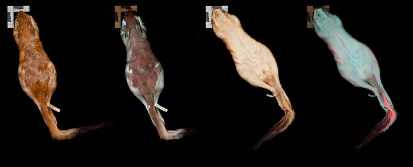
www.nature.com/scientificreports/

                                  Figure 2.  Spectra of biofluorescence observed in East African springhare. (Top panel) Spectra of
                                  biofluorescence on the ventral side of two different P. surdaster specimens (red and blue lines) in comparison
                                  to the spectrum of the light source taken against a diffuse reflectance standard (black line). (Bottom panel)
                                  Spectra of biofluorescence from the dorsal side of five different P. surdaster specimens. Peaks in the visible range
                                  (400–750 nm) represent biofluorescence.

                                  mixture of porphyrins that peaked at approximately two minutes, which may also play a role in the biofluores-
                                  cence observed (Fig. 5; Supplementary Figs S1 and S2).
                                     For P. capensis and P. surdaster, all individuals exhibited orange to red biofluorescence, although we did
                                  observe variation in the intensity of biofluorescence across individuals. Biofluorescence was pronounced on
                                  both the dorsal and ventral surfaces (Fig. 1), and fluorescence intensity for the dorsal surfaces was often more
                                  intense in the head and posterior region (Figs. 1 and 3). Ventrally, biofluorescence occurred predominantly
                                  along the inner thigh and tail (Fig. 1). We observed notable patchiness in the biofluorescence in both museum
                                  and captive specimens (Figs. 1 and 3).

                                  Discussion
                                  We have discovered a funky and vivid porphyrin-based biofluorescence in Pedetidate, representing the first well-
                                  documented biofluorescence of an Old World eutherian mammal. While we do not have a large enough sample
                                  size to draw conclusions about the frequency of this trait in wild populations, we did consistently observe the
                                  trait in six captive individuals, as well as, 14 museum specimens collected at different times over a 58-yr. period
                                  and across seven separate locations in four countries (Fig. 3). Due to the spatial, temporal, and contextual (i.e.,
                                  captive or wild) diversity of our specimens, we suspect this trait is not environmental. Both male and female
                                  specimens fluoresced in the same regions and with the same intensity, generally; therefore, we suspect that the
                                  trait is not sexually dimorphic. The fact that biofluorescence was not easily removed via washing and was pre-
                                  sent on museum specimens from 1905 suggests that the biofluorescence is a part of the physical anatomy of the
                                  hair fibers for Pedetidae. Biofluorescence appeared more vivid in living individuals than in museum specimens,
                                  potentially indicating some degradation over time (Figs. 1 and 3).

Scientific Reports |   (2021) 11:4125 |                https://doi.org/10.1038/s41598-021-83588-0                                                    3

                                                                                                                                                Vol.:(0123456789)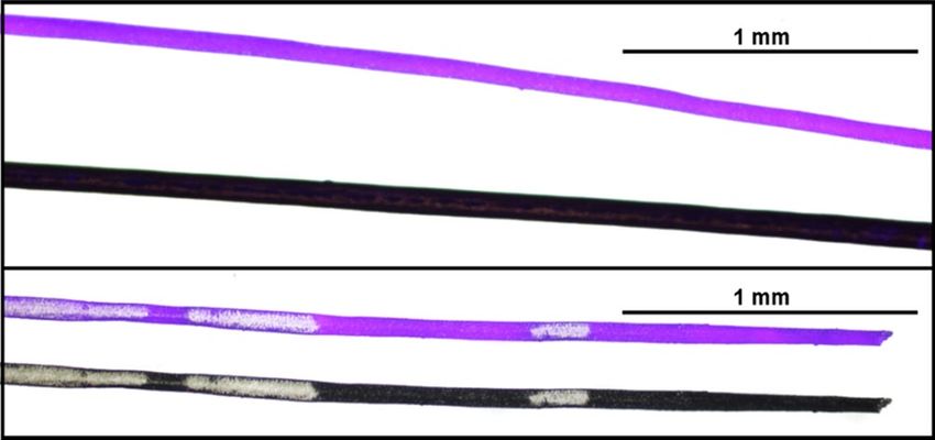
www.nature.com/scientificreports/
Figure 3. Biofluorescence in captive springhare. Pedetes capensis photographed under visible light (three
insets) and under 395 nm ultraviolet light with a 470 nm longpass filter to documenting the orange to red
biofluorescence (~ 650 nm) of springhare. Images contain two different captive-bred, captive individuals
(Individual #4, female, bottom two larger images under ultraviolet light; Individual #2, male, all remaining
images) from the Omaha’s Henry Doorly Zoo & Aquarium in Omaha, Nebraska, USA.
We detected multiple species of porphyrin in extracts from springhare hair samples. Porphyrin-based bio-
fluorescence has been suspected or confirmed in many marine invertebrates26,27, the plumage of many bird
species18,28–30, the bones of at least one species of r odent31, and at least one species of P latyhelminthes9 (Pla-
tydemus manokwari). Here we detected uroporphyin-I, -III, heptacarboxylporphyrin, hexacarboxylporphyrin,
coproporphyrin-I, and one unassigned molecule absent from our standard porphyrin mixture. The isolated
porphyrins are formed by oxidation of porphyrinogens, which are intermediates in the biosynthetic pathway of
heme32. Uroporphyrin and coproporphyrin fluoresce between 570 and 720 nm in various conditions33,34, suggest-
ing that at least both uroporphyrin and coproporphyrin play a role in causing biofluorescence in springhare. We
recommend future studies be done to determine whether this biofluorescence is an advantageous evolutionary
trait or a disease, such as porphyrias as seen in fox squirrels (Sciurus niger)31,35,36, canefield rats (Rattus sordidus)37,
and humans (Homo sapiens)32,38.
Biofluorescence in both species of Pedetes was notably patchy (Figs. 1 and 3), and observations of captive
individuals indicated that the areas most impacted by grooming and intra-specific interactions, i.e., reproduc-
tion, appear to overlap relatively strongly with areas most consistently exhibiting biofluorescence; this would
suggest that the fluorescence might be externally applied to the fur during certain behaviors. However, thorough
Scientific Reports | (2021) 11:4125 | https://doi.org/10.1038/s41598-021-83588-0 4
Vol:.(1234567890)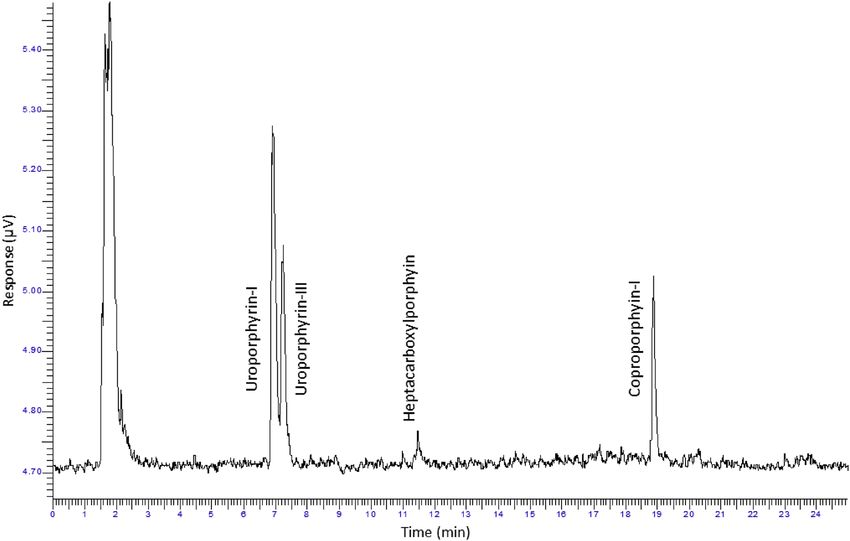
www.nature.com/scientificreports/

                                  Figure 4.  Biofluorescent hair of springhare. (Top panel) Biofluorescent hair of Pedetes capensis compared to
                                  the non-biofluorescent hair of a human photographed under 395 nm ultraviolet light at 4× magnification with a
                                  100 ms exposure, and (bottom panel) a single strand of P. capensis hair photographed under 395 nm ultraviolet
                                  light and visible light at 4× magnification with a 100 ms exposure; highlighting variation observed in hair
                                  pigmentation and fluorescence.

                                  Figure 5.  Identification of the porphyrins responsible for biofluorescence in Pedetidae. High performance
                                  liquid chromatograph of extracted pigment from captive Pedetes capensis fur. The response times in this
                                  sample were compared to that of a standard mixture of porphyrins (Supplementary Fig S1) which allowed
                                  for identification of uroporphyrin-I, uroporphyrin-III, heptacarboxylporphyrin, and coproporphyrin-I in
                                  the sample. The peak centered around two minutes has not been assigned and does not correspond to any
                                  of the porphyrins in the standard mix (uroporphyrin, heptacarboxylporphyrin, hexacarboxylporphyin,
                                  pentacarboxylporphyrin, coproporphyrin, and mesoporphyrin).

                                  washing of the fur did not result in the removal or transfer of the fluorescence, and observations of enclosures of
                                  captive individuals did not reveal any transferred fluorescence. Additionally, UV images of a single live individual
                                  taken 14 months apart indicate that the individual patterns are relatively consistent, further suggesting that an
                                  alternative explanation for the patchiness of biofluorescence in springhare (Fig. 3) may exist. Springhares are
                                  predominantly solitary and tend to forage in more open areas with sparse vegetation and, therefore, have greater
                                  exposure to predators due to the lack of cover or group vigilance. Thus, we hypothesize that the patchiness of
                                  biofluorescence in springhares could function as a camouflage of sorts, but this would depend on the UV sen-
                                  sitivity of their predators. We recommend additional behavioral and biological research to further understand
                                  the potential ecological relevance of springhare biofluorescence and patchiness thereof.
                                      The ecological implications of biofluorescence in springhare remain unknown. However, like other bio-
                                  fluorescent animals, springhares are nocturnal. Biofluorescence in mammals has been detected mainly in

Scientific Reports |   (2021) 11:4125 |                https://doi.org/10.1038/s41598-021-83588-0                                                   5

                                                                                                                                               Vol.:(0123456789)
www.nature.com/scientificreports/
Step Time (min) Flow (ml/min) Mobile phase A Mobile phase B Curve
0 2.0 1.20 100 0 0.0
1 2.0 1.20 85 15 6.0
2 18.0 1.20 0 100 2.0
3 5.0 1.20 100 0 2.0
Table 1. Gradient elution program for high-performance liquid chromatography analysis of extracts from hair
samples to determine the basis of biofluorescence in springhare (Pedetes capensis).
nocturnal-crepuscular19–22 and UV-sensitive7,39 species, and UV-color vision appears to be ecologically important
to many nocturnal-crepuscular mammals1. While we cannot determine why Pedetidae exhibits biofluorescence,
our observations add further support for the hypothesis that biofluorescence and UV wavelengths of light may
be ecologically important for nocturnal-crepuscular m ammals1,9,19,22. Our observations also suggest that biofluo-
rescence may be more broadly distributed throughout Mammalia than previously thought22.
Methods
All methods were carried out in accordance with the relevant guidelines and regulations. Protocol for captive
animal observation and photography was evaluated and considered exempt from the Omaha’s Henry Doorly
Zoo & Aquarium’s ACUC. Protocol for hair collection from the deceased (due to natural causes) individual was
evaluated by Mesker Park Zoo & Botanic Garden Research Committee and considered exempt from the Mesker
Park Zoo & Botanic Garden ACUC. Protocol for the microscopy and photography of a human hair was evaluated
and considered exempt from the Northland College IRB. Informed consent was obtained for the microscopy
and photography of the strand of human hair.
Fluorescence spectroscopy. We used an Ocean Optics Flame-S-UV-VIS-ES (Largo, Florida, USA) in
fluorescence mode (integration time = 1 s, and 5 scans per spectrum) and an Ocean Optics DH-2000-BAL deute-
rium light source (Largo, Florida, USA) to take fluorescence spectra. We selected an intense fluorescent spot on a
subset of the Pedetidae specimens and placed a probe holder directly on that spot with the probe at 45° relative to
the sample. Fluorescence spectra were taken at five different places within that spot, and these five spectra were
averaged. We recorded the light source spectrum against a polytetrafluoroethylene diffuse reflectance standard.
Extraction of biofluorescent compounds. We collected hair samples from the recently deceased cap-
tive individual of P. capensis at the Mesker Park Zoo & Botanic Garden. We attempted to determine if the fluo-
rescence could be removed via washing with Dawn dish soap (Cincinnati, Ohio, USA). We combined samples
of fluorescent fur (washed and unwashed) into two samples (68.8 mg and 95.1 mg for sample 1 and sample 2,
respectively), and each sample was refluxed in a 25 mL round bottom flask, with 1 mL of 0.5 M NaOH until all
hair was dissolved and the solvent appeared pink under UV light (approx. 16 min). We separated the solution
using thin layer chromatography and a mixture of 4 mL DMF, 35 mL MeOH, 6 mL ethylene glycol, 0.4 mL glacial
acetic acid, 18 mL 1-chlorobutane, and 20 mL C HCl3 as the solvent40. The thin layer chromatography plates had
aluminum backs and were coated with silica gel 60 F 254. After separation, three distinct pink bands were visible
under a handheld UV light. We collected and washed the pink silica with acetone and deionized water until the
silica was no longer pink. Then, we filtered and reduced the extracts under pressure until a yellow oil remained
which was sent for HPLC analysis.
Characterization of fluorescent compounds. We used a Perkin Elmer Series 200 HPLC instrument
(Waltham, MA, USA) connected in series with a C18 reversed phase Onyx Monolithic HD-C18 liquid chro-
matography column (100 mm length × 4.6 mm internal diameter, Phenomenex Inc., Torrance, CA, USA), and a
Perkin Elmer Series 200 fluorescence detector to characterize the extracts. All solvents were of HPLC grade and
purchased from Fisher Scientific, Inc., USA. The components of mobile phase A (pH = 5.16) contained 800 mL
of Milli-Q water mixed with 100 mL acetonitrile, 55 ml glacial acetic acid, and 45 ml concentrated ammonium
hydroxide. The components of mobile Phase B contained 900 ml methanol mixed with 100 ml acetonitrile.
We dissolved extracted materials in 1 mL of 1 M hydrochloric acid, centrifuged and transferred the superna-
tant to amber colored auto-sampler vials. For the identification of porphyrin carboxylic acids, we used a chroma-
tographic marker kit containing 5 micromole mixture of each octa-, hepta-, hexa-, penta-, tretra-, and di-carboxyl
porphyrin acids (Frontier Scientific, Inc., Salt Lake City, UT, USA). Octa- and tetra-carboxyl porphyrin acids are
conventionally called as Uropophyrin and Coproporphyrin, respectively. We dissolved the standards mixture of
porphyrins in the tube in 10 mL of 1 M hydrochloric acid (High Purity grade from Fisher Scientific, Inc., Salt
Lake City, UT, USA), and considered this mix as the HIGH standards mix. We created a ten times dilution of
the HIGH standards mix and considered this the LOW standards mix.
We used a gradient elution program with an injection volume set to 100 µl and total run time 36 min41,42
(Table 1). Two vials of porphyrin carboxylic acid of the (HIGH and LOW concentrations) standards were also
analyzed with each batch of samples. We identified the HPLC peaks in samples by matching retention times
(min) of sample peaks with carboxyl porphyrin acid peaks (Supplementary Fig S1).
Scientific Reports | (2021) 11:4125 | https://doi.org/10.1038/s41598-021-83588-0 6
Vol:.(1234567890)www.nature.com/scientificreports/
Data availability
All data produced in this study are included in the text and supplementary information documents.
Received: 27 October 2020; Accepted: 1 January 2021
References
1. Zhao, H. et al. The evolution of color vision in nocturnal mammals. PNAS 106, 8980–8985 (2009).
2. Douglas, R. H. & Jeffery, G. The spectral transmission of ocular media suggests ultraviolet sensitivity is widespread among mam-
mals. Proc. R. Soc. B. 281, 1471–2954. https://doi.org/10.1098/rspb.2013.2995 (2014).
3. Pearn, S. M., Bennett, A. T. & Cuthill, I. C. Ultraviolet vision, fluorescence and mate choice in a parrot, the budgerigar Melopsit-
tacus undulates. Proc. R. Soc. B. 268, 2273–2279. https://doi.org/10.1098/rspb.2001.1813 (2001).
4. Olofsson, M., Vallin, A., Jakobsson, S. & Wiklund, C. Marginal eyespots on butterfly wings deflect bird attacks under low light
intensities with UV wavelengths. PLoS ONE 5, e10798. https://doi.org/10.1371/journal.pone.0010798 (2010).
5. Honkavaara, J., Koivula, M., Korpimaki, E., Siitari, H. & Viitala, J. Ultraviolet vision and foraging in terrestrial vertebrates. Oikos
98, 505–511. https://doi.org/10.1034/j.1600-0706.2002.980315.x (2008).
6. McDonald, B., Geiger, B. & Vrla, S. Ultraviolet vision in Ord’s kangaroo rat (Dipodomys ordii). J. Mammal. https: //doi.org/10.1093/
jmammal/gyaa083 (2020).
7. Hunt, D. M., Carvalho, L. S., Cowing, J. A. & Davies, W. L. Evolution and spectral tuning of visual pigments in birds and mammals.
Phil. Trans. R. Soc. B. 364, 2941–2955. https://doi.org/10.1098/rstb.2009.0044 (2009).
8. Davies, W. L. et al. Visual pigments of the platypus: a novel route to mammalian colour vision. Curr. Biol. 17, R161–R163. https://
doi.org/10.1016/j.cub.2007.01.037 (2007).
9. Jeng, M.-L. Biofluorescence in terrestrial animals, with emphasis on fireflies: A review and field observation. In Bioluminescence
– analytical applications and basic biology (ed. Suzuki, H.) Ch. 6, https://doi.org/10.5772/intechopen.86029 (IntechOpen, 2019).
10. Sparks, J. S. et al. The covert world of fish biofluorescence: A phylogenetically widespread and phenotypically variable phenomenon.
PLoS ONE https://doi.org/10.1371/journal.pone.0083259 (2014).
11. Park, H. B. et al. Bright green biofluorescence in sharks derives from Bromo-kynurenine metabolism. iScience 19, 1277–1286.
https://doi.org/10.1016/j.isci.2019.07.019 (2019).
12. Gruber, D. F. & Sparks, J. S. First observation of fluorescence in marine turtles. Am. Mus. Novit. 3845, 1–8. https://doi.
org/10.1206/3845.1 (2015).
13. Taboada, C. et al. Naturally occurring fluorescence in frogs. PNAS 114, 3672–3677. https://doi.org/10.1073/pnas.1701053114
(2017).
14. Prötzel, D. et al. Widespread bone-based fluorescence in chameleons. Sci. Rep. 8, 698. https://doi.org/10.1038/s41598-017-19070
-7 (2018).
15. Goutte, S. et al. Intense bone fluorescence reveals hidden patterns in pumpkin toadlets. Sci. Rep. 9, 5388. https://doi.org/10.1038/
s41598-019-41959-8 (2019).
16. Lamb, J. Y. & Davis, M. P. Salamanders and other amphibians are aglow with biofluorescence. Sci. Rep. 10, 2821. https://doi.
org/10.1038/s41598-020-58528-9 (2020).
17. Weidensaul, C. S., Colvin, B. A., Brinker, D. F. & Huy, J. S. Use of ultraviolet light as an aid in age classification of owls. Wilson J
Ornithol. 123, 373–377. https://doi.org/10.1676/09-125.1 (2011).
18. Camacho, C., Negro, J. J., Redondo, I., Palacios, S. & Sáez-Gómez, P. Correlates of individual variation in the porphyrin-based
fluorescence of red-necked nightjars. Sci. Rep. 9, 19115. https://doi.org/10.1038/s41598-019-55522-y (2019).
19. Kohler, A. M., Olson, E. R., Martin, J. G. & Anich, P. S. Ultraviolet fluorescence discovered in New World flying squirrels (Glau-
comys). J. Mammal. 100, 21–30. https://doi.org/10.1093/jmammal/gyy177 (2019).
20. Meisner, D. H. Psychedelic opossums: fluorescence of the skin and fur of Didelphis virginiana Kerr. Ohio J. Sci. 83, 4 (1983).
21. Pine, R. H., Rice, J. E., Bucher, J. E., Tank, D. J. Jr. & Greenhall, A. M. Labile pigments and fluorescent pelage in Didelphid marsupi-
als. Mammalia 49, 249–256 (1985).
22. Anich, P. S. et al. Biofluorescence in the platypus (Orinthorhynchus anatinus). Mammalia https: //doi.org/10.1515/mammal ia-2020-
0027 (2020).
23. Matthee, C. A. & Robinson, T. J. Mitochondrial DNA phylogeography and comparative cytogenetics of the springhare, Pedetes
capensis (Mammalia: Reodentia). J. Mammal. Evol. 4, 53–73. https://doi.org/10.1023/A:1027331727034 (1997).
24. Augustine, D. J., Manzon, A., Klopp, C. & Elter, J. Habitat selection and group foraging of the springhare, Pedetes capensis larvalis
Hollister, East Africa. Afr. J. Ecol. 33, 347–357 (1995).
25. Peinke, D. M. & Brown, C. R. Habitat use by the southern springhare (Pedetes capensis) in the Eastern Cape Province, South Africa.
S. Afr. J. Wildl. Res. 36(2), 103–111 (2006).
26. Kennedy, G. Y. & Vevers, H. G. The occurrence of porphyrins in certain marine invertebrates. J. Mar. Biol. Ass. UK 33, 663–576
(1954).
27. Comfort, A. The pigmentation of molluscan shells. Biol. Rev. 26, 285–301. https://doi.org/10.1111/j.1469-185X.1951.tb01358.x
(1951).
28. Thomas, D. B., McGoverin, C. M., McGraw, K. J., James, H. F. & Madden, O. Vibrational spectroscopic analyses of unique yellow
feather pigments (spheniscins) in penguins. J. R. Soc. Interface 10, 20121065. https://doi.org/10.1098/rsif.2012.1065 (2012).
29. With, T. K. On porphyrins in feathers of owls and bustards. Int. J. Biochem. 9, 893–895 (1978).
30. With, T. K. Pure unequivocal uroporphyrin III simplified method of preparation from turaco feathers. J. Clin. Lab Invest. 9, 398–401
(1957).
31. Dooley, A. C. Jr. & Moncrief, N. D. Fluorescence provides evidence of congenital erythropoietic porphyria in 7000-year-old
specimens of the eastern fox squirrel (Sciurus niger) from the Devil’s Den. J. Vert. Paleontol. 32, 495–497 (2012).
32. Ajioka, R. S., Phillips, J. D. & Kushner, J. P. Biosynthesis of heme in mammals. Biochem. Biophys. Acta. 1763, 723–736. https://doi.
org/10.1016/j.bbamcr.2006.05.005 (2006).
33. Seo, I., Tseng, S. H., Cula, G. O., Bargo, P. R. & Kollias, N. Fluorescence spectroscopy for endogenous porphyrins in human facial
skin. Proc. SPIE. https://doi.org/10.1117/12.811913 (2009).
34. Heckl, C. et al. Rapid spectrophotometric quantification of urinary porphyrins and porphobilinogen as screening tool for attacks
of acute porphyria. Proc. SPIE. https://doi.org/10.1117/12.2527105 (2019).
35. Levin, E. Y. & Flyger, V. Erythropoietic Porphyria of Fox Squirrel Sciurus niger. J. Clin. Invest. 52, 96–105 (1973).
36. Turner, W. J. Studies on porphyria. I. Observations on the fox squirrel, Sciurus niger. J. Biol. Chem. 118, 519–530 (1937).
37. Rivera, D. F. & Leung, L.K.-P. A rare autosomal recessive condition, congenital erythropoietic porphyria, found in canefield rat
Rattus sordidus Gould 1858. Integative Zool. 216–218, 2008. https://doi.org/10.1111/j.1749-4877.2008.00088.x (2008).
38. Bickers, D. R., Keogh, L., Rifkind, A. B., Harber, L. C. & Kappas, A. Studies in porphyria VI. Biosynthesis of porphyrins in mam-
malian skin and in the skin of porphyric patients. J. Invest. Dermatol. 68(1), 5–9. https://doi.org/10.1111/1523-1747.ep12485121
(1977).
Scientific Reports | (2021) 11:4125 | https://doi.org/10.1038/s41598-021-83588-0 7
Vol.:(0123456789)www.nature.com/scientificreports/
39. Yolton, R. L., Yolton, D. P., Renz, J. & Jacobs, G. H. Preretinal absorbance in sciurid eyes. J. Mammal. 55, 14–20 (1974).
40. Friedmann, H. C. & Baldwin, E. T. Reverse-phase purification and silica gel thin-layer chromatography of porphyrin carboxylic
acids. Anal. Biochem 137, 473–480 (1984).
41. Lim, C. K. & Peters, T. J. Urine and faecal porphyrin profiles by reversed-phase high performance liquid chromatography in the
porphyrias. Clin. Chim. Acta. 139, 55–63 (1984).
42. To-Figueras, J., Ozalla, D. & Mateu, C. H. Long-standing changes in the urinary profile of porphyrin isomers after clinical remis-
sion of porphyria cutanea tarda. Ann. Clin. Lab. Sci. 33, 251–256 (2003).
Acknowledgements
We thank the Field Museum of Natural History, especially L. Heaney, A. Ferguson, and L. Smith. We thank the
staff of Omaha’s Henry Doorly Zoo and Aquarium in Omaha, Nebraska and Mesker Park Zoo & Botanic Garden
in Evansville, Indiana. Special thanks to Dr. G. Vincent. This research was funded in part by Northland College.
Author contributions
E.R.O., P.S.A., J.G.M., and S.E.A. conceived this study. Museum specimen work was conducted by E.R.O., P.S.A.,
J.G.M., M.R.C., and S.E.A. Live specimen photography and observation was conducted by J.G.M., L.R., L.S., and
E.R.O. Fluorescent spectroscopy was conducted by V.M.S.R., S.E.A., M.R.C., and A.G. Microscopy was conducted
by A.H. and E.R.O. A.M.K. and P.S.A. conducted literature review. E.R.O., M.R.C., and V.M.S.R. drafted the
manuscript with contributions from P.S.A., J.G.M., and S.E.A.; figures were designed by J.G.M., S.E.A., M.R.C.,
V.M.S.R., and E.R.O. All authors reviewed the manuscript.
Competing interests
The authors declare no competing interests.
Additional information
Supplementary Information The online version contains supplementary material available at https://doi.
org/10.1038/s41598-021-83588-0.
Correspondence and requests for materials should be addressed to E.R.O.
Reprints and permissions information is available at www.nature.com/reprints.
Publisher’s note Springer Nature remains neutral with regard to jurisdictional claims in published maps and
institutional affiliations.
Open Access This article is licensed under a Creative Commons Attribution 4.0 International
License, which permits use, sharing, adaptation, distribution and reproduction in any medium or
format, as long as you give appropriate credit to the original author(s) and the source, provide a link to the
Creative Commons licence, and indicate if changes were made. The images or other third party material in this
article are included in the article’s Creative Commons licence, unless indicated otherwise in a credit line to the
material. If material is not included in the article’s Creative Commons licence and your intended use is not
permitted by statutory regulation or exceeds the permitted use, you will need to obtain permission directly from
the copyright holder. To view a copy of this licence, visit http://creativecommons.org/licenses/by/4.0/.
© The Author(s) 2021
Scientific Reports | (2021) 11:4125 | https://doi.org/10.1038/s41598-021-83588-0 8
Vol:.(1234567890)You can also read