THE MOLECULAR DOCKING STUDY OF POTENTIAL DRUG CANDIDATES SHOWING ANTI-COVID-19 ACTIVITY BY EXPLORING OF THERAPEUTIC TARGETS OF SARS-COV-2
←
→
Page content transcription
If your browser does not render page correctly, please read the page content below
DOI: 10.14744/ejmo.2020.31503
EJMO 2020;4(3):185–195

Research Article
The Molecular Docking Study of Potential Drug Candidates
Showing Anti-COVID-19 Activity by Exploring of Therapeutic
Targets of SARS-CoV-2
    Rohan R. Narkhede,1            Rameshwar S. Cheke,2              Jaya P Ambhore,2            Sachin D. Shinde3
1
 Department of Medicinal Chemistry, National Institute of Pharmaceutical Training and Research, Raebareli, Lucknow, India
2
 Department of Chemistry, Dr. Rajendra Gode College of Pharmacy Malkapur, Buldana, India
3
 Department of Pharmacology, Shri. R. D. Bhakt College of Pharmacy Jalna, India

            Abstract
            Objectives: The novel human coronavirus designated severe acute respiratory syndrome coronavirus 2 (SARS-CoV-2)
            first emerged in late 2019 in Wuhan, China. This virus spread rapidly around the globe, causing the respiratory illness
            called coronavirus disease 2019 (COVID-19). In view of the multiple threats and disorder posed by the pandemic, scien-
            tists around the world have been racing to understand SARS-CoV-2 and investigate the pathophysiology of this disease
            to find potential treatments and effective therapeutic drug candidates.
            Methods: The virtual interaction of the COVID-19 main protease (Mpro) in complex with the inhibitor N3 (Research
            Collaboratory for Structural Bioinformatics Protein Data Bank [PDB] ID: 6LU7) with antiviral and antimalarial drugs was
            measured, as well as that of the SARS spike glycoprotein-human angiotensin-converting enzyme II (ACE2) complex
            (PDB ID: 6CS2) with antimalarial drugs currently on the market using the AutoDock Vina suite (O. Trott, The Scripps
            Research Institute, La Jolla, CA, USA).
            Results: The binding energy result obtained from the docking of 6LU7 with ligands of oseltamivir, ritonavir, remdesivir,
            ribavirin, favipiravir, chloroquine, and hydroxychloroquine was found to be -4.7, -7.3, -6.5, -5.6, -5.4, -5.1, -5.3 kcal/mol,
            respectively. The binding energy from the docking of 6CS2 with ligands of chloroquine, and hydroxychloroquine was
            -7.1 and -6.8 kcal/mol, respectively. The docking results suggested drug molecules of oseltamivir, ritonavir, remdesivir,
            ribavirin, and favipiravir had a greater capability to inhibit SARS-CoV-2 since they demonstrated high affinity interac-
            tions with the COVID-19 Mpro in complex with the N3 inhibitor. Chloroquine and hydroxychloroquine also showed
            prominent binding interaction with the SARS spike glycoprotein-human ACE2 complex.
            Conclusion: The results of this study suggest that these drugs are promising candidates for antiviral treatment with
            high potential to fight the SARS-CoV-2 strain.
            Keywords: Antiviral drugs, hydroxychloroquine, SARS-CoV-2 protease

            Cite This Article: Narkhede RR, Cheke RS, Ambhore JP, Shinde SD. The Molecular Docking Study of Potential Drug Candidates
            Showing Anti-COVID-19 Activity by Exploring of Therapeutic Targets of SARS-CoV-2. EJMO 2020;4(3):185–195.

S  ince December 2019, much of the world has suffered
   from the outbreak of coronavirus disease 2019 (COV-
ID-19), the disease caused by a novel human coronavirus,
                                                                                had been reported worldwide, with 119,955 patient deaths
                                                                                and 445,405 patients who recovered.[3]
                                                                                Novel approaches to drug design and discovery are be-
severe acute respiratory syndrome coronavirus 2 (SARS-                          ing utilized to explore therapeutic drug candidates for
CoV-2).[1] From its origin in Wuhan, China, it spread rapidly                   COVID-19. Molecular docking is a promising tool for drug
around the globe to affect all but Antarctica.[2] The World                     discovery and development through the study of the inter-
Health Organization declared it a pandemic in March 2020.                       action of ligand (drug) molecules inside the binding pocket
As of April 14, 2020, 1,924,679 cases of COVID-19 infection                     of a target protein (receptor).[4] It offers the opportunity to

Address for correspondence: Rameshwar S. Cheke. Department of Chemistry, Dr. Rajendra Gode College of Pharmacy Malkapur, 443101 Buldana, India
Phone: +91-8390760975 E-mail: ramcheke23@gmail.com
Submitted Date: April 08, 2020 Accepted Date: May 06, 2020 Available Online Date: May 09, 2020
©
 Copyright 2020 by Eurasian Journal of Medicine and Oncology - Available online at www.ejmo.org
OPEN ACCESS This work is licensed under a Creative Commons Attribution-NonCommercial 4.0 International License.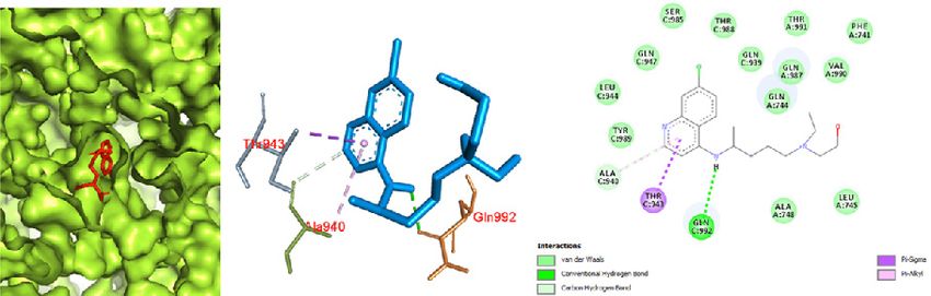
186                                   Narkhede et al., Docking Study of Candidate Proven to Show Anti-COVID-19 Activity / doi: 10.14744/ejmo.2020.31503

study factors such as the identification of hit molecules,                  [23]
                                                                                It has been reported that the S protein activated the
optimization of lead compounds, and virtual screening.[5–8]                 host immune response. This protein is considered a poten-
Several existing drugs have demonstrated potential effec-                   tial target for a therapeutic drug because the S1 domain
tiveness against COVID-19, including oseltamivir,[9] lopi-                  and host ACE2 for SARS-CoV, and dipeptidyl peptidase-4
navir,[10] ritonavir,[10] remdesivir,[11] favipiravir,[12] ribavirin,[12]   for Middle East respiratory syndrome coronavirus, are as-
chloroquine, and hydroxychloroquine.[13] Most of these                      sociated with host and viral membrane fusion mediated by
drugs are HIV protease inhibitors.[14] Docking with the CO-                 the S2 segment and enable the virus to release its RNA in
VID-19 Mpro in complex with the inhibitor N3 was explored                   the host cell.[23]
in this study.[15] It has also been reported that chloroquine               Proteases
and hydroxychloroquine showed anti-SARS-CoV action,
which may be attributed to depletion of angiotensin-con-                    Two polyproteins, PP1a and PP1b, in the coronavirus ge-
verting enzyme 2 (ACE2) glycosylation.[16] At low pH, these                 nome contain 16 non-structural polyproteins (NSPs) and
                                                                            are encoded by the replicase gene.[24] The release of these
drugs are also believed to interfere in post-translational
                                                                            NSPs is mediated by the action of proteases. The chymo-
modification in viral protease and glycosyl transferases in
                                                                            trypsin-like cysteine protease, also known as Mpro or 3-C
the endoplasmic reticulum or vesicles of the trans-Golgi
                                                                            like protease (3CLPro) cleaves at the C-terminal of polypro-
complex.[16] Therefore, docking of chloroquine and hy-
                                                                            teins. The papain-like protease (PLpro) facilitates cleavage
droxychloroquine with the COVID-19 Mpro (in complex
                                                                            of polyproteins at the N-terminal.[24] PLpro enables cleav-
with the inhibitor N3)[15] and the SARS spike glycoprotein-
                                                                            age at the first 3 polyprotein sites, whereas CLpro enables
human ACE2 complex was also performed.[17]
                                                                            cleavage at 11 sites.[25]
Some of the Potential Therapeutic Targets for                               The Cys-His dyad present on the active sites of 3CLPro/
SARS-CoV and SARS-CoV-2                                                     Mpro has been reported to show protease activity.[26] This
Several similarities have been recorded between SARS-                       protease has the ability to effect cleavage at 11 sites in
CoV-2 and SARS-CoV. Following the characterization of the                   the p1 region of PP1a and PP1ab. It can generate mature
genome sequencing of SARS-CoV-2,[18] various molecular                      protein, which facilitates replication[27, 28] and also helps to
modeling experiments have been launched in order to                         release NSPs.[29] Some of the HIV protease inhibitors, includ-
find a potential candidate to combat the novel coronavirus                  ing lopinavir and ritonavir, have also been found to inhibit
SARS-CoV-2. According to a phylogenetic analysis, SARS-                     the Mpro.[30]
CoV-2 is believed to have originated from a bat. Xu et al.[18, 19]          PLpro cleaves the pp1a polyprotein at the N-terminal to
performed molecular modeling experiments that revealed                      form NSP 1, 2, and 3.[27, 28] The catalytic domain of PLpro
similarity in the 3-dimensional structures of SARS-CoV-2 and                consists of 316 amino acids known to facilitate cleavage of
SARS-CoV in the receptor binding domain. This led to the                    substrates for replicase mediated by a consensus sequence
design of various approaches to find a potential target for a               (LXGG).[29] High doses of zinc and its conjugates have been
drug candidate to be used in the fight against SARS-CoV-2.                  reported to inhibit CLpro and PLpro. Several protease in-
The analysis of the crystal structure and several biochemical               hibitors, including combinations of ritonavir and lopinavir,
studies revealed that the S protein (spike protein) of SARS-                are being used to treat COVID‑19.[31]
CoV possesses a strong affinity for binding to human ACE2
receptors.[20] Some of the potential targets for drugs include              Nucleocapsid Protein
an S protein, envelope protein (E protein), membrane pro-                   The N protein consists of the N‑arm, the central linker (CL),
tein (M protein), protease, nucleocapsid protein (N protein),               and the C‑tail, which are also known as characteristic in-
hemagglutinin esterase, and helicase.[21]                                   trinsically disordered regions (IDRs).[32] The major structural
                                                                            and functional domains of the N protein are the N-terminal
Spike Protein                                                               domain (NTD) and the C-terminal domain (CTD). The NTD
The S protein consists of the ectodomain region (ED), the                   is known to facilitate RNA binding and the CTD plays an
intracellular domain, and the transmembrane (TM) region.                    important role in dimerization.[32, 33] The CL region includes
[22]
     The S protein is a clove-shaped type I TM protein.[22] The             several phosphorylation sites, as well as arginine-serine.
ED region is made up of 2 receptor binding domains (RBDs),                  [34]
                                                                                 The C-tail region plays a vital role in interactions of N-M
the S1 subunit and the S2 trimeric stalk, associated with the               proteins and oligomerization of N protein.[35] N protein has
C-terminal. The association of S proteins gives the virion a                been reported to cause inhibition of cell growth in humans
trimeric form, which gives it a crown-like structure and the                via inhibition of the cytokinesis process.[36] The N protein
name coronavirus.[22] The S protein has a role in viral entry.              peptide (N220) has been found to demonstrate a promis-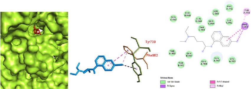
EJMO                                                                                                                           187

ing ability to destroy N protein-expressing cells in trans-          ated in future experimental studies. Molecular docking
genic animals. Thus, it may be a potential target for devel-         methodologies are a useful tool in drug discovery, as they
oping a DNA vaccine.[37]                                             permit rapid screening of candidates from drug libraries.[48,
                                                                     49]
                                                                         This docking study of some of the potential therapeu-
Envelope Protein                                                     tic drug candidates that are being used against COVID-19
The process of ion channel generation is mediated through            worldwide was performed using computer-based tech-
oligomerization of E proteins. E protein, the smallest trans-        niques to assess the structure of ligand-protein complexes
membrane structural protein of the coronaviruses, consists           and biochemical pathways.
of a hydrophobic domain and a cytoplasmic tail. It is 8.4-12
kDa in size.[38, 39] During viral assembly and release, E pro-       Methods
tein is known to facilitate viral morphogenesis. In the mam-         All of the docking experiments were performed using
malian cells expressing SARS‑CoV E protein, hexamethyle-             AutoDock Vina (O. Trott, The Scripps Research Institute, La
neamiloride has been found to inhibit E protein-mediated             Jolla, CA, USA) because (a) it offers more accuracy in pre-
ion channel activity.[40] Along with assembly and release, E         dicting ligand-protein interactions than the previous ver-
protein has also been found to be responsible for the viru-          sion, AutoDock 4.2, (b) it offers a shorter running time as a
lence of the virus.[39, 41]                                          result of multiple core processors, (c) and it offers greater
Membrane Protein                                                     accuracy for ligands with more than 20 rotatable bonds.
                                                                     All of the docking experiments were conducted using the
M protein modulates the shape of the envelope of the virus
                                                                     blind docking method: using a grid box large enough to
as a result of interaction with proteins in the Golgi com-
                                                                     cover the whole protein structure to include any possible
plex inside the virion and stabilizing the N protein.[39, 42] M
                                                                     protein-ligand interactions.
protein is known to promote intracellular homeostasis in a
virus via various protein interactions.[42] It consists of a short   All of the protein structures used in the docking experi-
N-terminal and a long C-terminal.[39] The entry of the virus         ments were retrieved from the Research Collaboratory for
takes place in association with the interaction of M–M, M–S,         Structural Bioinformatics Protein Data Bank (PDB). All of the
and M–N proteins. The introduction of a spike protein in a           ligand structures were drawn using ChemDraw 14 and con-
                                                                     verted into PDB format using Chem3D 12.0 (PerkinElmer,
new virus takes place via M-S interactions.[42] The stabiliza-
                                                                     Inc., Waltham, MA, USA). The ligands were converted to the
tion of the nucleocapsid-RNA complex (ribonucleoprotein
                                                                     energetically most stable structure using energy minimiza-
complex) is associated with M-N interactions. In addition
                                                                     tion with the Vega ZZ program (Drug Design Laboratory,
to regulating the shape of the virus, M and N proteins also
                                                                     University of Milan, Milan, Italy)[51] and the SP4 force field
facilitate the generation and release of virus-like particles.
                                                                     and a conjugate gradient. The ligand and protein mole-
M protein is known to potentiate sensitization of the host
                                                                     cules were converted to their proper readable file format
to the virus.[42]
                                                                     (pdbqt) using AutoDock tools 1.5.6. The docking was per-
Helicase                                                             formed using an exhaustiveness value of 8. All of the other
The SARS‑CoV helicase (NTPase) enzyme is a member of                 software parameters were the default values, and all of the
superfamily 1. It facilitates hydrolysis of all NTPs.[43] Helicase   bonds contained in the ligand were allowed to rotate freely
may be a potential target for numerous drug candidates               while the receptor was rigid. The final visualization of the
against various disorders.[44] Toxicity is a major concern,          docked structure was performed using Discovery Studio
however, in the design of helicase inhibitors, as non-speci-         Visualizer 2.5 (Accelrys Software Inc., San Diego, CA, USA).
ficity leads to a precipitation of toxic effects.[43]
                                                                     Results
Molecular Docking Study of Some Drug Candidates                      The results of these experiments revealed strong interac-
with Main Protease of SARS-CoV-2                                     tions of the potential drug candidates against the COVID-19
While there is as yet no specific treatment for COVID-19,            Mpro in complex with the inhibitor N3 and the SARS spike
several antiretroviral drugs have been reported to demon-            glycoprotein-human ACE2 complex of SARS-CoV-2. After
strate some effectiveness, including ritonavir,[45, 46] lopina-      successful docking of these drugs to the COVID-19 Mpro in
vir alone or in combination with oseltamivir,[46] remdesivir,        complex with an inhibitor N3, various modes of drug-pro-
chloroquine, and hydroxychloroquine.[47] Of these, ritona-           tein interactions are generated with a particular docking
vir, remdesivir, chloroquine, and hydroxychloroquine have            score (binding energy). The binding mode with the least
shown efficacy at the cellular level.[45] which will be evalu-       binding energy is regarded as the best mode of binding,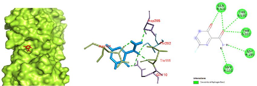
188                                               Narkhede et al., Docking Study of Candidate Proven to Show Anti-COVID-19 Activity / doi: 10.14744/ejmo.2020.31503

Table 1. Drugs (ligands) used for docking with the target protein

Drug                                    Structure                                                    Target (Protein)                          Protein Data Base Code

Oseltamivir		                                          O                        COVID-19 main protease in complex with an inhibitor N3[15]             6LU7
            O
                                                               O
                                   HN
             NH2
		         O
Ritonavir		                                                                     COVID-19 main protease in complex with an inhibitor N3[15]             6LU7
                               S
                               N
                                    N        O
                                    HN
                                        O        NH

                                             HO                    S
                                                  HN       O             N
		                                                     O
Remdesivir
           HN N                                                                 COVID-19 main protease in complex with an inhibitor N3[15]             6LU7
                       2
                                                   H       O
                           N        N          O   N
                                   N         O   P             O
                                                   O
                                   HO            O
                                        HO

Ribavirin		                                                                  COVID-19 main protease in complex with an inhibitor N3[15]                6LU7
                                             OH                O
                           HO
		                 N
                 N                                                  NH2
            HO
		             O     N
Favipiravir		                                                                COVID-19 main protease in complex with an inhibitor N3[15]                6LU7
                     O
                       F                 N
                                                                   NH2

                          N   O
		                        H
Chloroquine		      CI                                                        1. COVID-19 main protease in complex with an inhibitor N3[15]             6LU7
                            H
			                         N
                                                                   N              2. SARS Spike glycoprotein-Human ACE2 complex[17]                    6CS2
		                      N

Hydroxychloroquine
                  CI                                                           1. COVID-19 main protease in complex with an inhibitor N3[15]           6LU7
                          H
			                       N
                                N
                                                                       OH           2. SARS Spike glycoprotein-Human ACE2 complex[17]                  6CS2
		                    N

as it is most stable for the ligand. The binding energy re-                                        atoms for the particular interaction (Table 1).
sults observed are summarized in Table 2. The interaction
of specific amino acids taking part in the drug-protein in-                                        Visualization of Docking Results
teractions were also recorded. All of the docked structures                                        Chloroquine and hydroxychloroquine were docked with
were visualized in PyMOL 2.3 (Schrödinger LLC, New York,                                           the COVID-19 Mpro in complex with the inhibitor N3 as
NY, USA) and Discovery Studio 4.0.                                                                 well as the SARS spike glycoprotein-human ACE2 complex.
Three chains, A, B, and C, were present in the structure of                                        The lowest energy conformations of all of the ligand mol-
the SARS spike glycoprotein-human ACE2 complex (PDB                                                ecules were docked with the protein. The ligand was illus-
ID: 6CS2) with a central pocket for interaction with ligands.                                      trated as a red stick model and the protein is the surface.
The binding pocket of the COVID-19 Mpro in complex with                                            The amino acids taking part in the ligand-protein interac-
the inhibitor N3 (PDB ID: 6LU7) consists of 2 chains, chain                                        tion were shown with blue stick ligands surrounded by the
A and chain C, which may be part of interactions with the                                          amino acids. The interactions represented by green dashed
ligand. The ligand showed selective interaction with either                                        lines are the hydrogen-bonding interactions between the
or both of the chains, depending on the availability of the                                        ligand and protein. Amino acids involved in the ligand-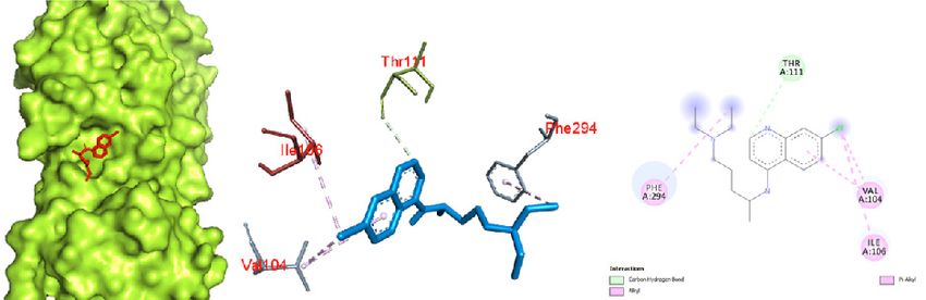
EJMO                                                                                                                                                  189

Table 2. Target (protein) and the drug candidates (ligands) undergoing docking experiment with their best docking score (lowest binding
energy)

Drug (ligand) Proteins (receptor)                                                                      Affinity      Distance from      Distance from
			                                                                                                  (kcal/mol)         rmsdl.b.          rmsdu.b.

Oseltamivir                    COVID-19 main protease in complex with inhibitor N3[15]                   -4.7             0.000              0.000
Ritonavir                      COVID-19 main protease in complex with inhibitor N3[15]                   -7.3             0.000              0.000
Remdesivir                     COVID-19 main protease in complex with inhibitor N3[15]                   -6.5             0.000              0.000
Ribavirin                      COVID-19 main protease in complex with inhibitor N3[15]                   -5.6             0.000              0.000
Favipiravir                    COVID-19 main protease in complex with inhibitor N3[15]                   -5.4             0.000              0.000
Chloroquine                   1. COVID-19 main protease in complex with inhibitor N3[15]                 -5.1             0.000              0.000
		                                2. SARS Spike glycoprotein-Human ACE2 complex[17]                      -7.1             0.000              0.000
Hydroxychloroquine            1. COVID-19 main protease in complex with inhibitor N3[15]                 -5.3             0.000              0.000
		                                2. SARS Spike glycoprotein-Human ACE2 complex[17]                      -6.8             0.000              0.000

                                                                               to the protein structure, ligands were shown as red sticks in
  a                                   b                                        the binding pocket of the protein.

                                                                               Discussion
                                                                               Successful docking of all the ligands revealed significant
                                                                               binding with the target proteins. After visualizing the pro-
                                                                               tein in Discovery Studio 12.0, it was found that the SARS
                                                                               spike glycoprotein-human ACE2 complex and the CO-
                                                                               VID-19 Mpro in complex with the inhibitor N3 consists of
                                                                               A, B, and C chains, as shown in (Fig. 1). In the COVID-19
                                                                               Mpro, only the A and C chains are involved in interaction
Figure 1. The ligand binding site of (a) SARS spike glycoprotein-hu-           with ligands. Oseltamivir docked in the COVID-19 Mpro in
man angiotensin-converting enzyme II complex with chain (labeled               complex with the inhibitor N3 showed significant binding,
in red) and (b) the COVID-19 main protease in complex with the in-             yielding a binding affinity of -6.1 kcal/mol. The interaction
hibitor N3. COVID-19: Coronavirus disease 2019; SARS: Severe acute
                                                                               of oseltamivir with the protease (Fig. 2) showed a high af-
respiratory syndrome.
                                                                               finity interaction in chain A, as the ligand fit inside the core
protein interactions were shown as sticks of different colors                  pocket region of the protease. This is further evidenced
and labeled in red. In order to visualize the ligands docked                   by hydrogen bonding between the oxygen of a carbonyl

  a                                                     b                                                       c

Figure 2. Oseltamivir docked in the COVID-19 main protease in complex with the inhibitor N3 (PDB ID: 6LU7) with (a) best binding mode in the
protein pocket (ligand illustrated as red sticks), (b) amino acid residues involved in the interaction (ligand as blue sticks), and (c) the binding inter-
action of oseltamivir with amino acid with a hydrogen bond (green dashed line). COVID-19: Coronavirus disease 2019.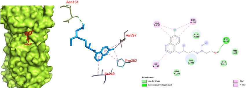
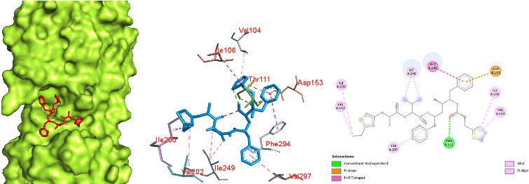
190                                    Narkhede et al., Docking Study of Candidate Proven to Show Anti-COVID-19 Activity / doi: 10.14744/ejmo.2020.31503

group (ester side chain) of oseltamivir with LYS 102 and SER                  The docking of remdesivir in the COVID-19 Mpro in com-
158. Some van der Waals interaction of oseltamivir with                       plex with the inhibitor N3 revealed significant interactions
VAL 104, ASP 153, ASN 151, THR 111, PHE 294, ILE 106, THR                     in the central pocket of chain A with an affinity of -6.5 kcal/
292, GLN 110, and GLN 107 was observed.                                       mol (Fig. 4). The major interaction between remdesivir and
The docking of ritonavir with the COVID-19 Mpro in com-                       the protease was characterized by hydrogen bonding be-
plex with the inhibitor N3 revealed that ritonavir (ligand)                   tween nitrogen of a cyno group and PHE 294, and oxygen
had a high-affinity interaction with the A chain of the pro-                  of a tetrahydrofuran ring with GLN 110. Some pi sigma
tein: -7.3 kcal/mol (Fig. 3). To interact with the protein, rito-             interactions of the aromatic ring and ILE 249 and VAL 104
navir acquired the central pocket surrounded by chains A,                     were observed.
B, and C, which led to several interactions between rito-                     Results obtained by docking ribavirin to the COVID-19
navir and the amino acid residues of the proteins. The in-                    Mpro in complex with the inhibitor N3 showed binding in
teraction resulted in a hydrogen bond between ritonavir                       the pocket of chain A with an affinity of -5.6 kcal/mol (Fig. 5).
and amino acid residues of the protein. The oxygen (amide                     Significant binding was observed, with 4 hydrogen bonds
group) demonstrated significant hydrogen bonding with                         between nitrogen of a dihydrotriazole ring and TYR 239,
THR 111. The benzene ring showed pi-anion and pi-pi in-                       hydrogen of an amide group (attached to dihydrotriazole)
teraction with ASP 153 and PHE 294, respectively.                             and TYR 237, hydrogen of a hydroxyl group (attached to a

  a                                                    b                                         c

Figure 3. Ritonavir docked in the COVID-19 main protease in complex with the inhibitor N3 (PDB ID: 6LU7) with (a) best binding mode in the pro-
tein pocket (ligand as red sticks), (b) amino acid residues involved in the interaction (ligand as blue sticks), and (c) binding interaction of ritonavir
with amino acid with a hydrogen bond (green dashed line). COVID-19: Coronavirus disease 2019.

   a                                                    b                                        c

Figure 4. Remdesivir docked in the COVID-19 main protease in complex with the inhibitor N3 (PDB ID: 6LU7) with (a) best binding mode in the
protein pocket (ligand as red sticks), (b) amino acid residues involved in the interaction (ligand as blue sticks), and (c) binding interaction of rem-
desivir with amino acid with a hydrogen bond (green dashed line). COVID-19: Coronavirus disease 2019.
EJMO 191
a b
c
Figure 5. Ribavirin docked in the COVID-19 main protease in complex with the inhibitor N3 (PDB ID:
6LU7) with (a) best binding mode in the protein pocket (ligand as red sticks), (b) amino acid residues
involved in the interaction (ligand as blue sticks), and (c) binding interaction of ribavirin with amino acid
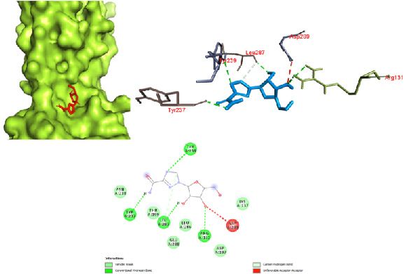
with a hydrogen bond (green dashed line). COVID-19: Coronavirus disease 2019.
tetrahydrofuran ring) and ARG 131 and oxygen (attached a binding pocket of chain A (Fig. 6). Thus, this interaction re-
to a tetrahydrofuran ring) with ARG 131. An unfavorable ac- sults in 6 hydrogen bonds, oxygen (attached to a pyrazine
ceptor interaction was also observed between oxygen (at- ring) and an oxygen carboxamide group (attached to a pyr-
tached to tetrahydrofuran ring) and ASP 289. azine ring) with GLN 110, THR 292, and THR 111. Similarly,
With 6 hydrogen bonds, favipiravir showed promising ac- hydrogen of a carboxamide group showed bonding with
tivity with the Mpro of COVID-19, with an affinity of -5.4 ASP 295 and ASN 151. Since the binding was character-
kcal/mol. It showed strong interaction with the protease in ized by 6 hydrogen bonds, this interaction can be seen as
a b c
Figure 6. Favipiravir docked in the COVID-19 main protease in complex with the inhibitor N3 (PDB ID: 6LU7) with (a) best binding mode in the
protein pocket (ligand illustrated as red sticks), (b) amino acid residues involved in the interaction (ligand as blue sticks), and (c) the binding inter-
action of favipirivir with amino acid with a hydrogen bond (green dashed line). COVID-19: Coronavirus disease 2019.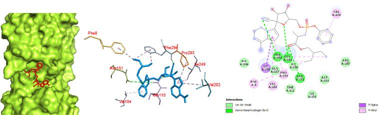
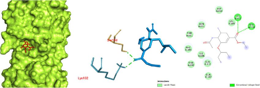
192                                   Narkhede et al., Docking Study of Candidate Proven to Show Anti-COVID-19 Activity / doi: 10.14744/ejmo.2020.31503

a possible mode of binding of favipiravir with the Mpro of                  SARS spike glycoprotein-human ACE2 complex was seen in
COVID-19.                                                                   pi-pi and pi-alkyl interactions between a benzene ring and
Docking of chloroquine was performed with both the CO-                      chlorine (attached to the benzene ring) with TYR 738 and
VID-19 Mpro in complex with the inhibitor N3 and the SARS                   PHE 952, respectively. A large number of amino acids were
spike glycoprotein-human ACE2 complex. The binding in-                      involved in van der Waals interactions, including GLN 987,
teraction of chloroquine with the COVID-19 Mpro was ob-                     THR 988, GLN 984, THR 988, SER 985, PHE 741, GLY 981, THR
served in the binding pocket of chain C with the affinity of                988, THR 991, GLN 987, GLN 984, and LEU 983. Thus, it was
-5.1 kcal/mol (Fig. 7). The interactions were primarily char-               observed that chloroquine favored the SARS spike glyco-
acterized by pi-alkyl interactions between a chlorobenzene                  protein-human ACE2 complex over the COVID-19 Mpro in
ring with VAL 104 and ILE 106. The prominent interaction                    complex with the inhibitor N3.
of chloroquine with the SARS spike glycoprotein-human                       Similarly, the docking study of hydroxychloroquine with
ACE2 complex was observed with a binding affinity of -70                    both the COVID-19 Mpro in complex with the inhibitor N3
kcal/mol (Fig. 8). The interaction of chloroquine with the                  and the SARS spike glycoprotein-human ACE2 complex

  a                                              b                                                            c

Figure 7. Chloroquine docked in the COVID-19 main protease in complex with the inhibitor N3 (PDB ID: 6LU7) with (a) best binding mode in
the protein pocket (ligand illustrated as red sticks), (b) amino acid residues involved in the interaction (ligand as blue sticks), and (c) the bind-
ing interaction of chloroquine with amino acid with a hydrogen bond (green dashed line). COVID-19: Coronavirus disease 2019.

  a                                        b                                                   c

Figure 8. Chloroquine docked in the SARS spike glycoprotein-human angiotensin-converting enzyme complex (PDB ID: 6CS2) with (a) best
binding mode in the protein pocket (ligand illustrated as red sticks), (b) amino acid residues involved in the interaction (ligand as blue sticks),
and (c) the binding interaction of chloroquine with amino acid with a hydrogen bond (green dashed line). COVID-19: Coronavirus disease
2019; SARS: Severe acute respiratory syndrome.
EJMO                                                                                                                                                  193

yielded several facts about binding interaction. Hydroxy-                      One hydrogen bond between secondary amine hydrogen
chloroquine docking with the COVID-19 Mpro revealed a                          and GLN 992 was observed. A pi-sigma interaction was
binding affinity of -5.3 kcal/mol with a hydrogen bonding                      seen between the ring and THR 943. Based on the results,
interaction between hydrogen of a hydroxyl group (in an                        it appeared that the SARS spike glycoprotein-human ACE2
aliphatic chain) with ASN 151 (Fig. 9). It also showed pi-pi                   complex was a promising target for hydroxychloroquine
and pi-alkyl interactions with residues of VAL 297, PRO 252                    against the COVID-19 Mpro in complex with the inhibitor
and ILE 249. Van der Waals interactions occurred with PRO                      N3. Based on this study, it can be said that hydroxychlo-
293, PHE 294, THR 111b and PHE 8. Similar to chloroquine,                      roquine favored the SARS spike glycoprotein-human ACE2
hydroxychloroquine also showed promising interaction                           complex over the protease for binding.
with the SARS spike glycoprotein-human ACE2 complex
(Fig. 10) with a binding affinity of -6.8 kcal/mol. This inter-                Conclusion
action is primarily attributed to the large number of amino                    The docking results yielded various kinds of binding inter-
acids involved in a van der Waals interaction due to a better                  actions of the drugs with the protein, and some were favor-
fit of hydroxychloroquine inside the pocket of the protein.                    able. The antiviral drugs oseltamivir, ritonavir, remdesivir,

  a                                  b                                                   c

Figure 9. Hydroxychloroquine docked in the COVID-19 main protease in complex with the inhibitor N3 (PDB ID: 6LU7) with (a) best binding mode
in the protein pocket (ligand illustrated as red sticks), (b) amino acid residues involved in the interaction (ligand as blue sticks), and (c) the binding
interaction of hydroxychloroquine with amino acid with a hydrogen bond (green dashed line). COVID-19: Coronavirus disease 2019.

    a                                 b                                                    c

Figure 10. Hydroxychloroquine docked in the SARS spike glycoprotein-human angiotensin-converting enzyme complex (PDB ID: 6CS2) with
(a) best binding mode in the protein pocket (ligand illustrated as red sticks), (b) amino acid residues involved in the interaction (ligand as blue
sticks), and (c) the binding interaction of hydroxychloroquine with amino acid with a hydrogen bond (green dashed line). COVID-19: Corona-
virus disease 2019; SARS: Severe acute respiratory syndrome.
194                                   Narkhede et al., Docking Study of Candidate Proven to Show Anti-COVID-19 Activity / doi: 10.14744/ejmo.2020.31503

ribavirin, and favipiravir showed high-affinity interactions                    2020;382:929–36.
with the COVID-19 Mpro in complex with the inhibitor N3,                    12. Wang M, Cao R, Zhang L, et al. Remdesivir and chloroquine
whereas the anti-malarial drugs chloroquine and hydroxy-                        effectively inhibit the recently emerged novel coronavirus
chloroquine demonstrated prominent interaction with the                         (2019-nCoV) in vitro. Cell Res 2020;30:269–71.
SARS spike glycoprotein-human ACE2 complex. It may be                       13. Yao X, Ye F, Zhang M, et al. In Vitro Antiviral Activity and Projec-
that the anti-COVID-19 activity of antiviral drugs is a result                  tion of Optimized Dosing Design of Hydroxychloroquine for
of interaction with the COVID-19 Mpro and that the anti-                        the Treatment of Severe Acute Respiratory Syndrome Corona-
COVID-19 effect of chloroquine and hydroxychloroquine is                        virus 2 (SARS-CoV-2). Clin Infect Dis an Off Publ Infect Dis Soc
due to a high-affinity interaction with the SARS spike glyco-                   Am [Epub ahead of print].
protein-human ACE2 complex.                                                 14. Lv Z, Chu Y, Wang Y. HIV protease inhibitors: a review of mo-
                                                                                lecular selectivity and toxicity. HIV AIDS (Auckl) 2015;795–104.
Disclosures                                                                 15. Jin Z, Du X, Xu Y, et al. Structure of Mpro from COVID-19 virus
Peer-review: Externally peer-reviewed.                                          and discovery of its inhibitors. Nature [Epub ahead of print].
Conflict of Interest: None declared.                                        16. Devaux CA, Rolain J-M, Colson P, Raoult D. New insights on the
Authorship Contributions: Concept – R.S.C., R.R.N., S.D.S.; De-                 antiviral effects of chloroquine against coronavirus: what to
sign – R.S.C., R.R.N., J.P.A.; Supervision – R.S.C., S.D.S.; Materials          expect for COVID-19? Int J Antimicrob Agents 2020;105938.
– R.S.C., R.R.N.; Data collection &/or processing – R.S.C., R.R.N.,         17. Kirchdoerfer RN, Wang N, Pallesen J, et al. Stabilized coronavi-
S.D.S., J.P.A.; Analysis and/or interpretation – R.S.C., R.R.N.; Litera-        rus spikes are resistant to conformational changes induced by
ture search – R.S.C., R.R.N., J.P.A.; Writing – R.S.C., R.R.N.; Critical        receptor recognition or proteolysis. Sci Rep 2018;8:15701.
review – R.S.C., S.D.S.                                                     18. Lu R, Zhao X, Li J, et al. Genomic characterisation and epi-
                                                                                demiology of 2019 novel coronavirus: implications for vi-
References
                                                                                rus origins and receptor binding. Lancet (London, England)
1. Zhou P, Yang X-L, Wang X-G, et al. A pneumonia outbreak as-                  2020;395:565–74.
    sociated with a new coronavirus of probable bat origin. Na-             19. Wan Y, Shang J, Graham R, Baric RS, Li F. Receptor Recogni-
    ture 2020;579:270–3.                                                        tion by the Novel Coronavirus from Wuhan: an Analysis Based
2. Wang C, Horby PW, Hayden FG, Gao GF. A novel coronavirus                     on Decade-Long Structural Studies of SARS Coronavirus. J Vi-
    outbreak of global health concern. The Lancet 2020;395:470–                 rol;94.
    3.                                                                      20. Li F, Li W, Farzan M, Harrison SC. Structure of SARS coronavirus
3. Coronavirus disease 2019 https://www.who.int/emergencies/                    spike receptor-binding domain complexed with receptor. Sci-
    diseases/novel-coronavirus-2019 Accessed: 2020-04-14.                       ence 2005;309:1864–8.
4. Mcconkey B, Sobolev V, Edelman M. The performance of cur-                21. Prajapat M, Sarma P, Shekhar N, et al. Drug targets for corona
    rent methods in ligand-protein docking. Curr Sci;83.                        virus: A systematic review. Indian J Pharmacol 2020;52:56–65.
5. Jorgensen WL. The many roles of computation in drug discov-              22. Belouzard S, Millet JK, Licitra BN, Whittaker GR. Mechanisms
    ery. Science 2004;303:1813–8.                                               of coronavirus cell entry mediated by the viral spike protein.
6. Bajorath J. Integration of virtual and high-throughput screen-               Viruses 2012;4:1011–33.
    ing. Nat Rev Drug Discov 2002;1:882–94.                                 23. Li F. Structure, Function, and Evolution of Coronavirus Spike
7. Langer T, Hoffmann RD. Virtual screening: an effective tool for              Proteins. Annu Rev Virol 2016;3:237–61.
    lead structure discovery? Curr Pharm Des 2001;7:509–527.                24. Lindner HA, Fotouhi-Ardakani N, Lytvyn V, Lachance P, Sulea
8. Kitchen DB, Decornez H, Furr JR, Bajorath J. Docking and scor-               T, Ménard R. The papain-like protease from the severe acute
    ing in virtual screening for drug discovery: methods and ap-                respiratory syndrome coronavirus is a deubiquitinating en-
    plications. Nat Rev Drug Discov 2004;3:935–949.                             zyme. J Virol 2005;79:15199–208.
9. Li G, De Clercq E. Therapeutic options for the 2019 novel                25. Jo S, Kim S, Shin DH, Kim M-S. Inhibition of SARS-CoV 3CL pro-
    coronavirus (2019-nCoV). Nature reviews. Drug discovery                     tease by flavonoids. J Enzyme Inhib Med Chem 2020;35:145–
    2020;19:149–50.                                                             51.
10. Lim J, Jeon S, Shin HY, et al. Case of the Index Patient Who            26. Shimamoto Y, Hattori Y, Kobayashi K, et al. Fused-ring struc-
    Caused Tertiary Transmission of COVID-19 Infection in Korea:                ture of decahydroisoquinolin as a novel scaffold for SARS 3CL
    the Application of Lopinavir/Ritonavir for the Treatment of                 protease inhibitors. Bioorg Med Chem 2015;23:876–90.
    COVID-19 Infected Pneumonia Monitored by Quantitative RT-               27. Hilgenfeld R. From SARS to MERS: crystallographic studies
    PCR. J Korean Med Sci 2020;35:e79.                                          on coronaviral proteases enable antiviral drug design. FEBS J
11. Holshue ML, DeBolt C, Lindquist S, et al. First Case of 2019                2014;281:4085–96.
    Novel Coronavirus in the United States. N Engl J Med                    28. Hsu M-F, Kuo C-J, Chang K-T, et al. Mechanism of the matu-
EJMO 195
ration process of SARS-CoV 3CL protease. J Biol Chem PLoS Pathog 2009;5:e1000511.
2005;280:31257–66. 41. Nieto-Torres JL, Dediego ML, Alvarez E, et al. Subcellular loca-
29. Barretto N, Jukneliene D, Ratia K, Chen Z, Mesecar AD, Bak- tion and topology of severe acute respiratory syndrome coro-
er SC. The papain-like protease of severe acute respiratory navirus envelope protein. Virology 2011;415:69–82.
syndrome coronavirus has deubiquitinating activity. J Virol 42. Schoeman D, Fielding BC. Coronavirus envelope protein: cur-
2005;79:15189–98. rent knowledge. Virol J 2019;16:69.
30. Liu X, Wang X-J. Potential inhibitors for 2019-nCoV coronavi- 43. Karpe YA, Lole KS. NTPase and 5’ to 3’ RNA duplex-unwind-
rus M protease from clinically approved medicines. bioRxiv ing activities of the hepatitis E virus helicase domain. J Virol
[Epub ahead of print]. 2010;84:3595–602.
31. Han Y-S, Chang G-G, Juo C-G, et al. Papain-like protease 2 44. Banerjee T, Aggarwal M, Sommers JA, Brosh RMJ. Biochemi-
(PLP2) from severe acute respiratory syndrome coronavirus cal and cell biological assays to identify and characterize DNA
(SARS-CoV): expression, purification, characterization, and in- helicase inhibitors. Methods 2016;108130–141.
hibition. Biochemistry 2005;44:10349–59. 45. Three drugs fairly effective on novel coronavirus at cellular
32. Chang C, Lo S-C, Wang Y-S, Hou M-H. Recent insights into the level - Xinhua | English.news.cn http://www.xinhuanet.com/
development of therapeutics against coronavirus diseases by english/2020-01/30/c_138742163.htm Accessed: 2020-04-11.
targeting N protein. Drug Discov Today 2016;21:562–72. 46. Coronavirus outbreak: Cocktail of flu, HIV drugs appears to
33. McBride R, van Zyl M, Fielding BC. The coronavirus nucleocap- help fight virus, say Thai doctors - World News https://www.
sid is a multifunctional protein. Viruses 2014;6:2991–3018. indiatoday.in/world/story/coronavirus-outbreak-cocktail-flu-
34. Lin S-Y, Liu C-L, Chang Y-M, Zhao J, Perlman S, Hou M-H. Struc- hiv-drugs-treatment-thai-doctors-1642783-2020-02-03 Ac-
tural basis for the identification of the N-terminal domain of cessed: 2020-04-11.
coronavirus nucleocapsid protein as an antiviral target. J Med 47. Singh AK, Singh A, Shaikh A, Singh R, Misra A. Chloroquine
Chem 2014;57:2247–57. and hydroxychloroquine in the treatment of COVID-19 with or
35. Chang C, Hou M-H, Chang C-F, Hsiao C-D, Huang T. The SARS without diabetes: A systematic search and a narrative review
coronavirus nucleocapsid protein--forms and functions. Anti- with a special reference to India and other developing coun-
viral Res 2014;10339–50. tries. Diabetes Metab Syndr [Epub ahead of print].
36. Zhou B, Liu J, Wang Q, et al. The nucleocapsid protein of se- 48. Warui DM, Baranger AM. Identification of specific small mole-
vere acute respiratory syndrome coronavirus inhibits cell cule ligands for stem loop 3 ribonucleic acid of the packaging
cytokinesis and proliferation by interacting with translation signal Psi of human immunodeficiency virus-1. J Med Chem
elongation factor 1alpha. J Virol 2008;82:6962–71. 2009;52:5462–473.
37. Cheung Y-K, Cheng SC-S, Sin FW-Y, Chan K-T, Xie Y. Induction 49. Cosconati S, Marinelli L, Trotta R, et al. Tandem application of
of T-cell response by a DNA vaccine encoding a novel HLA- virtual screening and NMR experiments in the discovery of
A*0201 severe acute respiratory syndrome coronavirus epit- brand new DNA quadruplex groove binders. J Am Chem Soc
ope. Vaccine 2007;25:6070–7. 2009;131:16336–7.
38. Kuo L, Hurst KR, Masters PS. Exceptional flexibility in the se- 50. Trott O, Olson AJ. AutoDock Vina: improving the speed and
quence requirements for coronavirus small envelope protein accuracy of docking with a new scoring function, efficient op-
function. J Virol 2007;81:2249–62. timization, and multithreading. J Comput Chem 2010;31:455–
39. Venkatagopalan P, Daskalova SM, Lopez LA, Dolezal KA, 61.
Hogue BG. Coronavirus envelope (E) protein remains at the 51. Pedretti A, Villa L, Vistoli G. VEGA-an open platform to de-
site of assembly. Virology 2015;47875–85. velop chemo-bio-informatics applications, using plug-in ar-
40. Pervushin K, Tan E, Parthasarathy K, et al. Structure and inhibi- chitecture and script programming. J Comput Aided Mol Des
tion of the SARS coronavirus envelope protein ion channel. 2004;18:167–73.You can also read