Cell Mediated Immunity as a Weapon to Fight Against COVID19: A Brief Review
←
→
Page content transcription
If your browser does not render page correctly, please read the page content below
MedDocs Publishers

                                                                                                                        ISSN: 2639-4391

                                                Annals of Epidemiology & Public Health
                                                                                                          Open Access | Review Article

      Cell Mediated Immunity as a Weapon to Fight
            Against COVID19: A Brief Review
Kalpana S1*; Raghul Ramachandran2
1
 Department of Epidemiology The Tamil Nadu Dr. M.G.R. Medical University Chennai, Tamil Nadu, India
2
 Madras Christian College, Chennai, Tamil Nadu, India

   *Corresponding Author(s): Kalpana S                                        Abstract
 Department of Epidemiology The Tamil Nadu Dr.                              COVID-19 is not the first severe respiratory disease out-
 M.G.R. Medical University Chennai, Tamil Nadu,                          break caused by the coronavirus. Just in the past two de-
 India                                                                   cades, coronaviruses have caused three epidemic diseases,
                                                                         namely, COVID-19, Severe Acute Respiratory Syndrome
  Email: drkalpanaphd@gmail.com
                                                                         (SARS) and Middle East Respiratory Syndrome (MERS). At
                                                                         present, the cases of COVID-19 have been found in many
                                                                         countries around the world. Here, we provide an outline
Received: May 25, 2020                                                   of the pathophysiology of SARS-CoV-2 infection. Also, we
Accepted: Jul 31, 2020                                                   describe the interaction of SARS-CoV-2 with the cell medi-
Published Online: Aug 05, 2020                                           ated immune system and the subsequent contribution of
                                                                         dysfunctional immune responses to disease progression.
Journal: Annals of Epidemiology and Public health                        Understanding the immunological response to the SARS-
Publisher: MedDocs Publishers LLC                                        CoV-2 viral infection will help us to develop successful treat-
Online edition: http://meddocsonline.org/                                ments and vaccines, identify vulnerable groups and help
                                                                         inform public health measures to control the coronavirus
Copyright: © Kalpana S (2020). This Article is                           outbreak.
distributed under the terms of Creative Commons
Attribution 4.0 International License

Keywords: COVID19; Respiratory disease; Respiratory
Syndrome

   Introduction
    Coronavirus disease 2019 (COVID-19) is a kind of viral pneu-        regions, but efforts to overcome the virus are hampered by a
monia which is caused by severe acute respiratory syndrome              lack of knowledge of several important aspects of SARS-CoV-2
coronavirus 2 (SARS-CoV-2). The emergence of SARS-CoV-2                 infection, ranging from pathogen biology to host response and
has been marked as the third introduction of a highly patho-            treatment options. Therefore, there is an urgent need to better
genic coronavirus into the human population after the severe            understand the host–pathogen biology of COVID-19 as this will
acute respiratory syndrome coronavirus (SARS-CoV) and the               offer important insights into treatment and management of the
Middle East respiratory syndrome coronavirus (MERS-CoV) in              disease, including identification of new therapies [2]. Everyday
the twenty-first century. By May 2020, SARS-CoV-2 had infected          reports of sharp rises in the number of new cases continue to
more than 26,49,542 people across 215 countries/regions and             emerge from many countries/regions, but efforts to overcome
killed more than 4,3960: a pandemic as declared by the World            the virus are hampered by a lack of knowledge of several im-
Health Organization [1]. Daily reports of sharp rises in the num-       portant aspects of SARS-CoV-2 infection, ranging from patho-
ber of new cases continue to emerge from many countries/                gen biology to host response and treatment options. Therefore,

              Cite this article: Kalpana S, Ramachandran R. Cell Mediated Immunity as a Weapon to Fight Against COVID19: A Brief
              review. A Epidemiol Public Health. 2020; 3(1): 1023.

                                                                    1
MedDocs Publishers

there is an urgent need to better understand the host–patho-                  Pathogenesis of COVID 19
gen biology of COVID-19 as this will offer important insights into
treatment and management of the disease, including identifica-                Patients with COVID-19 show clinical manifestations includ-
tion of new therapies. Here, we review the literature on SARS-             ing fever, nonproductive cough, dyspnea, myalgia, fatigue,
CoV-2 pathophysiology, its interaction with target cells and the           normal or decreased leukocyte counts, and radiographic evi-
immune response to the virus, including the contribution of                dence of pneumonia [10], which are similar to the symptoms
dysfunctional immune responses to disease progression. Spe-                of SARS-CoV and MERS-CoV infections [11]. Hence, although
cifically, we highlight the implications of specific features of the       the pathogenesis of COVID-19 is poorly understood, the similar
infection for promising therapeutic interventions that could tar-          mechanisms of SARS-CoV and MERS-CoV still can give us a lot
get the virus or the dysfunctional immune response. Moreover,              of information on the pathogenesis of SARS-CoV-2 infection to
we discuss about how studies focused on the pathogenesis and               facilitate our recognition of COVID-19.
immune response will be essential for the development of vac-                  Like the other respiratory coronaviruses, SARS-CoV-2 is
cines and therapeutic purposes.                                            transmitted primarily via respiratory droplets, with a possible,
   Structure of the COVID 19                                               but unproven, faecal–oral transmission route. On infection, the
                                                                           median incubation period is approximately 4–5 days before
   Coronaviruses (CoVs) are large enveloped viruses with a                 symptom onset [11,12], with 97.5% of symptomatic patients
single-stranded, nonsegmented, positive sense RNA genome                   developing symptoms within 11.5 days. At the point of hospital
that spans approximately 30 kilobases, making it the largest               admission, patients with COVID-19 typically exhibit a fever and
known genome of any RNA virus. Being RNA viruses, CoVs readily             dry cough; less commonly, patients also experience difficulty in
evolve by mutation and homologous and non-homologous                       breathing, muscle and/or joint pain, headache/dizziness, diar-
recombination, which expands their host range and facilitates              rhoea, nausea and the coughing up of blood [13,15]. Within
crossing of species barriers. Extensive animal reservoirs,                 5–6 days of symptom onset, SARS-CoV-2 viral load reaches its
especially among bats, genetic recombination among CoVs,                   peak significantly earlier than that of the related SARS-CoV,
and their plasticity in terms of receptor use renders CoVs highly          where viral load peaks at about 10 days after symptom onset
effective at host switching, sometimes across wide taxonomic               [16,17]. Severe COVID-19 cases progress to Acute Respiratory
distances [3,4].                                                           Distress Syndrome (ARDS), on average around 8–9 days after
                                                                           symptom onset [18] [20]. The pathophysiology of SARS-CoV-2
    Coronaviruses are spherical in shape. Their most prominent             infection very closely resembles that of SARS-CoV infection,
feature are club-like projections on the virus surface which are           with aggressive inflammatory responses strongly implicated
referred to as “spikes”. The virus membrane contains four struc-           in the resulting damage to the airways. Hence, disease sever-
tural components, the spike (S), envelope (E), membrane (M)                ity in patients is due to not only the viral infection but also the
and nucelocapsid (N) protein(5) (Figure 1 ). For SARS-CoV and              host response. The pattern of increasing severity with age is
SARS-CoV2, the S protein is the primary determinant for host               also broadly consistent with the epidemiology of SARS-CoV and
tropism and pathogenicity. It is the main target for neutralizing          MERS-CoV [21]. ARDS seen in severe COVID-19 is characterized
antibodies and therefore of great interest in terms of immuno-             by difficulty in breathing and low blood oxygen level. As a re-
logical response and vaccine design (6). The spike structure is            sult, some patients may succumb to secondary bacterial and
formed by homotrimers of S-glycoproteins, each of which con-               fungal infections. ARDS may lead directly to respiratory failure,
sists of two subunits, whereby S1 forms the part involved in re-           which is the cause of death in 70% of fatal COVID-19 cases. In
ceptor recognition, and S2 is highly conserved, anchors the pro-           addition, the vast release of cytokines by the immune system
tein in the viral membrane and facilitates viral fusion [7,8]. S1          in response to the viral infection and/or secondary infections
contains a hypervariable loop which differs greatly between be-            can result in a cytokine storm and symptoms of sepsis that are
tacoronaviruses on both size and sequence. Viral entry requires            the cause of death in 28% of fatal COVID-19 cases [21]. In these
the proteolysis of the S protein in two locations, a process that          cases, uncontrolled inflammation inflicts multi-organ damage
utilizes host proteases, and results in irreversible conformation-         leading to organ failure, especially of the cardiac, hepatic and
al changes of the S protein. Some anti-SARS-CoV antibodies in              renal systems (Figure 2). Most patients with SARS-CoV infection
humans mimic receptor engagement, thus modeling conforma-                  who progressed to renal failure eventually died [22]. Research
tional S protein changes upon antigen-antibody interaction. The            on real-life immunity to SARS-CoV-2 is in its preliminary stages,
amino acid sequence of receptor binding sites of SARS-CoV2 is              and uncertainties remain. It is possible that the antibodies that
74% homologous to that of SARS- CoV suggesting similar or                  someone develops against the virus could actually increase the
even identical cell entry mechanisms for both viruses [9].                 risk of the disease becoming worse.
                                                                              When severe acute respiratory syndrome coronavirus 2
                                                                           (SARS-CoV-2) infects cells expressing the surface receptors an-
                                                                           giotensin-converting enzyme 2 (ACE2) and TMPRSS2, the active
                                                                           replication and release of the virus cause the host cell to undergo
                                                                           pyroptosis and release damage-associated molecular patterns,
                                                                           including ATP, nucleic acids and ASC oligomers. These are rec-
                                                                           ognized by neighbouring epithelial cells, endothelial cells and
                                                                           alveolar macrophages, triggering the generation of pro-inflam-
                                                                           matory cytokines and chemokines (including IL-6, IP-10, mac-
                                                                           rophage inflammatory protein 1α (MIP1α), MIP1β and MCP1).
                                                                           These proteins attract monocytes, macrophages and T cells to
                                                                           the site of infection, promoting further inflammation (with the
                                                                           addition of IFNγ produced by T cells) and establishing a pro-

Annals of Epidemiology and Public health                               2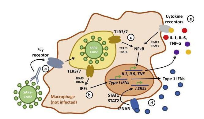
MedDocs Publishers

inflammatory feedback loop. In a defective immune response                   Further study of the nature of protective versus detrimental T
(left side) this may lead to further accumulation of immune                  cell responses is critically needed to determine the optimal T
cells in the lungs, causing overproduction of pro-inflammatory               cell engagement strategies for vaccines [30].
cytokines, which eventually damages the lung infrastructure.
The resulting cytokine storm circulates to other organs, leading
to multi-organ damage. In addition, non-neutralizing antibodies
produced by B cells may enhance SARS-CoV-2 infection through
Antibody-Dependent Enhancement (ADE), further exacerbat-
ing organ damage. Alternatively, in a healthy immune response
(right side), the initial inflammation attracts virus-specific T cells
to the site of infection, where they can eliminate the infected
cells before the virus spreads. Neutralizing antibodies in these
individuals can block viral infection, and alveolar macrophages
recognize neutralized viruses and apoptotic cells and clear them
by phagocytosis. Altogether, these processes lead to clearance
of the virus and minimal lung damage, resulting in recovery. G-
CSF, granulocyte colony-stimulating factor; TNF, tumour necro-
sis factor (Figure 2).

                                                                                Immune evasion strategies of SARS-CoV2.
                                                                                Cell-mediated immunity is essential in recovery from and
                                                                             control of viral infections, especially infections involving onco-
                                                                             genic viruses or viruses that spread directly from cell to con-
                                                                             tiguous cell. In these situations antibody cannot reach the virus
                                                                             but virally induced antigens on the surface of the infected cell
                                                                             can be recognized by different effector cells (e.g., cytotoxic T
                                                                             cells) , If the virus reaches target organs, it is more difficult to
                                                                             control. The host defenses that may play important roles in tar-
                                                                             get organs are initially inflammation, fever, and interferon and
                                                                             subsequently cell-mediated immunity. Together with reports of
   Development of immune elusion strategy (Figure 3)                         lymphopenia and reduced peripheral T cell levels in patients,
                                                                             these findings suggest that T cells are attracted away from the
    SARS-CoV-specific CD4+ T cells express IFNγ, TNF and IL-2,               blood and into the infected site to control the viral infection.
which suggests that patients with SARS-CoV infection exhibit a               In patients with COVID-19, increased T cell exhaustion and re-
TH1 cell response and mainly use cellular immunity to control                duced functional diversity predicted severe disease. Despite the
the infection [24,25]. Although this pro-inflammatory profile                impaired response, patients who recovered from SARS-CoV in-
may be an aggravating factor for immunepathogenesis, CD4+ T                  fection developed coronavirus-specific memory T cells, which
cells have been hypothesized to control SARS, as depletion of                were found up to 2 years after recovery.
these cells in mice resulted in slower clearance of the virus
from the host and severer lung inflammation. With the use of a                  Soluble mediators include immune interferon, chemotactic
mouse-adapted strain of SARS-CoV, immunization with dendrit-                 factors, macrophage migration inhibitory factor, and lympho-
ic cells bearing SARS-CoV peptides resulted in higher numbers                toxin; other lymphokines and monokines are not depicted. Cy-
of virus-specific CD4+ and CD8+ T cells that accumulated in the              totoxic effector lymphocytes, macrophages, and natural killer
lungs and increased survival [26] [27]. Also, transfer of SARS-              cells play complex but important roles in host defense. PMNs,
CoV-specific CD4+ and CD8+ T cells into immunodeficient mice                 Polymorphonuclear leukocytes. In some situations, cell-mediat-
resulted in better protection against a mouse-adapted strain of              ed immunity may develop before antibody production begins.
SARS-CoV [28].                                                               For example, cytotoxic effector T cells have been found in bron-
                                                                             chial washings 3 to 4 days after initiation of intranasal infection
   Despite evidence for an important role of T cells in control-             in mice; at this time, antibody cannot yet be detected. Cell-me-
ling infection, several vaccine formulations against SARS-CoV                diated immune responses can cause tissue damage; the lung le-
previously tested in animal models showed signs of immunopa-                 sions produced in influenza may be examples. Both T and B cell
thology associated with TH2 cell-mediated eosinophil infiltra-               responses against SARS-CoV-2 are detected in the blood around
tion [29]. In particular, aged mice that were vaccinated seemed              1 week after the onset of COVID-19 symptoms. CD8+ T cells are
to display increased immunopathology rather than protection.                 important for directly attacking and killing virus-infected cells,
Annals of Epidemiology and Public health                                 3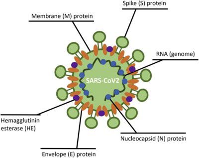
MedDocs Publishers
whereas CD4+ T cells are crucial to prime both CD8+ T cells and Coronavirus-specific T cells are clearly important in eliminat-
B cells. CD4+ T cells are also responsible for cytokine production ing the virus and controlling disease development and should
to drive immune cell recruitment. The first autopsy of a patient be considered in vaccine strategies. However, whether T cell
with COVID-19 revealed an accumulation of mononuclear cells responses alone are capable of preventing infection in human
(likely monocytes and T cells) in the lungs, coupled with low settings remains to be investigated. This knowledge will be im-
levels of hyperactive T cells in the peripheral blood [23]. portant for vaccine development.
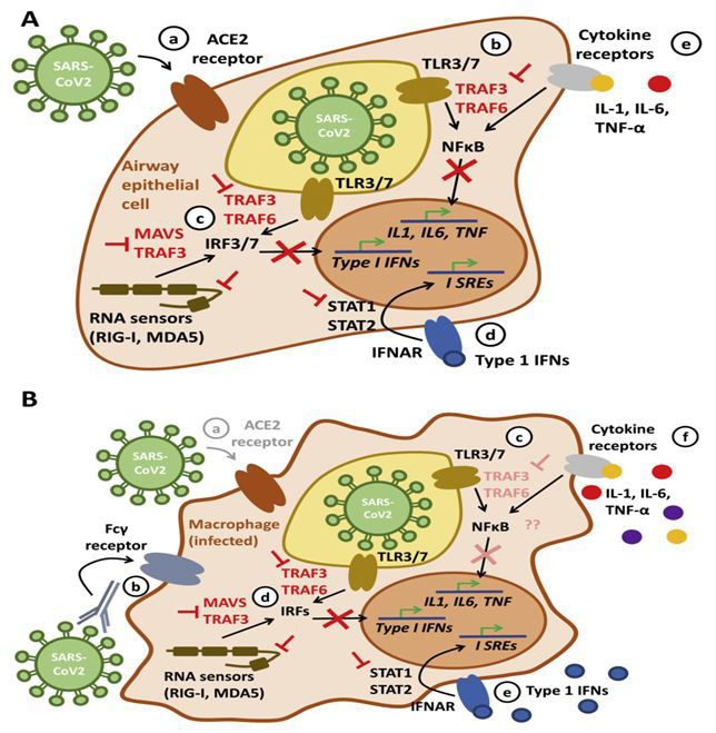
Fig-A describes the SARS-CoV2 infects airway epithelial cells Conclusion
through interactions with the trans-membrane enzyme ACE2 (a).
While RNA viruses usually activate TLR3 and/or 7 in endosomes As immunity does not exist and a considerable proportion
(b) and cytosolic RNA sensors RIG-I and MDA-5 (c), SARS-COV2 of humans develop severe disease, the novel coronavirus SARS-
effectively suppresses the activation of TNF receptor-associated CoV2 is a threat to millions globally. SARS-CoV2 has the capacity
factors (TRAF) 3 and 6, thereby limiting activation of the tran- to escape innate immune responses, which allows the patho-
scription factors NFκB and IRF3 and 7, thereby suppressing ear- gen to produce large copy numbers in primarily infected tissues,
ly pro-inflammatory responses through type I interferons (IFN) usually airway epithelia. Through the infection of innate immune
and pro-inflammatory effector cytokines IL-1, IL-6 and TNF-α cells, the conscription of uninfected cells from the circulation to
(red symbols). Furthermore, novel CoVs inhibit the activation of the primary site of infection, massive immune reactions may
STAT transcription factors (d) in response to type I IFN receptor induce hyperinflammation that can result in a cytokine storm
activation, which further limits antiviral response mechanisms. and many life-threatening complications. Researchers are still
Altogether, this prohibits virus containment through activation uncertain about what level of long-term immune memory is ad-
of anti-viral programs and the recruitment of immune cells. equate to protect against future coronavirus infection, and how
long it takes for the immune system to drop below that level.
Fig-B shows the tissue monocytes/macrophages express It is not even clear as of now whether someone with immunity
ACE2 to a significantly lower extent, making infection through could spread the coronavirus to others while fighting off a sec-
this route less likely (a). However, immune complexes consist- ond infection. If the immune response were strong enough to
ing of ineffective antibodies against e.g. seasonal CoVs and virus crush the virus quickly, the person probably wouldn't transmit
particles may be taken up by macrophages through Fcγ recep- it further. A weaker immune response that allowed some viral
tors resulting in their infection (b). In a process referred to as replication might not prevent transmission, though, particularly
Antibody Directed Enhancement (ADE), virions inhibit type I IFN since people without symptoms are known to pass the corona-
signaling in infected macrophages while allowing pro-inflamma- virus around. We are only beginning to understand host factors,
tory IL-1, IL-6 and TNF-α expression, which may contribute to such as differential expression of cell surface proteins that may
hyperinflammation and cytokine storm syndrome (c,d). Inhib- determine infection risk, disease presentation and outcomes.
ited type 1 IFN signaling suppresses anti-viral programs, while Unveiling tissue and stage specific factors contributing to pa-
increased IL-1, IL-6 and TNF-α expression auto-amplifies itself thology will result in new, effective and disease stage specific
through positive feedback loops (f). therapeutic approaches that control virus replication while lim-
iting inflammatory damage until vaccinations become available.
Cytokine strom in SARS-CoV2 (Figure 4). The association between immune dysfunction and outcome of
disease severity in patients with COVID-19 should serve as a
note of caution in vaccine development. Further studies of the
host immune response to SARS-CoV-2 are necessary, including
a in depth investigation of the determinants of healthy versus
dysfunctional outcomes. These will also help to identify bio-
markers to define immune correlates of protection and disease
severity for effective triage of patients.
References
1. World Health Organization. WHO Director-General’s opening re-
marks at the media briefing on COVID-19 – 11. 2020.
2. Tay, MZ, Poh, CM, Rénia L. et al. The trinity of COVID-19: immu-
nity, inflammation and intervention. Nat Rev Immunol. 2020.
3. Anthony SJ, Johnson CK, Greig DJ, Kramer S, Che X. Global pat-
terns in coronavirus diversity. Virus Evol. 2017; 3.
4. Kreuder Johnson C, Hitchens PL, Smiley Evans T, Goldstein T,
Thomas K. Clements A. Spillover and pandemic properties of
Inflammatory response through monocytes macrophages.
zoonotic viruses with high host plasticity. Sci. Rep. 2015; 5:
Uninfected monocytes/macrophages from the blood stream 14830.
invade the lungs where they detect virus particles and/or cyto-
plasmic and nuclear components. Within immune complexes, 5. Fehr AR, Perlman S. Coronaviruses: an overview of their replica-
these particles are taken up into the cell (a) where they are pre- tion and pathogenesis. Methods Mol. Biol. 2015; 1282: 1-23.
sented to TLRs, activating NFκB and/or IRF dependent pro-in- 6. Hulswit RJ, de Haan CA, Bosch BJ. Coronavirus Spike Protein and
flammatory pathways (b,c). As a result, uninfected monocytes/ Tropism Changes. Adv. Virus Res. 2016; 96: 29-57.
macrophages produce significant amounts of pro-inflammatory
cytokines (d,e) which recruit additional innate and adaptive im- 7. Tortorici MA, Veesler D. Structural insights into coronavirus en-
mune cells and cause additional tissue damage (Figure 4). try. Adv. Virus Res. 2019; 105: 93-116.
Annals of Epidemiology and Public health 4
MedDocs Publishers

8.    Walls AC, Xiong X, Park YJ, Tortorici MA, Snijder J, et al. Unex-          21.   Chen, N. et al. Epidemiological and clinical characteristics of 99
      pected Receptor Functional Mimicry Elucidates Activation of                      cases of 2019 novel coronavirus pneumonia in Wuhan, China: a
      Coronavirus Fusion. Cell. 2019; 176: 1026-1039.                                  descriptive study. Lancet. 2020; 395: 507-513.

9.    Bosch BJ, Smits SL, Haagmans BL. Membrane ectopeptidases                   22.   Chu, KH. et al. Acute renal impairment in coronavirus-associated
      targeted by human coronaviruses. Curr Opin Virol. 2014; 6: 55-                   severe acute respiratory syndrome. Kidney Int. 2005; 67: 698-
      60.                                                                              705.

10.   Huang C, Wang Y, XLi, et al. Clinical features of patients infected        23.   Xu, Z. et al. Pathological findings of COVID-19 associated with
      with 2019 novel coronavirus in Wuhan, China, Lancet. 2020.                       acute respiratory distress syndrome. Lancet Respir. Med. 2020;
                                                                                       8: 420-422.
11.   Peiris JS, Guan Y, KY. Yuen Severe acute respiratory syndrome
      Nat. Med. 2004.                                                            24.   Oh, HL J, Gan, KES, Bertoletti A. Understanding the T cell im-
                                                                                       mune response in SARS coronavirus infection. Emerg. Microbes.
12.   Guan, WJ. et al. Clinical characteristics of coronavirus disease                 Infect. 2012; 1: e23.
      2019 in China. N. Engl. J. Med. 2020.
                                                                                 25.   Shin, HS. et al. Immune responses to Middle East respiratory
13.   Lauer, SA. et al. The incubation period of coronavirus disease                   syndrome coronavirus during the acute and convalescent phas-
      (COVID-19) from publicly reported confirmed cases: estimation                    es of human infection. Clin. Infect. Dis. 2019; 68: 984-992.
      and application. Ann. Intern. Med. 2019.
                                                                                 26.   Roberts, A. et al. A mouse-adapted SARS-coronavirus causes dis-
14.    associated with the 2019 novel coronavirus indicating person-                   ease and mortality in BALB/c mice. PLoS Pathog. 2007; 3: e5.
      to-person transmission: a study of a family cluster. Lancet. 2020;
      395: 514–523.                                                              27.   Zhao, J., Zhao, J. Perlman, S. T cell responses are required for
                                                                                       protection from clinical disease and for virus clearance in severe
15.   Huang, C. et al. Clinical features of patients infected with 2019                acute respiratory syndrome coronavirus-infected mice. J. Virol.
      novel coronavirus in Wuhan, China. Lancet 395, 497–506. This                     2010; 84: 9318-9325.
      prospective study is the earliest to include an analysis of cy-
      tokine levels in severe and mild COVID-19, showing the pres-               28.   Bolles, M. et al. A double-inactivated severe acute respiratory
      ence of a cytokine storm analogous to that found for SARS-CoV                    syndrome coronavirus vaccine provides incomplete protection
      infection. 2020.                                                                 in mice and induces increased eosinophilic proinflammatory
                                                                                       pulmonary response upon challenge. J. Virol. 2011; 85: 12201-
16.   Chen, N. et al. Epidemiological and clinical characteristics of 99               12215.
      cases of 2019 novel coronavirus pneumonia in Wuhan, China: a
      descriptive study. Lancet. 2020; 395: 507-513.                             29.   Yasui, F. et al. Prior immunization with severe acute respiratory
                                                                                       syndrome (SARS)-associated coronavirus (SARS-CoV) nucleo-
17.   Zou, L. et al. SARS-CoV-2 viral load in upper respiratory speci-                 capsid protein causes severe pneumonia in mice infected with
      mens of infected patients. N. Engl. J. Med. 382: 1177-1179.                      SARS-CoV. J. Immunol. 2008; 181: 6337-6348.
18.   Kim, JY. et al. Viral load kinetics of SARS-CoV-2 infection in first       30.   Yasui, F. et al. Prior immunization with severe acute respiratory
      two patients in Korea. J. Korean Med. Sci. 2020; 35: e86.                        syndrome (SARS)-associated coronavirus (SARS-CoV) nucleo-
19.   Peiris, JS. et al. Clinical progression and viral load in a commu-               capsid protein causes severe pneumonia in mice infected with
      nity outbreak of coronavirus-associated SARS pneumonia: a pro-                   SARS-CoV. J. Immunol. 2008; 181: 6337-6348.
      spective study. Lancet.2003; 361: 1767-1772.

20.   Wang, D. et al. Clinical characteristics of 138 hospitalized pa-
      tients with 2019 novel coronavirus-infected pneumonia in Wu-
      han, China. JAMA. 2020; 323: 1061-1069.

Annals of Epidemiology and Public health                                     5
You can also read