STABILITY OF SARS-COV-2 RNA IN NONSUPPLEMENTED SALIVA
←
→
Page content transcription
If your browser does not render page correctly, please read the page content below
DISPATCHES

        Stability of SARS-CoV-2 RNA in
           Nonsupplemented Saliva
           Isabel M. Ott,1 Madison S. Strine,1 Anne E. Watkins, Maikel Boot, Chaney C. Kalinich,
          Christina A. Harden, Chantal B.F. Vogels, Arnau Casanovas-Massana, Adam J. Moore,
 M. Catherine Muenker, Maura Nakahata, Maria Tokuyama, Allison Nelson, John Fournier, Santos Bermejo,
   Melissa Campbell, Rupak Datta, Charles S. Dela Cruz, Shelli F. Farhadian, Albert I. Ko, Akiko Iwasaki,
         Nathan D. Grubaugh,2 Craig B. Wilen,2 Anne L. Wyllie,2 the Yale IMPACT Research team3

 The expense of saliva collection devices designed to sta-          an alternative sample type for antigen and antibody
 bilize severe acute respiratory syndrome coronavirus 2             testing (4,5). In addition, saliva collection is noninva-
 RNA is prohibitive to mass testing. However, virus RNA             sive, can be reliably performed without trained health
 in nonsupplemented saliva is stable for extended periods           professionals, and does not rely on a sometimes-lim-
 and at elevated temperatures. Simple plastic tubes for             ited swab supply. However, almost all saliva-based
 saliva collection will make large-scale testing and contin-        tests approved by the US Food and Drug Adminis-
 ued surveillance easier.                                           tration require specialized collection tubes containing
                                                                    stabilization or inactivation buffers that are costly and

D    espite increased diagnostic testing capacity for
     severe acute respiratory syndrome coronavirus
2 (SARS-CoV-2), testing in many countries, includ-
                                                                    not always available. Moreover, as saliva continues
                                                                    to gain popularity as a potential specimen to aid test-
                                                                    ing demands, standardized collection methods have
ing the United States, is still inadequate for slowing              not been defined for saliva collection as they have for
the coronavirus disease (COVID-19) pandemic. Many                   swab-based specimen collection. When true saliva is
persons still do not have access to SARS-CoV-2 testing,             not collected (e.g., if it contains sputum), which can
and for some that do, an imbalance between supply                   happen with COVID-19 inpatients when saliva is dif-
and demand at large testing centers leads to long de-               ficult to produce, specimens can be difficult to pipette
lays before results are received. The demand for test-              (6). Combined with untested concerns regarding
ing will only increase as many schools, colleges, and               SARS-CoV-2 RNA stability in saliva, using supple-
workplaces reopen. Ideally, specialized population                  ments to reduce degradation and improve sample
surveillance–oriented testing would require minimal                 processing has become common. Previous work with
diversion of resources from clinical diagnostic test-               saliva samples, however, has indicated that some
ing, be affordable and scalable, and enable rapid and               buffers optimized for host nucleic acid stabilization
reliable virus identification for persons with asymp-               may actually inhibit viral RNA detection (7) (S.B.
tomatic or subclinical infections. Thus, simplifying the            Griesemer et al., unpub. data, https://doi.org/10.1
sample collection and testing workflow is critical.                 101/2020.06.16.20133041), particularly in extraction-
     A simple solution is saliva collection. Saliva is a            free PCRs (D.R.E. D.R.E. Ranoa et al., unpub. data,
sensitive source for SARS-CoV-2 detection (1–3) and                 https://doi.org/10.1101/2020.06.18.159434). Thus, if
                                                                    true saliva (relatively easy to pipette) is being tested,
Author affiliations: Yale School of Public Health, New Haven,         the utility of collecting saliva in expensive tubes con-
Connecticut, USA (I.M. Ott, A.E. Watkins, C.C. Kalinich,            taining purported stabilization buffers comes into
C.A. Harden, C.B.F. Vogels, A. Casanovas-Massana,                   question. To explore the viability of broadly deploy-
A.J. Moore, M.C. Muenker, M. Nakahata, A.I. Ko, N.D. Grubaugh,      ing affordable saliva-based surveillance approaches
A.L. Wyllie); Yale School of Medicine, New Haven (M.S. Strine,      (8), we characterized SARS-CoV-2 RNA stability and
M. Boot, M. Tokuyama, A. Nelson, J. Fournier, S. Bermejo,           virus infectivity in saliva samples stored in widely
M. Campbell, R. Datta, C.S. Dela Cruz, S.F. Farhadian, A.I. Ko,
A. Iwasaki, C. Wilen); Howard Hughes Medical Institute, New         1
                                                                        These first authors contributed equally to this article.
Haven (A. Iwasaki)                                                  2
                                                                        These senior authors contributed equally to this article.
DOI: https://doi.org/10.3201/eid2704.204199
                                                                    3
                                                                        Team members team are listed at the end of this article.

1146                         Emerging Infectious Diseases • www.cdc.gov/eid • Vol. 27, No. 4, April 2021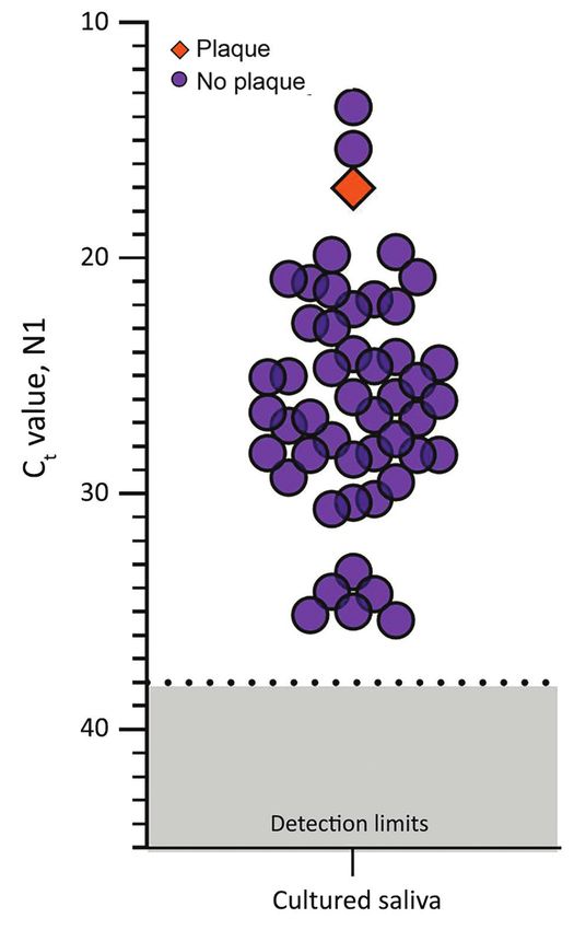
Stability of SARS-CoV-2 RNA in Nonsupplemented Saliva
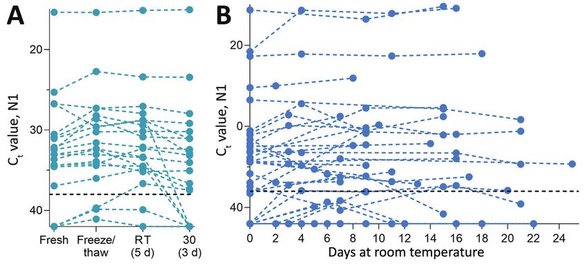
Figure 1. Stability of severe acute respiratory syndrome coronavirus 2 (SARS-CoV-2) RNA (N1) detection in saliva. A) Detection of
SARS-CoV-2 RNA in 20 saliva samples on day of sample collection (fresh) did not significantly change after storage at −80°C (to assess
the effect of a freeze/thaw cycle), 3 days at 30°C, or 5 days at RT (recorded as ≈19°C). Detection of N1 remained similar to that of
freshly collected samples, regardless of starting Ct value (Pearson r = −0.085, p = 0.518). B) At RT, detection remained stable for up to
25 days. Colored dashed lines track the same sample through different storage conditions. Black horizontal dashed lines represent Ct
38, which we applied as the cutoff to determine sample positivity. Samples that remained not detected after 45 cycles are depicted on
the x-axis. Ct, cycle threshold; RT, room temperature.
available, sterile, nuclease-free laboratory plastic a Ct increase of 0.973 (95% CI −0.252 to 2.197). More-
(polypropylene) tubes. over, SARS-CoV-2 RNA remained relatively stable in
saliva samples left at room temperature for up to 25
The Study days (Ct 0.027, 95% CI −0.019 to 0.071 Ct) (Figure 1,
We used saliva collected from COVID-19 inpatients panel B). Regardless of starting Ct value (viral load),
and at-risk healthcare workers into sterile wide-mouth this prolonged stability of SARS-CoV-2 RNA was also
containers (3) without preservatives (nonsupplement- observed when samples were stored for longer pe-
ed) to evaluate the temporal stability of SARS-CoV-2 riods at −80°C (maximum 92 days), 4°C (maximum
RNA at different holding temperatures (−80°C, 4°C, 21 days), and 30°C (maximum 16 days) (Appendix
≈19°C, 30°C) (Appendix, https://wwwnc.cdc.gov/ Figure 1).
EID/article/27/4/20-4199-App1.pdf). SARS-CoV-2 Although SARS-CoV-2 RNA from saliva re-
RNA from saliva was consistently detected at similar mained stable over time, we observed a decrease in
levels regardless of the holding time and temperatures human ribonuclease P at higher temperatures (room
tested. After RNA extraction and quantitative reverse temperature, Ct 1.837, 95% CI 0.468 to 3.188 Ct; 30°C,
transcription PCR (qRT-PCR) testing for SARS-CoV-2 Ct 3.526, 95% CI 1.750 to 5.349 Ct; Appendix Figure
on the day of saliva collection (3), we aliquoted and 2); the change in concentration was greater than that
stored the remaining 20 sample volumes at −80°C, observed for SARS-CoV-2 RNA (Appendix Figure 3).
room temperature (≈19°C), and 30°C. Whether stored Thus, although human RNA from saliva degrades
at −80°C, room temperature (5 days), or 30°C (3 days), without stabilization buffers, SARS-CoV-2 RNA re-
the qRT-PCR cycle threshold (Ct) values for the N1 mains protected even at warm temperatures suitable
region of the nucleocapsid protein did not differ sig- for nuclease activity.
nificantly from those for samples tested on the day of Because saliva has antiviral properties (9,10), we
collection (Figure 1, panel A). After the freeze/thaw explored the infectiousness of SARS-CoV-2 in saliva
cycle or storage at room temperature, we observed Ct samples. We inoculated Vero-E6 cells with 49 saliva
decreases of 1.058 (95% CI 2.289 to 0.141) for freeze/ samples with higher virus RNA titers (Ct range 13.57–
thaw and 0.960 (95% CI −2.219 to 0.266) for room tem- 35.32, median 26.01; Appendix Figure 4) because oth-
perature; however, the strength of this effect was low. ers have shown that SARS-CoV-2 isolation is uncom-
We saw a similar effect after incubation at 30°C, with mon from samples with low virus RNA titers (11,12;
Emerging Infectious Diseases • www.cdc.gov/eid • Vol. 27, No. 4, April 2021 1147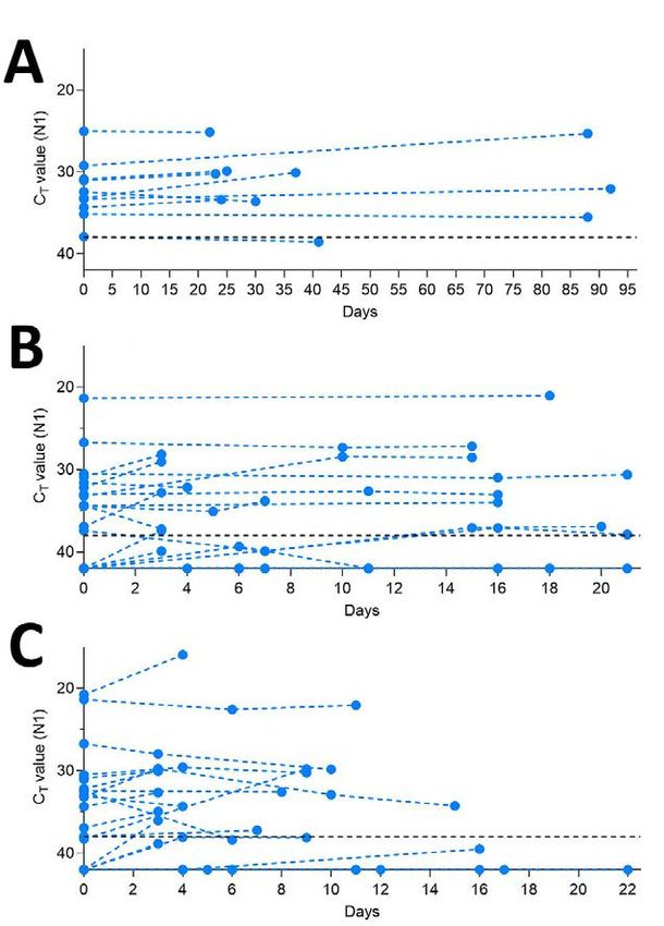
DISPATCHES

                                                                      and qRT-PCR). To determine whether this amplifica-
                                                                      tion resulted from detectable, active virus replication,
                                                                      we performed plaque assays in triplicate with cellular
                                                                      lysate from 72 hours after inoculation. Only 1 of these
                                                                      9 samples produced plaque-forming units; titer in-
                                                                      creased 3.79 × 10⁴ PFU/mL at 1 hour and at 72 hours
                                                                      after inoculation (Figure 2). This finding suggests
                                                                      that increased SARS-CoV-2 genome copies identi-
                                                                      fied by qRT-PCR may fall below the limit of detec-
                                                                      tion in plaque assay sensitivity (100 PFU/mL) until
                                                                      a certain reduction in Ct is reached (e.g., Ct reduction
                                                                      ≤12.90) or that components of saliva possibly inhibit
                                                                      active viral particle production and release in vitro.
                                                                      A similar result has been observed when attempting
                                                                      to perform plaque assays of virus from the colon (13),
                                                                      despite studies showing that SARS-CoV-2 infects gut
                                                                      enterocytes (14).

                                                                      Conclusions
                                                                      The cost of commercial tubes specialized for saliva
                                                                      collection and SARS-CoV-2 RNA stabilization (>$7/
                                                                      tube) (Table) can be prohibitive for mass testing. In-
                                                                      expensive saliva-based testing methods are urgently
                                                                      needed to help reach the capacity required to safely
                                                                      reopen schools and workplaces. We demonstrate
                                                                      the stability of SARS-CoV-2 RNA detection in saliva
                                                                      stored for prolonged periods in a variety of settings,
                                                                      which indicates that saliva can be simply collected
                                                                      without the need for additives.
                                                                           Previous studies have demonstrated the ease
                                                                      with which saliva can be collected into simple, wide-
Figure 2. Detection of severe acute respiratory syndrome
                                                                      mouth containers (3,15) and that buffers marketed
coronavirus 2 (SARS-CoV-2) in saliva samples tested for               for RNA stabilization may be detrimental to SARS-
infectious SARS-CoV-2. SARS-CoV-2 N1 detection (Ct values)            CoV-2 detection (S.B. Griesemer et al., unpub data,
measured by quantitative reverse transcription PCR for each           https://doi.org/10.1101/2020.06.16.20133041). Al-
saliva sample incubated with Vero-E6 cells for 72 hours. The          though some of these buffers are also marketed for
orange diamond depicts the only sample that produced plaque-
forming units (titer increase of 3.79 × 10⁴ PFU/mL; purple circles
                                                                      virus inactivation, SARS-CoV-2 is still considered a
indicate samples that did not produce plaque-forming units by         Biosafety Level 2 hazard, meaning that with or with-
72 h after inoculation; dashed lines indicate Ct 38 (the cutoff for   out buffer, any saliva sample should still be handled
sample positivity); gray shading indicates Cts below the limit of     with care. Without the need for RNA stabilization
detection. Ct, cycle threshold.                                       and given the limited evidence of virus replication
                                                                      in saliva samples, affordable alternatives to making
M.D. Folgueira, unpub. data, https://www.medrxiv.                     testing accessible throughout the country are simple,
org/content/10.1101/2020.06.10.20127837v1). By 72                     sterile, nuclease-free plastic containers.
hours after inoculation, Ct values were reduced in 9                       SARS-CoV-2 stability at room temperature and
(18.7%) of the 49 cultured saliva samples tested by                   at 30°C permits more affordable collection and trans-
qRT-PCR (−12.90, −11.53, −4.30, −3.68, −3.49, −2.88,                  port strategies without the need for expensive cooling
−2.81, −2.66, −2.40). Although these findings suggest                 strategies. Absence of the requirement for cold chain
an increased number of SARS-CoV-2 RNA copies                          handling also makes saliva testing easier in regions
by 72 hours, they may not definitively demonstrate                    with limited resources. Thus, one key for meeting
active virus replication. For instance, Ct reductions                 mass testing demands is collection of saliva in simple,
could also result from sampling artifacts or assay                    sterile, nuclease-fee tubes, negating the high costs as-
variations (disparities in inoculation, RNA extraction,               sociated with specialized collection devices.

 1148                            Emerging Infectious Diseases • www.cdc.gov/eid • Vol. 27, No. 4, April 2021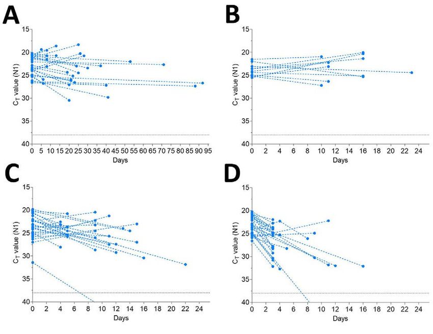
Stability of SARS-CoV-2 RNA in Nonsupplemented Saliva

Table. Possible saliva collection devices for severe acute respiratory virus coronavirus 2 RNA testing
                                                                                           Cost per
Tube type                            Collection                Buffer type               sample, USD                    Manufacturer
Oragene•Dx collection                 Funnel          Ethanol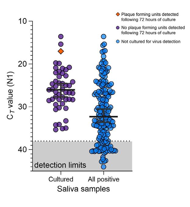
DISPATCHES

of Public Health, New Haven CT. Her work focuses on                           platform to enhance SARS-CoV-2 testing capacity. Med. 2020
developing diagnostic tools for and analyzing functional                      Dec 26 [Epub ahead of print]. https://doi.org/10.1016/
                                                                              j.medj.2020.12.010
evolution of viral pathogens, with particular focus on                   9.   Lieleg O, Lieleg C, Bloom J, Buck CB, Ribbeck K. Mucin
SARS-CoV-2 and endemic arboviruses.                                           biopolymers as broad-spectrum antiviral agents.
                                                                              Biomacromolecules. 2012;13:1724–32. https://doi.org/
                                                                              10.1021/bm3001292
References                                                              10.   Malamud D, Abrams WR, Barber CA, Weissman D,
1.     Hanson KE, Barker AP, Hillyard DR, Gilmore N,                          Rehtanz M, Golub E. Antiviral activities in human saliva.
       Barrett JW, Orlandi RR, et al. Self-collected anterior nasal           Adv Dent Res. 2011;23:34–7. https://doi.org/10.1177/
       and saliva specimens versus health care worker-collected               0022034511399282
       nasopharyngeal swabs for the molecular detection of SARS-        11.   Wölfel R, Corman VM, Guggemos W, Seilmaier M,
       CoV-2. J Clin Microbiol. 2020;58:e01824-20.                            Zange S, Müller MA, et al. Virological assessment of
2.     Byrne RL, Kay GA, Kontogianni K, Aljayyoussi G,                        hospitalized patients with COVID-2019. Nature.
       Brown L, Collins AM, et al. Saliva alternative to upper                2020;581:465–9. https://doi.org/10.1038/s41586-020-2196-x
       respiratory swabs for SARS-CoV-2 diagnosis. Emerg Infect         12.   Bullard J, Dust K, Funk D, Strong JE, Alexander D,
       Dis. 2020;26:2770–1.                                                   Garnett L, et al. Predicting infectious SARS-CoV-2 from
3.     Wyllie AL, Fournier J, Casanovas-Massana A, Campbell M,                diagnostic samples. Clin Infect Dis. 2020 May 22 [Epub ahead
       Tokuyama M, Vijayakumar P, et al. Saliva or nasopharyngeal             of print]. https://doi.org/10.1093/cid/ciaa638
       swab specimens for detection of SARS-CoV-2. N Engl J Med.        13.   Zang R, Gomez Castro MF, McCune BT, Zeng Q,
       2020;383:1283–6. https://doi.org/10.1056/NEJMc2016359                  Rothlauf PW, Sonnek NM, et al. TMPRSS2 and TMPRSS4
4.     Isho B, Abe KT, Zuo M, Jamal AJ, Rathod B, Wang JH, et al.             promote SARS-CoV-2 infection of human small intestinal
       Persistence of serum and saliva antibody responses to                  enterocytes. Sci Immunol. 2020;5:eabc3582. https://doi.org/
       SARS-CoV-2 spike antigens in COVID-19 patients.                        10.1126/sciimmunol.abc3582
       Sci Immunol. 2020;5:eabe5511.                                    14.   Lamers MM, Beumer J, van der Vaart J, Knoops K,
5.     Pisanic N, Randad PR, Kruczynski K, Manabe YC, Thomas DL,              Puschhof J, Breugem TI, et al. SARS-CoV-2 productively
       Pekosz A, et al. COVID-19 serology at population scale: SARS-          infects human gut enterocytes. Science. 2020;369:50–4.
       CoV-2-specific antibody responses in saliva. J Clin Microbiol.         https://doi.org/10.1126/science.abc1669
       2020;59:e02204–20. https://doi.org/10.1128/JCM.02204-20          15.   Wyllie AL, Chu MLJN, Schellens MHB, van Engelsdorp
6.     Landry ML, Criscuolo J, Peaper DR. Challenges in use of                Gastelaars J, Jansen MD, van der Ende A, et al. Streptococcus
       saliva for detection of SARS CoV-2 RNA in symptomatic                  pneumoniae in saliva of Dutch primary school children.
       outpatients. J Clin Virol. 2020;130:104567. https://doi.org/           PLoS One. 2014;9:e102045. https://doi.org/10.1371/
       10.1016/j.jcv.2020.104567                                              journal.pone.0102045
7.     Jones TH, Muehlhauser V. Effect of handling and storage
       conditions and stabilizing agent on the recovery of viral        Address for correspondence: Anne L. Wyllie and Nathan D.
       RNA from oral fluid of pigs. J Virol Methods. 2014;198:26–31.
       https://doi.org/10.1016/j.jviromet.2013.12.011                   Grubaugh, Yale School of Public Health, 60 College St,
8.     Vogels CBF, Watkins AE, Harden CA, Brackney DE, Shafer J,        New Haven, CT 06511, USA; email: anne.wyllie@yale.edu and
       Wang J, et al. SalivaDirect: a simplified and flexible           nathan.grubaugh@yale.edu

1150                            Emerging Infectious Diseases • www.cdc.gov/eid • Vol. 27, No. 4, April 2021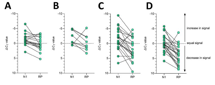
Article DOI: https://doi.org/10.3201/eid2704.204199

               Stability of SARS-CoV-2 RNA in
                  Nonsupplemented Saliva
Appendix

Methods

RNA extraction and SARS-CoV-2 detection

       Saliva samples were self-collected by COVID-19 inpatients and healthcare workers at the
Yale-New Haven Hospital (Yale Human Research Protection Program Institutional Review
Boards FWA00002571, Protocol ID. 2000027690) (2), into plain wide-mouth containers without
the addition of stabilizing buffers. RNA was extracted from saliva samples (1) and tested by RT-
qPCR for SARS-CoV-2 RNA (N1) and human RNase P (RP) (3) on day of collection (≤12 hours
post sample collection) and at various time points after the storage of the remaining,
unsupplemented samples at temperatures of -80°C, -20°C, +4°C, room temperature (measured at
an average of ~19ºC), or 30°C.

Cell culture

       Vero-E6 cells (ATCC) were cultured in Dulbecco’s Modified Eagle Medium (Gibco)
supplemented with 10% heat-inactivated fetal bovine serum (VWR), 1% Penicillin/Streptomycin
(Gibco), 100 µg/mL gentamicin (Gibco), and 0.5 µg/mL amphotericin B (Gibco). All cells were
incubated at 37°C and 5% CO2. All cell culture experiments were performed in a biosafety level
3 laboratory at Yale University and approved by the Yale University Biosafety Committee.

Saliva inoculation and virus culture in Vero-E6 cells

       Saliva samples were diluted 1:1 in 1X Dulbecco’s PBS (Gibco). Diluted saliva samples
were incubated for one hour at 37°C with 2.5x10⁵ Vero-E6 cells in a 24-well plate (Corning).
Unbound virus was aspirated and the media were replaced. Infected Vero-E6 cells were frozen at
-80°C at 1 and 72 hours post-inoculation. Thawed samples were used for plaque assays and RNA
extraction. Prior to RNA extraction (1) and RT-qPCR detection of SARS-CoV-2 RNA (3) the
Vero-E6 cells from 1 and 72 hours post-inoculation were thawed at room temperature and further

                                            Page 1 of 7
lysed by diluting 1:3 in MagMax Binding Solution (ThermoFisher). RNA was extracted from the
two timepoints and tested in RT-qPCR for SARS-CoV-2 N1. We interpreted a Ct reduction >2
as a difference which could potentially be explained by viral replication during the two
timepoints.
Plaque assay
Vero-E6 cells were seeded at 4x10⁵ cells/well in 12-well plates (Corning). The following
day, media were removed and replaced with 100 μl of 10-fold serial dilutions of thawed 1 hour
or 72 hour post-inoculation saliva samples. Plates were incubated at 37°C for 1 hour with gentle
rocking every 15 mins. Unbound inocula was aspirated from each well and overlay media
(DMEM, 2% FBS, 0.6% Avicel RC-581 (DuPont)) were added to each well. At 48 hours post-
infection, plates were fixed with 5-10% formaldehyde for 30 min then stained with crystal violet
solution (0.5% crystal violet in 20% ethanol) for 30 mins. Crystal violet solution was then
aspirated and plates were washed in tap water to visualize plaques.
Statistical analyses
We fit a linear regression to the experimental stability data to model the change in Ct
values of positive samples following stability conditions using the equation below. Let dct be the
change in Ct value from fresh testing following each storage condition and let condition be the
categorical storage condition (e.g. freeze/thaw, room temperature, 30°C, etc).
dct ~ condition
Robust confidence intervals were simulated from this model using the mvrnorm, in the R
package “MASS”, and quantile functions. This regression was also used to model the effect of
prolonged storage in stability conditions on RP.
For extended timepoint analyses of N1 we used a linear mixed effects model to predict
the change in Ct values of positive samples under each stability condition for greater durations of
time using the equation below. Let timepoint be the number of days under stability conditions
and let sample be the patient number.
dct ~ timepoint + (1|sample)
Confidence intervals were computed for this model using confint.merMod, in the R
package “lme4”.
Page 2 of 7Further statistical analyses were conducted in GraphPad Prism 8.0.0 as described in the
text and figure legends.
References
1. Ott I, Vogels C, Grubaugh N, Wyllie A. Saliva Collection and RNA Extraction for SARS-CoV-2
Detection v1 (protocols.io.bg3pjymn) [cited 2021 Jan 7].
http://dx.doi.org/10.17504/protocols.io.bg3pjymn
2. Wyllie AL, Fournier J, Casanovas-Massana A, Campbell M, Tokuyama M, Vijayakumar P, et al.
Saliva or nasopharyngeal swab specimens for detection of SARS-CoV-2. N Engl J Med.
2020;383:1283–6. PubMed https://doi.org/10.1056/NEJMc2016359
3. Vogels CBF, Brito AF, Wyllie AL, Fauver JR, Ott IM, Kalinich CC, et al. Analytical sensitivity and
efficiency comparisons of SARS-CoV-2 RT-qPCR primer-probe sets. Nat Microbiol.
2020;5:1299–305. PubMed https://doi.org/10.1038/s41564-020-0761-6
Page 3 of 7Appendix Figure 1. Stability of SARS-CoV-2 RNA (N1) detection in saliva. SARS-CoV-2 RNA detection
in saliva on day of sample collection (0) or after prolonged storage at -80°C, 4°C or 30°C. Ct values from
the same original sample are connected by a dotted line. The -80°C and 4°C conditions were found to
have a weakly beneficial effect on signal detection by the mixed effects model, while the 30°C condition
resulted in a slight increase in Ct. The -80°C storage alone did not cross zero suggesting a mildly
stronger effect than the other conditions (95% CI: -0.038, -0.010). The black dashed line represents Ct 38
which we applied as the cut-off to determine sample positivity. Samples that remained not detected (ND)
after 45 cycles are depicted as Ct 42.
Page 4 of 7Appendix Figure 2. Detection of human RNase P (RP) declines over time when stored in saliva in
warmer conditions. Detection of human RP in saliva on day of collection (0) or after prolonged storage at -
80°C, 4°C, room temperature (~19°C) or 30°C. Ct values from the same original sample are connected by
a dotted line. Prolonged storage at -80°C and 4°C had minimal effect on RP detection with Ct changes of
0.832 (95% CI: -0.402, 2.038) and -0.315 (95% CI: -2.336, 1.687), respectively. However, storage at
room temperature (Ct +1.837, 95% CI: 0.468, 3.188) and 30°C (Ct +3.526, 95% CI: 1.750, 5.349) was
detrimental to RP, exhibiting a more substantial decrease in signal at these warmer conditions. The black
dashed line represents Ct 38 which we applied as the cut-off to determine sample positivity. Samples that
remained not detected (ND) after 45 cycles are below the y-axis limit.
Page 5 of 7Appendix Figure 3. Detection of SARS-CoV-2 RNA (N1) in saliva remained more stable over time than
human RNase P (RP). Delta Ct was calculated as the difference in Ct value from the day of saliva
collection and after storage at -80°C, 4°C, room temperature (~19°C) or 30°C. Delta Ct values from the
same sample are joined by a solid line. While the change in detection of SARS-CoV-2 N1 and RP was
similar in saliva samples stored at 4°C (Wilcoxon signed rank test, p = 0.129), a greater difference was
observed between the change in N1 and RP for samples stored at -80°C (p = 0.001), room temperature
(p = 0.001) and 30°C (p < 0.0001).
Page 6 of 7Appendix Figure 4. Saliva samples of relatively high viral load were cultured to evaluate the infectiousness of SARS-CoV-2 in saliva. Saliva samples cultured on Vero-E6 to test for infectious virus were of higher SARS-CoV-2 RNA (N1) load as compared to the overall saliva samples collected by Yale IMPACT (2) which tested positive for SARS-CoV-2 (Mann-Whitney, p =
You can also read